Article

Effects of Citrus kawachiensis Peel in Frailty-like Model Mice Induced by Low Protein Nutrition Disorders

Department of Pharmaceutical Pharmacology, College of Pharmaceutical Sciences, Matsuyama University, 4-2 Bunkyo-cho, Matsuyama 790-8578, Japan
*
Author to whom correspondence should be addressed.
Antioxidants 2023, 12(3), 779; https://doi.org/10.3390/antiox12030779
Submission received: 28 February 2023 / Revised: 20 March 2023 / Accepted: 21 March 2023 / Published: 22 March 2023

Abstract

:
“Frailty” caused by a decline in physiological reserve capacity, chronic inflammation, and oxidative stress in the elderly has recently become a major social issue. The present study examined the effects of the peel of Citrus kawachiensis (CK), which exhibits anti-inflammatory, antioxidant, and pro-neurogenesis activities in frailty-like model mice. Male C57BL/6 mice (15 weeks old) were fed an 18% protein diet (CON), a 2.5% protein diet (PM), and PM mixed with 1% dried CK peel powder for approximately 1 month. Mice were euthanized 2 or 8 days after a single intraperitoneal administration of lipopolysaccharide (LPS) and tissues were dissected. Among peripheral tissues, muscle weight, liver weight, and blood glucose levels were significantly higher in the PM–LPS–CK group than in the PM–LPS group. In the behavioral analysis, locomotive activity was significantly lower in the PM–LPS group than in the PM group. The reduction in locomotive activity in the PM–LPS–CK group was significantly smaller than that in the PM–LPS group. The quantification of microglia in the hippocampal stratum lacunosum-moleculare revealed that increases in the PM–LPS group were significantly suppressed by the dried CK peel powder. Furthermore, the quantification of synaptic vesicle membrane proteins in the hippocampal CA3 region showed down-regulated expression in the PM–LPS group, which was significantly ameliorated by the administration of the dried CK peel powder. Collectively, these results suggest that CK inhibits inflammation and oxidative stress induced by PM and LPS in the central nervous system and peripheral tissue. Therefore, C. kawachiensis is highly effective against “frailty”.

Graphical Abstract

1. Introduction

Lifespans have been increasing worldwide with advances in health care and better diets, and many countries are now experiencing population aging. Aging is associated with an increased risk of a few diseases due to chronic inflammation, oxidative stress, and a decline in physiological reserve capacity. Aging decreases physical activity and food intake, resulting in protein-energy malnutrition, and has a negative impact on immunity and muscle strength. An increased susceptibility to stress, chronic inflammation, oxidative stress, and other factors may lead to a decline in the activities of daily living, the need for nursing care, and even death, and this is called “frailty” [1,2,3]. The repetition of these conditions leads to a vicious cycle, referred to as the “frailty cycle”, which has become a major social issue. Frailty includes physical frailty, such as sarcopenia and locomotive syndrome, and mental and psychological frailty related to cognitive dysfunction and depression-like symptoms, both of which may exacerbate the frailty cycle and, thus, must be carefully managed [1,2,3]. Moreover, oxidative stress caused by inflammatory cytokines and free radicals due to chronic inflammation has been implicated in various chronic diseases, such as autoimmune diseases, cancer, neurological diseases, and lifestyle-related diseases [4,5,6]. It is an important factor that accelerates the frailty cycle by creating an internal environment in which compensatory mechanisms are impaired. Furthermore, the propagation of inflammation not only to the periphery but also to the central nervous system has been suggested to accelerate brain aging and is also associated with mental disorders and neurodegenerative-related diseases, including Alzheimer’s disease [7], in which neuronal damage due to microglial activation and decreased brain-derived neurotrophic factor (BDNF) production are prominent features.
Citrus fruits contain a variety of bioactive components, especially polyphenols and flavonoids. In recent years, several citrus components have been reported to have a potential to prevent or treat not only peripheral diseases, but also central nervous system degenerative diseases caused by inflammation and oxidative stress [8]. Our laboratory extracted functional components from various citrus peels and screened them using cultured neurons, and we found that the components in Citrus kawachiensis peel were highly active in terms of brain-protective effects [9]. Therefore, our research has been focusing on the effects of peel components of Citrus kawachiensis, a specialty citrus fruit in Japan [10], on the suppression of central nervous system dysfunction induced by chronic inflammation or oxidative stress. The peel of C. kawachiensis contains larger amounts of bioactive components, such as 3,5,6,7,8,3′,4′-heptamethoxyflavone (HMF), auraptene (AUR), and naringin (NGI), than other citrus varieties [10,11], which are expected to exert ameliorative effects on the functional decline of the central nervous system and peripheral tissue. We previously reported that C. kawachiensis exhibited various anti-inflammatory, antioxidant, and brain-protective activities, such as the suppression of microglial activation and reductions in glutathione levels in the brain, in global cerebral ischemia model mice [12]. It also inhibited microglial activation and tau phosphorylation and enhanced neurogenesis in the brains of hyperglycemia model mice [13] and aging-accelerated model mice [14].
A protein malnutrition model has been reported, and the experiment was conducted by feeding a low-protein diet for four weeks [15]. The model showed hippocampal protein thiols decrease with a low protein diet and dramatic reactive gliosis accompanied by extensive neuronal loss following ischemia [15]; in addition, protein malnutrition has also been reported to decrease antioxidant capacity in the brain and may lead to increased oxidative stress [16], so a low protein diet impairs functional outcomes. Lipopolysaccharide has also been reported to induce oxidative stress and exacerbate inflammation in the body in animal models [17], suggesting that protein malnutrition and lipopolysaccharide administration-induced chronic inflammation reproduce frailty in the body. In the present study, we investigated the effects of dried C. kawachiensis peel powder on the central nervous system and peripheral tissues of frailty-like model mice induced by low-protein nutritional disorders.

2. Materials and Methods

2.1. Animals

All animal experiments were conducted in accordance with the guidelines of Matsuyama University (protocol # 18-008). Nine-week-old male C57BL/6N mice were purchased from Japan SLC (Hamamatsu, Japan). Mice were kept at 23 ± 1 °C with a light/dark cycle of 12 h (light period 8:00–20:00, dark period 20:00–8:00). Animals were kept with free access to water and food for the duration of the experiment.

2.2. Sample Treatment

The control group (CON) was fed an 18% protein control diet, the protein malnutrition (PM) group a 2.5% low-protein diet, and the PM–CK group a PM diet mixed with 1% dried C. kawachiensis (CK) peel powder for the four-week feeding period. Then, the PM–LPS and PM–LPS–CK groups were intraperitoneally administered lipopolysaccharide (LPS) once (1 mg/kg of mouse), and mice were dissected either 2 or 8 days later. The group not administered LPS was treated with saline.

2.3. Food Intake

Mice were housed four per cage, and food consumption was measured each week. The amount of food consumed by one mouse was estimated by dividing the total food intake each day by the number of mice.

2.4. The Y-maze Test

Mice were placed at the tip of one of three arms (each arm was designated as A, B, or C) of a Y-shaped Y-maze device (length of 35 cm/width of 8.5 cm/height of 15 cm) and allowed to freely explore for 8 min. Mouse behavior was analyzed for the total distance travelled and immobility time using the ANY-maze Video Tracking System (Stoelting, Wood Dale, IL, USA) connected to a USB digital camera. A behavioral analysis was performed in a dark place with indirect illumination, feces and urine were processed for each test, and the apparatus was disinfected with alcohol each time to keep the inside clean.

2.5. Dissection

Blood was collected from the heart under anesthesia and followed by the transcardial perfusion of heparinized phosphate-buffered saline. The brain was removed and halved according to the cerebral longitudinal fissure. One half of the brain was soaked in 4% of paraformaldehyde for 2 days and then in 15 and 30% sucrose solutions for 1 day each. Brains were embedded in OCT compound, and 30 µm thick frozen brain sections (sagittal plane) were prepared using a cryostat (CM3050; Leica Microsystems, Heidelberger, Germany). The liver and gastrocnemius muscle were also collected. Blood samples were left to stand at room temperature for 30 min and then centrifuged at 1200× g at 4 °C for 20 min.

2.6. Blood Glucose Measurement

LabAssay™ Glucose (Mutarotase-GOD method; Wako, Osaka, Japan) was used to measure blood glucose levels. The experimental procedure was performed using the protocol in this kit, which involved measuring the absorbance of the sample and standard solutions at 570 nm with a microplate reader (Thermo Fisher, Tokyo, Japan).

2.7. Immunohistochemistry for Optical Microscopy

Thirty-micrometer-thick brain sections were immersed in 3% hydrogen peroxide solution for 20 min to remove endogenous peroxidase and then blocked in 2% skim milk for 60 min followed by 5% normal goat serum solution for 60 min. Sections were incubated with a rabbit polyclonal antibody against ionized calcium-binding adaptor 1 (Iba1, 1:1000; Wako, Osaka, Japan), which is specifically expressed in microglia, with shaking at 4 °C overnight. Brain sections were reacted with an Envision-plus system-HRP-labeled polymer as a secondary antibody for 60 min. The DAB substrate was used to develop staining, followed by 95% ethanol, 100% ethanol, and xylene × 2 in that order, and then sealed with a cover glass. Images of stained brain sections were captured with an optical microscope (CX21; Olympus, Tokyo, Japan). Multiple brain sections from each mouse were stained.

2.8. Immunofluorescence for Confocal Microscopy

Thirty-micrometer-thick brain sections were immersed in HistoVT One (Nacalai Tesque, Kyoto, Japan) and heated at 70 °C for antigen retrieval. Sections were then cooled to room temperature and blocked with 2% skim milk for 30 min followed by 5% normal goat serum solution for 60 min. Sections were incubated with a mouse monoclonal antibody against a synaptic vesicle membrane protein (synaptophysin, 1:1000; Sigma-Aldrich, St. Louis, MO, USA) as the primary antibody with shaking at 4 °C overnight. The next day, sections were reacted with Alexa Fluor 488 goat anti-mouse IgG (H + L) (1:300; Invitrogen, Carlsbad, CA, USA) as a secondary antibody and shaken for 60 min in the dark. Brain sections were placed on glass slides using mounting medium with DAPI (Vectashield; Vector Laboratories, Burlingame, CA, USA). Stained brain sections were then observed, and images were captured using a confocal fluorescence microscopy system (LSM800, Zeiss, Oberkochen, Germany). Multiple brain sections from each mouse were stained.

2.9. Image Analysis

A quantitative analysis of immune-positive signals in the images was performed using ImageJ software (NIH, Bethesda, MD, USA). The particle analysis tool was used for the analysis, and the pixels of the immune-positive signals were accumulated.

2.10. Statistical Analysis

Data for individual groups are expressed as means ± SEM. Data were statistically analyzed between two groups using the t-test. A value of p < 0.05 was significant.

3. Results

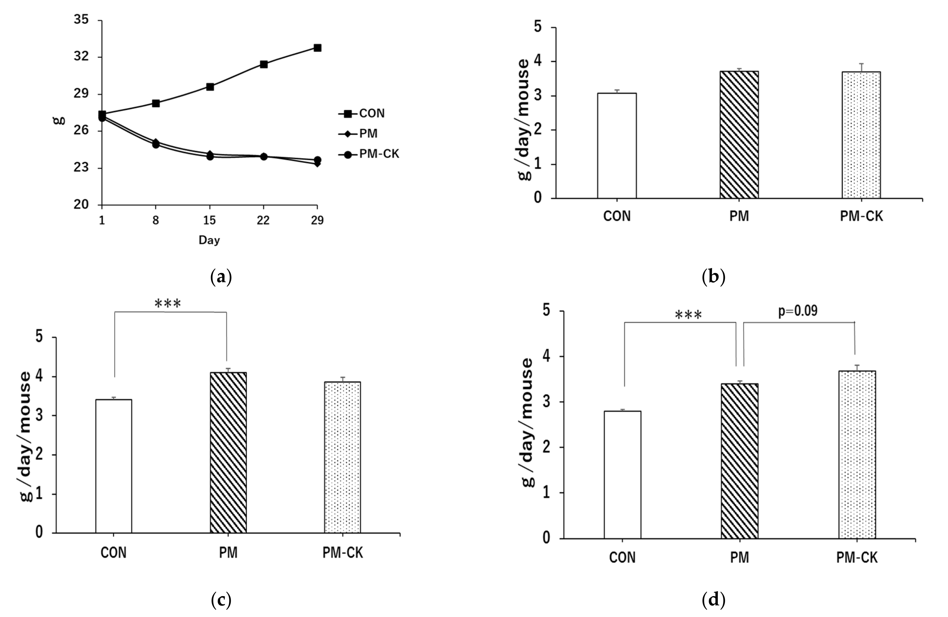
3.1. Changes in Body Weight and Food Intake in Different Experimental Groups

Body weight showed normal increases in the CON diet group but decreased in the PM group (Figure 1a). However, food intake was significantly higher in the PM group than in the CON group (Figure 1b–d; *** p < 0.001). Furthermore, a comparison of food intake in the fourth week showed that intake was slightly higher in the PM–CK group than in the PM group (Figure 1d; p < 0.09).

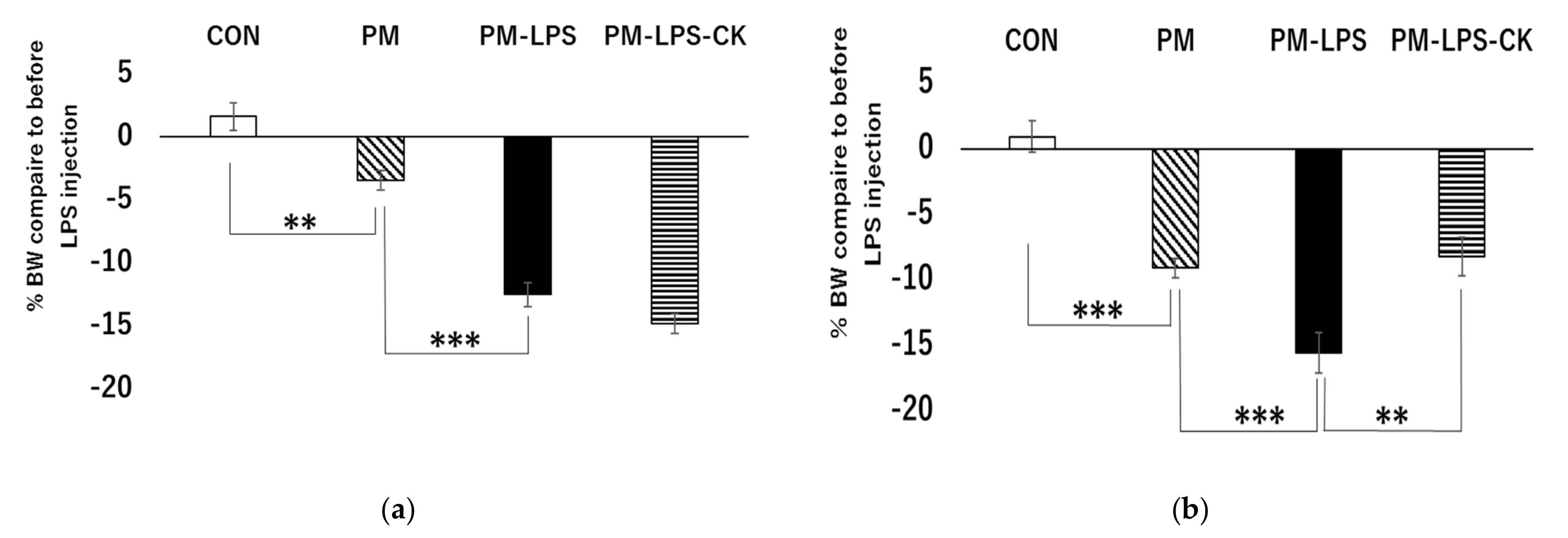
3.2. Percent Weight Loss 2 or 8 Days after the Administration of LPS

Comparisons of the CON and PM groups treated with saline showed that weight loss was significantly greater in the PM group than in the CON group 2 and 8 days after the administration of LPS (Figure 2a,b; ** p < 0.01, *** p < 0.001). Two days after the administration of LPS, weight loss was significantly greater in the PM–LPS group than in the PM group (Figure 2a; *** p < 0.001) and was not ameliorated by the dried C. kawachiensis peel powder (Figure 2a). Eight days after the administration of LPS, weight loss was significantly greater in the PM–LPS group than in the PM group (Figure 2b; *** p < 0.001). Body weight loss was significantly lower in the PM–LPS–CK group than in the PM–LPS group (Figure 2b; ** p < 0.01).

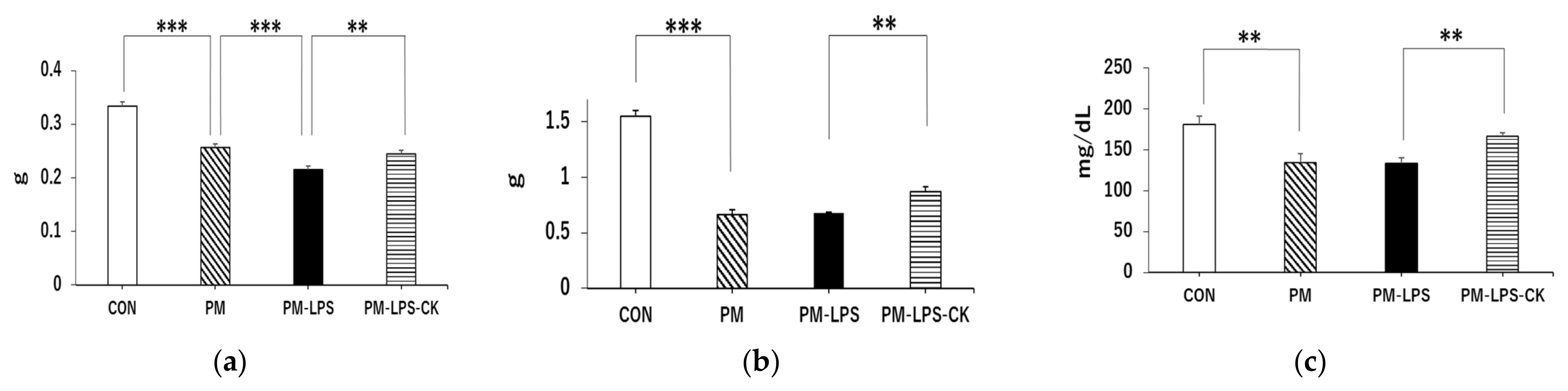
3.3. Ameliorative Effects of C. kawachiensis on Peripheral Dysfunction 8 Days after the Administration of LPS

We collected gastrocnemius muscle, liver, and serum samples to examine the peripheral effects of the PM diet and inflammation. Gastrocnemius muscle weight was significantly lower in the PM group than in the CON group (Figure 3a; *** p < 0.001) and significantly lower in the PM–LPS group than in the PM group (Figure 3a; *** p < 0.001). However, gastrocnemius muscle weight loss was significantly lower in the PM–LPS–CK group than in the PM–LPS group (Figure 3a; ** p < 0.01). Liver weight was significantly lower in the PM group than in the CON group (Figure 3b; *** p < 0.001) but was similar in the PM–LPS group and PM group. On the other hand, liver weight loss was significantly lower in the PM–LPS–CK group than in the PM–LPS group (Figure 3b, ** p < 0.01). Blood glucose levels were also significantly lower in the PM group than in the CON group (Figure 3c; *** p < 0.001). Furthermore, the decrease observed in the PM–LPS group was significantly lower in the PM–LPS–CK group (Figure 3c; *** p < 0.001).

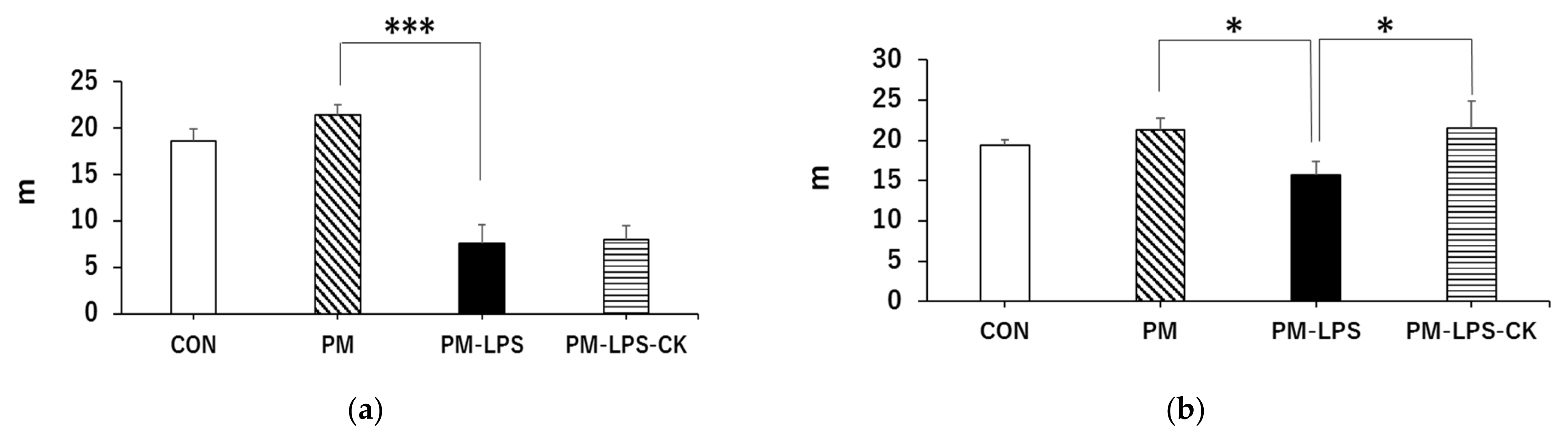
3.4. Ameliorative Effects of C. kawachiensis on Spontaneous Behavior

We performed the Y-maze test to assess behavioral activity. The total distance traveled in 8 min did not significantly differ between the CON and PM groups 2 days after the administration of LPS (Figure 4a); however, it was significantly shorter in the PM–LPS group than in the PM group (Figure 4a; *** p < 0.001). Additionally, reductions in the total distance traveled were not attenuated by the dried C. kawachiensis peel powder. No significant differences were observed in immobility times 2 days after the administration of LPS between the CON and PM groups (Figure 4c), whereas it was significantly longer in the PM–LPS group than in the PM group (Figure 4c; *** p < 0.001). In addition, the immobility time in the PM–LPS–CK group was not suppressed (Figure 4c), as in the previous results. Eight days after the administration of LPS, no significant differences were noted in the total distance traveled between the CON and PM groups (Figure 4b); however, it was significantly shorter in the PM–LPS group than in the PM group (Figure 4b; * p < 0.05). The reduction observed in the total distance travelled in the PM–LPS group was significantly smaller in the PM–LPS–CK group (Figure 4b; * p < 0.05). Furthermore, no significant differences were noted in immobility times between the CON and PM groups (Figure 4d), whereas they were significantly longer in the PM–LPS group than in the PM group (Figure 4d; * p < 0.05). The longer immobility time observed in the PM–LPS group was reduced in the PM–LPS–CK group (Figure 4d; * p < 0.05).

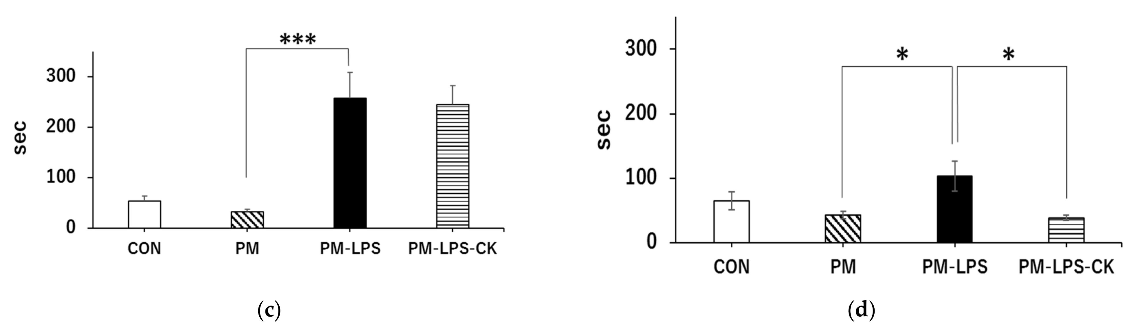
3.5. Suppressive Effects of C. kawachiensis on Microglial Activation

The quantification of microglia in the stratum lacunosum-moleculare of the hippocampus revealed no significant differences between the CON and PM groups (Figure 5a,b); however, the total area of activated microglia was significantly higher in the PM–LPS group than in the PM group (Figure 5b; ** p < 0.01). This increase was significantly smaller in the PM–LPS–CK group (Figure 5b; * p < 0.05).

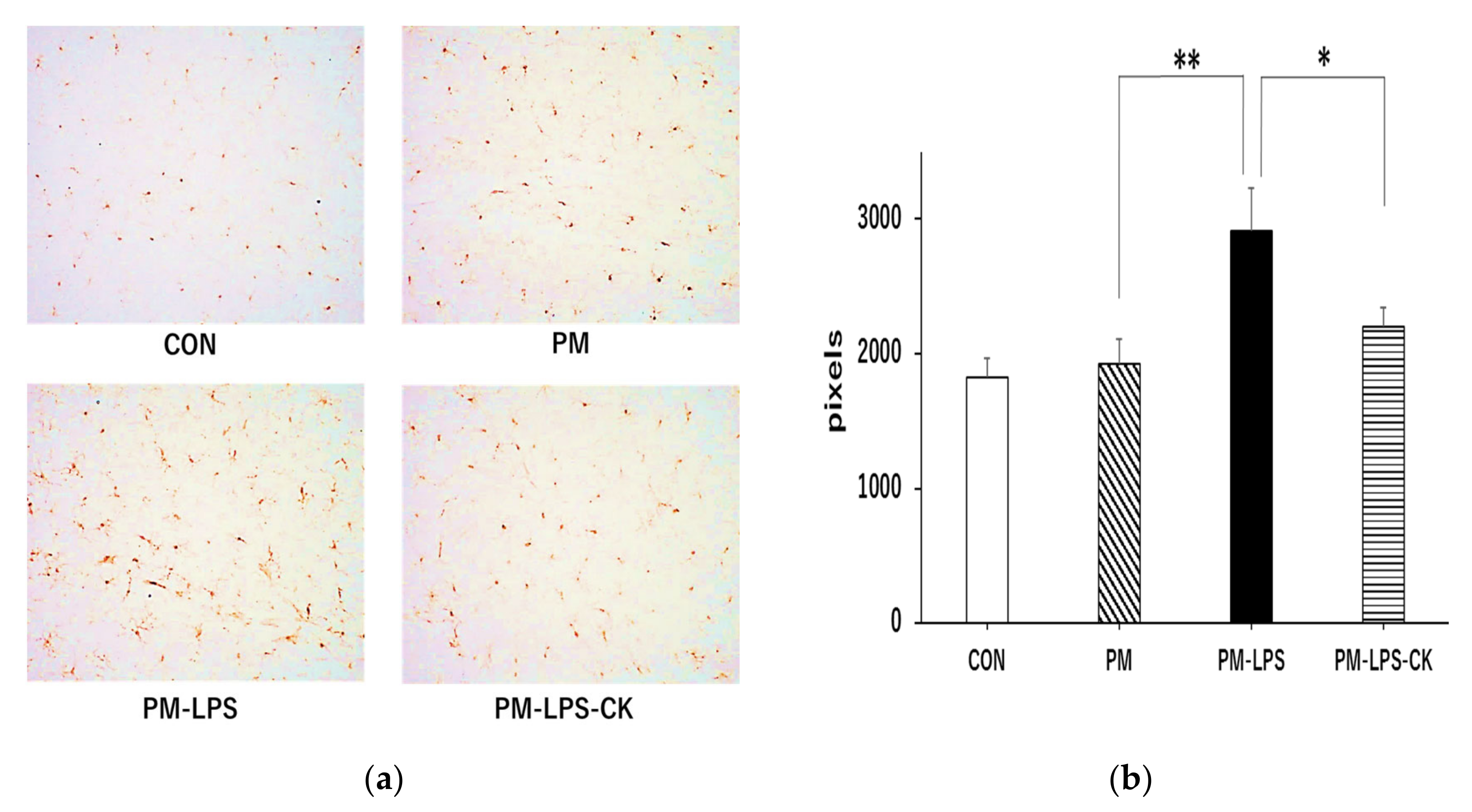
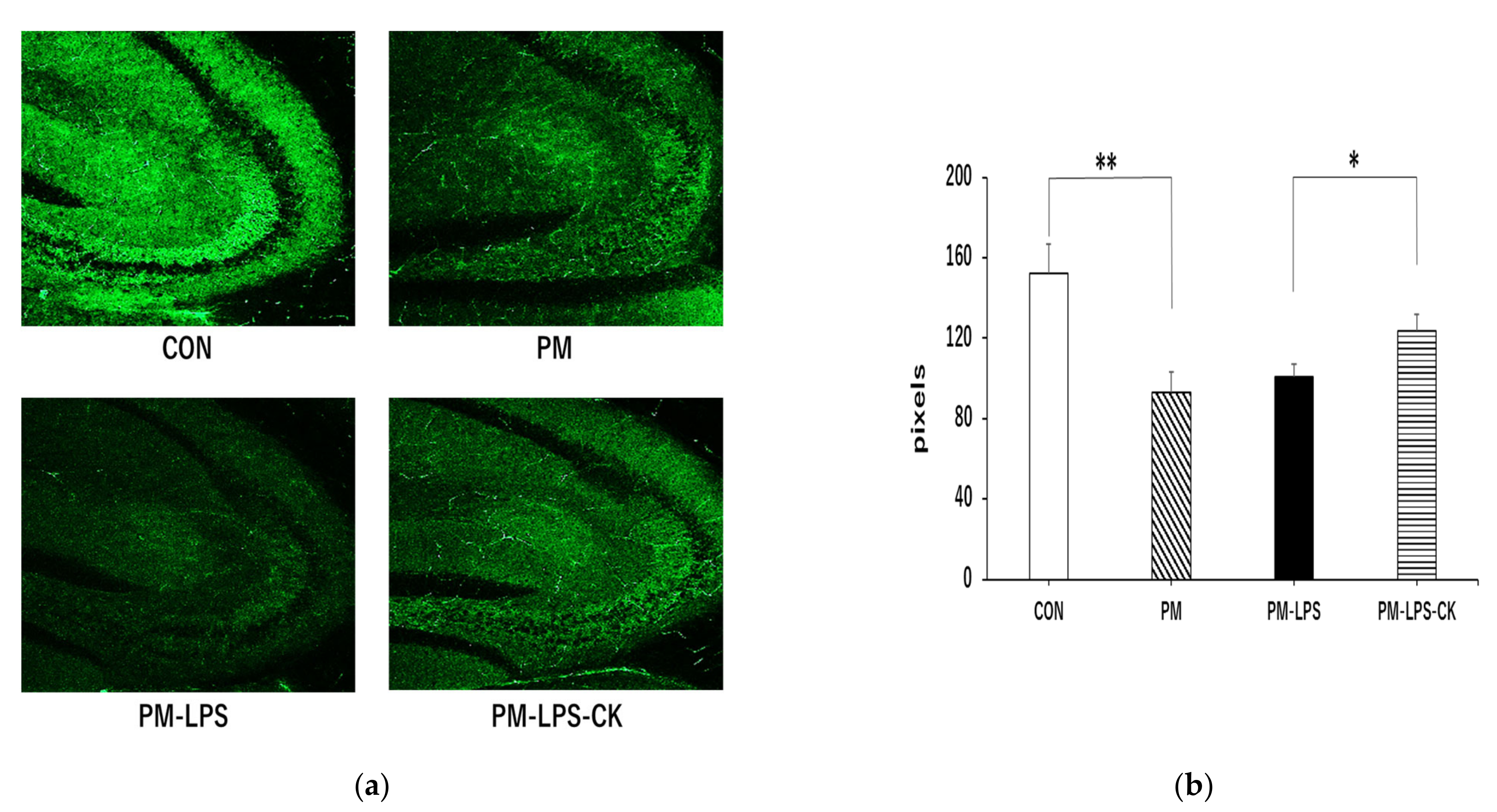
3.6. Protective Effects of C. kawachiensis on Neuronal Cell Function in the Hippocampus

Synaptophysin as an indicator of synaptic vesicle membrane proteins in neurons was examined and quantified in the stratum lucidum of the hippocampal CA3 region. As shown in Figure 6, a significantly stronger synaptophysin signal was observed in the CON group than in the PM group (Figure 6a,b; *** p < 0.001). Although there was no significant difference between the PM and PM–LPS groups (Figure 6b), synaptophysin signaling was significantly stronger in the PM–LPS–CK group than in the PM–LPS group (Figure 6a,b; * p < 0.05).

4. Discussion

The peel of C. kawachiensis has been shown to exert protective effects on the brains of various pathological mouse models, including global cerebral ischemia, type 2 diabetes, and senescence-accelerated models, through anti-inflammatory, antioxidant, and neuroprotective activities [10,12,13,14]. HPLC and NMR analyses have revealed that the peel of C. kawachiensis contains larger amounts of bioactive components, such as HMF, AUR, and NGI, than other citrus varieties [11]. A previous study reported that the dried peel of C. kawachiensis contained 0.27 mg/g of HMF, 4.07 mg/g of AUR, and 44.02 mg/kg of NGI [10]. Furthermore, these components exert various effects. HMF exhibits anti-inflammatory activity, inhibits neuronal cell death, promotes BDNF production, stimulates neurogenesis, improves memory impairment, and exerts antidepressant effects [18,19,20,21,22,23,24]; AUR exhibits anti-inflammatory activity, inhibits neuronal cell death, promotes neurogenesis, and suppresses tau phosphorylation [25,26,27,28,29]. NGI also exhibits anti-inflammatory activity, inhibits neuronal cell death, promotes neurogenesis, suppresses tau phosphorylation, and exerts antioxidant effects [28,29,30].
Frailty is the result of the interplay of various factors, including chronic inflammation and oxidative stress associated with aging, which reduces physiological reserve capacity and increases vulnerability to stress [1,2,3]. Many patients with frailty have a poor nutritional status, particularly protein-energy malnutrition. These factors contribute not only to sarcopenia and locomotive syndrome, but also to diseases of the central nervous system, such as cognitive dysfunction and mental disorders. However, frailty is a reversible disease because improvements in any of these factors may lead to recovery; therefore, appropriate interventions are needed [1,2,3]. Protein malnutrition alters various parameters of oxidative stress [15,16]; furthermore, inflammation caused by LPS administration aggravates the pathology and produces a frailty model. This may replicate the chronic disease conditions in the body of the elderly.
In the present study, food intake was slightly higher in the group fed dried C. kawachiensis peel than in the PM group (Figure 1d). These results are consistent with the effects of “Chinpi”, the dried peel of C. unshiu, an herbal medicine with stomachic effects. Chinpi is a component of the Kampo medicines Rikkunshito and Ninjin’yoeito, both of which enhance digestive function and are effective against anorexia. Previous studies suggested that the flavonoids present in citrus peels increase the level of ghrelin, an appetite-stimulating hormone [31,32,33]. Furthermore, the peel of C. unshiu contains HMF, which enhances appetite through 5-HT2 receptor antagonism, and HMF in dried C. kawachiensis peel powder has also been suggested to increase food intake [34]. Various mechanisms for age-related sarcopenia have been reported, including anorexia, a decrease in anabolic hormones such as growth hormone and insulin-like growth factor, and damage caused by inflammatory cytokines [35]. An increased food intake may ameliorate the losses in liver and muscle weights caused by a PM diet, and the HMF-induced increase in ghrelin levels also stimulates growth hormone secretion via the growth hormone secretagogue receptor [32,33,36]. Ghrelin has also been reported to exert anabolic effects on protein and suppress inflammatory cytokines [32,35,37], and these combined effects may have prevented muscle and liver weight losses in the PM–LPS–CK group (Figure 3a,b). Blood glucose levels are maintained by glycogenolysis and glycogenesis in the liver even during fasting. In the present study, endogenous glucose production may have been reduced in the PM group due to a decrease in glycogen storage, a smaller liver volume, and a lack of the amino acids needed for glycogenesis [38,39]. In contrast, the repair of peripheral tissue function by C. kawachiensis as described above may have increased blood glucose levels (Figure 3c).
In the behavioral analysis, inflammation caused by LPS decreased activity 2 days after its administration, which may have been due to sickness behavior during the acute phase of inflammation (Figure 4a,c), consistent with previous findings [40]. The decrease observed in behavior in the PM–LPS group 8 days after the administration of LPS was attributed to the previously reported spread of inflammation from the periphery to the brain, which led to brain dysfunction and depression-like symptoms [40,41]. On the other hand, muscle and brain improvements associated with anti-inflammatory, antioxidant, neuroprotective, and anti-depressive effects in peripheral tissues and the central nervous system in the PM–LPS–CK group may have contributed to the recovery of behavioral activity (Figure 4b,d, Figure 5 and Figure 6) [12,14,19]. In the brain tissue analysis, the quantification of microglia in the acute inflammatory phase showed no significant inflammation in the PM group; however, inflammation was confirmed after the administration of LPS. This result also demonstrated that peripheral inflammation propagates to the central nervous system [40,41]; however, the administration of the dried peel of C. kawachiensis significantly suppressed microglial activation in the brain, which is consistent with previous findings on the effects of the peel in the brain (Figure 5a,b) [12,14]. In addition, the quantification of synaptic vesicle membrane proteins, a marker of neuronal cell function, showed low immunoreactivity signals in the PM group. In spite of our expectations, no further deterioration was observed with the administration of LPS. These results suggest that malnutrition due to protein deficiency alone may induce significant damage to the central nervous system, which is regarded as a major factor accelerating frailty [1,2,3,42]. However, the dried peel of C. kawachiensis significantly inhibited neuronal cell dysfunction induced by the PM diet and administration of LPS (Figure 6a,b), suggesting that it maintained brain function.
Nobiletin, which is structurally similar to HMF and a polymethoxyflavone found in C. unshiu and C. depressa, was recently shown to ameliorate memory impairment by reducing Aβ in the brain of animal models of Alzheimer’s disease and has anti-inflammatory and antioxidant effects [43,44]. Furthermore, AUR was found to exert antidepressant effects by lowering malondialdehyde (MDA) and nitrite concentrations in the brain, thereby enhancing antioxidant capacity [27]. Furthermore, NGI suppressed cognitive impairment and oxidative stress by reducing MDA and glutathione levels in the brain [30]. It has been also reported that many polyphenols as ingredients of citrus and other plants, including naringin, could increase PPAR-gamma, which works as an antioxidant [28,45]. The anti-inflammatory, antioxidant, and neuroprotective effects of dried C. kawachiensis peel powder, which is rich in these bioactive components, have been confirmed and other effects are expected [12,13,14].

5. Conclusions

Dried C. kawachiensis peel powder has the potential to attenuate peripheral tissue and central nervous system dysfunctions under frailty-like conditions caused by protein-deficient malnutrition. Furthermore, it may be an effective food material for physical and psychosomatic frailty. We have confirmed the effect of the dried peel of C. kawachiensis in various disease model mice, and we were able to confirm the effect of the dried peel of C. kawachiensis in this frailty-like model as well.

Author Contributions

T.O. and S.O. designed and performed the research and wrote the manuscript; S.O. acquired the funding; A.S. performed the research; M.N. and Y.F. provided research advice. All authors have read and agreed to the published version of the manuscript.

Funding

Some of this work was supported by The Research Promotion Fund of the Ehime Industrial Promotion Foundation (#167; 17 July 2019).

Institutional Review Board Statement

All animal experiments were performed in accordance with the Guidelines for Animal Experimentation, approved by the Animal Care and Use Committee, and implemented using the approved protocol (#18-008).

Data Availability Statement

Data is contained within the article.

Acknowledgments

The peel of C. kawachiensis was supplied by Ehime Beverage Inc. (Matsuyama, Japan).

Conflicts of Interest

S.O., M.N. and Y.F. are the inventor of patent JP6994191B2.

References

  1. Sacha, J.; Sacha, M.; Sobon, J.; Borysiuk, Z.; Feusette, P. Is It Time to Being a Public Campaign Concerning Frailty and Pre-Frailty? A Review Article. Front. Physiol. 2017, 8, 484. [Google Scholar] [CrossRef]
  2. Shinkai, S.; Yoshida, H.; Taniguchi, Y.; Murayama, H.; Nishi, M.; Amano, H.; Nofuji, Y.; Seino, S.; Fujiwara, Y. Public health approach to preventing frailty in the community and its effect on healthy aging in Japan. Geriatr. Gerontol. Int. 2016, 16, 87–97. [Google Scholar] [CrossRef] [Green Version]
  3. Panza, F.; Lozupone, M.; Solfrizzi, V.; Sardone, R.; Dibello, V.; Lena, L.D.; D’Urso, F.; Stallone, R.; Petruzzi, M.; Giannelli, G.; et al. Different Cognitive Frailty Models and Health-and Cognitive-related Outcomes in Older Age: From Epidemiology to Prevention. J. Alzheimer’s Dis. 2018, 62, 993–1012. [Google Scholar] [CrossRef] [PubMed] [Green Version]
  4. Smallwood, M.J.; Nissim, A.; Knight, A.R.; Whiteman, M.; Haigh, R.; Winyard, P.G. Oxidative stress in autoimmune rheumatic disease. Free Radic. Biol. Med. 2018, 125, 3–14. [Google Scholar] [CrossRef] [PubMed]
  5. Reuter, S.; Gupta, S.C.; Chaturvedi, M.M.; Aggarwal, B.B. Oxidative stress, inflammation, and cancer: How are they linked? Free Radic. Biol. Med. 2010, 49, 1603–1616. [Google Scholar] [CrossRef] [PubMed] [Green Version]
  6. Moylan, J.S.; Reid, M.B. Oxidative stress, chronic disease, and muscle wasting. Muscle Nerve 2007, 35, 411–429. [Google Scholar] [CrossRef]
  7. Fischer, R.; Maier, O. Interrelation of Oxidative Stress and Inflammation in Neurodegenerative Disease: Role of TNF. Oxid. Med. Cell. Longev. 2015, 18, 610813. [Google Scholar] [CrossRef] [Green Version]
  8. Melrose, J. The Potential of Flavonoids and Flavonoid Metabolites in the Treatment of Neurodegenerative Pathology in Disorders of Cognitive Decline. Antioxidants 2023, 12, 663. [Google Scholar] [CrossRef]
  9. Furukawa, Y.; Okuyama, S.; Amakura, Y.; Watanabe, S.; Fukata, T.; Nakajima, M.; Yoshimura, M.; Yoshida, T. Isolation and Characterization of Activators of ERK/MAPK from Citrus Plants. Int. J. Mol. Sci. 2012, 13, 1832–1845. [Google Scholar] [CrossRef] [Green Version]
  10. Okuyama, S.; Yamamoto, K.; Mori, H.; Toyoda, N.; Yoshimura, M.; Amakura, Y.; Yoshida, T.; Sugawara, K.; Sudo, M.; Nakajima, M.; et al. Auraptene in the Peels of Citrus kawachiensis (Kawachi bankan) Ameliorates Lipopolysaccharide-Induced Inflammation in the Mouse Brain. Evid. Based Complement. Altern. Med. 2014, 2014, 408503. [Google Scholar] [CrossRef] [Green Version]
  11. Amakura, Y.; Yoshimura, M.; Ouchi, K.; Okuyama, S.; Furukawa, Y.; Yoshida, T. Characterization of Constituents in the Peel of Citrus kawachiensis (Kawachi bankan). Biosci. Biotechnol. Biochem. 2013, 77, 1977–1980. [Google Scholar] [CrossRef] [PubMed]
  12. Okuyama, S.; Yamamoto, K.; Mori, H.; Sawamoto, A.; Amakura, Y.; Yoshimura, Y.; Tamanaha, A.; Ohkubo, Y.; Sugawara, K.; Sudo, M.; et al. Neuroprotective effect of Citrus kawachiensis (Kawachi Bankan) peels, a rich source of naringin, against global cerebral ischemia/reperfusion injury in mice. Biosci. Biotechnol. Biochem. 2018, 82, 1216–1224. [Google Scholar] [CrossRef] [PubMed]
  13. Okuyama, S.; Shinoka, W.; Nakamura, K.; Kotani, M.; Sawamoto, A.; Sugawara, K.; Sudo, M.; Nakajima, M.; Furukawa, Y. Suppressive effects of the peel of Citrus kawachiensis (Kawachi bankan) on astroglial activation, tau phosphorylation, and inhibition of neurogenesis in the hippocampus of type 2 diabetic db/db mice. Biosci. Biotechnol. Biochem. 2018, 82, 1384–1395. [Google Scholar] [CrossRef]
  14. Okuyama, S.; Kotani, Y.; Yamamoto, K.; Sawamoto, A.; Sugawara, K.; Sudo, M.; Ohkubo, Y.; Tamanaha, A.; Nakajima, M.; Furukawa, Y. The peel of Citrus kawachiensis (Kawachi Bankan) ameliorates microglial activation, tau hyper-phosphorylation, and suppression of neurogenesis in the hippocampus of senescence-accelerated mice. Biosci. Biotechnol. Biochem. 2018, 82, 869–878. [Google Scholar] [CrossRef]
  15. Boban, P.J.; Corbett, D.; Saucier, D.M.; Noyan-Ashraf, M.H.; Juurlink, B.H.J.; Paterson, P.G. Protein-energy malnutrition impairs functional outcome in global ischemia. Exp. Neurol. 2005, 196, 308–315. [Google Scholar] [CrossRef] [PubMed]
  16. Feoli, A.M.; Siqueira, I.R.; Almeida, L.; Tramontina, A.C.; Vanzella, C.; Sbaraini, S.; Schweigert, I.D.; Netto, C.A.; Perry, M.L.S.; Gonḉalves, C.A. Effects of protein malnutrition on oxidative status in rat brain. Nutrition 2006, 22, 160–165. [Google Scholar] [CrossRef] [PubMed]
  17. Noworyta-Sokołowska, K.; Gόrska, A.; Gołembiowska, K. LPS-induced oxidative stress and inflammatory reaction in the rat striatum. Pharmacol. Rep. 2013, 65, 863–869. [Google Scholar] [CrossRef]
  18. Okuyama, S.; Shimada, N.; Kaji, M.; Morita, M.; Miyoshi, K.; Minami, S.; Amakura, Y.; Yoshimura, M.; Yoshida, T.; Watanabe, S.; et al. Heptamethoxyflavone, a citrus flavonoid, enhances brain-derived neurotrophic factor production and neurogenesis in the hippocampus following cerebral global ischemia in mice. Neurosci. Lett. 2012, 528, 190–195. [Google Scholar] [CrossRef]
  19. Sawamoto, A.; Okuyama, S.; Yamamoto, K.; Amakura, Y.; Yoshimura, M.; Nakajima, M.; Furukawa, Y. 3,5,6,7,8,3′,4′-Heptamethoxyflavone, a Citrus Flavonoid, Ameliorates Corticosterone-Induced Depression-like Behavior and Restores Brain-Derived Neurotrophic Factor Expression, Neurogenesis, and Neuroplasticity in the Hippocampus. Molecules 2016, 21, 541. [Google Scholar] [CrossRef] [Green Version]
  20. Okuyama, S.; Morita, M.; Miyoshi, K.; Nishigawa, Y.; Kaji, M.; Sawamoto, A.; Terugo, T.; Toyoda, N.; Makihata, N.; Amakura, Y.; et al. 3,5,6,7,8,3′,4′-Heptamethoxyflavone, a citrus flavonoid, on protection against memory impairment and neuronal cell death in a global cerebral ischemia mouse model. Neurochem. Int. 2014, 70, 30–38. [Google Scholar] [CrossRef] [Green Version]
  21. Sawamoto, A.; Okuyama, S.; Amakura, Y.; Yoshimura, M.; Yamada, T.; Yokogoshi, H.; Nakajima, M.; Furukawa, Y. 3,5,6,7,8,3′,4′-Heptamethoxyflavone Ameliorates Depression-Like Behavior and Hippocampal Neurochemical Changes in Chronic Unpredictable Mild Stressed Mice by Regulating the Brain-Derived Neurotrophic Factor: Requirement for ERK Activation. Int. J. Mol. Sci. 2017, 18, 2133. [Google Scholar] [CrossRef] [Green Version]
  22. Okuyama, S.; Miyoshi, K.; Tsumura, Y.; Amakura, Y.; Yoshimura, M.; Yoshida, T.; Nakajima, M.; Furukawa, Y. 3,5,6,7,8,3′,4′-Heptamethoxyflavone, a Citrus Polymethoxylated Flavone, Attenuates Inflammtion in the Mouse Hippocampus. Brain Sci. 2015, 5, 118–129. [Google Scholar] [CrossRef] [Green Version]
  23. Ihara, H.; Yamamoto, H.; Ida, T.; Tsutsuki, H.; Sakamoto, T.; Fujita, T.; Okada, T.; Kozaki, S. Inhibition of Nitric Oxide Production and Inducible Nitric Oxide Synthase Expression by a Polymethoxyflavone from Young Fruits of Citrus unshiu in Rat Primary Astrocytes. Biosci. Biotechnol. Biochem. 2012, 76, 1843–1848. [Google Scholar] [CrossRef] [PubMed] [Green Version]
  24. Sawamoto, A.; Okuyama, S.; Nakajima, M.; Furukawa, Y. Citrus flavonoid 3,5,6,7,8,3′,4′-heptamethoxyflavone induces BDNF via cAMP/ERK/CREB signaling and reduces phosphodiesterase activity in C6 cells. Pharmacol. Rep. 2019, 71, 653–658. [Google Scholar] [CrossRef]
  25. Okuyama, S.; Semba, T.; Toyoda, N.; Epifano, F.; Genovese, S.; Fiorito, S.; Taddeo, V.A.; Sawamoto, A.; Nakajima, M.; Furukawa, Y. Auraptene and Other Prenyloxyphenylpropanoids Suppress Microglial Activation and Dopaminergic Neuronal Cell Death in a Lipopolysaccharide-Induced Model of Parkinson’s Disease. Int. J. Mol. Sci. 2016, 17, 1716. [Google Scholar] [CrossRef] [PubMed] [Green Version]
  26. Lee, M.J.; Jang, Y.; Zhu, J.; Namgung, E.; Go, D.; Seo, C.; Ju, X.; Cui, J.; Lee, Y.L.; Kang, H.; et al. Auraptene Enhances Junction Assembly in Cerebrovascular Endothelial Cells by Promoting Resilience to Mitochondrial Stress through Activation of Antioxidant Enzymes and mtUPR. Antioxidants 2021, 10, 475. [Google Scholar] [CrossRef] [PubMed]
  27. Amini-Khoei, H.; Boroujeni, S.N.; Maghsoudi, F.; Rahimi-Madiseh, M.; Bijad, E.; Moradi, M.; Lorigooini, Z. Possible involvement of L-arginine-nitric oxide pathway in the antidepressant activity of Auraptene in mice. Behav. Brain Funct. 2022, 18, 4. [Google Scholar] [CrossRef]
  28. Okuyama, S.; Nakashima, T.; Nakamura, K.; Shinoka, W.; Kotani, M.; Sawamoto, A.; Nakajima, M.; Furukawa, Y. Inhibitory Effects of Auraptene and Naringin on Astroglial Activation, Tau Hyperphosphorylation, and Suppression of Neurogenesis in the Hippocampus of Streptozotocin-Induced Hyperglycemic Mice. Antioxidants 2018, 7, 109. [Google Scholar] [CrossRef] [PubMed] [Green Version]
  29. Okuyama, S.; Katoh, M.; Kanzaki, T.; Kotani, Y.; Amakura, Y.; Yoshimura, M.; Fukuda, N.; Tamai, T.; Sawamoto, A.; Nakajima, M.; et al. Auraptene/Naringin-Rich Fruit Juice of Citrus kawachiensis (Kawachi Bankan) Prevents Ischemia-Induced Neuronal Cell Death in Mouse Brain through Anti-Inflammatory Responces. J. Nutr. Sci. Vitaminol. 2019, 65, 66–71. [Google Scholar] [CrossRef] [Green Version]
  30. Golechha, M.; Chaudhry, U.; Bhatia, J.; Saluja, D.; Arya, D.S. Naringin Protects against Kainic Acid-Induced Status Epilepticus in Rats: Evidence for an Antioxidant, Anti-inflammatory and Neuroprotective Intervention. Biol. Pharm. Bull. 2011, 34, 360–365. [Google Scholar] [CrossRef] [Green Version]
  31. Yamada, C.; Saegusa, Y.; Nakagawa, K.; Ohnishi, S.; Muto, S.; Nahata, M.; Sadakane, C.; Hattori, T.; Sakamoto, N.; Takeda, H. Rikkunshito, a Japanese Kampo Medicine, Ameliorates Decreased Feeding Behavior via Ghrelin and Serotonin 2B Receptor Signaling in a Novelty Stress Murine Model. BioMed Res. Int. 2013, 2013, 792940. [Google Scholar] [CrossRef] [PubMed]
  32. Fujitsuka, N.; Uezono, Y. Rikkunshito, a ghrelin potentiator, ameliorates anorexia-cachexia syndrome. Front. Pharmacol. 2014, 5, 271. [Google Scholar] [CrossRef] [PubMed] [Green Version]
  33. Uto Sameshima, N.; Amitani, H.; Atobe, Y.; Sameshima, Y.; Sakaki, M.; Rokot, N.; Ataka, K.; Amitani, M.; Inui, A. Herbal Medicine Ninjin’yoeito in the Treatment of Sarcopenia and Frailty. Front. Nutr. 2018, 5, 126. [Google Scholar]
  34. Takeda, H.; Sadakane, C.; Hattori, T.; Katsurada, T.; Ohkawara, T.; Nagai, K.; Asaka, M. Rikkunshito, an Herbal Medicine, Suppresses Cisplatin-Induced Anorexia in Rats Via 5-HT2 Receptor Antagonism. Gastroenterology 2008, 134, 2004–2013. [Google Scholar] [CrossRef]
  35. Guillory, B.; Splenser, A.; Garcia, J. The Role of Ghrelin in Anorexia-Cachexia Syndromes. Vitam. Horm. 2013, 92, 61–106. [Google Scholar]
  36. Amitani, M.; Amitani, H.; Cheng, K.; Kairupan, T.S.; Sameshima, N.; Shimoshikiryo, I.; Mizuma, K.; Rokot, N.T.; Nerome, Y.; Owaki, T.; et al. The Role of Ghrelin and Ghrelin Signaling in Aging. Int. J. Mol. Sci. 2017, 18, 1511. [Google Scholar] [CrossRef]
  37. Amitani, M.; Asakawa, A.; Amitani, H.; Inui, A. Control of food intake and muscle wasting in cachexia. Int. J. Biochem. Cell Biol. 2013, 45, 2179–2185. [Google Scholar] [CrossRef] [PubMed]
  38. Bandsma, R.H.J.; Mendel, M.; Spoelstra, M.N.; Reijngoud, D.; Boer, T.; Stellaard, F.; Brabin, B.; Schellekens, R.; Senga, E.; Heikens, G.T. Mechanisms Behind Decreased Endogenous Glucose Production in Malnourished Children. Pediatr. Res. 2010, 68, 423–428. [Google Scholar] [CrossRef] [Green Version]
  39. Heard, C.R.C.; Frangi, S.M.; Wright, P.M.; McCartney, P.R. Biochemical characteristic of different forms of protein-energy malnutrition: An experimental model using young rats. Br. J. Nutr. 1977, 37, 1–21. [Google Scholar] [CrossRef] [Green Version]
  40. Biesmans, S.; Meert, T.F.; Bouwknecht, J.A.; Acton, P.D.; Davoodi, N.; Haes, P.D.; Kuijlaars, J.; Langlois, X.; Matthews, L.J.R.; Donck, L.V.; et al. Systemic Immune Activation Leads to Neuroinflammation and Sickness Behavior in Mice. Mediat. Inflamm. 2013, 2013, 271359. [Google Scholar] [CrossRef]
  41. Zhao, X.; Cao, F.; Liu, Q.; Li, X.; Xu, G.; Liu, G.; Zhang, Y.; Yang, X.; Yi, S.; Xu, F.; et al. Behavioral, inflammatory and neurochemical disturbances in LPS and UCMS-induced mouse models of depression. Behav. Brain Res. 2019, 364, 494–502. [Google Scholar] [CrossRef] [PubMed]
  42. Martone, A.M.; Onder, G.; Vetrano, D.L.; Ortolani, E.; Tosato, M.; Marzetti, E.; Landi, F. Anorexia of Aging: A Modifiable Risk Factor for Frailty. Nutrients 2013, 5, 4126–4133. [Google Scholar] [CrossRef] [PubMed] [Green Version]
  43. Onozuka, H.; Nakajima, A.; Matsuzaki, K.; Shin, R.; Ogino, K.; Saigusa, D.; Tetsu, N.; Yokosuka, A.; Sashida, Y.; Mimaki, Y.; et al. Nobiletin, a Citrus Flavonoid, Improves Memory Impairment and Aβ Pathology in a Transgenic Mouse Model of Alzheimer’s Disease. J. Pharmacol. Exp. Ther. 2008, 326, 739–744. [Google Scholar] [CrossRef]
  44. Nakajima, A.; Ohizumi, Y.; Yamada, K. Anti-dementia Activity of Nobiletin, a Citrus Flavonoid: A Review of Animal Studies. Clin. Psychopharmacol. Neurosci. 2014, 12, 75–82. [Google Scholar] [CrossRef] [Green Version]
  45. Choi, M.; Lee, E.; Park, J.; Kim, S.; Park, E.; Kim, H. Anti-inflammatory mechanism of galangin in lipopolysaccharide-stimulated microglia: Critical role of PPAR-γ signaling pathway. Biochem. Pharmacol. 2017, 144, 120–131. [Google Scholar] [CrossRef] [PubMed]
Figure 1. Body weight changes based on different experimental diets and average food intake per mouse before the administration of LPS. (a) Body weight changes during the experimental diet feeding period. The number of mice in each group is as follows: CON: n = 32, PM: n = 32, PM–CK: n = 32. Food intake in the (b) second, (c) third, and (d) fourth week after starting the experimental diet. Values are means ± SEM. Symbols show significant differences for the following conditions: CON vs. PM (*** p < 0.001). The number of samples in each group is as follows: CON: n = 4, PM: n = 4, PM–CK: n = 4.
Antioxidants 12 00779 g001
Figure 2. Body weight changes after the administration of LPS. The ratio of body weight changes (a) 2 days and (b) 8 days after the administration of LPS. Values are means ± SEM. Symbols show significant differences for the following conditions: CON vs. PM (** p < 0.01, *** p < 0.001). PM vs. PM–LPS (*** p < 0.001), and PM–LPS vs. PM–LPS–CK (** p < 0.01). The number of mice in each group is as follows: (a) CON: n = 8, PM: n = 8, PM–LPS: n = 7, PM–LPS–CK: n = 8, and (b) CON: n = 8, PM: n = 8, PM–LPS: n = 7, PM–LPS–CK: n = 7.
Antioxidants 12 00779 g002
Figure 3. Effects of dried C. kawachiensis peel powder on protein malnutrition or LPS-induced peripheral dysfunction. Comparison of collected (a) gastrocnemius weight, (b) liver weight, and (c) blood glucose concentration in the 8-day group. Values are means ± SEM. Symbols show significant differences for the following conditions: CON vs. PM (*** p < 0.001, ** p < 0.01), PM vs. PM–LPS (*** p < 0.001), and PM–LPS vs. PM–LPS–CK (** p < 0.01, *** p < 0.001). The number of mice in each group is as follows: CON: n = 8, PM: n = 8, PM–LPS: n = 7, PM–LPS–CK: n = 7.
Antioxidants 12 00779 g003
Figure 4. Effects of dried C. kawachiensis peel powder in the behavioral test. Measurement of total distance traveled in the (a) 2-day or (b) 8-day group in the Y-maze device. Measurement of immobility time in the (c) 2-day or (d) 8-day group using the ANY-maze Video Tracking System. Values are means ± SEM. Symbols show significant differences for the following conditions: PM vs. PM–LPS (* p < 0.05, *** p < 0.001), and PM–LPS vs. PM–LPS–CK (* p < 0.05). The number of mice in each group is as follows: (a,c) CON: n = 8, PM: n = 8, PM–LPS: n = 7, PM–LPS–CK: n = 8, and (b,d) CON: n = 8, PM: n = 8, PM–LPS: n = 7, PM–LPS–CK: n = 7.
Antioxidants 12 00779 g004aAntioxidants 12 00779 g004b
Figure 5. Effects of dried C. kawachiensis peel powder on microglial activity in the hippocampus of the 2-day group. (a) Representative microglia pictures stained with anti-Iba1 antibody. (b) Quantitative analysis of the area of Iba1-positive signals using ImageJ software. Values are means ± SEM. Symbols show significant differences for the following conditions: PM vs. PM–LPS (** p < 0.01), and PM–LPS vs. PM–LPS–CK (* p < 0.05). The number of samples in each group is as follows: CON: n = 23, PM: n = 23, PM–LPS: n = 23, PM–LPS–CK: n = 23.
Antioxidants 12 00779 g005
Figure 6. Effects of dried C. kawachiensis peel powder on synaptophysin immunoreactivity in the hippocampus of the 8-day group. (a) Representative synaptophysin antibody pictures stained with anti-synaptophysin antibody. (b) Quantitative analysis of the area of synaptophysin-positive signals using ImageJ software. Values are means ± SEM. Symbols show significant differences for the following conditions: CON vs. PM (** p < 0.01), and PM–LPS vs. PM–LPS–CK (* p < 0.05). The number of samples in each group is as follows: CON: n = 12, PM: n = 12, PM–LPS: n = 11, PM–LPS–CK: n = 10.
Antioxidants 12 00779 g006
Disclaimer/Publisher’s Note: The statements, opinions and data contained in all publications are solely those of the individual author(s) and contributor(s) and not of MDPI and/or the editor(s). MDPI and/or the editor(s) disclaim responsibility for any injury to people or property resulting from any ideas, methods, instructions or products referred to in the content.

