Review

Advances of H2S in Regulating Neurodegenerative Diseases by Preserving Mitochondria Function

Departments of Anesthesiology, Center for Brain Science, The First Affiliated Hospital of Xi’an Jiaotong University, Xi’an 710061, China
*
Author to whom correspondence should be addressed.
Antioxidants 2023, 12(3), 652; https://doi.org/10.3390/antiox12030652
Submission received: 2 December 2022 / Revised: 22 February 2023 / Accepted: 3 March 2023 / Published: 6 March 2023
(This article belongs to the Special Issue Oxidative Stress-Induced Neurotoxicity and Mitochondrial Dysfunction)

Abstract

:
Neurotoxicity is induced by different toxic substances, including environmental chemicals, drugs, and pathogenic toxins, resulting in oxidative damage and neurodegeneration in mammals. The nervous system is extremely vulnerable to oxidative stress because of its high oxygen demand. Mitochondria are the main source of ATP production in the brain neuron, and oxidative stress-caused mitochondrial dysfunction is implicated in neurodegenerative diseases. H2S was initially identified as a toxic gas; however, more recently, it has been recognized as a neuromodulator as well as a neuroprotectant. Specifically, it modulates mitochondrial activity, and H2S oxidation in mitochondria produces various reactive sulfur species, thus modifying proteins through sulfhydration. This review focused on highlighting the neuron modulation role of H2S in regulating neurodegenerative diseases through anti-oxidative, anti-inflammatory, anti-apoptotic and S-sulfhydration, and emphasized the importance of H2S as a therapeutic molecule for neurological diseases.

1. Introduction

Neurodegenerative diseases in both humans and animals are usually complicated. They can be induced by pharmaceuticals and various environmental chemicals, including antiviral drug tilorone, typhaneoside, metronidazole, homocysteine, paraquat and some heavy metals [1]. In the brain, mitochondria with striking structure are mainly responsible for cellular energy production, which occurs through oxidative phosphorylation [2]. They are the primary target for toxin-induced bioenergetic failure, subsequently resulting in oxidative stress and mitochondrial dysfunction [3]. Accordingly, oxidative damage has long been recognized as the initial cause of neurodegenerative diseases, including Alzheimer’s disease (AD), Huntington’s disease (HD) and Parkinson’s disease (PD) [4,5]. In addition, mitochondrial dysfunction has been described as a pathological hallmark of neurotoxicity development and cognitive impairment [5,6]. Thus far, the mechanisms involved in modulating cellular redox homeostasis and protecting mitochondrial function during neurotoxicity, and efficient mechanism-based therapies have not been fully elucidated.
In recent years, hydrogen sulfide (H2S) has been accepted as a novel gas transmitter, followed by nitric oxide (NO) and carbon monoxide (CO). The earliest discovery of H2S in 1989 reported the endogenous production of H2S and its bioactive properties in mitochondria fraction of rats’ brain [7], implying that H2S may exert physiological functions in the mammalian central nervous system (CNS). Numerous studies have reported that H2S can be endogenously produced by three main enzymes in mammalian cells, i.e., cystathionine β synthase (CBS), cystathionine γ lyase (CSE), and 3-mercaptopyruvate sulfur transferase (3-MST) [8]. Moreover, a newly reported enzyme, D-amino acid oxidase (DAO), has been confirmed to promote H2S production in mitochondria [9]. The molecular mechanism mediated by H2S is the S-sulfhydation, which modifies the thiol groups of specific cysteine residues of target proteins, resulting in alterations of protein structure, enzymatic activity, or translocation [10]. The reactive sulfur species required for posttranslational modification mediated by protein persulfidation are generated from mitochondria through H2S oxidation [11]. The cytoprotective functions of H2S reported in mammals mainly include antioxidant defense, autophagy induction, neurotransmission, vasorelaxation, lifespan extension, etc. [12,13,14,15]. In 1996, Kimura et al. reported that H2S functions as a neuromodulator to induce LTP (long-term potentiation) via activating NMDA receptors [16]. Furthermore, numerous studies on the pharmacological and physiological role of H2S characterized H2S as having anti-inflammatory and antioxidant properties in brain neuron systems [17,18]. It has been recently reported that H2S protects animals from homocysteine-induced neurodegeneration by attenuating oxidative DNA damage, mitochondrial disorders and mitochondria-mediated apoptosis [19]. Collectively, research of H2S-mitochondrial in the brain advanced our understanding of mechanisms of neuron diseases, and specific compounds releasing H2S into mitochondria could be developed as therapeutic molecules for different neurodegenerative diseases in the future.
This review mainly focused on H2S as a protective gaseous signaling molecule in the development of neurodegenerative diseases. First, we provided an overview of neurodegenerative diseases associated with mitochondria dysfunction and oxidative stress. Furthermore, we highlighted the biological effect and regulatory mechanisms of H2S on different neurodegenerative diseases. Finally, we provide perspectives on potential therapeutic treatment using H2S-releasing drugs in modulating neurological diseases.

2. Induction of Neurotoxicity

Different toxic substances, including natural products, environmental chemical compounds, and pharmaceuticals, can affect the nervous system, inducing neurotoxicity in humans and laboratory animals. Micronutrient homocysteine (Hcy) is a sulfur-containing amino acid derived from methionine metabolism. The earliest study of Hcy-induced neurotoxicity was reported in patients with a deficiency of cystathionine beta-synthase (CBS) [20,21]. It was later found that the CBS enzyme was mainly expressed in the human brain [22]. It was also found to have the ability to convert Hcy into cysteine and glutathione; thus, Hcy is accumulated in the brain under a deficiency of CBS. This was further confirmed in CBS knockout mice (Cbs−/+ or Cbs−/−), in which the Hcy level increased approximately 2–50-fold compared with wild-type mice, leading to oxidative stress and neuronal death [23,24]. An accumulating body of literature has reported on neurotoxic mechanisms induced by Hcy. In rat ischemic brain cells, accumulation of Hcy influences mitochondrial ultrastructure, mitochondrial complex I-III enzymatic activities and phosphorylation of mitochondrial STAT3 (mitoStat3), resulting in mitochondrial injury and oxidative stress, ultimately leading to neurotoxicity [25]. Similarly, impairment of mitochondrial activity and oxidative damage were also considered as the prime mechanisms of neurodegeneration in the Hcy-induced Parkinson’s disease (PD) rat model [5]. These findings further indicated that mitochondrial dysfunction is the major cause of different neurodegenerative diseases. In addition, excess Hcy can accumulate cytokine levels in the brain and disturb the inflammatory system [26]. Furthermore, hyperhomocysteinemia (HHcy) induces excitotoxicity and cognitive impairment by disturbing redox potentials and activating N-methyl-D-aspartate (NMDA) receptor [27]. Meanwhile, HHcy was found to impair activity of cytochrome c oxidase (COX) in mitochondria and induce reactive oxygen species (ROS) accumulation, subsequently resulting in apoptotic cell death and neurological dysfunction in primary neurons of rats and humans [28]. Thereby, oxidative damage and mitochondrial dysfunction are considered the most important pathomechanisms in Hcy/HHcy-induced apoptosis and neurotoxicity.
Glutamate is a major excitatory neurotransmitter in the brain. Glutamate-induced excitotoxicity mainly includes calcium imbalance and mitochondrial swelling [29]. It was reported that typhaneoside, a flavonoid compound, suppresses glutamate release in rat cerebrocortical nerve terminals by inhibiting voltage-dependent calcium entry [30]. Glutamate accumulation can over stimulate NMDA receptors, leading to excitotoxicity and disruption of learning and memory [31]. Meanwhile, treatment of the herbicide paraquat promotes the accumulation of β-amyloid and tau protein [32], which have been recognized as pathophysiological events associated with the development of AD.
Organophosphorus pesticides (OPs), which are widely used for controlling pests worldwide, induce neurotoxicity by disturbing mitochondrial function and inducing oxidative stress [33]. Arsenic is a toxic metalloid widely distributed in groundwater. It has been found that arsenic exposure induces neurotoxicity and cognitive impairment by damaging mitochondrial biogenesis [34,35]. Aluminium is a toxic metal ubiquitously present on earth that impairs mitochondrial bioenergetics and leads to cognitive disorder once it enters the brain [36]. Moreover, it has been found that N-methyl-4-phenylpyridinium (MPP+) related compounds are dopaminergic toxins that can induce mitochondrial oxidative stress and dysfunction [37]. Thus, they are usually used to induce animal models of PD. Generally, these compounds disturb intracellular redox status and mitochondrial activity, ultimately leading to neuronal death and cognitive impairment. Hence, preserving cellular redox homeostasis and mitochondria function can be regarded as the major therapeutic target for different neurodegenerative diseases.

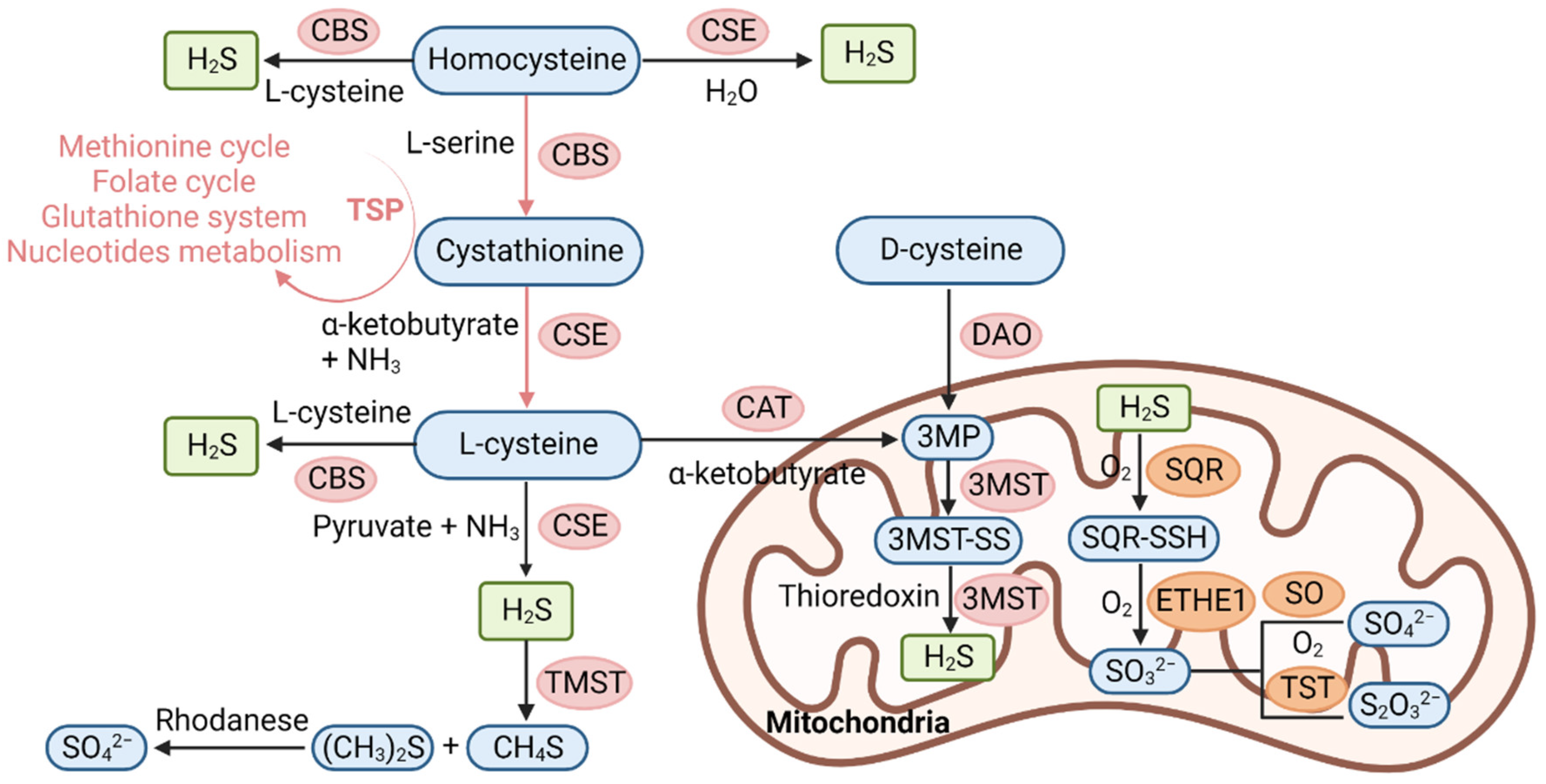
3. Biosynthesis and Metabolism of H2S

3.1. Physiological Function of H2S
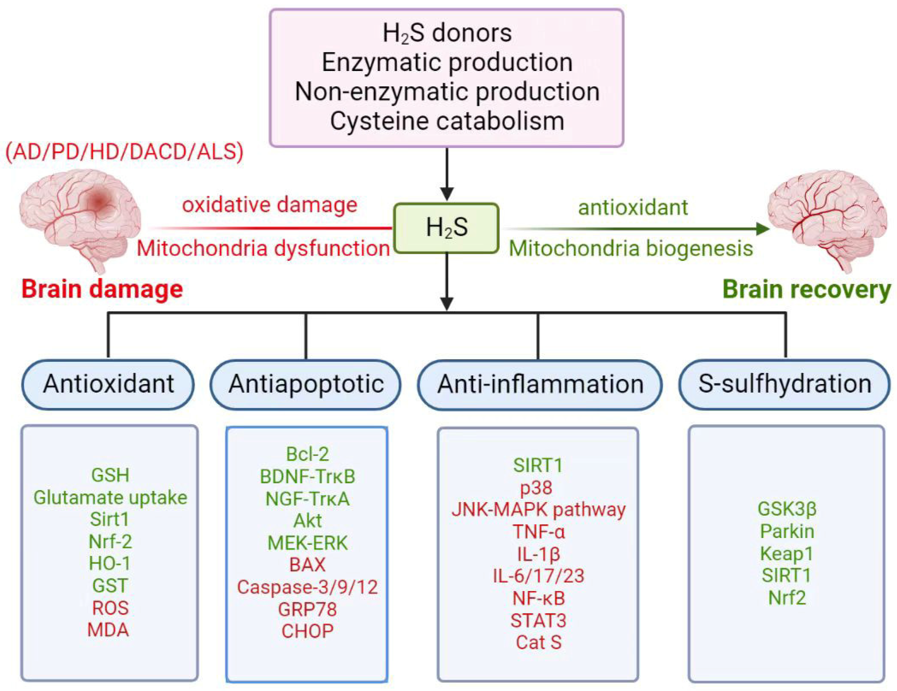
While H2S is colorless, it is characterized by an offensive smell of rotten eggs [38]. It can freely penetrate cell membranes due to the lipid solubility property [39]. At avery early stage, H2S was considered toxic due to the inhibition of mitochondria cytochrome c oxidase, which causes respiratory depression, pulmonary edema and central neuronal system damage [7,40,41,42]. However, in 1989 and 1990, high levels of sulfide in the brain of rats, humans, and bovines were detected, indicating they possibly have a neuromodulator role of H2S in neurons [7,42,43]. In 1996, it was found that the appropriate application of H2S donor sodium hydrosulfide (NaHS) facilitates hippocampal LTP by enhancing the activity of the NMDA receptor [16]. These findings preliminarily suggested that H2S acting as a novel signaling molecule may have a physiological role in modulating neuronal activity.
In mammalian cells, pyridoxal-5′-phosphate (PLP)-dependent enzymes cystathionine-β-synthase (CBS) and cystathionine-γ-lyase (γ-cystathionase CSE) have important roles in generating H2S from L-cysteine (Cys) (Figure 1) [16,38]. Recently, it was reported that 3-mercaptopyruvate sulfurtransferase (3-MST) could also produce H2S from L-cysteine in combination with cysteine aminotransferase (CAT) in brain nervous system (Figure 1) [44]. Moreover, a new pathway involved in H2S generation in mammals revealed that peroxisome-dependent DAO uses D-cysteine to produce 3MP, and subsequently producing H2S in mitochondria (Figure 1) [9]. Except for enzyme-dependent production of H2S, it was also found in 2009 that H2S can be stored as bound sulfane sulfur by adding sulfur to specific cysteine residues of target proteins, which may have an important role in the response to physiological stimuli via releasing H2S in the brain [45,46,47]. Thereby, H2S and polysulfides may function as signal molecules in the nervous system.
The above-mentioned enzymes responsible for H2S production are tissue-specific enzymes involving certain biochemical pathways in humans [48]. The expression of CBS, which is mainly responsible for H2S production in the brain, is higher in the hippocampus and cerebellum than the CSE. It is especially expressed in radial glia and astrocyte in the central neuron system of the brain [22,49]. This is further confirmed by the evidence that H2S production remains unchanged in the brain through using CSE inhibitors D, L-propargylglycine (PAG) and β-cyano-L-alanine (β-CNA), although they suppress H2S generation in the liver and kidney [16,50]. Moreover, it has been found that CBS knockout mice had abnormal development of cerebellar morphology [49]. Additionally, the 3-MST is also mainly localized in brain tissues, including hippocampal pyramidal neurons, mitral cells in the olfactory bulb and cerebellar Purkinje cells [44]. Because the H2S produced by 3-MST/CAT is mainly localized to mitochondria, it can attenuate mitochondria dysfunction and protect cells from oxidative stress by scavenging reactive oxygen species [6]. Moreover, D-cysteine-dependent H2S production preferentially operates in the cerebellum and the kidney via attenuating oxidative stress [9]. Thus, these discoveries suggest that H2S acts as an antioxidant by regulating cellular redox status in the brain.
It is well documented that H2S is toxic at high concentrations and beneficial at low concentrations. These investigations emphasize the importance of identifying a strict fine balance of physiological H2S for therapeutic use. The endogenous steady-state level of H2S is mainly dependent on its oxidation and metabolism in mitochondria. In the mitochondrial matrix, sulfane sulfur is generated from sulfide quinone oxidoreductase (SQR)-mediated H2S oxidation [51,52,53]. This reaction forms persulfide (R-SSH) and releases two electrons to the electron transport chain (ETC) via coenzyme Q (CoQ). After that, ethylmalonic encephalopathy 1 (ETHE1) oxidizes the persulfide to generate sulfite, which is further oxidized to SO42− by SO or to S2O32− by TST (Figure 1) [54]. In addition to the oxidation of H2S, H2S can also be catabolized through methylation via thiol S-methyltransferase (TSMT) and the H2S-scavenging pathway via metalloproteins [55,56]. More importantly, the H2S-mediated transsulfuration pathway can also integrate with other sulfide-linked metabolic pathways, including the folate cycle, glutathione system, and nucleotide metabolism [57]. Thus, the cellular H2S levels are tightly controlled via H2S biosynthesis and catabolism pathway in order to maintain its physiological protective effect and cellular metabolic profile.
H2S synthesis are mainly catalyzed by the enzymes CBS, CSE and 3-MST. DAO is a newly discovered enzyme that can produce H2S in mitochondria. H2S oxidation occurs in mitochondria, and H2S is oxidized to persulfide (SQR-SSH) by SQR via accepting a thiol (-SH), after which the persulfide is oxidized to SO32− by ETHE1. The SO32− is oxidized to SO42− and S2O32− by SO and TST, respectively. In addition, H2S is methylated by thiol S-methyltransferase (TMST) to form methanethiol and dimethyl sulfide. The dimethyl sulfide is oxidized to SO42− by rhodanese. Transsulfuration pathway (TSP), which involves the transfer of sulfur from homocysteine to cysteine, can integrate with sulfide-associated metabolic pathways, including methionine cycle, folate cycle, glutathione synthesis and nucleotides metabolism to maintain a physiological balance of H2S. H2S, hydrogen sulfide; CBS, cystathionine β-synthase; CSE, cystathionine γ-lyase; CAT, cysteine aminotransferase; DAO, D-amino acid oxidase; 3MST, 3-mercaptopyruvate sulfurtransferase; SQR, sulfide quinone oxidoreductase; ETHE1, ethylmalonic encephalopathy 1; TST, thiosulfate sulfurtransferase; SO32−, sulfite; SO42−, sulfate; S2O32−, thiosulfate; TSP, transsulfuration pathway. The pink arrows indicate the transsulfuration pathway (TSP) and related metabolic pathways. This model was created using BioRender (https://biorender.com/, accessed on 12 February 2023).

3.2. Donors of H2S

H2S donors NaHS and disodium sulfide (Na2S) can quickly release H2S and display cytoprotective effect by regulating mitochondrial biogenesis and function [58,59]. GYY4137 is believed to release H2S dependent on acidic pH and higher temperatures [60]. It can protect mitochondria and vascular endothelial cells from oxidative damage [61]. Furthermore, S-adenosylmethionine (SAM) is regarded as an activator of endogenous H2S generation, which could inhibit endothelial growth factor-A-related diseases [62]. Besides biochemical synthesized H2S donors, H2S can also be generated from natural plants. For example, diallyl disulfide (DADS) and diallyl trisulfide (DATS) derived from Allium plant garlic function as H2S donors, impairing the mitochondrial function [63] or decreasing the ROS production in mitochondria [64]. Concerning the contributions of H2S donors to clinical experiments, SG1002, a novel H2S prodrug, was clinically used in patients with heart failure. It was found that H2S is essential for vascular homeostasis by preserving mitochondrial functions and increasing myocardial vascular density [65,66]. MZe786, the ADTOH H2S donor, could improve preeclampsia state through lowering blood pressure and attenuating renal damage in mice model C57Bl/6 J of preeclampsia [67,68]. Meanwhile, the administration of H2S can protect mitochondrial activity against a high level of antiangiogenic-factor-sFlt-1-induced mitochondrial respiration inhibition and superoxide production in endothelial cells [69]. H2S producer sodium thiosulfate exhibits anti-inflammatory and antioxidative effects and has already been investigated in phase I trials for potential benefits in patients with an acute coronary syndrome undergoing coronary angiography [70].
Recently, it has been reported that both AP39 and AP123 are mitochondrial-targeted H2S donors whose application can regulate glucose-oxidase-induced oxidative stress and improve mitochondrial function in endothelial cells [71]. It has also been reported that both AP39 and AP123 can be targeted for mitochondrial decrease hyperpolarization of the mitochondrial membrane and increase the electron transport at respiratory complex III to improve cellular metabolism [72]. Thereby, mitochondria-targeted H2S donors can potentially be used in the treatment of neurological diseases in the near future.

4. Involvement of H2S in Neurological Diseases

The physiological levels of H2S in human brain tissue are nearly 50–160 μM, which suggests that H2S may exert a possible biological function in neurons [16]. Subsequently, numerous researchers have reported that the possible physiological functions of H2S in the brain mainly include potentiating long-term potential (LTP) through activation of the NMDA receptors [16], thus inhibiting mitochondrial dysfunction and oxidative damage by directly or indirectly scavenging free radicals and reactive species [73,74,75,76,77], activating Ca2+ influx waves in astrocytes [78], initiating anti-neuroinflammatory responses in both astrocyte and microglia [79,80,81], and modulating apoptotic response signaling in neurons [81,82]. These studies highlight the potential neuroprotective role of H2S in regulating neurodegenerative diseases through anti-oxidative, anti-inflammatory, or anti-apoptotic effects (Table 1).

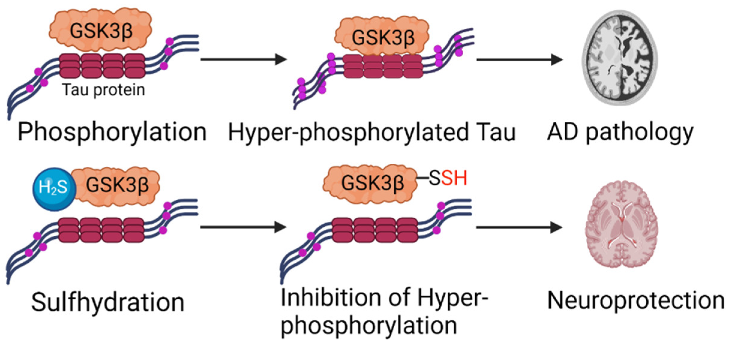
4.1. Alzheimer’s Disease (AD)

AD has been characterized by memory deterioration and cognition impairment, mainly caused by neuropathologic hallmark events, which include enlargement of β-amyloid plaque size and hyperphosphorylation of tau protein in the brain [83]. AD-related neurodegeneration is associated with oxidative stress [77]. It has been implied that soluble oligomers of Aβ can deregulate H2S signaling by inhibiting cysteine transporter excitatory amino acid transporter 3 (EAAT3/EAAC1), resulting in oxidative stress and neurodegeneration [84,85]. Thereby, H2S may have a vital role in AD. Indeed, it was found that the H2S levels in both plasma and the brain of AD patients were much lower than those in healthy individuals [86,87]. Furthermore, the plasma H2S level was negatively correlated with the severity of AD [87], because H2S is synthesized from homocysteine via CBS catalyzation in the transsulfuration pathway, and SAM is the activator of CBS [62]. In AD patients, the homocysteine level in the brain is high, and SAM content is low, whereas the expression level of CBS showed no difference between AD patients and the normal group [86]. Therefore, it has been concluded that the reduction in H2S might be associated with decreased activity of CBS [86,88], which further confirms the essential neuroprotective role of the CBS-H2S signaling axis in the AD brain. Thus, dietary adjustment has been proposed to improve symptoms of AD patients, as a diet rich in taurine, cysteine, folate, B12, and betaine could promote H2S synthesis in the brain [89].
Mitochondrial dysfunction is characterized as a pathogenesis of AD. Previous studies found that H2S protected PC12 cells against Hcy or formaldehyde (FA)-induced neurotoxicity by preserving mitochondrial membrane potential (MMP) and attenuating intracellular ROS accumulation [90,91]. AP39, a mitochondrially targeted H2S donor, has been found to exert a neuroprotective effect in a dose-dependent manner in APP/PS1 neurons by increasing energy production and cell viability, protecting mitochondrial DNA, and decreasing ROS accumulation at 100 nM concentrations, which reduces Aβ deposition in the brain, inhibits brain atrophy, and ameliorates memory deficiency in the Morris water maze experiment [92]. Meanwhile, the in vitro and in vivo experiments in mammals demonstrated that H2S-mediated sulfhydration of ATP synthase helps to maintain mitochondrial bioenergetics [93], implying the possible mechanism of H2S in modulating AD development. Thus, it is possible that the exogenous application of appropriate concentration of H2S preserves mitochondrial dysfunction in AD by improving mitochondrial redox homeostasis and maintaining energetic production.
Meanwhile, it was found that H2S treatment alleviated the excitotoxicity-triggered oxidative stress by decreasing levels of malondialdehyde in the cerebral cortex of the 3xTg-AD mice model [94,95]. Moreover, a novel neuroprotective mechanism of H2S has been explored, revealing that H2S exerts antioxidative effects by activating nuclear factor erythroid-2-related factor 2 (Nrf2), heme oxygenase-1 (HO-1) and glutathione S-transferase (GST) in APP/PS1 transgenic mice [96]. In addition, Hyperhomocysteinemia (HHcy) has been recognized as a potential risk factor for AD development. It was found that NaHS and MK801 treatments attenuate the high level of the homocysteine-induced blood–brain barrier (BBB) disruption, synaptic dysfunction and excitotoxicity in mice brains by modulating NMDA receptors [97]. It has also been reported that ACS6, an H2S donator, preserves mitochondrial function and prevents Hcy-induced apoptosis by inhibiting cytochrome C releasing, ROS accumulation, and caspase-3 activation in PC12 cells of the AD mouse model [98].
H2S exerts its anti-inflammatory effect via several signal pathways, including preserving mitochondrial function in a p38 and JNK-MAPK dependent manner in microglia [99], inhibiting activation of NF-κB pathway in the hippocampus [100], suppressing phosphorylation of signal transducer and activator of transcription 3 (STAT3) and cathepsin S (Cat S) activation [101] in mouse models of AD. In addition, H2S exerts protective effects through different neuroprotective pathways. For example, it was found that the application of NaHS and Tabiano’s spa water (rich in H2S) can slow down the progression of learning and memory impairment in three experimental models of AD, including brain injection of β-amyloid1-40 (Aβ) or streptozotocin-induced rat models, and in an AD mouse model harboring human transgenes APPSwe, PS1M146V and tauP301L (3xTg-AD mice), by lowering phosphorylation level of tau protein, preventing oxidative and nitrosative stresses in the cerebral cortex, upregulating Bcl-2 and downregulating BAX in the hippocampus [95]. Moreover, it has been confirmed that administration of an H2S donor (NaHS) into APP/PS1 transgenic mouse improves spatial memory acquisition through shifting from the plaque-forming β pathway to the non-plaque forming α pathway of APP cleavage, a non-amyloidogenic processing of APP [102]. It was also reported that H2S decreased Aβ deposition in mitochondria by inhibiting γ-secretase activity [103]. Overall, chronic treatment with H2S donor could clearly reduce the size of β-amyloid plaques, and reduce activities of c-jun N-terminal kinases (JNK), extracellular signal-regulated kinases and p38 involved in tau phosphorylation, inflammatory response and apoptosis in the cortex and hippocampus, thus finally protecting AD mice against cognitive impairment [104]. These discoveries show promising therapeutic prospects for H2S by targeting multiple pathophysiological events in AD.
Although H2S has been shown to exert a neuroprotective effect on account of its antioxidative, anti-inflammatory, and anti-apoptotic properties, and that administration of H2S donors is beneficial for AD, the exact molecular mechanisms underlying the neuroprotective effect are still largely unknown. Glycogen synthase kinase 3β (GSK3β) promotes hyperphosphorylation of tau protein, which finally leads to AD (Figure 2). Recently, it has been reported that H2S-mediated sulfhydration modifies GSK3β, eventually inhibiting tau’s hyperphosphorylation, which is a major contributor to AD development (Figure 2) [105]. Thereby, the application of H2S donor GYY4137 improves cognitive deficiency in the 3xTg-AD mouse model [105]. In summary, in comparison to a normal mouse, the H2S level was decreased, and sulfhydration was also diminished in the brain of the AD mouse model [88,105], implying the important role of H2S-mediated post-translational modification in regulating AD development.
GSK3β binds to Tau protein and hyper-phosphorylates Tau, which subsequently leads to AD pathology. The application of H2S sulfhydrates the GSK3β and inhibits the hyper-phosphorylation of Tau, which finally confers neuroprotection. The red fonts represent sulfhydryl group. This model was created using BioRender (https://biorender.com/, accessed on 19 November 2022).

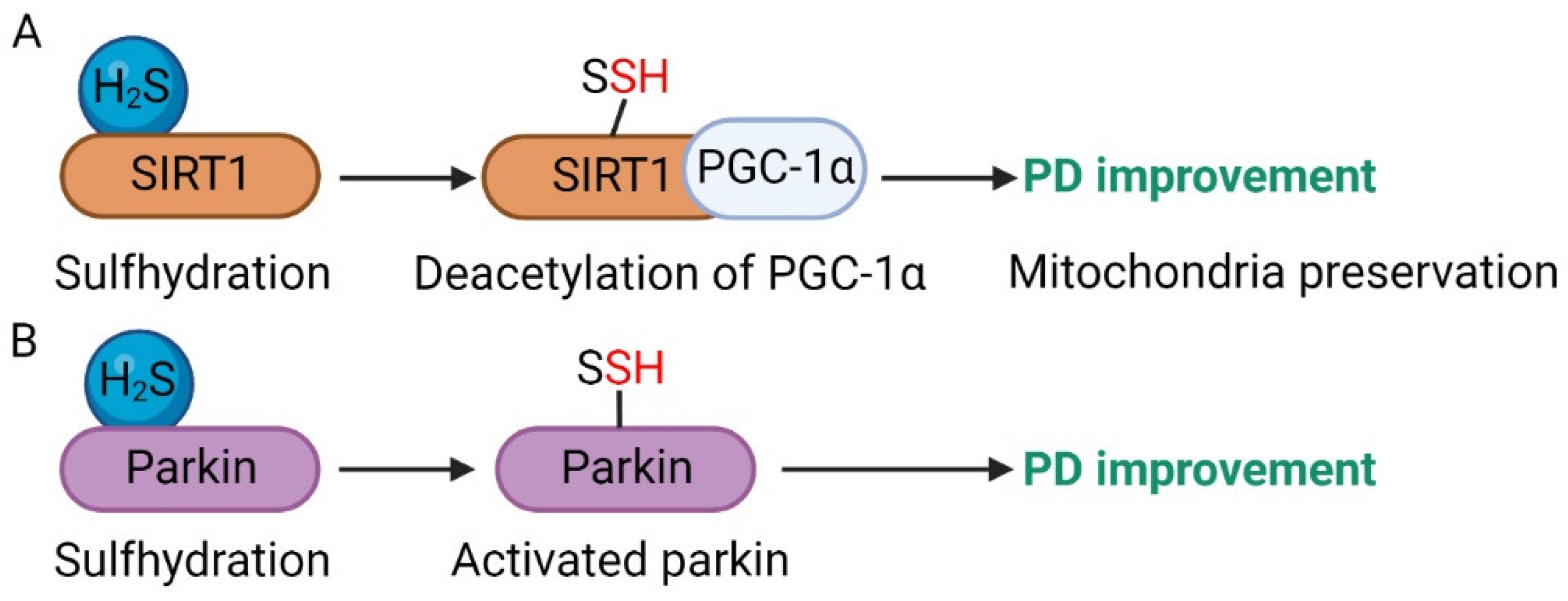
4.2. Parkinson’s Disease (PD)

PD has been characterized by the loss of dopaminergic neurons in the substantia nigra (SN) and dopamine deficiency in the striatum [106]. Oxidative stress, mitochondrial dysfunction, misfolded protein accumulation, and neuroinflammation are considered major pathogenesis contributors to PD, which eventually result in impaired movement [106,107]. Last few decades, a large number of discoveries have indicated that H2S has an essential anti-oxidative effect in dealing with PD patients suffering from oxidative stress [76,108,109]. These findings demonstrate that H2S exerts its neuroprotective effects by acting as an antioxidant in the brain of PD patients.
In addition, rotenone is used to establish models of PD disease in animals. It has been reported that H2S can attenuate rotenone-induced cell apoptosis by preserving mitochondrial function and regulating the JNK-MAPK pathway [82], which suggests that appropriate administration of NaHS is beneficial for PD treatment, and it can be regarded as a potential therapeutic strategy for neurodegenerative disease PD. Indeed, in 6-hydroxydopamine (6-OHDA)-induced PD rat model revealed that endogenous H2S level was decreased in the SN. On the other hand, the exogenous application of H2S promoted the increase in endogenous H2S and inhibited the movement disorder by reversing the depletion of tyrosine–hydroxylase-positive neurons in the SN. This protective effect may be mediated by promoting leptin signaling and increasing malondialdehyde levels in the striatum [110,111]. NaHS application could also protect SH-SY5Y cells against 1-methyl-4-phenylpyridinium ion (MPP+)-induced mitochondrial transmembrane potential loss, oxidative stress and cell apoptotic in the PD cell model [109]. Accordingly, inhibition of H2S production from the CBS-H2S axis enhances 1-methy-4-phenyl-1,2,3,6-tetrahydropyridine (MPTP) or MPP+-induced neurotoxicity in the PD model [112,113]. Moreover, injection of GYY4137 (an H2S donor) or overexpression of CBS in the striatum attenuates hallmark pathological event of nitrated α-synuclein in the PD model and rescues motor disorder induced by MPTP [112,114]. Since the N-terminus of α-synuclein serves as a mitochondrial targeting sequence peptide and is associated with the regulation of mitochondrial function, it is possible that H2S application may preserve mitochondrial activity and cell viability in the brain of PD by attenuating nitrate stress induced by post-translational modification of α-synuclein.
Proper endogenous H2S production by strictly controlling CBS synthesis in astrocytes exhibits anti-inflammatory and neuroprotective effects [80]. It was reported that the application of H2S inhibited microglial activation in SN and prevented the accumulation of inflammatory factors in the striatum in PD model induced by rotenone [110]. However, recent studies have pointed out that excessive H2S generated from gut bacteria promotes the formation of α-synuclein fibrils by releasing cytochrome C from mitochondria and stimulating ROS accumulation [115], thus suggesting that H2S may contribute to PD disease. According to these findings, H2S exerts an essential physiological role in the brain, mainly depending on strictly controlling physiological H2S concentrations. Except for the application of H2S-releasing donors, it has been reported that inhaled H2S protects MPTP-induced movement disorder in PD mouse model against neurodegeneration via antioxidative, anti-inflammatory, and antiapoptotic signaling in the brain [116], which was further confirmed by the possible beneficial link between chronic inhalation of H2S in animal models and protection of dopaminergic neurons associated with PD [117].
The exact mechanism of H2S has been defined as the sulfhydration of specific target proteins. It has been recently reported that H2S increases expression level of silent information regulator-1 (sirtuin 1, SIRT1), and enhances the activity of SIRT1 through H2S-mediated sulfhydration, which could increase autophagy flux and attenuate cell injury in PD mouse model induced by MPP+ [118]. Although the SIRT1 is primarily expressed in the cell nucleus, its activity is largely associated with mitochondrial biogenesis and turnover by mitophagy [119]. Peroxisome proliferator-activated receptor γ co-activator 1α (PGC-1α), a metabolic coactivator, can be deacetylated by SIRT1 to modulate mitochondrial biogenesis and respiration [120]. Thus, it is possible that H2S-induced sulfhydration of SIRT1 promotes the deacetylation of PGC-1α, finally resulting in the amelioration of mitochondrial activity and improvement of PD (Figure 3A). Furthermore, it has been reported that the parkin protein, a neuroprotective ubiquitin E3 ligase, is depleted in the brains of patients affected by PD. Interestingly, it has been found that the parkin protein can be sulfhydrated by H2S, subsequently enhancing its activity (Figure 3B) [121]. Thus, modulating specific protein activities via H2S-mediated sulfhydration may be therapeutic in PD disease.

4.3. Huntington’s Disease

HD is characterized by damage to the corpus striatum of the brain, leading to involuntary movements and motor disorders along with psychiatric disturbances [122]. It occurs due to the gene encoding huntingtin (Htt) mutation, which results in the expansion of polyglutamine repeats and subsequent cellular processes, including oxidative stress, mitochondrial dysfunction, neurotoxicity, and behavioral dysfunction [123,124]. Previous studies reported that cystathionine γ-lyase (CSE) depletion could contribute to HD pathophysiology [124,125], considering that CSE promotes cysteine generation, and cysteine is a precursor of H2S. Depletion of CSE in neurons causes decreased H2S, which leads to oxidative stress and mitochondrial dysfunction. Furthermore, it has been reported that the transsulfuration pathway linked to cysteine and H2S metabolism is disrupted in HD [10]. Therefore, administration of cysteine through the use of N-acetylcysteine (NAC) can ameliorate the HD-associated excitotoxicity and depressive behaviors by specifically increasing glutamate in a glutamate transporter-dependent manner in the R6/1 transgenic mouse model of HD [126]. Furthermore, the transsulfuration pathway regulates signaling events through sulfhydration which is a posttranslational modification mediated by H2S [127,128]. Diminished levels of CSE leads to decreased levels of sulfhydration in striatal cell-line model Q111 of HD, suggesting that H2S may be decreased in the brain of HD [129]. In addition, the enzyme CBS affects H2S production by specifically interacting with Huntingtin protein [130]. Therefore, it is speculated that H2S may be involved in regulating HD, and treatment with H2S donors may benefit HD [108]. Recently, it was found that NaHS protect rats against 3-nitropropionic acid (3NP)-induced HD-like pathology through activation of anti-oxidative responses, and anti-inflammatory, as well as anti-apoptotic signals [131]. Moreover, it has been reported that 40 mg/kg H2S is clinically beneficial for people with HD disease [132].

4.4. Diabetes-Associated Cognitive Impairment and Other Neurodegenerative Diseases

Numerous studies have pointed out that patients with diabetes are more likely to develop cognitive impairment than healthy individuals [133], resulting in a serious social burden [134]. However, as the exact mechanism of diabetes-associated cognitive decline (DACD) is not completely clear, there is no effective treatment for the disease. Recent studies have indicated that ER stress in the hippocampus and synaptic dysfunction contribute to diabetes-related cognitive disorders [135,136,137,138,139]. More importantly, it has been reported that endogenous H2S production is decreased during the pathogenesis of diabetes [140,141], suggesting the possible physiological involvement of H2S in regulating diabetes-induced cognitive disorders. Accordingly, it was found that administration of NaHS promoted hippocampal endogenous H2S production in streptozotocin (STZ)-induced diabetic rat models, and significantly ameliorated the diabetic-associated cognitive disorders by inhibiting ER stress, including preventing expressions of glucose-regulated protein 78 (GRP78), cleaved caspase-12, and C/EBP homologous protein (CHOP) [142]. Meanwhile, He et al. found that NaHS could improve the cognitive dysfunction of STZ-diabetic rats by promoting the expression of SIRT1, a major regulator in synaptic plasticity [143]. In the db/db mice model, it has also been reported that exogenous injection of NaHS could decrease ER stress [144]. Considering that the upregulation of SIRT1 inhibits the ER stress pathway [145], it is possible that H2S could improve diabetes-associated cognitive deficits by inhibiting hippocampal ER stress through upregulating SIRT1. However, the mechanisms through which SIRT1 regulates ER still remain unclear. Because the activity of SIRT1 is associated with mitochondrial biogenesis and mitophagy, the ER and mitochondria are tightly contacted at specific subdomains via tethering mediated by mitochondria-associated ER membrane (MAM) proteins, which are critical for determining cell fate [146]. It can be hypothesized that H2S may modulate diabetes-related learning and memory decline by regulating ER-mitochondria signaling. Moreover, it was reported that exogenous H2S had anti-apoptotic and anti-inflammatory effects in ameliorating the diabetes-associated cognitive deficit by downregulating mitochondria-mediated pro-apoptotic genes cleaved Caspase-3, cleaved Caspase-9, Bax and cytochrome C, and expressions of IL-23/IL-17 [147]. Therefore, mitochondria-targeted H2S donors with the regulation of mitophagy could be used as a new strategy to improve diabetes-associated cognitive impairment.
As is well known, H2S exerts its antioxidant functions by ameliorating oxidative stress and preserving mitochondrial function in neurodegenerative diseases [75,108]. However, low physiological concentrations of H2S are generally cytoprotective, whereas higher doses are detrimental. For example, amyotrophic lateral sclerosis (ALS) is defined as the selective degeneration of upper and lower motor neurons caused by mutations of Cu/Zn superoxide dismutase 1 (SOD1) genes and dysregulation of mitochondrial complexes II and IV [10,75,148]. Studies performed in spinal cord cell culture lines, familial ALS (FALS) mouse model, and sporadic ALS patients, respectively, showed that H2S concentrations were increased in astrocytes and microglia and contributed to motor neuron death by inducing intracellular Ca2+ accumulation in motor neurons, and activating Nrf-2-mediated oxidative stress response and peroxiredoxins [149,150]. Interestingly, it was reported that treatment with amino-oxyacetic acid (AOA, a systemic dual inhibitor of CBS and CSE) only expanded the lifespan of female fALS mice [151]. Based on these findings, we can conclude that the therapeutic or deleterious effect of H2S on neurodegenerative diseases mainly depends on the type of H2S donors and modulators, and the timing of therapy [152,153,154].
Table 1. Summary of the beneficial effect of H2S in neurodegenerative diseases.
DiseasesBiological ModelTargetsReferences
ADPC12 cellsMitochondrial membrane potential, Redox steady state, Apoptosis pathway[90,91,98]
APP/PS1 neuronsEnergy production, Mitochondrial DNA, Redox steady state, APP pathway[92,102]
HepG2/HEK293 cellsMitochondrial bioenergetics[93]
3xTg-AD miceRedox steady state, GSK3β[94,95,105]
APP/PS1 transgenic miceNrf2, HO-1, GST[96]
Homocysteine-induced AD miceNMDA receptor[97]
Microglia cellsp38 and JNK-MAPK dependent pathway, STAT3, Cat S[99,101]
Hippocampus cellsNF-κB pathway[100]
PDRotenone-induced PD miceMitochondrial function, JNK-MAPK pathway, anti-inflammatory factors[82,110]
SH-SY5Y cellsMitochondrial transmembrane potential, Cell apoptotic, Redox steady state[109]
MPP+-induced PD mouseSIRT1 expression and sulfhydration, PGC-1α deacetylation[118,120]
6-OHDA-induced PD ratLeptin signaling, Redox steady state[110]
PD patientsParkin sulfhydration[121]
HD3-nitropropionic acid (3NP)-induced HD ratAntioxidative responses, anti-inflammatory and anti-apoptotic signaling[131]
DACDStreptozotocin (STZ)-induced diabetic ratGRP78, cleaved caspase-12 and CHOP expression, SIRT1 expression[142,143]
db/db miceSIRT1 expression, ER stress pathway, anti-apoptotic and anti-inflammatory pathway[145,147]

5. Conclusions and Perspective

This review summarized the general toxic substances that contribute to neurotoxicity, and its possible mechanisms resulting in neurodegenerative diseases, including oxidative stress and mitochondrial dysfunction. Furthermore, we described the physiological roles of novel gas transmitter H2S in protecting neurons by regulating antioxidative, anti-inflammatory and anti-apoptotic responses (Table 1, Figure 4). Although the mechanism of H2S-mediated protein persulfidation has been reported in modulating some neurodegenerative diseases, many unanswered questions remain, from those related to its biological functions to specific target proteins of sulfhydration. Furthermore, most previous studies mainly focused on the beneficial effect of exogenous H2S application on neurodegenerative diseases, while overlooking the exploration of exact technology for measuring and modulating its intracellular production and metabolism. In addition, we concluded that the basic endogenous level of H2S varies in different tissues, and different neurodegenerative diseases have different requirements for H2S and polysulfides.
The impairment of mitochondrial function mainly includes disruptions of mitochondrial enzyme activities, reduction in circulating mitochondrial DNA content, loss of circular structure and dysregulation of biogenesis. Further investigation of the effect of H2S on mitochondrial activity in neurodegenerative diseases is expected to advance our understanding of future drug targets. Future research will increase the identification of additional targets of S-sulfhydration, and perhaps also reveal the extended conservation of this process through interacting with other signal pathways in different diseases. In addition, iron dyshomeostasis is implicated in various neurodegenerative diseases. Endogenous H2S production is regulated by iron via non-enzymatic reactions, implying the inter-relationship between H2S and iron in neuron diseases. However, the exact molecular mechanisms for the interplay between H2S and iron, and their therapeutic applications in neuron diseases need to be further explored. Thereby, research on the supplementation of H2S-releasing drugs or modulation of its metabolism would be benefit future therapeutic strategies of neurodegenerative diseases in clinic.
Oxidative damage and mitochondria dysfunction are regarded as the primary cause of neurodegenerative diseases, including AD, PD, HD, DACD, and ALS. H2S exerts its neuroprotective effects through upregulating (marked in green) or downregulating (marked in red) the antioxidant, antiapoptotic, and anti-inflammation genes, or influencing activities of specific proteins through sulfhydration. Strictly controlling the endogenous H2S production through the administration of mitochondrial-targeted H2S donors and modulation of enzymatic or non-enzymatic mediated H2S metabolism would be beneficial for antioxidative stress and preserving mitochondria activity and biogenesis, thus subsequently improving neurological diseases. This model was created using BioRender (https://biorender.com/, accessed on 20 November 2022).

Author Contributions

L.Z. wrote the manuscript. Q.W. read and revised the final manuscript. All authors have read and agreed to the published version of the manuscript.

Funding

This research was funded by the National Natural Science Foundation of China (Grant Nos. 81974540, 82274290), the Natural Science Foundation of Shaanxi (Grant No. 2023-JC-QN-0257), Key Research & Development Program of Shaanxi (Program No. 2022ZDLSF02-09), and the Innovation Capability Support Program of Shaanxi (Program No. 2021TD-58).

Conflicts of Interest

The authors declare no conflict of interest.

References

  1. Richardson, J.R.; Wang, Y. Neurotoxicology. Chem. Res. Toxicol. 2021, 34, 1197. [Google Scholar] [CrossRef]
  2. Harris, J.J.; Jolivet, R.; Attwell, D. Synaptic energy use and supply. Neuron 2012, 75, 762–777. [Google Scholar] [CrossRef] [Green Version]
  3. Wallace, K.B.; Starkov, A.A. Mitochondrial targets of drug toxicity. Annu. Rev. Pharmacol. Toxicol. 2000, 40, 353–388. [Google Scholar] [CrossRef] [Green Version]
  4. Lu, M.; Hu, L.F.; Hu, G.; Bian, J.S. Hydrogen sulfide protects astrocytes against H2O2-induced neural injury via enhancing glutamate uptake. Free Radic. Biol. Med. 2008, 45, 1705–1713. [Google Scholar] [CrossRef]
  5. Bhattacharjee, N.; Borah, A. Oxidative stress and mitochondrial dysfunction are the underlying events of dopaminergic neurodegeneration in homocysteine rat model of Parkinson’s disease. Neurochem. Int. 2016, 101, 48–55. [Google Scholar] [CrossRef]
  6. Kimura, H. Hydrogen sulfide: From brain to gut. Antioxid. Redox Signal. 2010, 12, 1111–1123. [Google Scholar] [CrossRef]
  7. Warenycia, M.W.; Goodwin, L.R.; Benishin, C.G.; Reiffenstein, R.J.; Francom, D.M.; Taylor, J.D.; Dieken, F.P. Acute hydrogen sulfide poisoning. Demonstration of selective uptake of sulfide by the brainstem by measurement of brain sulfide levels. Biochem. Pharmacol. 1989, 38, 973–981. [Google Scholar] [CrossRef]
  8. Kimura, H. Hydrogen sulfide: Its production, release and functions. Amino Acids 2011, 41, 113–121. [Google Scholar] [CrossRef]
  9. Shibuya, N.; Koike, S.; Tanaka, M.; Ishigami-Yuasa, M.; Kimura, Y.; Ogasawara, Y.; Fukui, K.; Nagahara, N.; Kimura, H. A novel pathway for the production of hydrogen sulfide from D-cysteine in mammalian cells. Nat. Commun. 2013, 4, 1366. [Google Scholar] [CrossRef] [Green Version]
  10. Paul, B.D.; Snyder, S.H. Gasotransmitter hydrogen sulfide signaling in neuronal health and disease. Biochem. Pharmacol. 2018, 149, 101–109. [Google Scholar] [CrossRef]
  11. Kimura, H. Signaling of hydrogen sulfide and polysulfides. Antioxid. Redox Signal. 2015, 22, 347–349. [Google Scholar] [CrossRef] [Green Version]
  12. Paul, B.D.; Snyder, S.H. H2S: A Novel Gasotransmitter that Signals by Sulfhydration. Trends Biochem. Sci. 2015, 40, 687–700. [Google Scholar] [CrossRef] [Green Version]
  13. Kamat, P.K.; Kalani, A.; Tyagi, N. Role of hydrogen sulfide in brain synaptic remodeling. Methods Enzymol. 2015, 555, 207–229. [Google Scholar]
  14. Panthi, S.; Manandhar, S.; Gautam, K. Hydrogen sulfide, nitric oxide, and neurodegenerative disorders. Transl. Neurodegener. 2018, 7, 3. [Google Scholar] [CrossRef]
  15. Aroca, A.; Gotor, C. Hydrogen Sulfide: A Key Role in Autophagy Regulation from Plants to Mammalians. Antioxidants 2022, 11, 327. [Google Scholar] [CrossRef]
  16. Abe, K.; Kimura, H. The possible role of hydrogen sulfide as an endogenous neuromodulator. J. Neurosci. 1996, 16, 1066–1071. [Google Scholar] [CrossRef] [Green Version]
  17. Li, L.; Bhatia, M.; Zhu, Y.Z.; Zhu, Y.C.; Ramnath, R.D.; Wang, Z.J.; Anuar, F.B.; Whiteman, M.; Salto-Tellez, M.; Moore, P.K. Hydrogen sulfide is a novel mediator of lipopolysaccharide-induced inflammation in the mouse. FASEB J. 2005, 19, 1196–1198. [Google Scholar] [CrossRef]
  18. Zanardo, R.C.; Brancaleone, V.; Distrutti, E.; Fiorucci, S.; Cirino, G.; Wallace, J.L. Hydrogen sulfide is an endogenous modulator of leukocyte-mediated inflammation. FASEB J. 2006, 20, 2118–2120. [Google Scholar] [CrossRef]
  19. Kumar, M.; Ray, R.S.; Sandhir, R. Hydrogen sulfide attenuates homocysteine-induced neurotoxicity by preventing mitochondrial dysfunctions and oxidative damage: In vitro and in vivo studies. Neurochem. Int. 2018, 120, 87–98. [Google Scholar] [CrossRef]
  20. Grieco, A.J. Homocystinuria: Pathogenetic mechanisms. Am. J. Med. Sci. 1977, 273, 120–132. [Google Scholar] [CrossRef]
  21. Mudd, S.H.; Skovby, F.; Levy, H.L.; Pettigrew, K.D.; Wilcken, B.; Pyeritz, R.E.; Andria, G.; Boers, G.H.; Bromberg, I.L.; Cerone, R.; et al. The natural history of homocystinuria due to cystathionine beta-synthase deficiency. Am. J. Hum. Genet. 1985, 37, 1–31. [Google Scholar]
  22. Ichinohe, A.; Kanaumi, T.; Takashima, S.; Enokido, Y.; Nagai, Y.; Kimura, H. Cystathionine beta-synthase is enriched in the brains of Down’s patients. Biochem. Biophys. Res. Commun. 2005, 338, 1547–1550. [Google Scholar] [CrossRef]
  23. Kamath, A.F.; Chauhan, A.K.; Kisucka, J.; Dole, V.S.; Loscalzo, J.; Handy, D.E.; Wagner, D.D. Elevated levels of homocysteine compromise blood-brain barrier integrity in mice. Blood 2006, 107, 591–593. [Google Scholar] [CrossRef] [Green Version]
  24. Vitvitsky, V.; Dayal, S.; Stabler, S.; Zhou, Y.; Wang, H.; Lentz, S.R.; Banerjee, R. Perturbations in homocysteine-linked redox homeostasis in a murine model for hyperhomocysteinemia. Am. J. Physiol. Regul. Integr. Comp. Physiol. 2004, 287, R39–R46. [Google Scholar] [CrossRef] [Green Version]
  25. Chen, S.; Dong, Z.; Zhao, Y.; Sai, N.; Wang, X.; Liu, H.; Huang, G.; Zhang, X. Homocysteine induces mitochondrial dysfunction involving the crosstalk between oxidative stress and mitochondrial pSTAT3 in rat ischemic brain. Sci. Rep. 2017, 7, 6932. [Google Scholar] [CrossRef] [Green Version]
  26. Kamat, P.K.; Kalani, A.; Givvimani, S.; Sathnur, P.B.; Tyagi, S.C.; Tyagi, N. Hydrogen sulfide attenuates neurodegeneration and neurovascular dysfunction induced by intracerebral-administered homocysteine in mice. Neuroscience 2013, 252, 302–319. [Google Scholar] [CrossRef] [Green Version]
  27. Obeid, R.; Herrmann, W. Mechanisms of homocysteine neurotoxicity in neurodegenerative diseases with special reference to dementia. FEBS Lett. 2006, 580, 2994–3005. [Google Scholar] [CrossRef] [Green Version]
  28. Linnebank, M.; Lutz, H.; Jarre, E.; Vielhaber, S.; Noelker, C.; Struys, E.; Jakobs, C.; Klockgether, T.; Evert, B.O.; Kunz, W.S.; et al. Binding of copper is a mechanism of homocysteine toxicity leading to COX deficiency and apoptosis in primary neurons, PC12 and SHSY-5Y cells. Neurobiol. Dis. 2006, 23, 725–730. [Google Scholar] [CrossRef]
  29. Zieminska, E.; Matyja, E.; Kozlowska, H.; Stafiej, A.; Lazarewicz, J.W. Excitotoxic neuronal injury in acute homocysteine neurotoxicity: Role of calcium and mitochondrial alterations. Neurochem. Int. 2006, 48, 491–497. [Google Scholar] [CrossRef]
  30. Chiu, K.M.; Lin, T.Y.; Lee, M.Y.; Lu, C.W.; Wang, M.J.; Wang, S.J. Typhaneoside Suppresses Glutamate Release Through Inhibition of Voltage-Dependent Calcium Entry in Rat Cerebrocortical Nerve Terminals. Chem. Res. Toxicol. 2021, 34, 1286–1295. [Google Scholar] [CrossRef]
  31. Li, Q.Q.; Chen, J.; Hu, P.; Jia, M.; Sun, J.H.; Feng, H.Y.; Qiao, F.C.; Zang, Y.Y.; Shi, Y.Y.; Chen, G.; et al. Enhancing GluN2A-type NMDA receptors impairs long-term synaptic plasticity and learning and memory. Mol. Psychiatry 2022, 27, 3468–3478. [Google Scholar] [CrossRef] [PubMed]
  32. Moyano, P.; Sanjuna, J.; Garcia, J.M.; Garcia, J.; Frejo, M.T.; Naval, M.V.; Del Pino, J. Paraquat Treatment Compromises the Clearance of β-Amyloid and Tau Proteins and Induces Primary Hippocampal Neuronal Cell Death through HSP70, P20S, and TFEB Disruption. Chem. Res. Toxicol. 2021, 34, 1240–1244. [Google Scholar] [CrossRef] [PubMed]
  33. Farkhondeh, T.; Mehrpour, O.; Forouzanfar, F.; Roshanravan, B.; Samarghandian, S. Oxidative stress and mitochondrial dysfunction in organophosphate pesticide-induced neurotoxicity and its amelioration: A review. Environ. Sci. Pollut. Res. Int. 2020, 27, 24799–24814. [Google Scholar] [CrossRef] [PubMed]
  34. Prakash, C.; Soni, M.; Kumar, V. Mitochondrial oxidative stress and dysfunction in arsenic neurotoxicity: A review. J. Appl. Toxicol. 2016, 36, 179–188. [Google Scholar] [CrossRef] [PubMed]
  35. Garza-Lombó, C.; Pappa, A.; Panayiotidis, M.I.; Gonsebatt, M.E.; Franco, R. Arsenic-induced neurotoxicity: A mechanistic appraisal. J. Biol. Inorg. Chem. 2019, 24, 1305–1316. [Google Scholar] [CrossRef]
  36. Kumar, V.; Gill, K.D. Oxidative stress and mitochondrial dysfunction in aluminium neurotoxicity and its amelioration: A review. Neurotoxicology 2014, 41, 154–166. [Google Scholar] [CrossRef]
  37. Murphy, D.; Patel, H.; Wimalasena, K. Caenorhabditis elegans Model Studies Show MPP(+) Is a Simple Member of a Large Group of Related Potent Dopaminergic Toxins. Chem. Res. Toxicol. 2021, 34, 1275–1285. [Google Scholar] [CrossRef]
  38. Kabil, O.; Banerjee, R. Redox biochemistry of hydrogen sulfide. J. Biol. Chem. 2010, 285, 21903–21907. [Google Scholar] [CrossRef] [Green Version]
  39. Qu, K.; Lee, S.W.; Bian, J.S.; Low, C.M.; Wong, P.T. Hydrogen sulfide: Neurochemistry and neurobiology. Neurochem. Int. 2008, 52, 155–165. [Google Scholar] [CrossRef] [PubMed]
  40. Reiffenstein, R.J.; Hulbert, W.C.; Roth, S.H. Toxicology of hydrogen sulfide. Annu. Rev. Pharmacol. Toxicol. 1992, 32, 109–134. [Google Scholar] [CrossRef] [PubMed]
  41. Burnett, W.W.; King, E.G.; Grace, M.; Hall, W.F. Hydrogen sulfide poisoning: Review of 5 years’ experience. Can. Med. Assoc. J. 1977, 117, 1277–1280. [Google Scholar]
  42. Beauchamp, R.O., Jr.; Bus, J.S.; Popp, J.A.; Boreiko, C.J.; Andjelkovich, D.A. A critical review of the literature on hydrogen sulfide toxicity. Crit. Rev. Toxicol. 1984, 13, 25–97. [Google Scholar] [CrossRef] [PubMed]
  43. Savage, J.C.; Gould, D.H. Determination of sulfide in brain tissue and rumen fluid by ion-interaction reversed-phase high-performance liquid chromatography. J. Chromatogr. 1990, 526, 540–545. [Google Scholar] [CrossRef] [PubMed]
  44. Shibuya, N.; Tanaka, M.; Yoshida, M.; Ogasawara, Y.; Togawa, T.; Ishii, K.; Kimura, H. 3-Mercaptopyruvate sulfurtransferase produces hydrogen sulfide and bound sulfane sulfur in the brain. Antioxid. Redox Signal. 2009, 11, 703–714. [Google Scholar] [CrossRef]
  45. Kimura, H. Hydrogen sulfide and polysulfides as signaling molecules. Proc. Jpn. Acad. Ser. B Phys. Biol. Sci. 2015, 91, 131–159. [Google Scholar] [CrossRef] [PubMed] [Green Version]
  46. Mustafa, A.K.; Gadalla, M.M.; Sen, N.; Kim, S.; Mu, W.; Gazi, S.K.; Barrow, R.K.; Yang, G.; Wang, R.; Snyder, S.H. H2S signals through protein S-sulfhydration. Sci. Signal. 2009, 2, ra72. [Google Scholar] [CrossRef] [Green Version]
  47. Ishigami, M.; Hiraki, K.; Umemura, K.; Ogasawara, Y.; Ishii, K.; Kimura, H. A source of hydrogen sulfide and a mechanism of its release in the brain. Antioxid. Redox Signal. 2009, 11, 205–214. [Google Scholar] [CrossRef] [Green Version]
  48. Panthi, S.; Chung, H.J.; Jung, J.; Jeong, N.Y. Physiological Importance of Hydrogen Sulfide: Emerging Potent Neuroprotector and Neuromodulator. Oxidative Med. Cell. Longev. 2016, 2016, 9049782. [Google Scholar] [CrossRef] [Green Version]
  49. Enokido, Y.; Suzuki, E.; Iwasawa, K.; Namekata, K.; Okazawa, H.; Kimura, H. Cystathionine beta-synthase, a key enzyme for homocysteine metabolism, is preferentially expressed in the radial glia/astrocyte lineage of developing mouse CNS. FASEB J. 2005, 19, 1854–1856. [Google Scholar] [CrossRef]
  50. Stipanuk, M.H.; Beck, P.W. Characterization of the enzymic capacity for cysteine desulphhydration in liver and kidney of the rat. Biochem. J. 1982, 206, 267–277. [Google Scholar] [CrossRef] [Green Version]
  51. Marutani, E.; Ichinose, F. Emerging pharmacological tools to control hydrogen sulfide signaling in critical illness. Intensive Care Med. Exp. 2020, 8, 5. [Google Scholar] [CrossRef] [PubMed] [Green Version]
  52. Caliendo, G.; Cirino, G.; Santagada, V.; Wallace, J.L. Synthesis and biological effects of hydrogen sulfide (H2S): Development of H2S-releasing drugs as pharmaceuticals. J. Med. Chem. 2010, 53, 6275–6286. [Google Scholar] [CrossRef] [PubMed]
  53. Szabo, C.; Ransy, C.; Módis, K.; Andriamihaja, M.; Murghes, B.; Coletta, C.; Olah, G.; Yanagi, K.; Bouillaud, F. Regulation of mitochondrial bioenergetic function by hydrogen sulfide. Part I. Biochemical and physiological mechanisms. Br. J. Pharmacol. 2014, 171, 2099–2122. [Google Scholar] [CrossRef] [PubMed] [Green Version]
  54. Picton, R.; Eggo, M.C.; Merrill, G.A.; Langman, M.J.; Singh, S. Mucosal protection against sulphide: Importance of the enzyme rhodanese. Gut 2002, 50, 201–205. [Google Scholar] [CrossRef] [Green Version]
  55. Donnarumma, E.; Trivedi, R.K.; Lefer, D.J. Protective Actions of H2S in Acute Myocardial Infarction and Heart Failure. Compr. Physiol. 2017, 7, 583–602. [Google Scholar]
  56. Whiteman, M.; Moore, P.K. Hydrogen sulfide and the vasculature: A novel vasculoprotective entity and regulator of nitric oxide bioavailability? J. Cell. Mol. Med. 2009, 13, 488–507. [Google Scholar] [CrossRef]
  57. González-García, P.; Hidalgo-Gutiérrez, A.; Mascaraque, C.; Barriocanal-Casado, E.; Bakkali, M.; Ziosi, M.; Abdihankyzy, U.B.; Sánchez-Hernández, S.; Escames, G.; Prokisch, H.; et al. Coenzyme Q10 modulates sulfide metabolism and links the mitochondrial respiratory chain to pathways associated to one carbon metabolism. Hum. Mol. Genet. 2020, 29, 3296–3311. [Google Scholar] [CrossRef]
  58. Untereiner, A.A.; Fu, M.; Módis, K.; Wang, R.; Ju, Y.; Wu, L. Stimulatory effect of CSE-generated H2S on hepatic mitochondrial biogenesis and the underlying mechanisms. Nitric Oxide 2016, 58, 67–76. [Google Scholar] [CrossRef]
  59. Elrod, J.W.; Calvert, J.W.; Morrison, J.; Doeller, J.E.; Kraus, D.W.; Tao, L.; Jiao, X.; Scalia, R.; Kiss, L.; Szabo, C.; et al. Hydrogen sulfide attenuates myocardial ischemia-reperfusion injury by preservation of mitochondrial function. Proc. Natl. Acad. Sci. USA 2007, 104, 15560–15565. [Google Scholar] [CrossRef] [Green Version]
  60. Li, L.; Whiteman, M.; Guan, Y.Y.; Neo, K.L.; Cheng, Y.; Lee, S.W.; Zhao, Y.; Baskar, R.; Tan, C.H.; Moore, P.K. Characterization of a novel, water-soluble hydrogen sulfide-releasing molecule (GYY4137): New insights into the biology of hydrogen sulfide. Circulation 2008, 117, 2351–2360. [Google Scholar] [CrossRef] [Green Version]
  61. Xie, L.; Feng, H.; Li, S.; Meng, G.; Liu, S.; Tang, X.; Ma, Y.; Han, Y.; Xiao, Y.; Gu, Y.; et al. SIRT3 Mediates the Antioxidant Effect of Hydrogen Sulfide in Endothelial Cells. Antioxid. Redox Signal. 2016, 24, 329–343. [Google Scholar] [CrossRef] [Green Version]
  62. Yan, Y.; Yan, Q.; Qian, L.; Jiang, Y.; Chen, X.; Zeng, S.; Xu, Z.; Gong, Z. S-adenosylmethionine administration inhibits levodopa-induced vascular endothelial growth factor-A expression. Aging 2020, 12, 21290–21307. [Google Scholar] [CrossRef] [PubMed]
  63. Caro, A.A.; Adlong, L.W.; Crocker, S.J.; Gardner, M.W.; Luikart, E.F.; Gron, L.U. Effect of garlic-derived organosulfur compounds on mitochondrial function and integrity in isolated mouse liver mitochondria. Toxicol. Lett. 2012, 214, 166–174. [Google Scholar] [CrossRef] [Green Version]
  64. Hao, Y.; Liu, H.M.; Wei, X.; Gong, X.; Lu, Z.Y.; Huang, Z.H. Diallyl trisulfide attenuates hyperglycemia-induced endothelial apoptosis by inhibition of Drp1-mediated mitochondrial fission. Acta Diabetol. 2019, 56, 1177–1189. [Google Scholar] [CrossRef] [PubMed] [Green Version]
  65. Polhemus, D.J.; Li, Z.; Pattillo, C.B.; Gojon, G., Sr.; Gojon, G., Jr.; Giordano, T.; Krum, H. A novel hydrogen sulfide prodrug, SG1002, promotes hydrogen sulfide and nitric oxide bioavailability in heart failure patients. Cardiovasc. Ther. 2015, 33, 216–226. [Google Scholar] [CrossRef]
  66. Polhemus, D.; Kondo, K.; Bhushan, S.; Bir, S.C.; Kevil, C.G.; Murohara, T.; Lefer, D.J.; Calvert, J.W. Hydrogen sulfide attenuates cardiac dysfunction after heart failure via induction of angiogenesis. Circ. Heart Fail. 2013, 6, 1077–1086. [Google Scholar] [CrossRef] [Green Version]
  67. Saif, J.; Ahmad, S.; Rezai, H.; Litvinova, K.; Sparatore, A.; Alzahrani, F.A.; Wang, K.; Ahmed, A. Hydrogen sulfide releasing molecule MZe786 inhibits soluble Flt-1 and prevents preeclampsia in a refined RUPP mouse model. Redox Biol. 2021, 38, 101814. [Google Scholar] [CrossRef]
  68. Rezai, H.; Ahmad, S.; Alzahrani, F.A.; Sanchez-Aranguren, L.; Dias, I.H.; Agrawal, S.; Sparatore, A.; Wang, K.; Ahmed, A. MZe786, a hydrogen sulfide-releasing aspirin prevents preeclampsia in heme oxygenase-1 haplodeficient pregnancy under high soluble flt-1 environment. Redox Biol. 2021, 38, 101768. [Google Scholar] [CrossRef]
  69. Sanchez-Aranguren, L.C.; Rezai, H.; Ahmad, S.; Alzahrani, F.A.; Sparatore, A.; Wang, K.; Ahmed, A. MZe786 Rescues Cardiac Mitochondrial Activity in High sFlt-1 and Low HO-1 Environment. Antioxidants 2020, 9, 598. [Google Scholar] [CrossRef] [PubMed]
  70. De Koning, M.L.Y.; Assa, S.; Maagdenberg, C.G.; van Veldhuisen, D.J.; Pasch, A.; van Goor, H.; Lipsic, E.; van der Harst, P. Safety and Tolerability of Sodium Thiosulfate in Patients with an Acute Coronary Syndrome Undergoing Coronary Angiography: A Dose-Escalation Safety Pilot Study (SAFE-ACS). J. Interv. Cardiol. 2020, 2020, 6014915. [Google Scholar] [CrossRef]
  71. Szczesny, B.; Módis, K.; Yanagi, K.; Coletta, C.; Le Trionnaire, S.; Perry, A.; Wood, M.E.; Whiteman, M.; Szabo, C. AP39, a novel mitochondria-targeted hydrogen sulfide donor, stimulates cellular bioenergetics, exerts cytoprotective effects and protects against the loss of mitochondrial DNA integrity in oxidatively stressed endothelial cells in vitro. Nitric Oxide 2014, 41, 120–130. [Google Scholar] [CrossRef] [Green Version]
  72. Gerő, D.; Torregrossa, R.; Perry, A.; Waters, A.; Le-Trionnaire, S.; Whatmore, J.L.; Wood, M.; Whiteman, M. The novel mitochondria-targeted hydrogen sulfide (H2S) donors AP123 and AP39 protect against hyperglycemic injury in microvascular endothelial cells in vitro. Pharmacol. Res. 2016, 113 Pt A, 186–198. [Google Scholar] [CrossRef]
  73. Kimura, Y.; Kimura, H. Hydrogen sulfide protects neurons from oxidative stress. FASEB J. 2004, 18, 1165–1167. [Google Scholar] [CrossRef] [PubMed]
  74. Kimura, Y.; Goto, Y.; Kimura, H. Hydrogen sulfide increases glutathione production and suppresses oxidative stress in mitochondria. Antioxid. Redox Signal. 2010, 12, 1–13. [Google Scholar] [CrossRef] [PubMed]
  75. Tabassum, R.; Jeong, N.Y. Potential for therapeutic use of hydrogen sulfide in oxidative stress-induced neurodegenerative diseases. Int. J. Med. Sci. 2019, 16, 1386–1396. [Google Scholar] [CrossRef] [PubMed] [Green Version]
  76. Tabassum, R.; Jeong, N.Y.; Jung, J. Protective effect of hydrogen sulfide on oxidative stress-induced neurodegenerative diseases. Neural Regen. Res. 2020, 15, 232–241. [Google Scholar] [PubMed]
  77. Tabassum, R.; Jeong, N.Y.; Jung, J. Therapeutic importance of hydrogen sulfide in age-associated neurodegenerative diseases. Neural Regen. Res. 2020, 15, 653–662. [Google Scholar]
  78. Kumar, M.; Sandhir, R. Hydrogen Sulfide in Physiological and Pathological Mechanisms in Brain. CNS Neurol. Disord. Drug Targets 2018, 17, 654–670. [Google Scholar] [CrossRef]
  79. Hu, L.F.; Wong, P.T.; Moore, P.K.; Bian, J.S. Hydrogen sulfide attenuates lipopolysaccharide-induced inflammation by inhibition of p38 mitogen-activated protein kinase in microglia. J. Neurochem. 2007, 100, 1121–1128. [Google Scholar] [CrossRef]
  80. Lee, M.; Schwab, C.; Yu, S.; McGeer, E.; McGeer, P.L. Astrocytes produce the antiinflammatory and neuroprotective agent hydrogen sulfide. Neurobiol. Aging 2009, 30, 1523–1534. [Google Scholar] [CrossRef]
  81. Aschner, M.; Skalny, A.V.; Ke, T.; da Rocha, J.B.; Paoliello, M.M.; Santamaria, A.; Bornhorst, J.; Rongzhu, L.; Svistunov, A.A.; Djordevic, A.B.; et al. Hydrogen sulfide (H2S) signaling as a protective mechanism against endogenous and exogenous neurotoxicants. Curr. Neuropharmacol. 2022, 20, 1908–1924. [Google Scholar] [CrossRef] [PubMed]
  82. Hu, L.F.; Lu, M.; Wu, Z.Y.; Wong, P.T.; Bian, J.S. Hydrogen sulfide inhibits rotenone-induced apoptosis via preservation of mitochondrial function. Mol. Pharmacol. 2009, 75, 27–34. [Google Scholar] [CrossRef] [PubMed] [Green Version]
  83. Bellenguez, C.; Küçükali, F.; Jansen, I.E.; Kleineidam, L.; Moreno-Grau, S.; Amin, N.; Naj, A.C.; Campos-Martin, R.; Grenier-Boley, B.; Andrade, V.; et al. New insights into the genetic etiology of Alzheimer’s disease and related dementias. Nat. Genet. 2022, 54, 412–436. [Google Scholar] [CrossRef] [PubMed]
  84. Hodgson, N.; Trivedi, M.; Muratore, C.; Li, S.; Deth, R. Soluble oligomers of amyloid-β cause changes in redox state, DNA methylation, and gene transcription by inhibiting EAAT3 mediated cysteine uptake. J. Alzheimers Dis. 2013, 36, 197–209. [Google Scholar] [CrossRef]
  85. Aoyama, K.; Suh, S.W.; Hamby, A.M.; Liu, J.; Chan, W.Y.; Chen, Y.; Swanson, R.A. Neuronal glutathione deficiency and age-dependent neurodegeneration in the EAAC1 deficient mouse. Nat. Neurosci. 2006, 9, 119–126. [Google Scholar] [CrossRef]
  86. Eto, K.; Asada, T.; Arima, K.; Makifuchi, T.; Kimura, H. Brain hydrogen sulfide is severely decreased in Alzheimer’s disease. Biochem. Biophys. Res. Commun. 2002, 293, 1485–1488. [Google Scholar] [CrossRef]
  87. Liu, X.Q.; Liu, X.Q.; Jiang, P.; Huang, H.; Yan, Y. Plasma levels of endogenous hydrogen sulfide and homocysteine in patients with Alzheimer’s disease and vascular dementia and the significance thereof. Zhonghua Yi Xue Za Zhi 2008, 88, 2246–2249. [Google Scholar]
  88. Peng, S.Y.; Wu, X.; Lu, T.; Cui, G.; Chen, G. Research progress of hydrogen sulfide in Alzheimer’s disease from laboratory to hospital: A narrative review. Med. Gas Res. 2020, 10, 125–129. [Google Scholar] [CrossRef]
  89. McCarty, M.F.; O’Keefe, J.H.; DiNicolantonio, J.J. A diet rich in taurine, cysteine, folate, B(12) and betaine may lessen risk for Alzheimer’s disease by boosting brain synthesis of hydrogen sulfide. Med. Hypotheses 2019, 132, 109356. [Google Scholar] [CrossRef]
  90. Tang, X.Q.; Shen, X.T.; Huang, Y.E.; Ren, Y.K.; Chen, R.Q.; Hu, B.; He, J.Q.; Yin, W.L.; Xu, J.H.; Jiang, Z.S. Hydrogen sulfide antagonizes homocysteine-induced neurotoxicity in PC12 cells. Neurosci. Res. 2010, 68, 241–249. [Google Scholar] [CrossRef]
  91. Tang, X.Q.; Ren, Y.K.; Zhou, C.F.; Yang, C.T.; Gu, H.F.; He, J.Q.; Chen, R.Q.; Zhuang, Y.Y.; Fang, H.R.; Wang, C.Y. Hydrogen sulfide prevents formaldehyde-induced neurotoxicity to PC12 cells by attenuation of mitochondrial dysfunction and pro-apoptotic potential. Neurochem. Int. 2012, 61, 16–24. [Google Scholar] [CrossRef]
  92. Zhao, F.L.; Fang, F.; Qiao, P.F.; Yan, N.; Gao, D.; Yan, Y. AP39, a Mitochondria-Targeted Hydrogen Sulfide Donor, Supports Cellular Bioenergetics and Protects against Alzheimer’s Disease by Preserving Mitochondrial Function in APP/PS1 Mice and Neurons. Oxidative Med. Cell. Longev. 2016, 2016, 8360738. [Google Scholar] [CrossRef] [PubMed] [Green Version]
  93. Módis, K.; Ju, Y.; Ahmad, A.; Untereiner, A.A.; Altaany, Z.; Wu, L.; Szabo, C.; Wang, R. S-Sulfhydration of ATP synthase by hydrogen sulfide stimulates mitochondrial bioenergetics. Pharmacol. Res. 2016, 113 Pt A, 116–124. [Google Scholar] [CrossRef] [Green Version]
  94. Wei, H.J.; Li, X.; Tang, X.Q. Therapeutic benefits of H₂S in Alzheimer’s disease. J. Clin. Neurosci. 2014, 21, 1665–1669. [Google Scholar] [CrossRef] [PubMed]
  95. Giuliani, D.; Ottani, A.; Zaffe, D.; Galantucci, M.; Strinati, F.; Lodi, R.; Guarini, S. Hydrogen sulfide slows down progression of experimental Alzheimer’s disease by targeting multiple pathophysiological mechanisms. Neurobiol. Learn. Mem. 2013, 104, 82–91. [Google Scholar] [CrossRef] [PubMed]
  96. Liu, Y.; Deng, Y.; Liu, H.; Yin, C.; Li, X.; Gong, Q. Hydrogen sulfide ameliorates learning memory impairment in APP/PS1 transgenic mice: A novel mechanism mediated by the activation of Nrf2. Pharmacol. Biochem. Behav. 2016, 150–151, 207–216. [Google Scholar] [CrossRef]
  97. Kamat, P.K.; Kyles, P.; Kalani, A.; Tyagi, N. Hydrogen Sulfide Ameliorates Homocysteine-Induced Alzheimer’s Disease-Like Pathology, Blood-Brain Barrier Disruption, and Synaptic Disorder. Mol. Neurobiol. 2016, 53, 2451–2467. [Google Scholar] [CrossRef]
  98. Tang, X.Q.; Chen, R.Q.; Ren, Y.K.; Soldato, P.D.; Sparatore, A.; Zhuang, Y.Y.; Fang, H.R.; Wang, C.Y. ACS6, a Hydrogen sulfide-donating derivative of sildenafil, inhibits homocysteine-induced apoptosis by preservation of mitochondrial function. Med. Gas Res. 2011, 1, 20. [Google Scholar] [CrossRef] [Green Version]
  99. Liu, Y.Y.; Bian, J.S. Hydrogen sulfide protects amyloid-β induced cell toxicity in microglia. J. Alzheimers Dis. 2010, 22, 1189–1200. [Google Scholar] [CrossRef]
  100. Fan, H.; Guo, Y.; Liang, X.; Yuan, Y.; Qi, X.; Wang, M.; Ma, J.; Zhou, H. Hydrogen sulfide protects against amyloid beta-peptide induced neuronal injury via attenuating inflammatory responses in a rat model. J. Biomed. Res. 2013, 27, 296–304. [Google Scholar]
  101. Cao, L.; Cao, X.; Zhou, Y.; Nagpure, B.V.; Wu, Z.Y.; Hu, L.F.; Yang, Y.; Sethi, G.; Moore, P.K.; Bian, J.S. Hydrogen sulfide inhibits ATP-induced neuroinflammation and Aβ(1-42) synthesis by suppressing the activation of STAT3 and cathepsin S. Brain Behav. Immun. 2018, 73, 603–614. [Google Scholar] [CrossRef] [PubMed]
  102. He, X.L.; Yan, N.; Zhang, H.; Qi, Y.W.; Zhu, L.J.; Liu, M.J.; Yan, Y. Hydrogen sulfide improves spatial memory impairment and decreases production of Aβ in APP/PS1 transgenic mice. Neurochem. Int. 2014, 67, 1–8. [Google Scholar] [CrossRef] [PubMed]
  103. Zhao, F.L.; Qiao, P.F.; Yan, N.; Gao, D.; Liu, M.J.; Yan, Y. Hydrogen Sulfide Selectively Inhibits γ-Secretase Activity and Decreases Mitochondrial Aβ Production in Neurons from APP/PS1 Transgenic Mice. Neurochem. Res. 2016, 41, 1145–1159. [Google Scholar] [CrossRef]
  104. Vandini, E.; Ottani, A.; Zaffe, D.; Calevro, A.; Canalini, F.; Cavallini, G.M.; Rossi, R.; Guarini, S.; Giuliani, D. Mechanisms of Hydrogen Sulfide against the Progression of Severe Alzheimer’s Disease in Transgenic Mice at Different Ages. Pharmacology 2019, 103, 50–60. [Google Scholar] [CrossRef] [PubMed]
  105. Giovinazzo, D.; Bursac, B.; Sbodio, J.I.; Nalluru, S.; Vignane, T.; Snowman, A.M.; Albacarys, L.M.; Sedlak, T.W.; Torregrossa, R.; Whiteman, M.; et al. Hydrogen sulfide is neuroprotective in Alzheimer’s disease by sulfhydrating GSK3β and inhibiting Tau hyperphosphorylation. Proc. Natl. Acad. Sci. USA 2021, 118, e2017225118. [Google Scholar] [CrossRef]
  106. Moore, D.J.; West, A.B.; Dawson, V.L.; Dawson, T.M. Molecular pathophysiology of Parkinson’s disease. Annu. Rev. Neurosci. 2005, 28, 57–87. [Google Scholar] [CrossRef] [PubMed] [Green Version]
  107. Parkinson, J. An essay on the shaking palsy. 1817. J. Neuropsychiatry Clin. Neurosci. 2002, 14, 223–236; discussion 222. [Google Scholar] [CrossRef]
  108. Shefa, U.; Kim, M.S.; Jeong, N.Y.; Jung, J. Antioxidant and Cell-Signaling Functions of Hydrogen Sulfide in the Central Nervous System. Oxidative Med. Cell. Longev. 2018, 2018, 1873962. [Google Scholar] [CrossRef] [Green Version]
  109. Liu, L.; Wang, J.; Wang, H. Hydrogen sulfide alleviates oxidative stress injury and reduces apoptosis induced by MPP(+) in Parkinson’s disease cell model. Mol. Cell Biochem. 2020, 472, 231–240. [Google Scholar] [CrossRef]
  110. Hu, L.F.; Lu, M.; Tiong, C.X.; Dawe, G.S.; Hu, G.; Bian, J.S. Neuroprotective effects of hydrogen sulfide on Parkinson’s disease rat models. Aging Cell 2010, 9, 135–146. [Google Scholar] [CrossRef]
  111. Jiang, W.; Zou, W.; Hu, M.; Tian, Q.; Xiao, F.; Li, M.; Zhang, P.; Chen, Y.J.; Jiang, J.M. Hydrogen sulphide attenuates neuronal apoptosis of substantia nigra by re-establishing autophagic flux via promoting leptin signalling in a 6-hydroxydopamine rat model of Parkinson’s disease. Clin. Exp. Pharmacol. Physiol. 2022, 49, 122–133. [Google Scholar] [CrossRef]
  112. Yuan, Y.Q.; Wang, Y.L.; Yuan, B.S.; Yuan, X.; Hou, X.O.; Bian, J.S.; Liu, C.F.; Hu, L.F. Impaired CBS-H2S signaling axis contributes to MPTP-induced neurodegeneration in a mouse model of Parkinson’s disease. Brain Behav. Immun. 2018, 67, 77–90. [Google Scholar] [CrossRef]
  113. Tang, X.Q.; Fan, L.L.; Li, Y.J.; Shen, X.T.; Zhuan, Y.Y.; He, J.Q.; Xu, J.H.; Hu, B.; Li, Y.J. Inhibition of hydrogen sulfide generation contributes to 1-methy-4-phenylpyridinium ion-induced neurotoxicity. Neurotox. Res. 2011, 19, 403–411. [Google Scholar] [CrossRef]
  114. Hou, X.; Yuan, Y.; Sheng, Y.; Yuan, B.; Wang, Y.; Zheng, J.; Liu, C.F.; Zhang, X.; Hu, L.F. GYY4137, an H2S Slow-Releasing Donor, Prevents Nitrative Stress and α-Synuclein Nitration in an MPTP Mouse Model of Parkinson’s Disease. Front. Pharmacol. 2017, 8, 741. [Google Scholar] [CrossRef] [Green Version]
  115. Murros, K.E. Hydrogen Sulfide Produced by Gut Bacteria May Induce Parkinson’s Disease. Cells 2022, 11, 978. [Google Scholar] [CrossRef]
  116. Kida, K.; Yamada, M.; Tokuda, K.; Marutani, E.; Kakinohana, M.; Kaneki, M.; Ichinose, F. Inhaled hydrogen sulfide prevents neurodegeneration and movement disorder in a mouse model of Parkinson’s disease. Antioxid. Redox Signal. 2011, 15, 343–352. [Google Scholar] [CrossRef] [PubMed] [Green Version]
  117. Cakmak, Y.O. Rotorua, hydrogen sulphide and Parkinson’s disease-A possible beneficial link? N. Z. Med. J. 2017, 130, 123–125. [Google Scholar] [PubMed]
  118. Li, J.; Li, M.; Wang, C.; Zhang, S.; Gao, Q.; Wang, L.; Ma, L. NaSH increases SIRT1 activity and autophagy flux through sulfhydration to protect SH-SY5Y cells induced by MPP+. Cell Cycle 2020, 19, 2216–2225. [Google Scholar] [CrossRef] [PubMed]
  119. Tang, B.L. Sirt1 and the Mitochondria. Mol. Cells 2016, 39, 87–95. [Google Scholar]
  120. Gerhart-Hines, Z.; Rodgers, J.T.; Bare, O.; Lerin, C.; Kim, S.H.; Mostoslavsky, R.; Alt, F.W.; Wu, Z.; Puigserver, P. Metabolic control of muscle mitochondrial function and fatty acid oxidation through SIRT1/PGC-1alpha. EMBO J. 2007, 26, 1913–1923. [Google Scholar] [CrossRef]
  121. Vandiver, M.S.; Paul, B.D.; Xu, R.; Karuppagounder, S.; Rao, F.; Snowman, A.M.; Ko, H.S.; Lee, Y.I.; Dawson, V.L.; Dawson, T.M.; et al. Sulfhydration mediates neuroprotective actions of parkin. Nat. Commun. 2013, 4, 1626. [Google Scholar] [CrossRef] [Green Version]
  122. Bates, G.P.; Dorsey, R.; Gusella, J.F.; Hayden, M.R.; Kay, C.; Leavitt, B.R.; Nance, M.; Ross, C.A.; Scahill, R.I.; Wetzel, R.; et al. Huntington disease. Nat. Rev. Dis. Prim. 2015, 1, 15005. [Google Scholar] [CrossRef]
  123. MacDonald, M.E.; Ambrose, C.M.; Duyao, M.P.; Myers, R.H.; Lin, C.; Srinidhi, L.; Barnes, G.; Taylor, S.A.; James, M.; Groot, N.; et al. A novel gene containing a trinucleotide repeat that is expanded and unstable on Huntington’s disease chromosomes. Cell 1993, 72, 971–983. [Google Scholar] [CrossRef] [PubMed]
  124. Paul, B.D.; Sbodio, J.I.; Xu, R.; Vandiver, M.S.; Cha, J.Y.; Snowman, A.M.; Snyder, S.H. Cystathionine γ-lyase deficiency mediates neurodegeneration in Huntington’s disease. Nature 2014, 509, 96–100. [Google Scholar] [CrossRef] [PubMed] [Green Version]
  125. Paul, B.D.; Snyder, S.H. Neurodegeneration in Huntington’s disease involves loss of cystathionine γ-lyase. Cell Cycle 2014, 13, 2491–2493. [Google Scholar] [CrossRef] [Green Version]
  126. Wright, D.J.; Gray, L.J.; Finkelstein, D.I.; Crouch, P.J.; Pow, D.; Pang, T.Y.; Li, S.; Smith, Z.M.; Francis, P.S.; Renoir, T.; et al. N-acetylcysteine modulates glutamatergic dysfunction and depressive behavior in Huntington’s disease. Hum. Mol. Genet. 2016, 25, 2923–2933. [Google Scholar] [CrossRef] [Green Version]
  127. Paul, B.D. Cysteine metabolism and hydrogen sulfide signaling in Huntington’s disease. Free Radic. Biol. Med. 2022, 186, 93–98. [Google Scholar] [CrossRef]
  128. Sbodio, J.I.; Snyder, S.H.; Paul, B.D. Regulators of the transsulfuration pathway. Br. J. Pharmacol. 2019, 176, 583–593. [Google Scholar] [CrossRef] [Green Version]
  129. Zivanovic, J.; Kouroussis, E.; Kohl, J.B.; Adhikari, B.; Bursac, B.; Schott-Roux, S.; Petrovic, D.; Miljkovic, J.L.; Thomas-Lopez, D.; Jung, Y.; et al. Selective Persulfide Detection Reveals Evolutionarily Conserved Antiaging Effects of S-Sulfhydration. Cell Metab. 2019, 30, 1152–1170.e1113. [Google Scholar] [CrossRef] [PubMed]
  130. Boutell, J.M.; Wood, J.D.; Harper, P.S.; Jones, A.L. Huntingtin interacts with cystathionine beta-synthase. Hum. Mol. Genet. 1998, 7, 371–378. [Google Scholar] [CrossRef] [Green Version]
  131. Mohammed, R.A.; Mansour, S.M. Sodium hydrogen sulfide upregulates cystathionine β-synthase and protects striatum against 3-nitropropionic acid-induced neurotoxicity in rats. J. Pharm. Pharmacol. 2021, 73, 310–321. [Google Scholar] [CrossRef] [PubMed]
  132. Dubinsky, R.; Gray, C. CYTE-I-HD: Phase I dose finding and tolerability study of cysteamine (Cystagon) in Huntington’s disease. Mov. Disord. 2006, 21, 530–533. [Google Scholar] [CrossRef] [PubMed]
  133. Yuan, C.L.; Yi, R.; Dong, Q.; Yao, L.F.; Liu, B. The relationship between diabetes-related cognitive dysfunction and leukoaraiosis. Acta Neurol. Belg. 2021, 121, 1101–1110. [Google Scholar] [CrossRef] [PubMed]
  134. You, Y.; Liu, Z.; Chen, Y.; Xu, Y.; Qin, J.; Guo, S.; Huang, J.; Tao, J. The prevalence of mild cognitive impairment in type 2 diabetes mellitus patients: A systematic review and meta-analysis. Acta Diabetol. 2021, 58, 671–685. [Google Scholar] [CrossRef]
  135. Kong, F.J.; Ma, L.L.; Guo, J.J.; Xu, L.H.; Li, Y.; Qu, S. Endoplasmic reticulum stress/autophagy pathway is involved in diabetes-induced neuronal apoptosis and cognitive decline in mice. Clin. Sci. 2018, 132, 111–125. [Google Scholar] [CrossRef]
  136. Wu, X.L.; Deng, M.Z.; Gao, Z.J.; Dang, Y.Y.; Li, Y.C.; Li, C.W. Neferine alleviates memory and cognitive dysfunction in diabetic mice through modulation of the NLRP3 inflammasome pathway and alleviation of endoplasmic-reticulum stress. Int. Immunopharmacol. 2020, 84, 106559. [Google Scholar] [CrossRef] [PubMed]
  137. Ye, T.; Meng, X.; Zhai, Y.; Xie, W.; Wang, R.; Sun, G.; Sun, X. Gastrodin Ameliorates Cognitive Dysfunction in Diabetes Rat Model via the Suppression of Endoplasmic Reticulum Stress and NLRP3 Inflammasome Activation. Front. Pharmacol. 2018, 9, 1346. [Google Scholar] [CrossRef]
  138. Trujillo-Estrada, L.; Nguyen, C.; da Cunha, C.; Cai, L.; Forner, S.; Martini, A.C.; Ager, R.R.; Prieto, G.A.; Cotman, C.W.; Baglietto-Vargas, D. Tau underlies synaptic and cognitive deficits for type 1, but not type 2 diabetes mouse models. Aging Cell 2019, 18, e12919. [Google Scholar] [CrossRef] [Green Version]
  139. Wu, Y.J.; Lin, C.C.; Yeh, C.M.; Chien, M.E.; Tsao, M.C.; Tseng, P.; Huang, C.W.; Hsu, K.S. Repeated transcranial direct current stimulation improves cognitive dysfunction and synaptic plasticity deficit in the prefrontal cortex of streptozotocin-induced diabetic rats. Brain Stimul. 2017, 10, 1079–1087. [Google Scholar] [CrossRef]
  140. Szabo, C. Roles of hydrogen sulfide in the pathogenesis of diabetes mellitus and its complications. Antioxid. Redox Signal. 2012, 17, 68–80. [Google Scholar] [CrossRef]
  141. Gheibi, S.; Samsonov, A.P.; Gheibi, S.; Vazquez, A.B.; Kashfi, K. Regulation of carbohydrate metabolism by nitric oxide and hydrogen sulfide: Implications in diabetes. Biochem. Pharmacol. 2020, 176, 113819. [Google Scholar] [CrossRef] [PubMed]
  142. Zou, W.; Yuan, J.; Tang, Z.J.; Wei, H.J.; Zhu, W.W.; Zhang, P.; Gu, H.F.; Wang, C.Y.; Tang, X.Q. Hydrogen sulfide ameliorates cognitive dysfunction in streptozotocin-induced diabetic rats: Involving suppression in hippocampal endoplasmic reticulum stress. Oncotarget 2017, 8, 64203–64216. [Google Scholar] [CrossRef] [PubMed] [Green Version]
  143. He, J.; Chen, Z.; Kang, X.; Wu, L.; Jiang, J.M.; Liu, S.M.; Wei, H.J.; Chen, Y.J.; Zou, W.; Wang, C.Y.; et al. SIRT1 Mediates H(2)S-Ameliorated Diabetes-Associated Cognitive Dysfunction in Rats: Possible Involvement of Inhibiting Hippocampal Endoplasmic Reticulum Stress and Synaptic Dysfunction. Neurochem. Res. 2021, 46, 611–623. [Google Scholar] [CrossRef]
  144. Sun, Y.; Zhang, L.; Lu, B.; Wen, J.; Wang, M.; Zhang, S.; Li, Q.; Shu, F.; Lu, F.; Liu, N.; et al. Hydrogen sulphide reduced the accumulation of lipid droplets in cardiac tissues of db/db mice via Hrd1 S-sulfhydration. J. Cell. Mol. Med. 2021, 25, 9154–9167. [Google Scholar] [CrossRef] [PubMed]
  145. Zhang, J.; Wang, L.; Gong, D.; Yang, Y.; Liu, X.; Chen, Z. Inhibition of the SIRT1 signaling pathway exacerbates endoplasmic reticulum stress induced by renal ischemia/reperfusion injury in type 1 diabetic rats. Mol. Med. Rep. 2020, 21, 695–704. [Google Scholar] [CrossRef] [Green Version]
  146. Zheng, P.; Chen, Q.; Tian, X.; Qian, N.; Chai, P.; Liu, B.; Hu, J.; Blackstone, C.; Zhu, D.; Teng, J.; et al. DNA damage triggers tubular endoplasmic reticulum extension to promote apoptosis by facilitating ER-mitochondria signaling. Cell Res. 2018, 28, 833–854. [Google Scholar] [CrossRef] [Green Version]
  147. Ma, S.; Zhong, D.; Ma, P.; Li, G.; Hua, W.; Sun, Y.; Liu, N.; Zhang, L.; Zhang, W. Exogenous Hydrogen Sulfide Ameliorates Diabetes-Associated Cognitive Decline by Regulating the Mitochondria-Mediated Apoptotic Pathway and IL-23/IL-17 Expression in db/db Mice. Cell. Physiol. Biochem. 2017, 41, 1838–1850. [Google Scholar] [CrossRef] [Green Version]
  148. Rosen, D.R.; Siddique, T.; Patterson, D.; Figlewicz, D.A.; Sapp, P.; Hentati, A.; Donaldson, D.; Goto, J.; O’Regan, J.P.; Deng, H.X.; et al. Mutations in Cu/Zn superoxide dismutase gene are associated with familial amyotrophic lateral sclerosis. Nature 1993, 362, 59–62. [Google Scholar] [CrossRef] [PubMed]
  149. Davoli, A.; Greco, V.; Spalloni, A.; Guatteo, E.; Neri, C.; Rizzo, G.R.; Cordella, A.; Romigi, A.; Cortese, C.; Bernardini, S.; et al. Evidence of hydrogen sulfide involvement in amyotrophic lateral sclerosis. Ann. Neurol. 2015, 77, 697–709. [Google Scholar] [CrossRef]
  150. Greco, V.; Spalloni, A.; Corasolla Carregari, V.; Pieroni, L.; Persichilli, S.; Mercuri, N.B.; Urbani, A.; Longone, P. Proteomics and Toxicity Analysis of Spinal-Cord Primary Cultures upon Hydrogen Sulfide Treatment. Antioxidants 2018, 7, 87. [Google Scholar] [CrossRef] [Green Version]
  151. Spalloni, A.; Greco, V.; Ciriminna, G.; Corasolla Carregari, V.; Marini, F.; Pieroni, L.; Mercuri, N.B.; Urbani, A.; Longone, P. Impact of Pharmacological Inhibition of Hydrogen Sulphide Production in the SOD1G93A-ALS Mouse Model. Int. J. Mol. Sci. 2019, 20, 2550. [Google Scholar] [CrossRef] [PubMed] [Green Version]
  152. Gopalakrishnan, P.; Shrestha, B.; Kaskas, A.M.; Green, J.; Alexander, J.S.; Pattillo, C.B. Hydrogen sulfide: Therapeutic or injurious in ischemic stroke? Pathophysiology 2019, 26, 1–10. [Google Scholar] [CrossRef]
  153. Chan, S.J.; Wong, P.T. Hydrogen sulfide in stroke: Protective or deleterious? Neurochem. Int. 2017, 105, 78–87. [Google Scholar] [CrossRef] [PubMed]
  154. Dou, Y.; Wang, Z.; Chen, G. The role of hydrogen sulfide in stroke. Med. Gas Res. 2016, 6, 79–84. [Google Scholar] [PubMed] [Green Version]
Figure 1. Cellular biosynthesis and oxidation of H2S.
Antioxidants 12 00652 g001
Figure 2. Proposed model of neuroprotection of AD afforded by H2S.
Antioxidants 12 00652 g002
Figure 3. Proposed neuroprotective mechanisms of H2S in regulating PD. (A) H2S sulfhydrates the SIRT1 and promotes the activation of SIRT1, which subsequently activates the deacetylation of PGC-1α, resulting in mitochondria preservation and PD improvement. (B) H2S promotes the activation of parkin through sulfhydration, which finally leads to PD improvement. The red fonts represent sulfhydryl group. This model was created using BioRender (https://biorender.com/, accessed on 19 November 2022).
Antioxidants 12 00652 g003
Figure 4. Overview of H2S in regulating neurodegenerative diseases.
Antioxidants 12 00652 g004
Disclaimer/Publisher’s Note: The statements, opinions and data contained in all publications are solely those of the individual author(s) and contributor(s) and not of MDPI and/or the editor(s). MDPI and/or the editor(s) disclaim responsibility for any injury to people or property resulting from any ideas, methods, instructions or products referred to in the content.

