
Isolation, Physicochemical Characterization, and Biological Properties of Inotodiol, the Potent Pharmaceutical Oxysterol from Chaga Mushroom
 , and
, and
Abstract
:1. Introduction
2. Materials and Methods
2.1. Materials and Reagents
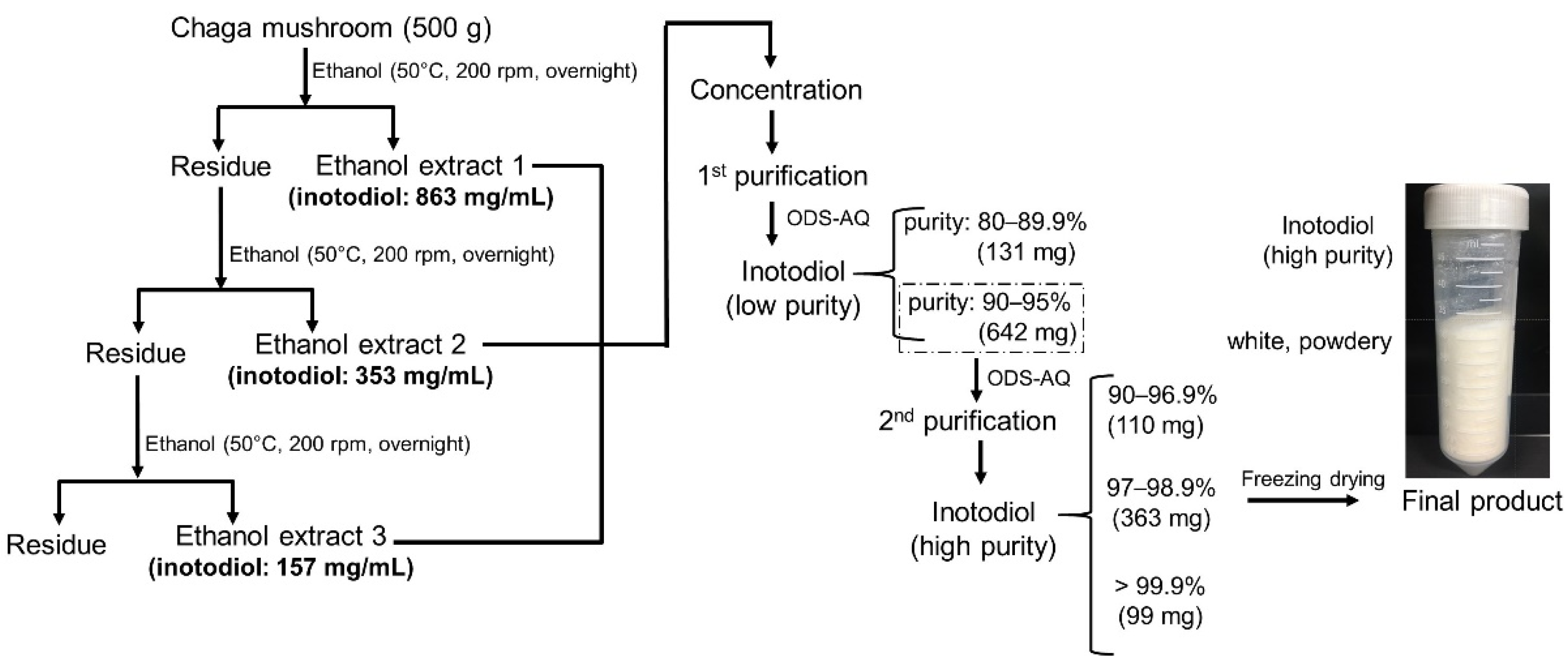
2.2. Inotodiol Extraction and Purification
2.3. Analytic Methodology
2.3.1. High-Performance Liquid Chromatography Evaporative Light-Scattering Detector (HPLC-ELSD)
2.3.2. High-Performance Liquid Chromatography-Tandem Mass Spectrometry (HPLC-MS/MS)
2.3.3. Nuclear Magnetic Resonance (NMR)
2.4. Physicochemical Properties
2.4.1. Thermal Analysis
2.4.2. Solubility Analyses in Organic Solvents
2.4.3. Density Functional Theory (DFT) Calculations
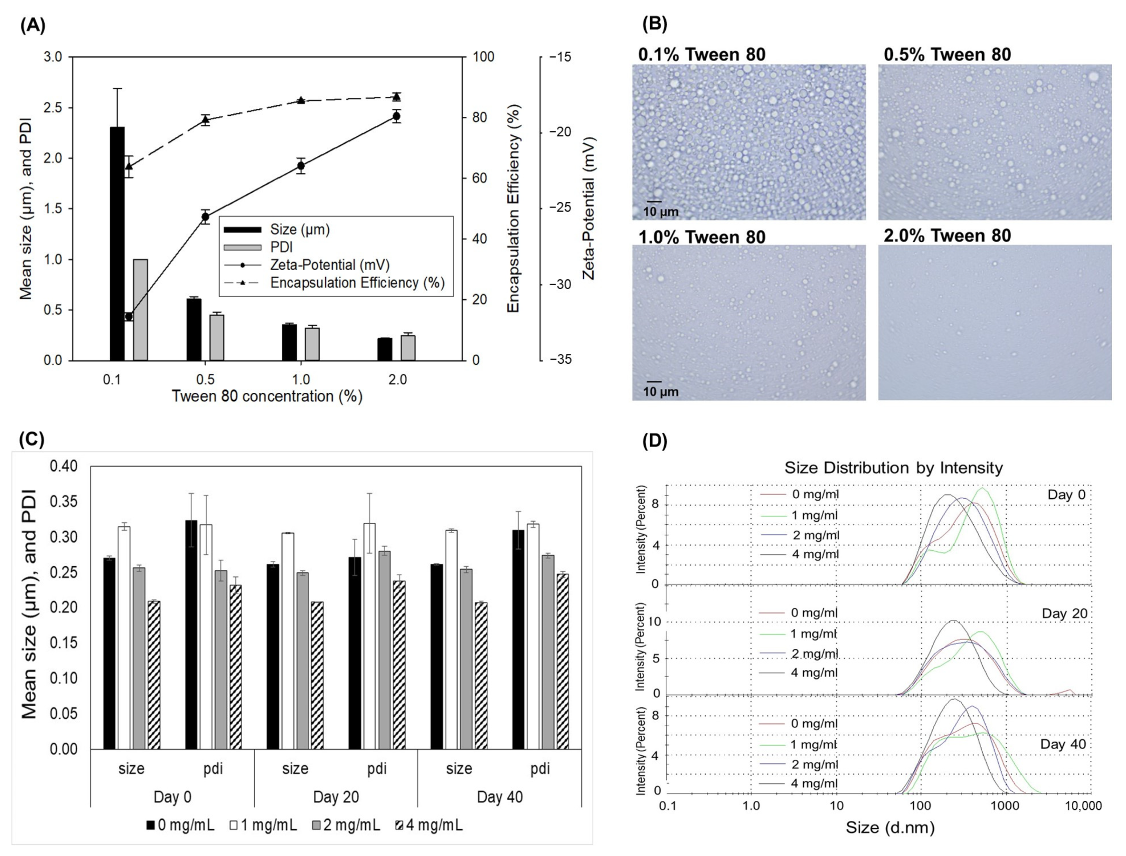
2.5. Inotodiol Emulsion Study
2.5.1. Inotodiol Emulsion Preparation
2.5.2. Encapsulation Efficiency (EE)
2.5.3. Microstructure, Mean Size, Polydispersity Index (PDI), and Zeta Potential Analysis
2.6. Pharmacokinetic Study and Analysis
2.6.1. Animals
2.6.2. Determination of Inotodiol Concentration in Plasma
2.6.3. Data Analysis
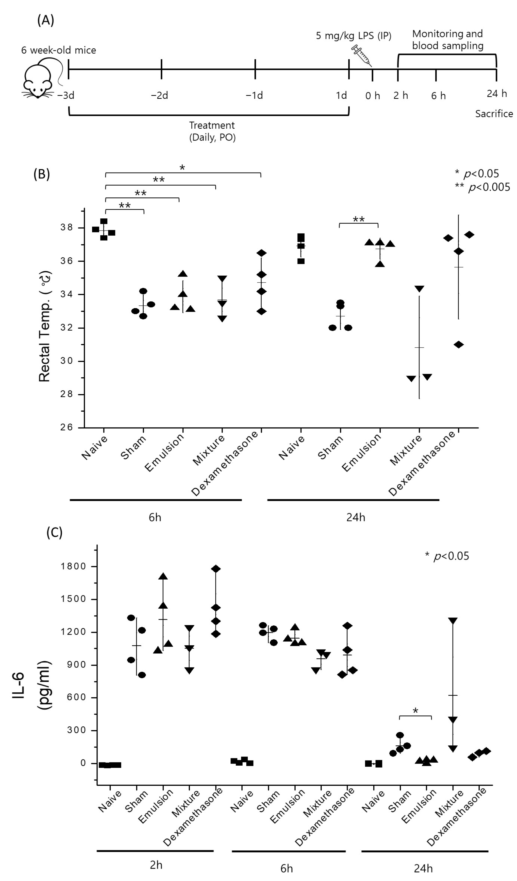
2.7. Acute Sepsis Mouse Model
3. Results and Discussion
3.1. Selection of the Extraction Solvent

| Solvent | Relative Polarity (i) | Log P (ii) | Toxicity (iii) | Solubility of Inotodiol (mg/mL) |
|---|---|---|---|---|
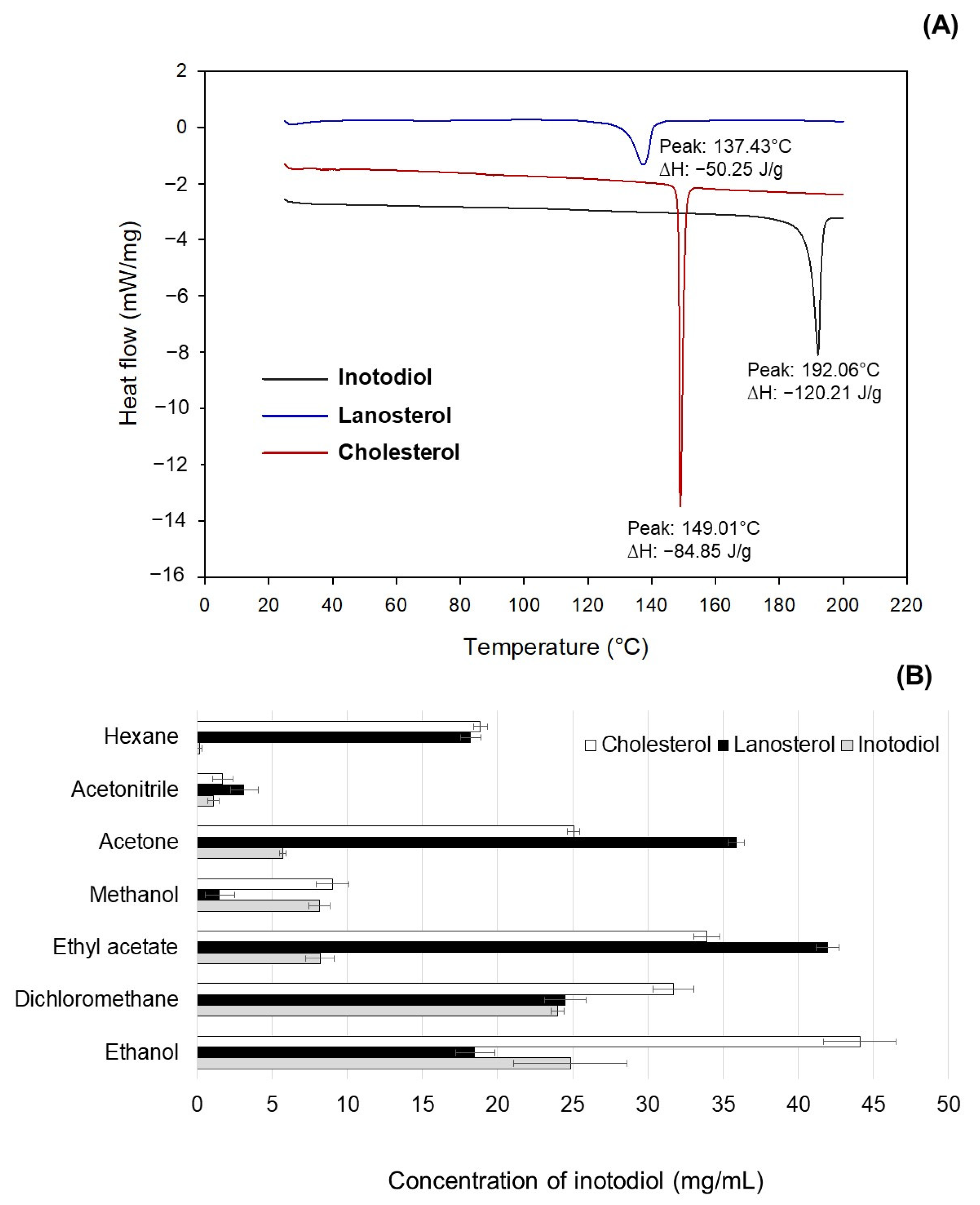
| Acetone | 0.355 | −0.23 | class 3 | 5.70 ± 0.39 |
| Acetonitrile | 0.460 | −0.33 | class 2 | 1.11 ± 0.19 |
| Dichloromethane | 0.309 | 0.60 | class 2 | 24.00 ± 0.95 |
| Ethanol | 0.654 | −0.24 | class 3 | 24.84 ± 0.43 |
| Ethyl acetate | 0.228 | 0.68 | class 3 | 8.18 ± 0.71 |
| Methanol | 0.762 | −0.76 | class 2 | 8.13 ± 0.24 |
| n-Hexane | 0.009 | 3.50 | class 2 | 0.15 ± 0.07 |
3.2. Large-Scale Extraction and Purification of Inotodiol
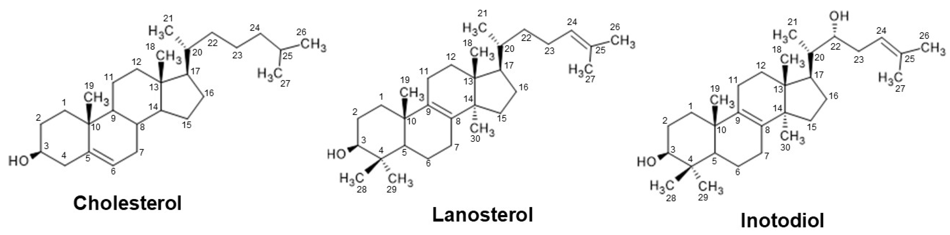
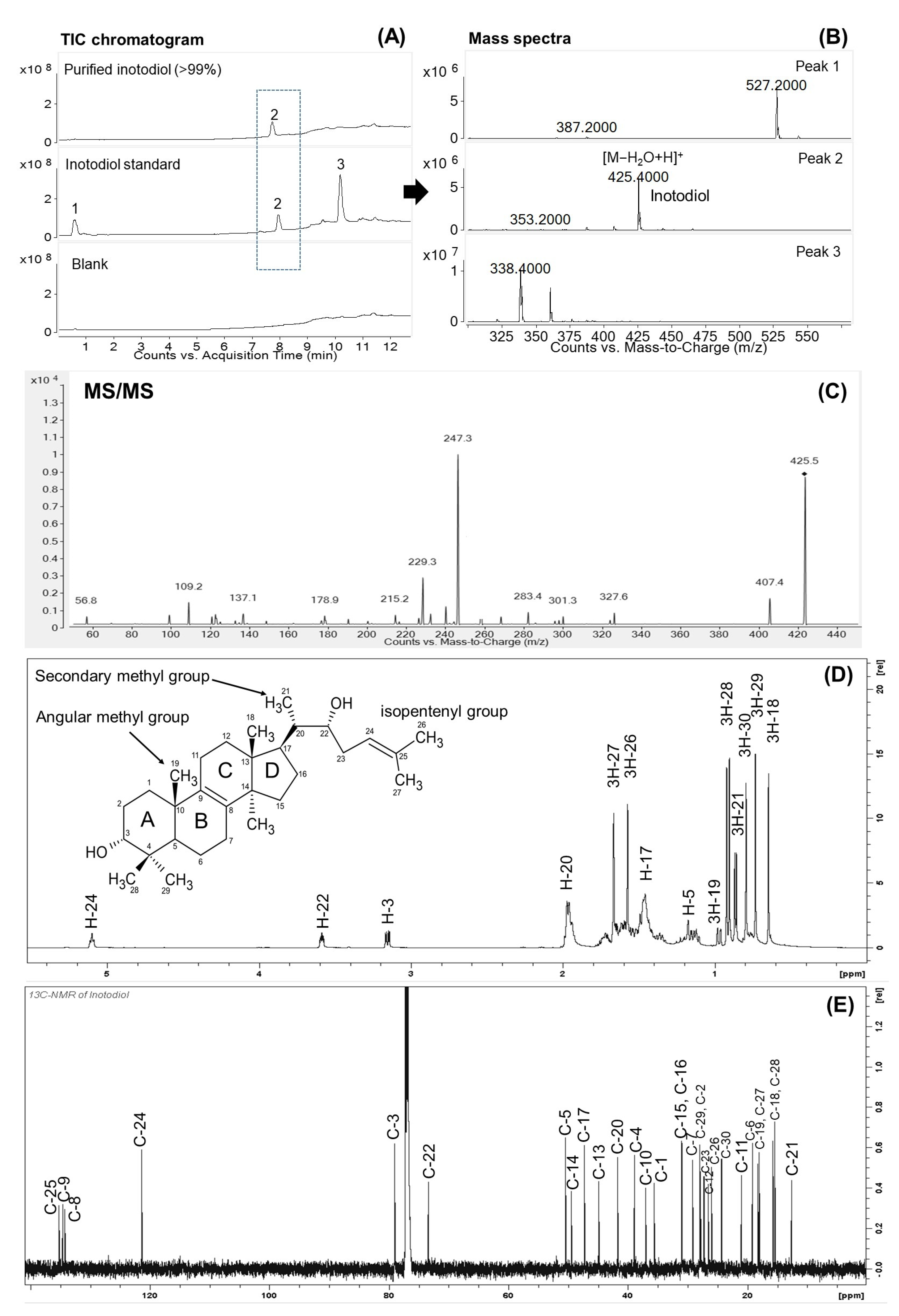
3.3. Structure Characterization
3.4. Physicochemical Properties of Inotodiol in Comparison with Cholesterol and Lanosterol
3.4.1. Thermal Property
3.4.2. Solubility in Organic Solvents
3.4.3. DFT Calculation of Molecular Interaction
3.5. Characteristics of Inotodiol Emulsion
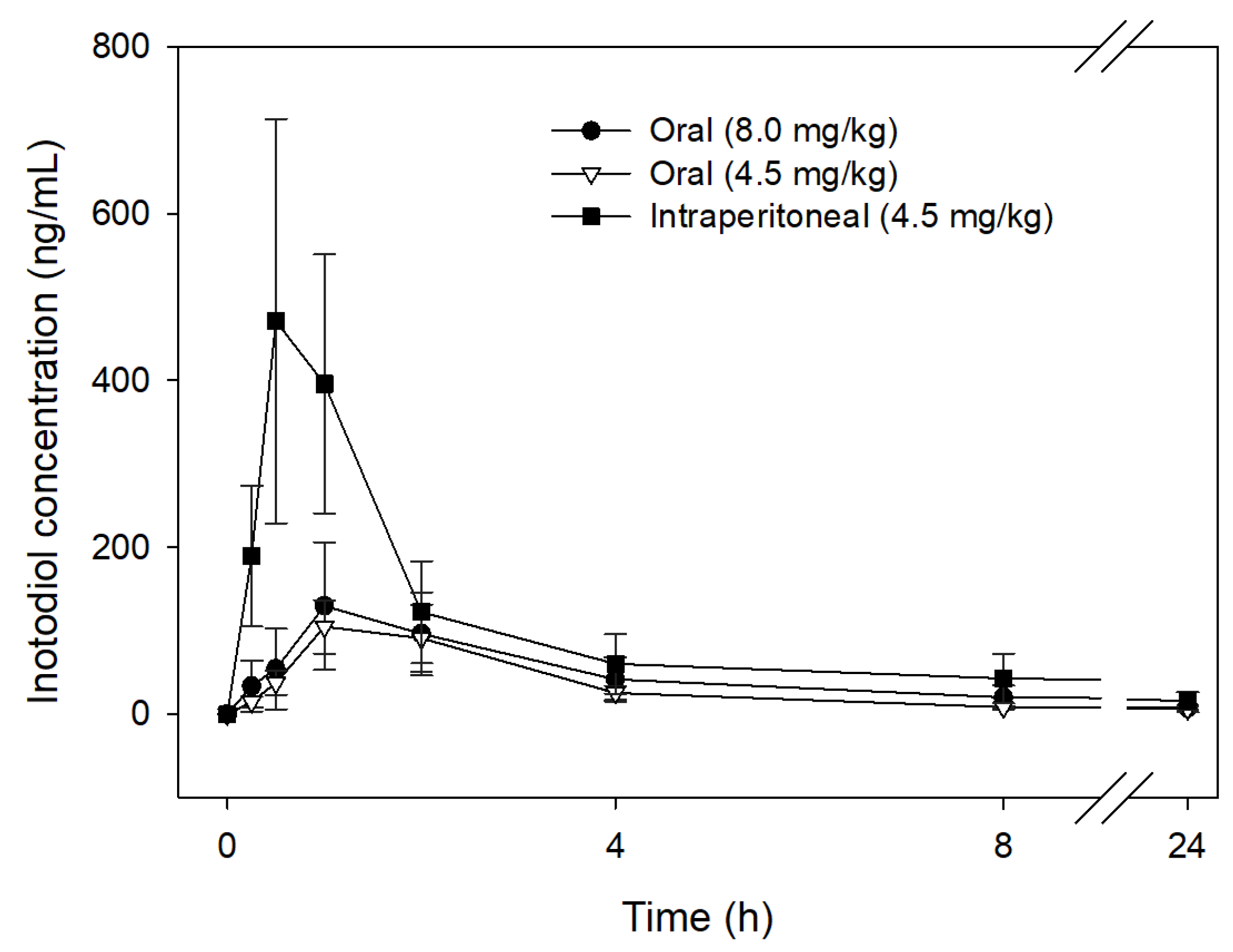
3.6. Pharmacokinetics of Inotodiol
3.7. Anti-Sepsis Properties of Inotodiol
4. Conclusions
Supplementary Materials
Author Contributions
Funding
Institutional Review Board Statement
Informed Consent Statement
Data Availability Statement
Conflicts of Interest
References
- Chung, M.J.; Chung, C.K.; Jeong, Y.; Ham, S.S. Anticancer activity of subfractions containing pure compounds of Chaga mushroom (Inonotus obliquus) extract in human cancer cells and in Balbc/c mice bearing Sarcoma-180 cells. Nutr. Res. Pract. 2010, 4, 177–182. [Google Scholar] [CrossRef] [PubMed]
- Nomura, M.; Takahashi, T.; Uesugi, A.; Tanaka, R.; Kobayashi, S. Inotodiol, a lanostane triterpenoid, from Inonotus obliquus inhibits cell proliferation through caspase-3-dependent apoptosis. Anticancer Res. 2008, 28, 2691–2696. [Google Scholar] [PubMed]
- Zhang, S.D.; Yu, L.; Wang, P.; Kou, P.; Li, J.; Wang, L.T.; Wang, W.; Yao, L.P.; Zhao, X.H.; Fu, Y.J. Inotodiol inhibits cells migration and invasion and induces apoptosis via p53-dependent pathway in HeLa cells. Phytomedicine 2019, 60, 152957. [Google Scholar] [CrossRef]
- Li, Y.; Zhang, W.; Chen, C.; Zhang, C.; Duan, J.; Yao, H.; Wei, Q.; Meng, A.; Shi, J. Inotodiol protects PC12 cells against injury induced by oxygen and glucose deprivation/restoration through inhibiting oxidative stress and apoptosis. J. Appl. Biomed. 2018, 16, 126–132. [Google Scholar] [CrossRef]
- Lee, S.H.; Won, G.W.; Choi, S.H.; Kim, M.Y.; Oh, C.H.; Park, J.T.; Park, J.I. Antiaging effect of inotodiol on oxidative stress in human dermal fibroblasts. Biomed. Pharmacother. 2022, 153, 113311. [Google Scholar] [CrossRef]
- Nguyen, T.M.N.; Ban, S.Y.; Park, K.B.; Lee, C.K.; Lee, S.W.; Lee, Y.J.; Baek, S.M.; Park, J.K.; Nguyen, M.T.T.; Kim, J.; et al. Evaluation of Toxicity and Efficacy of Inotodiol as an Anti-Inflammatory Agent Using Animal Model. Molecules 2022, 27, 4704. [Google Scholar] [CrossRef]
- Nguyet, T.M.N.; Lomunova, M.; Le, B.V.; Lee, J.S.; Park, S.K.; Kang, J.S.; Kim, Y.H.; Hwang, I. The mast cell stabilizing activity of Chaga mushroom critical for its therapeutic effect on food allergy is derived from inotodiol. Int. Immunopharmacol. 2018, 54, 286–295. [Google Scholar] [CrossRef]
- Nguyen, T.M.N.; Le, H.S.; Le, B.V.; Kim, Y.H.; Hwang, I. Anti-allergic effect of inotodiol, a lanostane triterpenoid from Chaga mushroom, via selective inhibition of mast cell function. Int. Immunopharmacol. 2020, 81, 106244. [Google Scholar] [CrossRef]
- Youssef, J.; Novosad, S.A.; Winthrop, K.L. Infection risk and safety of corticosteroid use. Rheum. Dis. Clin. N. Am. 2016, 42, 157–176. [Google Scholar] [CrossRef]
- Nikitina, S.A.; Habibrakhmanova, V.R.; Sysoeva, M.A. Chemical composition and biological activity of triterpenes and steroids of chaga mushroom. Biochem. (Mosc.) Suppl. B Biomed. Chem. 2016, 10, 63–69. [Google Scholar] [CrossRef]
- Nguyen, P.C.; Nguyen, M.T.T.; Lee, C.K.; Oh, I.N.; Kim, J.H.; Hong, S.T.; Park, J.T. Enzymatic synthesis and characterization of maltoheptaose-based sugar esters. Carbohydr. Polym. 2019, 218, 126–135. [Google Scholar] [CrossRef] [PubMed]
- Frisch, M.; Trucks, G.; Schlegel, H.; Scuseria, G.; Robb, M.; Cheeseman, J.; Scalmani, G.; Barone, V.; Petersson, G.; Nakatsuji, H. Gaussian 16 Revision C. 01; Gaussian Inc.: Wallingford, CT, USA, 2016. [Google Scholar]
- Marenich, A.V.; Cramer, C.J.; Truhlar, D.G. Universal solvation model based on solute electron density and on a continuum model of the solvent defined by the bulk dielectric constant and atomic surface tensions. J. Phys. Chem. B 2009, 113, 6378–6396. [Google Scholar] [CrossRef] [PubMed]
- Sugumar, S.; Ghosh, V.; Nirmala, M.J.; Mukherjee, A.; Chandrasekaran, N. Ultrasonic emulsification of eucalyptus oil nanoemulsion: Antibacterial activity against Staphylococcus aureus and wound healing activity in Wistar rats. Ultrason. Sonochem. 2014, 21, 1044–1049. [Google Scholar] [CrossRef] [PubMed]
- Nguyen, P.C.; Nguyen, M.T.T.; Kim, J.H.; Hong, S.T.; Kim, H.L.; Park, J.T. A novel maltoheptaose-based sugar ester having excellent emulsifying properties and optimization of its lipase-catalyzed synthesis. Food Chem. 2021, 352, 129358. [Google Scholar] [CrossRef] [PubMed]
- Zhang, Y.; Huo, M.; Zhou, J.; Xie, S. PKSolver: An add-in program for pharmacokinetic and pharmacodynamic data analysis in Microsoft Excel. Comput. Methods Programs Biomed. 2010, 99, 306–314. [Google Scholar] [CrossRef]
- Roosendaal, J.; Groenland, S.L.; Rosing, H.; Lucas, L.; Venekamp, N.; Nuijen, B.; Huitema, A.D.; Beijnen, J.H.; Steeghs, N. Determination of the absolute bioavailability of oral imatinib using a stable isotopically labeled intravenous imatinib-d8 microdose. Eur. J. Clin. Pharmacol. 2020, 76, 1075–1082. [Google Scholar] [CrossRef]
- FDA, U.S. Q3C-Tables and List Guidance for Industry. 2017. Available online: https://www.fda.gov/media/71737/download (accessed on 12 January 2023).
- Fridén, M.E.; Jumaah, F.; Gustavsson, C.; Enmark, M.; Fornstedt, T.; Turner, C.; Sjöberg, P.J.; Samuelsson, J. Evaluation and analysis of environmentally sustainable methodologies for extraction of betulin from birch bark with a focus on industrial feasibility. Green Chem. 2016, 18, 516–523. [Google Scholar] [CrossRef]
- Şoica, C.M.; Dehelean, C.A.; Peev, C.; Aluas, M.; Zupkó, I.; Kása Jr, P.; Alexa, E. Physico-chemical comparison of betulinic acid, betulin and birch bark extract and in vitro investigation of their cytotoxic effects towards skin epidermoid carcinoma (A431), breast carcinoma (MCF7) and cervix adenocarcinoma (HeLa) cell lines. Nat. Prod. Res. 2012, 26, 968–974. [Google Scholar] [CrossRef]
- Cai, C.; Ma, J.; Han, C.; Jin, Y.; Zhao, G.; He, X. Extraction and antioxidant activity of total triterpenoids in the mycelium of a medicinal fungus, Sanghuangporus sanghuang. Sci. Rep. 2019, 9, 7418. [Google Scholar] [CrossRef]
- Reichardt, C.; Welton, T. Solvents and Solvent Effects in Organic Chemistry, 4th ed.; Wiley: Hoboken, NJ, USA, 2010. [Google Scholar]
- Hazarika, S.; Goswami, P.; Dutta, N.N.; Hazarika, A.K. Ethyl oleate synthesis by Porcine pancreatic lipase in organic solvents. Chem. Eng. J. 2002, 85, 61–68. [Google Scholar] [CrossRef]
- Laane, C.; Boeren, S.; Vos, K.; Veeger, C. Rules for optimization of biocatalysis in organic solvents. Biotechnol. Bioeng. 1987, 30, 81–87. [Google Scholar] [CrossRef] [PubMed]
- Huynh, N.; Beltrame, G.; Tarvainen, M.; Suomela, J.P.; Yang, B. Supercritical CO2 Extraction of Triterpenoids from Chaga Sterile Conk of Inonotus obliquus. Molecules 2022, 27, 1880. [Google Scholar] [CrossRef] [PubMed]
- Won, H.J.; Lee, S.M.; Kim, D.Y.; Kwon, O.K.; Park, M.H.; Kim, J.H.; Ryu, H.W.; Oh, S.R. Rapid securing of reference substances from Peucedanum japonicum Thunberg by recycling preparative high-performance liquid chromatography. J. Chromatogr. B 2019, 1133, 121835. [Google Scholar] [CrossRef]
- Rafferty, J.L.; Siepmann, J.I.; Schure, M.R. Mobile phase effects in reversed-phase liquid chromatography: A comparison of acetonitrile/water and methanol/water solvents as studied by molecular simulation. J. Chromatogr. A 2011, 1218, 2203–2213. [Google Scholar] [CrossRef] [PubMed]
- Münger, L.H.; Boulos, S.; Nyström, L. UPLC-MS/MS based identification of dietary steryl glucosides by investigation of corresponding free sterols. Front. Chem. 2018, 6, 342. [Google Scholar] [CrossRef]
- Géry, A.; Dubreule, C.; André, V.; Rioult, J.P.; Bouchart, V.; Heutte, N.; Eldin de Pécoulas, P.; Krivomaz, T.; Garon, D. Chaga (Inonotus obliquus), a future potential medicinal fungus in oncology? A chemical study and a comparison of the cytotoxicity against human lung adenocarcinoma cells (A549) and human bronchial epithelial cells (BEAS-2B). Integr. Cancer Ther. 2018, 17, 832–843. [Google Scholar] [CrossRef]
- Kim, Y.J.; Park, J.; Min, B.S.; Shim, S.H. Chemical constituents from the sclerotia of Inonotus obliquus. J. Korean Soc. Appl. Biol. Chem. 2011, 54, 287–294. [Google Scholar] [CrossRef]
- Du, D.; Zhu, F.; Chen, X.; Ju, X.; Feng, Y.; Qi, L.W.; Jiang, J. Rapid isolation and purification of inotodiol and trametenolic acid from Inonotus obliquus by high-speed counter-current chromatography with evaporative light scatting detection. Phytochem. Anal. 2011, 22, 419–423. [Google Scholar] [CrossRef]
- Li, K.; Forciniti, D. Solubility of Lanosterol in Organic Solvents and in Water–Alcohol Mixtures at 101.8 kPa. J. Chem. Eng. Data. 2020, 65, 436–445. [Google Scholar] [CrossRef]
- Williams, J.H.; Kuchmak, M.; Witter, R.F. Purity of cholesterol to be used as a primary standard. J. Lipid Res. 1965, 6, 461–465. [Google Scholar] [CrossRef]
- Chu, K.A.; Yalkowsky, S.H. An interesting relationship between drug absorption and melting point. Int. J. Pharm. 2009, 373, 24–40. [Google Scholar] [CrossRef]
- Ponnuchamy, V.; Gordobil, O.; Diaz, R.H.; Sandak, A.; Sandak, J. Fractionation of lignin using organic solvents: A combined experimental and theoretical study. Int. J. Biol. Macromol. 2021, 168, 792–805. [Google Scholar] [CrossRef] [PubMed]
- Da Silva, H.C.; Paluch, A.S.; Costa, L.T.; De Almeida, W.B. Thermodynamic and structural description of relative solubility of the flavonoid rutin by DFT calculations and molecular dynamics simulations. J. Mol. Liq. 2021, 341, 117214. [Google Scholar] [CrossRef]
- Kim, J.H.; Gao, D.; Cho, C.W.; Hwang, I.; Kim, H.M.; Kang, J.S. A Novel Bioanalytical Method for determination of inotodiol isolated from inonotus obliquus and its application to pharmacokinetic study. Plants 2021, 10, 1631. [Google Scholar] [CrossRef] [PubMed]
- Roldan-Cruz, C.; Vernon-Carter, E.J.; Alvarez-Ramirez, J. Assessing the stability of Tween 80-based O/W emulsions with cyclic voltammetry and electrical impedance spectroscopy. Colloids Surf. A Physicochem. Eng. Asp. 2016, 511, 145–152. [Google Scholar] [CrossRef]
- Forgács, E.; Cserháti, T.; Farkas, O.; Eckhardt, A.; Miksik, I.; Deyl, Z. Interaction Between Cholesterol and Non-ionic Surfactants Studied by Thin-Layer Chromatography. J. Liq. Chromatogr. Relat. Technol. 2009, 27, 1981–1992. [Google Scholar] [CrossRef]
- Ostlund Jr, R.E.; McGill, J.B.; Zeng, C.M.; Covey, D.F.; Stearns, J.; Stenson, W.F.; Spilburg, C.A. Gastrointestinal absorption and plasma kinetics of soy Δ5-phytosterols and phytostanols in humans. Am. J. Physiol. Endocrinol. Metab. 2002, 282, E911–E916. [Google Scholar] [CrossRef]
- Meaney, S.; Hassan, M.; Sakinis, A.; Lütjohann, D.; von Bergmann, K.; Wennmalm, Å.; Diczfalusy, U.; Björkhem, I. Evidence that the major oxysterols in human circulation originate from distinct pools of cholesterol: A stable isotope study. J. Lipid Res. 2001, 42, 70–78. [Google Scholar] [CrossRef]
- Meaney, S.; Bodin, K.; Diczfalusy, U.; Björkhem, I. On the rate of translocation in vitro and kinetics in vivo of the major oxysterols in human circulation: Critical importance of the position of the oxygen function. J. Lipid Res. 2002, 43, 2130–2135. [Google Scholar] [CrossRef]
- Godugu, C.; Patel, A.R.; Doddapaneni, R.; Somagoni, J.; Singh, M. Approaches to improve the oral bioavailability and effects of novel anticancer drugs berberine and betulinic acid. PLoS ONE 2014, 9, e89919. [Google Scholar] [CrossRef] [Green Version]
- Jeong, D.W.; Kim, Y.H.; Kim, H.H.; Ji, H.Y.; Yoo, S.D.; Choi, W.R.; Lee, S.M.; Han, C.K.; Lee, H.S. Dose-linear pharmacokinetics of oleanolic acid after intravenous and oral administration in rats. Biopharm. Drug Dispos. 2007, 28, 51–57. [Google Scholar] [CrossRef] [PubMed]
- Supajatura, V.; Ushio, H.; Nakao, A.; Okumura, K.; Ra, C.; Ogawa, H. Protective roles of mast cells against enterobacterial infection are mediated by Toll-like receptor 4. J. Immunol. 2001, 167, 2250–2256. [Google Scholar] [CrossRef] [PubMed]
- Nautiyal, K.M.; McKellar, H.; Silverman, A.J.; Silver, R. Mast cells are necessary for the hypothermic response to LPS-induced sepsis. Am. J. Physiol. Regul. Integr. Comp. Physiol. 2009, 296, R595–R602. [Google Scholar] [CrossRef] [PubMed] [Green Version]


| Parameters | Units | Oral (8.0 mg/kg) | Oral (4.5 mg/kg) | Intraperitoneal (4.5 mg/kg) |
|---|---|---|---|---|
| ka | 1/h | 1.60 ± 1.29 | 0.77 ± 0.20 | 2.14 ± 1.32 |
| t1/2ka | h | 0.65 ± 0.34 | 0.95 ± 0.19 | 0.40 ± 0.14 |
| Tmax | h | 1.28 ± 0.48 | 1.44 ± 0.20 | 0.70 ± 0.17 |
| Cmax | ng/mL | 116.25 ± 43.54 | 88.05 ± 17.06 | 412.81 ± 155.12 |
| AUC0–24h | ng·h/mL | 489.45 ± 252.66 | 341.81 ± 86.77 | 827.20 ± 216.68 |
| F | % | 33.28 | 41.32 | - |
Disclaimer/Publisher’s Note: The statements, opinions and data contained in all publications are solely those of the individual author(s) and contributor(s) and not of MDPI and/or the editor(s). MDPI and/or the editor(s) disclaim responsibility for any injury to people or property resulting from any ideas, methods, instructions or products referred to in the content. |
© 2023 by the authors. Licensee MDPI, Basel, Switzerland. This article is an open access article distributed under the terms and conditions of the Creative Commons Attribution (CC BY) license (https://creativecommons.org/licenses/by/4.0/).
Share and Cite
Nguyen, P.C.; Nguyen, M.T.T.; Truong, B.T.; Kim, D.-R.; Shin, S.; Kim, J.-E.; Park, K.-B.; Park, J.-H.; Tran, P.L.; Ban, S.-Y.; et al. Isolation, Physicochemical Characterization, and Biological Properties of Inotodiol, the Potent Pharmaceutical Oxysterol from Chaga Mushroom. Antioxidants 2023, 12, 447. https://doi.org/10.3390/antiox12020447
Nguyen PC, Nguyen MTT, Truong BT, Kim D-R, Shin S, Kim J-E, Park K-B, Park J-H, Tran PL, Ban S-Y, et al. Isolation, Physicochemical Characterization, and Biological Properties of Inotodiol, the Potent Pharmaceutical Oxysterol from Chaga Mushroom. Antioxidants. 2023; 12(2):447. https://doi.org/10.3390/antiox12020447
Chicago/Turabian StyleNguyen, Phu Cuong, My Tuyen Thi Nguyen, Ba Tai Truong, Dae-Ryeol Kim, Sujin Shin, Ju-Eun Kim, Kyu-Been Park, Ji-Hyun Park, Phuong Lan Tran, So-Young Ban, and et al. 2023. "Isolation, Physicochemical Characterization, and Biological Properties of Inotodiol, the Potent Pharmaceutical Oxysterol from Chaga Mushroom" Antioxidants 12, no. 2: 447. https://doi.org/10.3390/antiox12020447