
Static Magnetic Fields Protect against Cisplatin-Induced Kidney Toxicity
 , , , , ,
Abstract
:1. Introduction
2. Materials and Methods
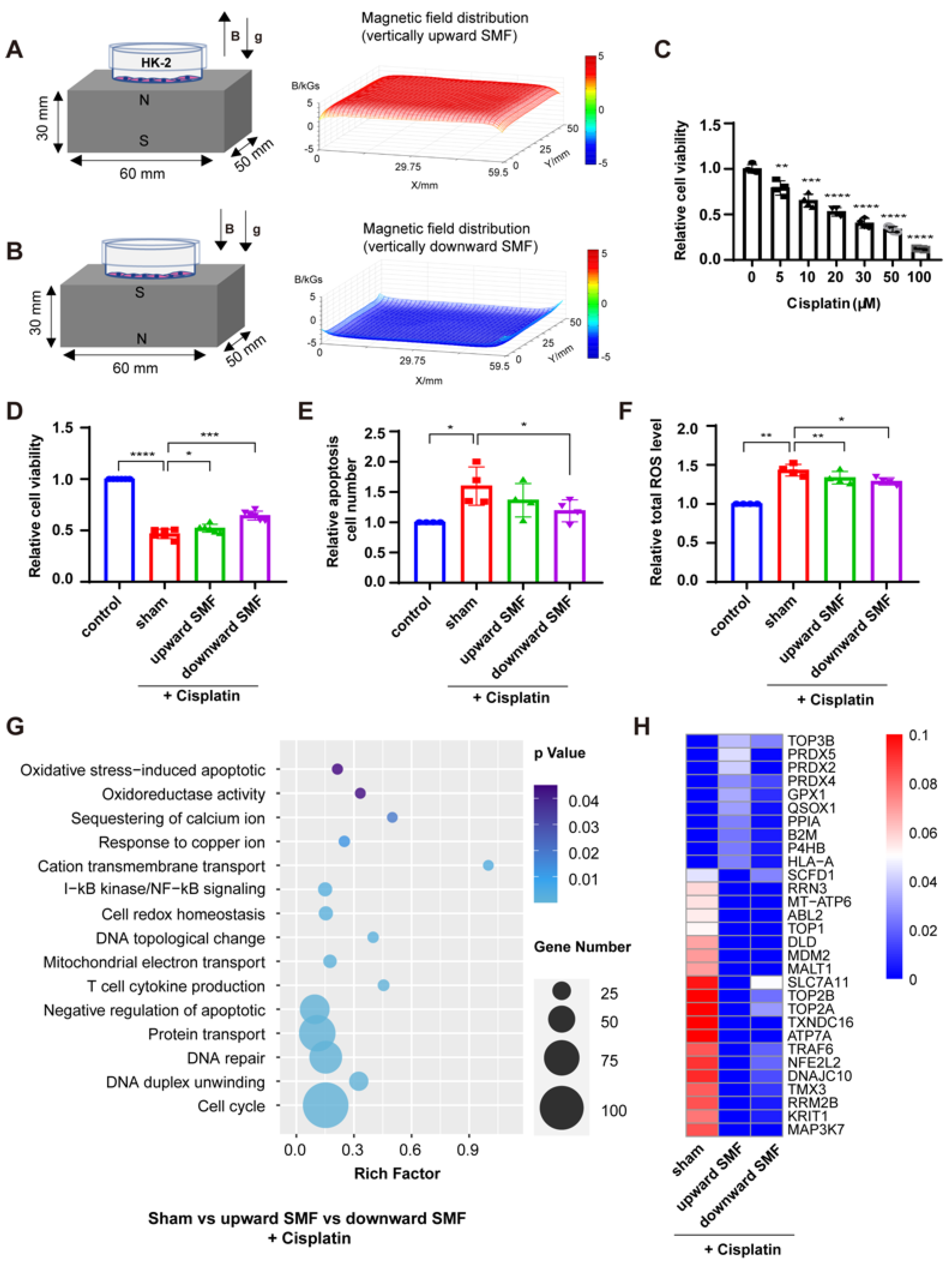
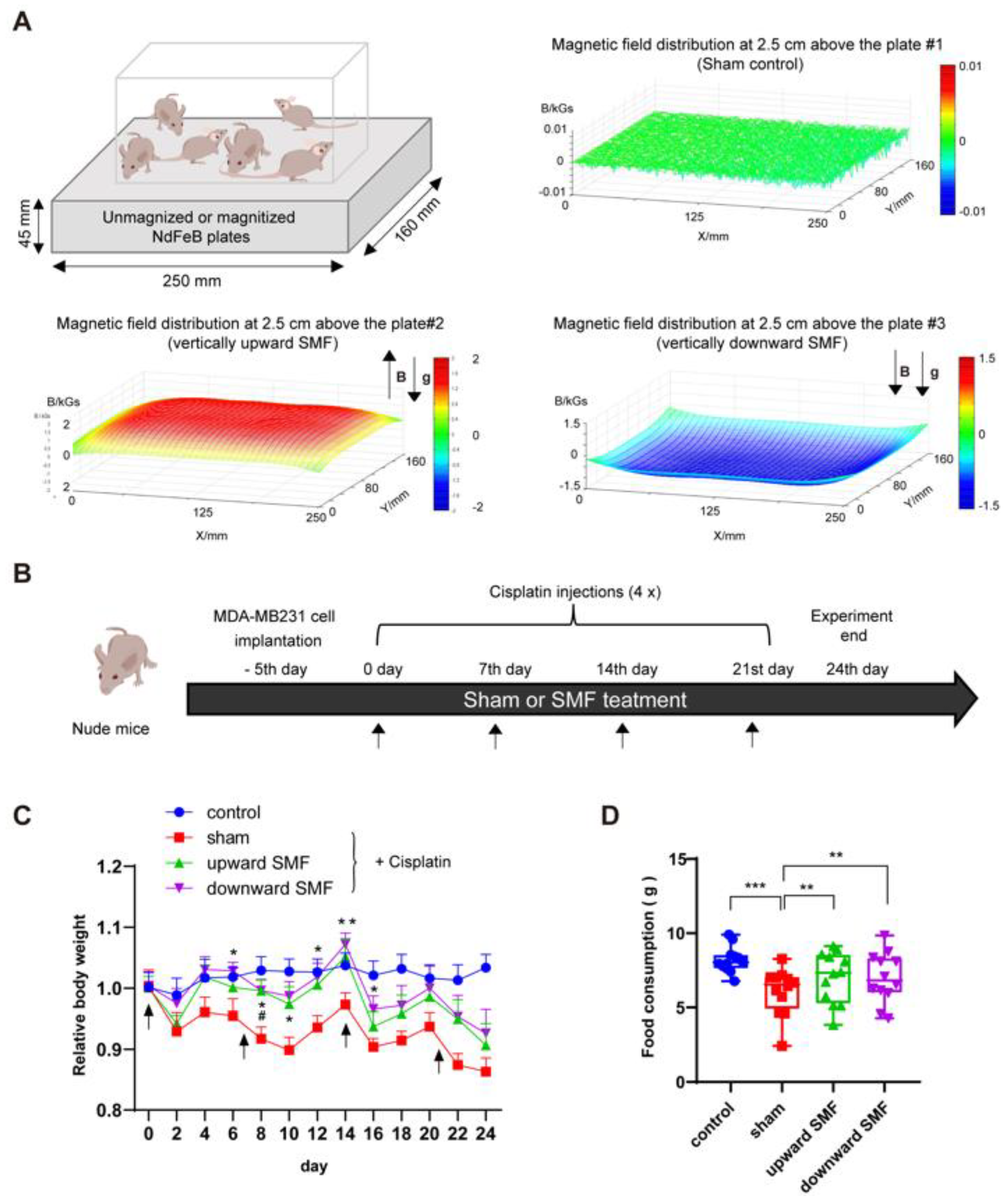
2.1. Static Magnetic Field Exposure
2.2. Cell Culture
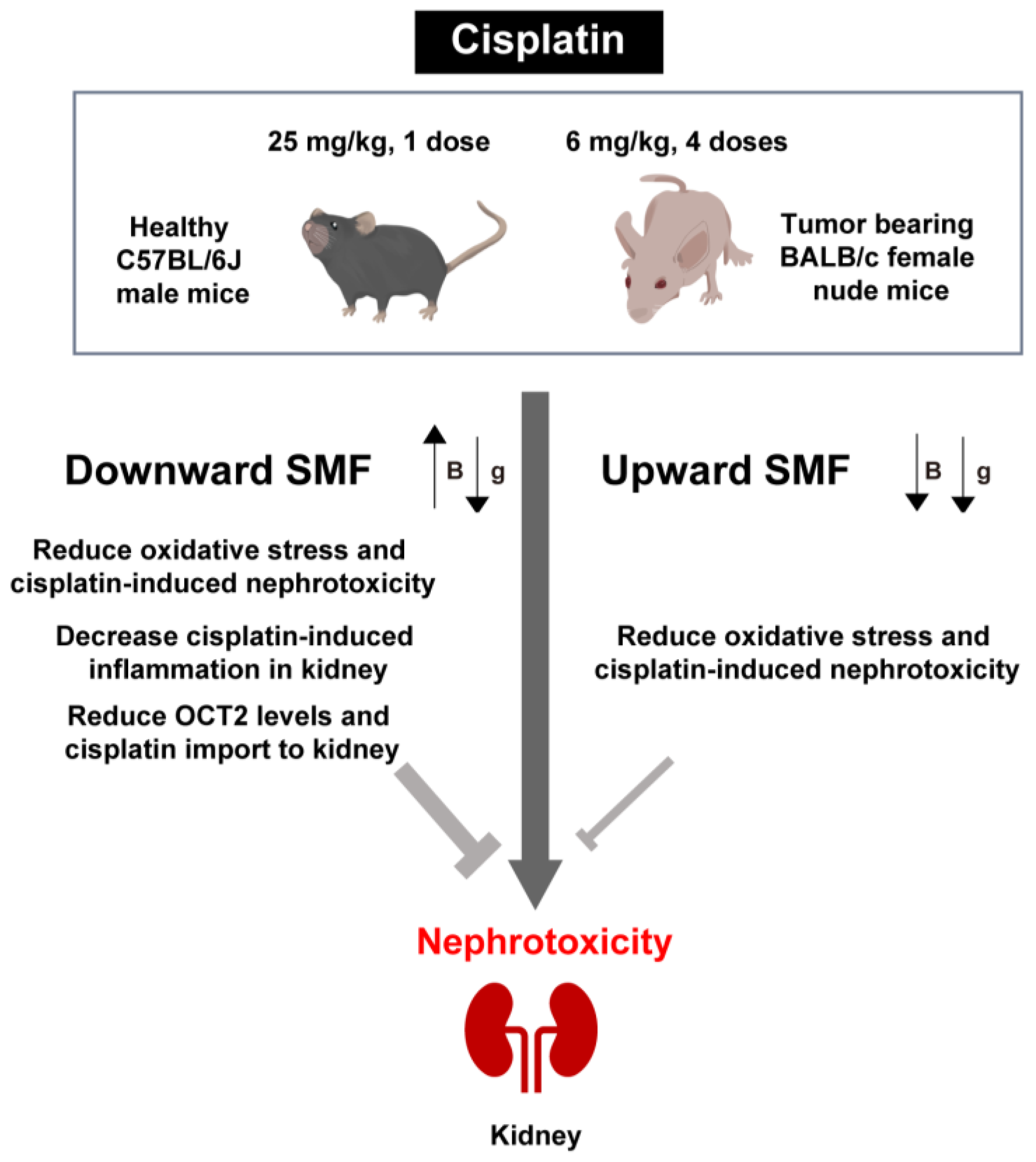
2.3. Animal Model
2.4. Histological Analysis
2.5. Immunohistochemistry
2.6. Terminal Deoxynucleotidyl Transferase dUTP Nick-End Labeling (TUNEL) Assay
2.7. DHE Staining
2.8. Measurement of Platinum Concentration
2.9. Cell Viability Analysis
2.10. Cell Apoptosis Analysis
2.11. Cellular ROS Detection
2.12. Mice Behavior Tests—Open Field Test (OFT)
2.13. Western Blotting
2.14. RNA Extraction Library Construction and Sequencing
2.15. Statistical Analysis
3. Results
3.1. SMFs Protect HK-2 Cells from Cisplatin-Induced Cytotoxicity
3.2. SMFs Improve the Physiological State of Tumor-Bearing Mice Treated with Cisplatin
3.3. SMFs Enhanced the Locomotive and Exploratory Activities of Cisplatin-Treated Tumor-Bearing Mice
3.4. SMFs Protect against Cisplatin-Induced Kidney Injury in Tumor-Bearing Mice
3.5. Short-Term SMF Treatment can Effectively Ameliorate Cisplatin-Induced Acute Kidney Injury in Healthy Mice
4. Discussion
5. Conclusions
Supplementary Materials
Author Contributions
Funding
Institutional Review Board Statement
Informed Consent Statement
Data Availability Statement
Acknowledgments
Conflicts of Interest
References
- Rottenberg, S.; Disler, C.; Perego, P. The rediscovery of platinum-based cancer therapy. Nat. Rev. Cancer 2020, 21, 37–50. [Google Scholar] [CrossRef] [PubMed]
- Manohar, S.; Leung, N. Cisplatin nephrotoxicity: A review of the literature. J. Nephrol. 2017, 31, 15–25. [Google Scholar] [CrossRef] [PubMed]
- Pabla, N.; Dong, Z. Cisplatin nephrotoxicity: Mechanisms and renoprotective strategies. Kidney Int. 2008, 73, 994–1007. [Google Scholar] [CrossRef] [PubMed] [Green Version]
- Campbell, A.B.; Kalman, S.M.; Jacobs, C. Plasma platinum levels: Relationship to cisplatin dose and nephrotoxicity. Cancer Treat. Rep. 1983, 67, 169–172. [Google Scholar] [PubMed]
- Santoso, J.T.; Lucci, J.A.; Coleman, R.L.; Schafer, I.; Hannigan, E.V. Saline, mannitol, and furosemide hydration in acute cisplatin nephrotoxicity: A randomized trial. Cancer Chemother. Pharmacol. 2003, 52, 13–18. [Google Scholar] [CrossRef]
- Crona, D.J.; Faso, A.; Nishijima, T.F.; McGraw, K.A.; Galsky, M.D.; Milowsky, M.I. A Systematic Review of Strategies to Prevent Cisplatin-Induced Nephrotoxicity. Oncologist 2017, 22, 609–619. [Google Scholar] [CrossRef] [Green Version]
- Volarevic, V.; Djokovic, B.; Jankovic, M.G.; Harrell, C.R.; Fellabaum, C.; Djonov, V.; Arsenijevic, N. Molecular mechanisms of cisplatin-induced nephrotoxicity: A balance on the knife edge between renoprotection and tumor toxicity. J. Biomed. Sci. 2019, 26, 1–14. [Google Scholar] [CrossRef] [Green Version]
- Lewis, A.M.; Fay, T.P.; Manolopoulos, D.E.; Kerpal, C.; Richert, S.; Timmel, C.R. On the low magnetic field effect in radical pair reactions. J. Chem. Phys. 2018, 149, 034103. [Google Scholar] [CrossRef] [Green Version]
- Ikeya, N.; Woodward, J.R. Cellular autofluorescence is magnetic field sensitive. Proc. Natl. Acad. Sci. USA 2021, 118, e2018043118. [Google Scholar] [CrossRef]
- Zhang, X.; Yarema, K.; Xu, A. Biological Effects of Static Magnetic Fields; Springer: Singapore, 2017. [Google Scholar] [CrossRef]
- Tian, X.; Wang, C.; Yu, B.; Fan, Y.; Zhang, L.; Zhang, X. 9.4 T static magnetic field ameliorates imatinib mesylate-induced toxicity and depression in mice. Eur. J. Pediatr. 2022. Online ahead of print. [Google Scholar] [CrossRef]
- Yu, B.; Liu, J.; Cheng, J.; Zhang, L.; Song, C.; Tian, X.; Fan, Y.; Lv, Y.; Zhang, X. A Static Magnetic Field Improves Iron Metabolism and Prevents High-Fat-Diet/Streptozocin-Induced Diabetes. Innovation 2021, 2, 100077. [Google Scholar] [CrossRef] [PubMed]
- Koepsell, H. Organic cation transporters in intestine, kidney, liver, and brain. Annu. Rev. Physiol. 1998, 60, 243–266. [Google Scholar] [CrossRef] [PubMed]
- Miller, R.P.; Tadagavadi, R.K.; Ramesh, G.; Reeves, W.B. Mechanisms of Cisplatin Nephrotoxicity. Toxins 2010, 2, 2490–2518. [Google Scholar] [CrossRef] [PubMed] [Green Version]
- Ozkok, A.; Edelstein, C.L. Pathophysiology of cisplatin-induced acute kidney injury. BioMed Res. Int. 2014, 2014, 967826. [Google Scholar] [CrossRef] [PubMed] [Green Version]
- Wang, H.; Zhang, X. Magnetic Fields and Reactive Oxygen Species. Int. J. Mol. Sci. 2017, 18, 2175. [Google Scholar] [CrossRef] [Green Version]
- Carter, C.S.; Huang, S.; Searby, C.; Cassaidy, B.; Miller, M.; Grzesik, W.; Piorczynski, T.; Pak, T.; Walsh, S.; Acevedo, M.; et al. Exposure to Static Magnetic and Electric Fields Treats Type 2 Diabetes. Cell Metab. 2020, 32, 561–574.e7. [Google Scholar] [CrossRef]
- Van Huizen, A.V.; Morton, J.M.; Kinsey, L.J.; Von Kannon, D.G.; Saad, M.A.; Birkholz, T.R.; Czajka, J.M.; Cyrus, J.; Barnes, F.S.; Beane, W.S. Weak magnetic fields alter stem cell–mediated growth. Sci. Adv. 2019, 5, eaau7201. [Google Scholar] [CrossRef] [Green Version]
- Feng, C.; Yu, B.; Song, C.; Wang, J.; Zhang, L.; Ji, X.; Wang, Y.; Fang, Y.; Liao, Z.; Wei, M.; et al. Static Magnetic Fields Reduce Oxidative Stress to Improve Wound Healing and Alleviate Diabetic Complications. Cells 2022, 11, 443. [Google Scholar] [CrossRef]
- Shine, M.; Guruprasad, K.; Anand, A. Effect of stationary magnetic field strengths of 150 and 200 mT on reactive oxygen species production in soybean. Bioelectromagnetics 2012, 33, 428–437. [Google Scholar] [CrossRef]
- Jouni, F.J.; Abdolmaleki, P.; Ghanati, F. Oxidative stress in broad bean (Vicia faba L.) induced by static magnetic field under natural radioactivity. Mutat. Res. Toxicol. Environ. Mutagen. 2012, 741, 116–121. [Google Scholar] [CrossRef]
- Timmel, C.; Till, U.; Brocklehurst, B.; Mclauchlan, K.; Hore, P. Effects of weak magnetic fields on free radical recombination reactions. Mol. Phys. 1998, 95, 71–89. [Google Scholar] [CrossRef]
- Ciarimboli, G.; Ludwig, T.; Lang, D.; Pavenstädt, H.; Koepsell, H.; Piechota, H.-J.; Haier, J.; Jaehde, U.; Zisowsky, J.; Schlatter, E. Cisplatin Nephrotoxicity Is Critically Mediated via the Human Organic Cation Transporter 2. Am. J. Pathol. 2005, 167, 1477–1484. [Google Scholar] [CrossRef] [PubMed] [Green Version]
- Yonezawa, A.; Masuda, S.; Nishihara, K.; Yano, I.; Katsura, T.; Inui, K.-I. Association between tubular toxicity of cisplatin and expression of organic cation transporter rOCT2 (Slc22a2) in the rat. Biochem. Pharmacol. 2005, 70, 1823–1831. [Google Scholar] [CrossRef] [PubMed]
- Lu, G.; Opella, S.J. Resonance assignments of a membrane protein in phospholipid bilayers by combining multiple strategies of oriented sample solid-state NMR. J. Biomol. NMR 2013, 58, 69–81. [Google Scholar] [CrossRef] [PubMed] [Green Version]
- Vergallo, C.; Dini, L. Comparative analysis of biological effects induced on different cell types by magnetic fields with magnetic flux densities in the range of 1–60 mT and frequencies up to 50 Hz. Sustainability 2018, 10, 2776. [Google Scholar] [CrossRef] [Green Version]
- Calabrò, E.; Condello, S.; Currò, M.; Ferlazzo, N.; Caccamo, D.; Magazù, S.; Ientile, R. Effects of low intensity static magnetic field on FTIR spectra and ROS production in SH-SY5Y neuronal-like cells. Bioelectromagnetics 2013, 34, 618–629. [Google Scholar] [CrossRef]
- Hsu, S.-H.; Chang, J.-C. The static magnetic field accelerates the osteogenic differentiation and mineralization of dental pulp cells. Cytotechnology 2010, 62, 143–155. [Google Scholar] [CrossRef] [Green Version]
- Dini, L.; Dwikat, M.; Panzarini, E.; Vergallo, C.; Tenuzzo, B. Morphofunctional study of 12-O-tetradecanoyl-13-phorbol acetate (TPA)-induced differentiation of U937 cells under exposure to a 6 mT static magnetic field. Bioelectromagnetics 2009, 30, 352–364. [Google Scholar] [CrossRef]
- Chionna, A.; Dwikat, M.; Panzarini, E.; Tenuzzo, B.; Verri, T.; Pagliara, P.; Abbro, L.; Dini, L. Cell shape and plasma membrane alterations after static magnetic fields exposure. Eur. J. Histochem. 2003, 47, 299–308. [Google Scholar] [CrossRef]
- Chionna, A.; Tenuzzo, B.; Panzarini, E.; Dwikat, M.B.; Abbro, L.; Dini, L. Time dependent modifications of Hep G2 cells during exposure to static magnetic fields. Bioelectromagnetics 2005, 26, 275–286. [Google Scholar] [CrossRef]
- Braganza, L.F.; Blott, B.H.; Coe, T.J.; Melville, D. The superdiamagnetic effect of magnetic fields on one and two component multilamellar liposomes. Biochim. Biophys. Acta (BBA) Gen. Subj. 1984, 801, 66–75. [Google Scholar] [CrossRef]
- Torbati, M.; Mozaffari, K.; Liu, L.; Sharma, P. Coupling of mechanical deformation and electromagnetic fields in biological cells. Rev. Mod. Phys. 2022, 94, 025003. [Google Scholar] [CrossRef]
- Tasić, T.; Lozić, M.; Glumac, S.; Stanković, M.; Milovanovich, I.; Djordjevich, D.M.; Trbovich, A.M.; Japundžić-Žigon, N.; De Luka, S.R. Static magnetic field on behavior, hematological parameters and organ damage in spontaneously hypertensive rats. Ecotoxicol. Environ. Saf. 2020, 207, 111085. [Google Scholar] [CrossRef] [PubMed]
- Jin, Y.; Guo, W.; Hu, X.; Liu, M.; Xu, X.; Hu, F.; Lan, Y.; Lv, C.; Fang, Y.; Liu, M.; et al. Static magnetic field regulates Arabidopsis root growth via auxin signaling. Sci. Rep. 2019, 9, 14384. [Google Scholar] [CrossRef] [Green Version]
- Tian, X.; Wang, D.; Zha, M.; Yang, X.; Ji, X.; Zhang, L.; Zhang, X. Magnetic field direction differentially impacts the growth of different cell types. Electromagn. Biol. Med. 2018, 37, 114–125. [Google Scholar] [CrossRef] [PubMed]
- Yang, X.; Song, C.; Zhang, L.; Wang, J.; Yu, X.; Yu, B.; Zablotskii, V.; Zhang, X. An upward 9.4 T static magnetic field inhibits DNA synthesis and increases ROS-P53 to suppress lung cancer growth. Transl. Oncol. 2021, 14, 101103. [Google Scholar] [CrossRef] [PubMed]
- Raylman, R.R.; Clavo, A.C.; Wahl, R.L. Exposure to strong static magnetic field slows the growth of human cancer cells in vitro. Bioelectromagnetics 1996, 17, 358–363. [Google Scholar] [CrossRef]
- Tofani, S.; Barone, D.; Cintorino, M.; De Santi, M.M.; Ferrara, A.; Orlassino, R.; Ossola, P.; Peroglio, F.; Rolfo, K.; Ronchetto, F. Static and ELF magnetic fields induce tumor growth inhibition and apoptosis. Bioelectromagnetics 2001, 22, 419–428. [Google Scholar] [CrossRef] [PubMed]
- Zhang, L.; Yang, X.; Liu, J.; Luo, Y.; Li, Z.; Ji, X.; Wang, W.; Zhang, X. 1 T moderate intensity static magnetic field affects Akt/mTOR pathway and increases the antitumor efficacy of mTOR inhibitors in CNE-2Z cells. Sci. Bull. 2015, 60, 2120–2128. [Google Scholar] [CrossRef]
- Tatarov, I.; Panda, A.; Petkov, D.; Kolappaswamy, K.; Thompson, K.; Kavirayani, A.; Lipsky, M.M.; Elson, E.; Davis, C.C.; Martin, S.S.; et al. Effect of magnetic fields on tumor growth and viability. Comp. Med. 2011, 61, 339–345. [Google Scholar]
- Zhang, K.; Chen, W.; Bu, T.; Qi, H.; Sun, R.; He, X. Decreased P-glycoprotein is associated with the inhibitory effects of static magnetic fields and cisplatin on K562 cells. Bioelectromagnetics 2014, 35, 437–443. [Google Scholar] [CrossRef] [PubMed]
- Chen, W.-F.; Qi, H.; Sun, R.-G.; Liu, Y.; Zhang, K.; Liu, J.-Q. Static Magnetic Fields Enhanced the Potency of Cisplatin on K562 Cells. Cancer Biother. Radiopharm. 2010, 25, 401–408. [Google Scholar] [CrossRef] [PubMed]
- Jalali, A.; Zafari, J.; Jouni, F.J.; Abdolmaleki, P.; Shirazi, F.H.; Khodayar, M.J. Combination of static magnetic field and cisplatin in order to reduce drug resistance in cancer cell lines. Int. J. Radiat. Biol. 2019, 95, 1194–1201. [Google Scholar] [CrossRef] [PubMed]
- Kamalipooya, S.; Abdolmaleki, P.; Salemi, Z.; Jouni, F.J.; Zafari, J.; Soleimani, H. Simultaneous application of cisplatin and static magnetic field enhances oxidative stress in HeLa cell line. In Vitro Cell. Dev. Biol. Anim. 2017, 53, 783–790. [Google Scholar] [CrossRef]
- Luo, Y.; Ji, X.; Liu, J.; Li, Z.; Wang, W.; Chen, W.; Wang, J.; Liu, Q.; Zhang, X. Moderate intensity static magnetic fields affect mitotic spindles and increase the antitumor efficacy of 5-FU and Taxol. Bioelectrochemistry 2016, 109, 31–40. [Google Scholar] [CrossRef]
- Vergallo, C.; Ahmadi, M.; Mobasheri, H.; Dini, L. Impact of Inhomogeneous Static Magnetic Field (31.7–232.0 mT) Exposure on Human Neuroblastoma SH-SY5Y Cells during Cisplatin Administration. PLoS ONE 2014, 9, e113530. [Google Scholar] [CrossRef] [PubMed]
- Liu, Y.; Qi, H.; Sun, R.-G.; Chen, W.-F. An investigation into the combined effect of static magnetic fields and different anticancer drugs on K562 cell membranes. Tumori J. 2011, 97, 386–392. [Google Scholar] [CrossRef] [PubMed]


Disclaimer/Publisher’s Note: The statements, opinions and data contained in all publications are solely those of the individual author(s) and contributor(s) and not of MDPI and/or the editor(s). MDPI and/or the editor(s) disclaim responsibility for any injury to people or property resulting from any ideas, methods, instructions or products referred to in the content. |
© 2022 by the authors. Licensee MDPI, Basel, Switzerland. This article is an open access article distributed under the terms and conditions of the Creative Commons Attribution (CC BY) license (https://creativecommons.org/licenses/by/4.0/).
Share and Cite
Yu, X.; Ji, X.; Fan, Y.; Yu, B.; Wang, X.; Feng, C.; Zhang, L.; Song, C.; Zhang, X. Static Magnetic Fields Protect against Cisplatin-Induced Kidney Toxicity. Antioxidants 2023, 12, 73. https://doi.org/10.3390/antiox12010073
Yu X, Ji X, Fan Y, Yu B, Wang X, Feng C, Zhang L, Song C, Zhang X. Static Magnetic Fields Protect against Cisplatin-Induced Kidney Toxicity. Antioxidants. 2023; 12(1):73. https://doi.org/10.3390/antiox12010073
Chicago/Turabian StyleYu, Xin, Xinmiao Ji, Yixiang Fan, Biao Yu, Xinyu Wang, Chuanlin Feng, Lei Zhang, Chao Song, and Xin Zhang. 2023. "Static Magnetic Fields Protect against Cisplatin-Induced Kidney Toxicity" Antioxidants 12, no. 1: 73. https://doi.org/10.3390/antiox12010073