Article

Comprehensive Effect of Carbon Tetrachloride and Reversal of Gandankang Formula in Mice Liver: Involved in Oxidative Stress, Excessive Inflammation, and Intestinal Microflora

1
Department of Clinical Veterinary Medicine, College of Veterinary Medicine, China Agricultural University, Beijing 100193, China
2
Key Biology Laboratory of Chinese Veterinary Medicine, Ministry of Agriculture and Rural Affairs, Beijing 100193, China
3
China Institute of Veterinary Drug Control, Beijing 100081, China
*
Author to whom correspondence should be addressed.
Antioxidants 2022, 11(11), 2234; https://doi.org/10.3390/antiox11112234
Submission received: 21 October 2022 / Revised: 10 November 2022 / Accepted: 10 November 2022 / Published: 12 November 2022
(This article belongs to the Special Issue Oxidative Stress and Chronic Liver Diseases)

Abstract

:
Aim: To systematically evaluate the effect of Gandankang (GDK) aqueous extract in alleviating acute and chronic liver injury. Forty-one chemical compounds were identified by ultra-high performance liquid chromatography-linear trap quadrupole-orbitrap-tandem mass spectrometry (UHPLC-LTQ-Orbitrap-MS) from GDK. All dosages of GDK and Biphenyl diester (BD) improved CCl4-induced acute and chronic liver injury. GDK curbed liver fibrosis and blocked the NF-κB pathway to effectively inhibit the hepatic inflammatory response. Additionally, GDK treatment reduced the abundance of Phascolarctobacterium, Turicibacter, Clostridium_xlva, Atoprostipes, and Eubacterium, in comparison with those in the CCl4 mice and elevated the abundance of Megamonas and Clostridium_IV as evident from 16S rDNA sequencing. Correlation analysis showed that the abundance of Eubacterium and Phascolarctobacterium was positively correlated with inflammation, fibrosis, and oxidation indexes. This indicates that GDK ameliorates chronic liver injury by mitigating fibrosis and inflammation. Nrf2 pathway is the key target of GDK in inhibiting liver inflammation and ferroptosis. Eubacterium and Phascolarctobacterium played a vital role in attenuating liver fibrosis.

1. Introduction

Chronic liver injuries are highly prevalent worldwide, and they severely affect the daily life of individuals. According to the World Health Organization report released on 23 April 2022, as of 21 April 2022, 11 European countries, including the UK, Spain, Israel, France, and the United States, reported at least 169 cases of acute hepatitis of unknown origin in children [1]. Subsequently, many cases of acute liver injury of unknown etiology have been reported in Asia. Liver fibrosis, which occurs in many liver diseases, is critical in the progression of liver injury to liver cirrhosis and hepatocellular carcinoma [2,3]. Multiple cell types, including hepatocytes, Kupffer cells, and stellate cells, play essential roles in the pathogenesis of liver fibrosis [2]. Damaged hepatocytes are the “initiators” of liver fibrosis, which trigger the release of excessive reactive oxygen species (ROS) and chemotactic cytokines to continuously activate the cascade of inflammation and fibrosis [4]. Despite large investments in new therapies by pharmaceutical industries, no therapy targeting liver fibrosis has been approved yet [5]. Unraveling the mechanism of liver fibrosis and devising safe and effective treatment strategies is, therefore, of high value.
Oxidative stress, mainly due to abundant ROS production and weakening of the antioxidant capacity, is important in the progression of liver fibrosis [6]. Nuclear factor erythroid 2-related factor 2 (Nrf2) is a key factor in regulating the oxidative stress response [7,8]. It binds to the cytoplasmic protein Kelch-like ECH-associated protein 1 (Keap1) under normal conditions [9]. In the presence of excessive free radicals, Keap1 detaches from Nrf2, and free cytoplasmic Nrf2 is transferred to the nucleus, where it activates the genes encoding antioxidant enzymes, such as heme oxygenase (HO-1), NAD(P)H: Quinone oxidoreductase 1 (NQO1), and glutamate-cysteine ligase catalytic subunit (GCLC) [7,10]. In the liver fibrosis model induced by chemical toxins, such as carbon tetrachloride (CCl4) [11], thioacetyl [12], and acetaminophen [13], Nrf2 activation effectively protects against liver fibrosis. Contrarily, deletion of Nrf2 leads to chemically induced disorders of cellular redox homeostasis and xenobiotic metabolism in the liver, resulting in excessive accumulation of ROS, irreversible oxidative damage to DNA, and liver fibrosis [13,14,15,16]. Xu et al. [17] found that in Nrf2-knockout mice, liver injury and fibrosis induced by CCl4 were more serious than in wild-type mice. Similarly, genetic inhibition of Nrf2 worsened inflammation and spontaneous liver fibers in a mouse model of hereditary hemochromatosis [18]. Although the specific role of Nrf2 in the progression of liver fibrosis has been studied, its exact function in complex pathophysiological processes is unclear.
Inflammatory responses also cause liver fibrosis [19]. Persistent liver injury leads to inflammatory pathology and infiltration of inflammatory cells [20]. A key factor in hepatocyte-driven liver fibrosis is the activation of key proinflammatory cytokines regulated by the NF-κB signaling pathway in hepatocytes [21]. The activation of NF-κB in injured hepatocytes causes secretion of various proinflammatory cytokines and chemokines, including interleukin-1 β (IL-1β), tumor necrosis factor-α (TNF-α), IL-6, and monocyte chemoattractant protein-1 (MCP-1) [22]. The NF-κB signaling pathway also plays an important role in the activation of hepatic stellate cell fibrosis [23]. Although activated NF-κB is involved in the development of fibrosis, its exact contribution remains unknown.
The intestinal microbiota is also related to liver injury and fibrosis in patients with chronic hepatotoxicity [24,25]. The degree of liver fibrosis is closely associated with the bacterial composition of feces in patients with non-alcoholic fatty liver disease. There is sufficient evidence that metagenomic characteristics of the intestinal microbiome indicate the level of cirrhosis [26]. Recent studies show that members of Lactobacillus in the intestine can activate the Nrf2 signaling pathway, prompting protection against liver injury and reducing liver fibrosis [27].
Many herbal remedies used in traditional Chinese medicine have antifibrotic effects [28]. Gandankang (GDK) granule emerged from the traditional prescribing of Yinchenhao, a decoction approved by the Chinese National Medical Products Administration for clinically treating chronic hepatitis and pancreatitis in China [29,30,31,32,33]. Yinchenhao decoction, used in liver fibrosis treatment, was reported to regulate multiple targets, especially those affecting apoptosis-related signaling pathways [34]. Our preliminary research reported that GDK inhibits oxidative stress and inflammation in liver to alleviate liver injury in mice. However, the efficacy of GDK on hepatic fibrosis and the underlying mechanism remain unclear.
In this study, we report that GDK effectively inhibits CCl4-induced liver fibrosis by inhibiting inflammation and oxidative stress. Mechanistically, the attenuation of liver fibrosis by GDK largely depends on Nrf2 activation and subsequent inhibition of the NF-kB pathway. We also found that the intestinal flora plays an important role in the alleviation of liver fibrosis by GDK. We propose that GDK granules may be a promising drug for the treatment of liver fibrosis.

2. Materials and Methods

2.1. LC-MS Conditions and Chemical Composition Analysis

GDK formula (consisting of 6 herbs: Artemisiae Scopariae Herba, Scutellariae Barbatae Herba, Schisandrae Chinensis Fructus, Gardeniae Fructus, licorice, and Isatidis Radix) was purchased from China Beijing Tongrentang Co., Ltd. A Waters AQUITYUPLCHSST3C18 (2.1 × 100 mm, 1.8 μm) column was used with the mobile phase of (A) 0.1% formic acid water and (B) acetonitrile. The gradient elution is 0~7 min (5–15% B), 7~14 min (15–20% B), 14~21 min (20–23% B), 21~28 min (23–40% B), 28~35 min (40–70% B), 35~42 min (70–95% B), 42~50 min (95% B), and 50~58 min (5% B), respectively. The flow rate was 0.25 mL/min, the column temperature was 35 °C, and the injection volume was 3 μL. In the negative ion mode, the best operating parameters were: Sheath gas flow 30 arb, auxiliary gas flow 10 arb, capillary voltage −35V, electrospray voltage 3.0 kV, tube lens voltage −110V, and capillary temperature 300 °C.

2.2. Establishment of Chronic Liver Injury Models

Eight-week-old Institute of Cancer Research (ICR) mice of Specific Pathogen Free (SPF) grade were purchased from Speyford (Beijing, China) Experimental Animal Science and Technology Co., Ltd. (license SCXK (Beijing, China) 2019–0010). Mice were separated into six groups (n = 8). Mice in the control group were perfused with 0.2 mL distilled water for four weeks and treated with corn oil (10 µL/g body weight, BW, i.p) on days 1, 3, and 5. The mice in the model group were given equal volumes of distilled water for four weeks and treated with 10% CCl4 solution (10 µL/g BW) three times a week [35,36]. In the low, medium, and high dosage GDK groups, the mice were orally administered 1.17, 2.34, and 4.68 mg/kg BW GDK, respectively, for four weeks, followed by 10% CCl4 solution on the same day. Biphenyl diester (BD, 200 mg/kg) was used as the positive control in our study, and its treatment protocol was similar to that of GDK. All experiments involving animals were conducted according to the ethical policies of and procedures approved by the Institutional Animal Care and Use Committee of China Agricultural University (No. Aw20801202–2–2).

2.3. Measurement of Cytokines

Cytokines IL-1β (Cat. No. E-EL-M0037c), IL-6 (Cat. No. E-EL-M0044c), and TNF-α (Cat. No. E-EL-M3063) in the liver homogenate and serum were measured using kits purchased from Elabscience Biotechnology Co., Ltd. (Wuhan, China).

2.4. Hepatic Function Assays

Serum was obtained by centrifuging blood samples at 1500× g for 15 min. The levels of alanine aminotransferase (GPT/ALT, Cat. No. C009-2-1), aspartate aminotransferase (GOT/AST, Cat. No. C010-2-1), lactate dehydrogenase (LDH, Cat. No. A020-2-2), total bile acid (TBA, Cat. No. E003-2-1), and total bilirubin (TBIL, Cat. No. C019-1-1) in the serum or liver homogenate were measured using kits purchased from the Nanjing Jiancheng Bioengineering Institute.

2.5. Histopathology of Liver

Liver tissues were separated from each sample and fixed in 4% formaldehyde for 1 week at 25 °C and then embedded in paraffin and sliced into 4 μm sections. The sections were stained with Eosinophil Staining Kit (Solarbio, Cat. No. G3632) and Masson’s Trichrome Stain Kit (fast green FCF method; Solarbio, Cat. No. G1343). After sections were mounted and imaged under a light microscope (DS-Ri2, Nikon, Japan), the slices were graded with reference to Ishak scoring rules (Tables S1 and S2). The mean density of collagen volume fraction (CVF) and immunohistochemical reactions were measured by Fiji image analysis. Mean density was calculated as integrated optical density (IOD) divided by area.

2.6. Detection of Hepatic Fibrosis and Oxidation Parameters

The levels of hydroxyproline (HYP, Cat. No. A030-2-1), γ-glutamyltransferase (γ-GT, Cat. No. C017-2-1), and hyaluronic acid (HA, Cat. No. H141-1-2) were determined using kits with multi-wavelength measurement system (TECAN, Infinite M200Pro Nanoquant). The enzymatic activities of total superoxide dismutase (T-SOD, Cat. No. A001-1), catalase (CAT, Cat. No. A007-1-1), peroxidase (POD, Cat. No. A084-1-1), and glutathione peroxidase (GSH-px, Cat. No. A005-1-1), as well as the levels of malondialdehyde (MDA, Cat. No. A003-1-2) and glutathione (GSH, Cat. No. A006-2-1), were determined using respective kits, as described by the Nanjing Jiancheng Bioengineering Institute.

2.7. Western Blot Analysis

The liver tissue samples were lysed with RIPA buffer (containing 1% phenylmethyl-sulfonyl fluoride and 1% protease inhibitor cocktail) for 30 min on ice and then homogenized and centrifuged at 14,000× g for 30 min. The supernatant was collected and stored at 4 °C for western blot analysis. Nuclear Protein Extraction Kit was used for subcellular protein extraction, according to the manufacturer’s protocol.
The protein concentration was determined with a BCA kit. The protein samples were separated on 12% SDS-polyacrylamide gels and transferred onto a PVDF membrane (Millipore, Billerica, MA, USA, Cat. No. IPVH00010). The membrane was blocked with 5% BSA in TBST and then incubated with specific primary antibodies (Table S3) overnight at 4 °C followed by corresponding HRP-conjugated anti-rabbit secondary antibodies (1:5000) for 3 h at 21 °C. ECL Prime Western Blotting Detection Reagent (Cytiva, Marlborough, MA, USA; Shinjuku City, Japan, Cat. No. RPN2232) for visualization of protein bands with an imaging system (Cytiva) was used. The intensity of bands was analyzed using Fiji image analysis [37].

2.8. RNA Extraction, Reverse Transcription, and Q-PCR

Total RNA was extracted from the liver tissue using a total RNA extraction kit (Promega, Madison, WI, USA, Cat. No. LS1040). Single-strand cDNA was reverse-transcribed from the extracted RNA using the GoScript™ Reverse Transcription System according to the manufacturer’s instructions. q-PCR was performed on the CFX96 Touch Real-Time PCR Detection System (Bio-Red, Hercules, CA, USA) using SYBR Green reagent (Promega, Cat. No. A6001). The sequences of forward (F-) and reverse (R-) primers are listed in the Supplementary Materials (Table S4). The relative expression level of each gene was calculated using the 2−ΔΔCt method.

2.9. TUNEL Assay

Liver sections were prepared and dewaxed according to the instructions in the TUNEL kit manual (Boster, Pleasanton, CA, USA, Cat. No. MK1025) and immunohistochemistry (IHC) instructions (Solarbio, Beijing, China, Cat. No. SA0021). Briefly, endogenous peroxidase was quenched with 3% H2O2, the cell membrane was permeabilized with proteinase K treatment at 37 °C for 15 min, and the sections were washed thrice with TBS for 2 min, blocked with 5% BSA for 30 min, and incubated with biotinylated anti-digoxin antibody at 37 °C for 2 h. Next, the sections were incubated with TDT and DIG-d-UTP (1:100) at 37 °C for 2 h, washed thrice with TBS for 2 min, and then incubated with SABC. Finally, the stain was developed using a DAB system. Cells showing brown-yellow particles in the nuclei were considered apoptotic.

2.10. Determination of Fecal Flora Diversity

The method we used referred to our previous methods [38]. In brief, DNA was extracted from feces, and its quality was assessed using spectrophotometry. Polymerase chain reaction (PCR) was conducted using a diluted genomic DNA template, a KAPA HiFi HotStart ReadyMix PCR Kit, and DNA polymerase. The primer sequences for V3-V4 region amplification were as follows: (338F: 5′-ACTCCTACGGGAGGCAGCAG-3′ and 806R: 5′-GGACTACHVGGGTWTCTAAT-3′). The operational taxonomic unit clusters and species classification analyses, α- and β-diversity, were performed on the Sibiocore platform.

2.11. Statistical Analysis

All data are expressed as the mean ± SD, and statistical significance was determined using one-way ANOVA followed by Tukey’s multiple comparison procedure. p values are specified as follows: p < 0.05 (*), (#); p < 0.01 (**), (##).

3. Results

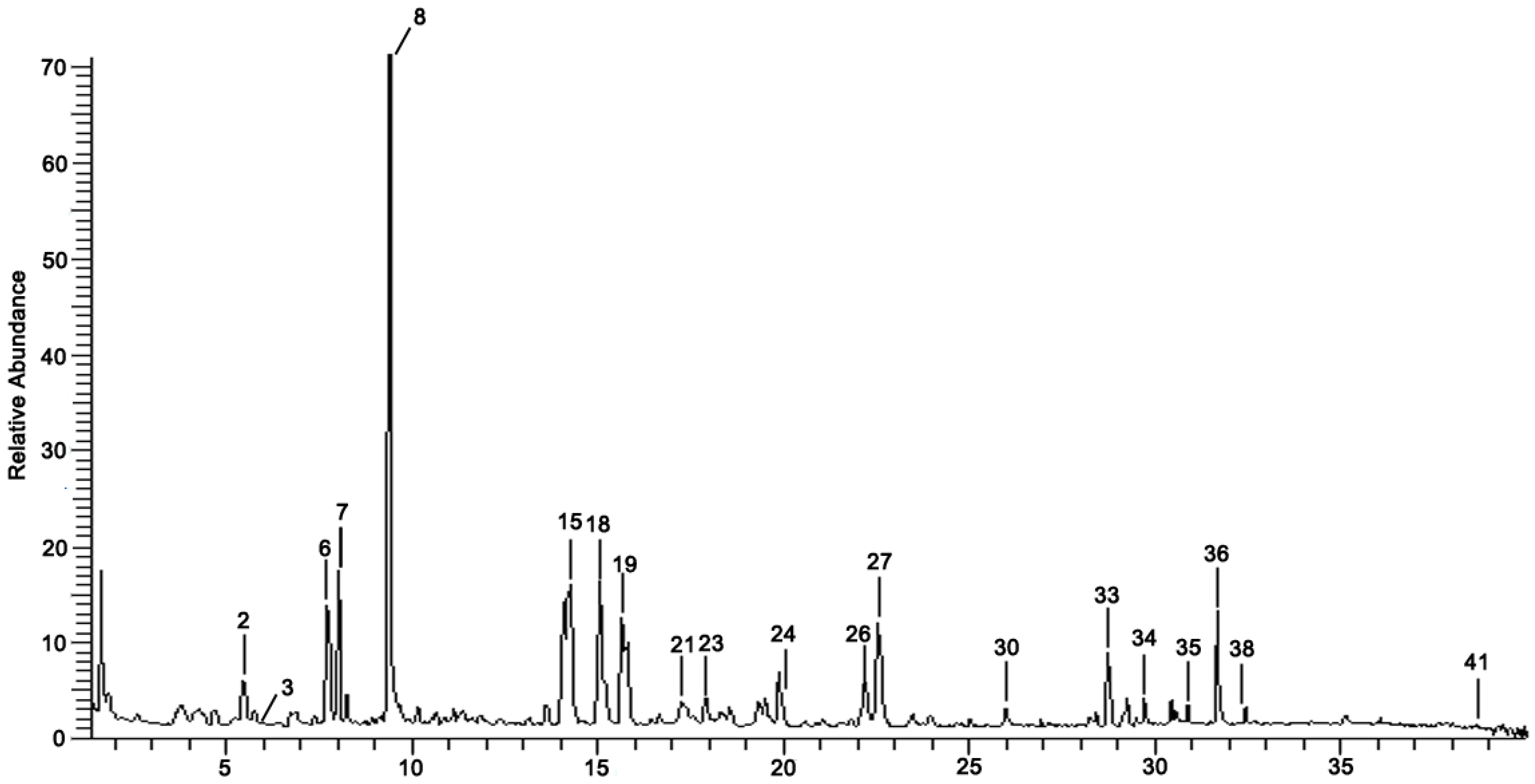
3.1. Chemical Composition Identification in GDK

To determine chemical ingredients in GDK, sample analysis was performed in the negative ion mode. After subtracting blank and removing peak adducts that were generated in source, the compounds were tentatively identified from their exact mass, isotope patterns, and mass fragmentation patterns. A total of 41 communal compounds were preliminarily identified by LC-Orbitrap-ESI-MS (Table 1, Figure 1). They contained various chemical compositions, such as Sebacic Acid, licoagrochalcone A, Gancaonin C, neoliquiritin, Rutin, Isovitexin, Gancaonin O, Quercetin, Gancaonin R, Chrysophanol, Isorhamnetin, Glycyrrhizic Acid, etc. (Table 1).

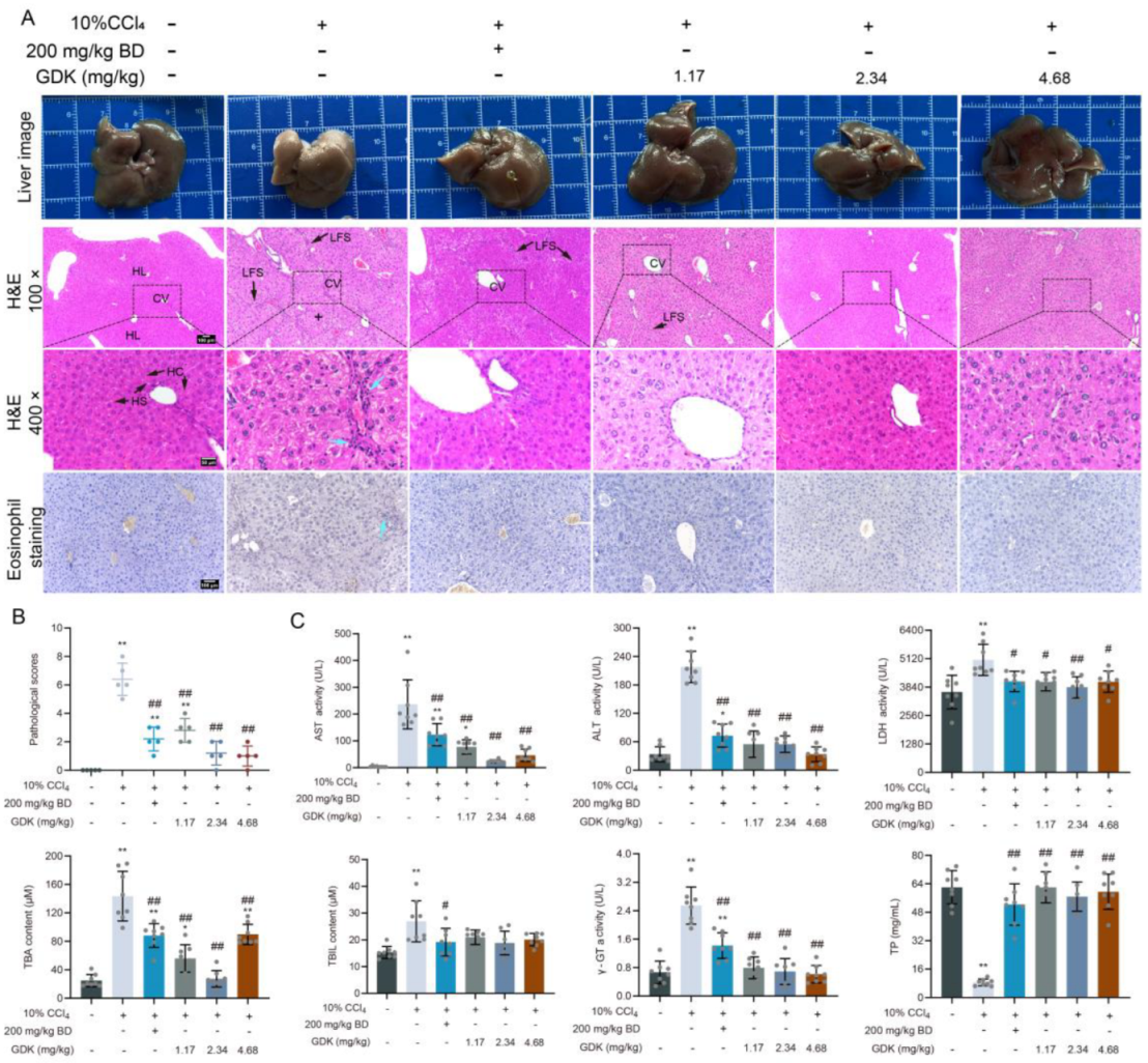
3.2. GDK Improves Hepatic Function and Structure in Chronic Liver Injury

Subsequently, we explored the effects of GDK on liver structure and function in chronic liver injury induced by 10% CCl4. After 10% CCl4 treatment, the liver became brittle and white, and the surface of the capsule was rough. BD and different doses of GDK considerably improved the appearance of liver fibrosis. The disordered arrangement of hepatocytes in the pseudolobule included steatosis, necrosis and regeneration, and deviation or absence of the central vein, lymphocytes, monocytes, and newborn bile ducts in the fibrous tissue around the pseudolobule. Furthermore, BD and GDK improved hepatic LFS broadening, pseudolobule formation, cellular lipid degeneration, and eosinophil infiltration induced by 10% CCl4 and decreased liver pathological scores (Figure 2A,B).
We analyzed the biochemical factors in serum to evaluate the status of liver function. The levels of serum AST, ALT, TBA, TBIL, γ-GT, and LDH were markedly increased by 10% CCl4. Administration of BD and different doses of GDK markedly decreased the levels of serum AST, ALT, LDH, TBA, and γ-GT. There was no significant difference in serum AST and ALT levels between 2.34, 4.68 mg/kg GDK, and the blank groups. CCl4 (10%) caused a considerable decrease in serum TP levels, whereas BD and different doses of GDK considerably increased its levels (Figure 2C).

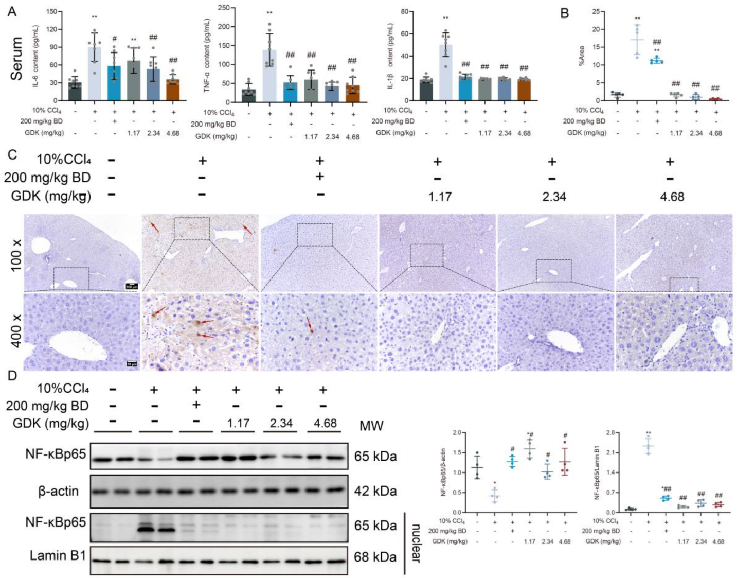
3.3. GDK Reduces Inflammation in CCl4-Induced Chronic Liver Injury

Because inflammation often accompanies liver disease, we detected the state of inflammation in chronic liver injury. First, the levels of serum inflammatory cytokines were detected. CCl4 (10%) remarkably increased the IL-6, IL-1β, and TNF-α levels. BD and different doses of GDK remarkably decreased the levels of IL-1β and TNF-α, and 2.34 mg/kg BD and 4.68 mg/kg GDK markedly decreased the IL-6 levels (Figure 3A). Additionally, 10% CCl4 caused apoptosis in the liver tissue, and different doses of GDK markedly inhibited liver apoptosis (Figure 3B,C).
In terms of proteins, 10% CCl4 markedly promoted the translocation of NF-κBp65 into the nucleus. BD and different doses of GDK considerably inhibited the translocation of NF-κBp65 into the nucleus (Figure 3D).

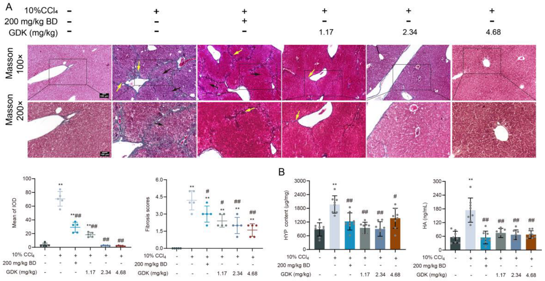
3.4. GDK Suppresses Fibrosis in CCl4-Induced Chronic Liver Injury

Fibrosis is a prominent feature of chronic liver injury, so we detected the fibrotic phenotype in the liver. Masson’s staining showed that intermittent treatment with 10% CCl4 induced fibrous tissue hyperplasia and pseudobular formation in the liver and increased the fibrosis score extensively. BD and 1.17 mg/kg GDK had a certain alleviating effect on fibrous tissue proliferation, but a trend of pseudolobular formation was still observed. GDK (2.34 and 4.68 mg/kg) considerably reduced the intrahepatic fibrous tissue proliferation and fibrosis scores and inhibited pseudolobular formation (Figure 4A). Predictably, the abnormal increase in liver tissue HYP levels and serum HA levels was markedly reversed by BD and different doses of GDK (Figure 4B).

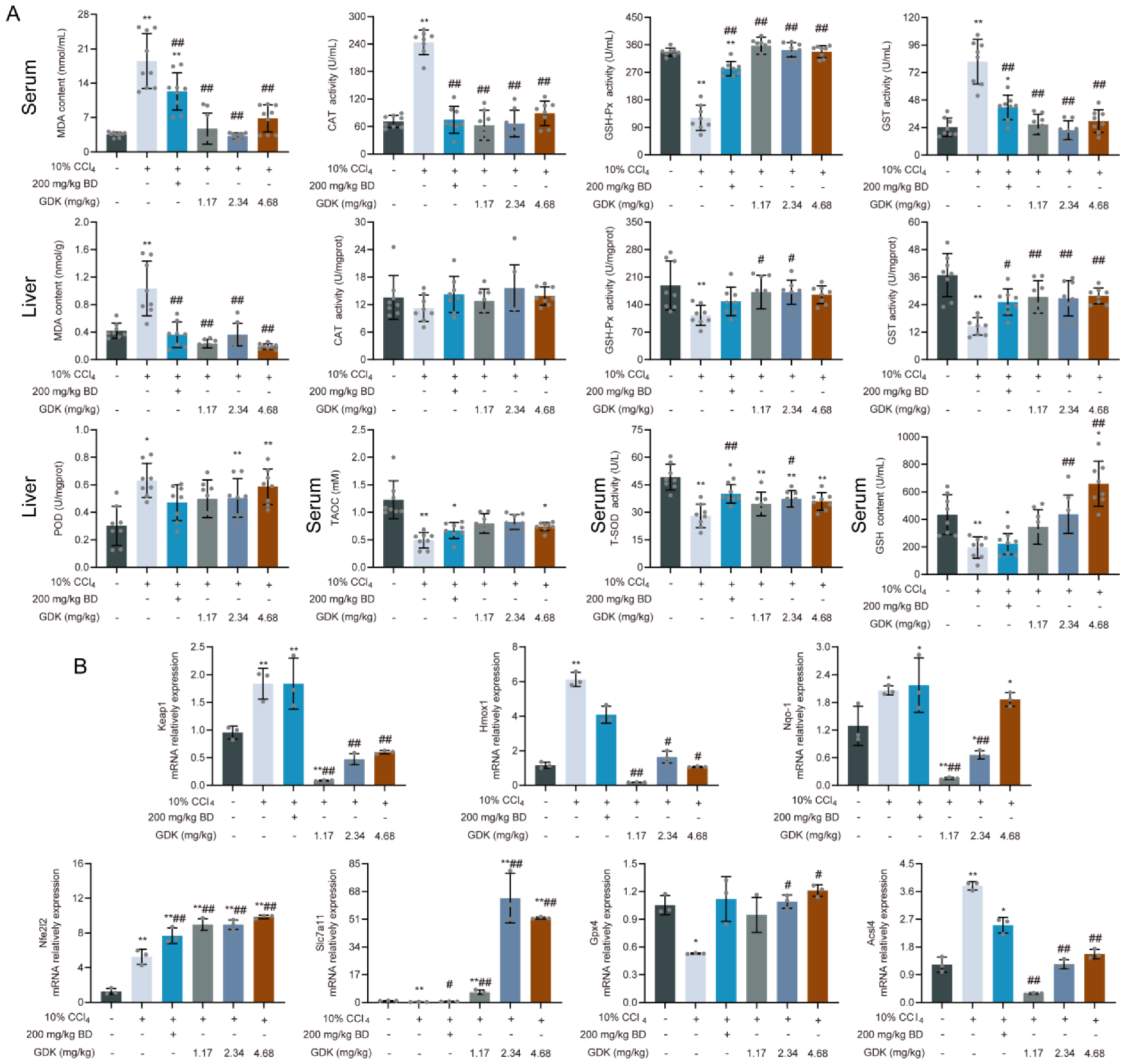
3.5. GDK Weakens Ferroptosis through the Antioxidant System in CCl4-Induced Chronic Liver Injury

Considering the above results, we hypothesized that oxidative stress and ferroptosis play key roles in chronic liver injury. To test this, we first detected the serum and tissue levels of oxidative products and antioxidants. BD and different doses of GDK considerably reduced MDA content in the serum and liver tissue (Figure 5A). Intriguingly, there was no significant difference in CAT levels in the liver tissue among the treatment groups. However, the activity of serum CAT in the CCl4 group was highest and was considerably reduced after the treatment. The GSH-Px activity in serum and liver tissue was markedly reduced by CCl4, whereas 1.17 and 2.34 mg/kg GDK markedly increased it. Interestingly, different trends were observed for the GST activity in tissue and serum. The activity in serum was considerably increased in the CCl4 group but considerably reduced in the liver tissue. BD and different doses of GDK markedly restored the abnormal expression of GST. In addition, the activity of POD in the liver tissue was also markedly increased in the CCl4, 2.34 mg/kg, and 4.68 mg/kg GDK groups. BD and 2.34 mg/kg GDK considerably increased the activity of serum T-SOD, and 2.34 and 4.68 mg/kg GDK considerably increased the content of serum GSH. No significant effect on serum T-AOC was observed in the administration group (Figure 5A).
We further explored the expression of key targets in oxidative stress and the ferroptosis signaling pathway. GDK (2.34 and 4.68 mg/kg) considerably promoted the expression of Nfe2l2, Slc7a11, and Gpx4, while down-regulated the expression of Keap-1, Hmox-1, Nqo-1, and Acsl4 (Figure 5B).

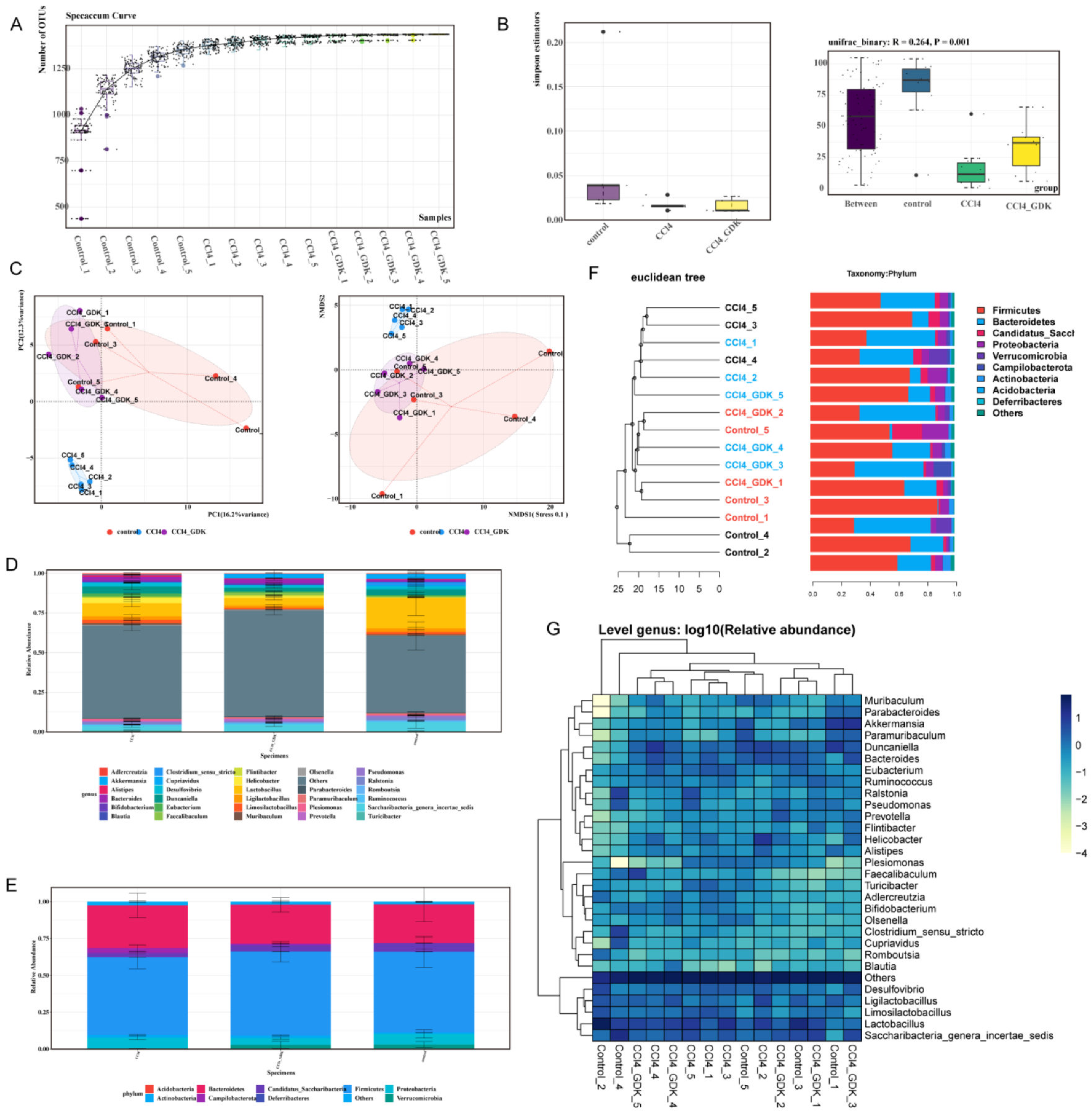
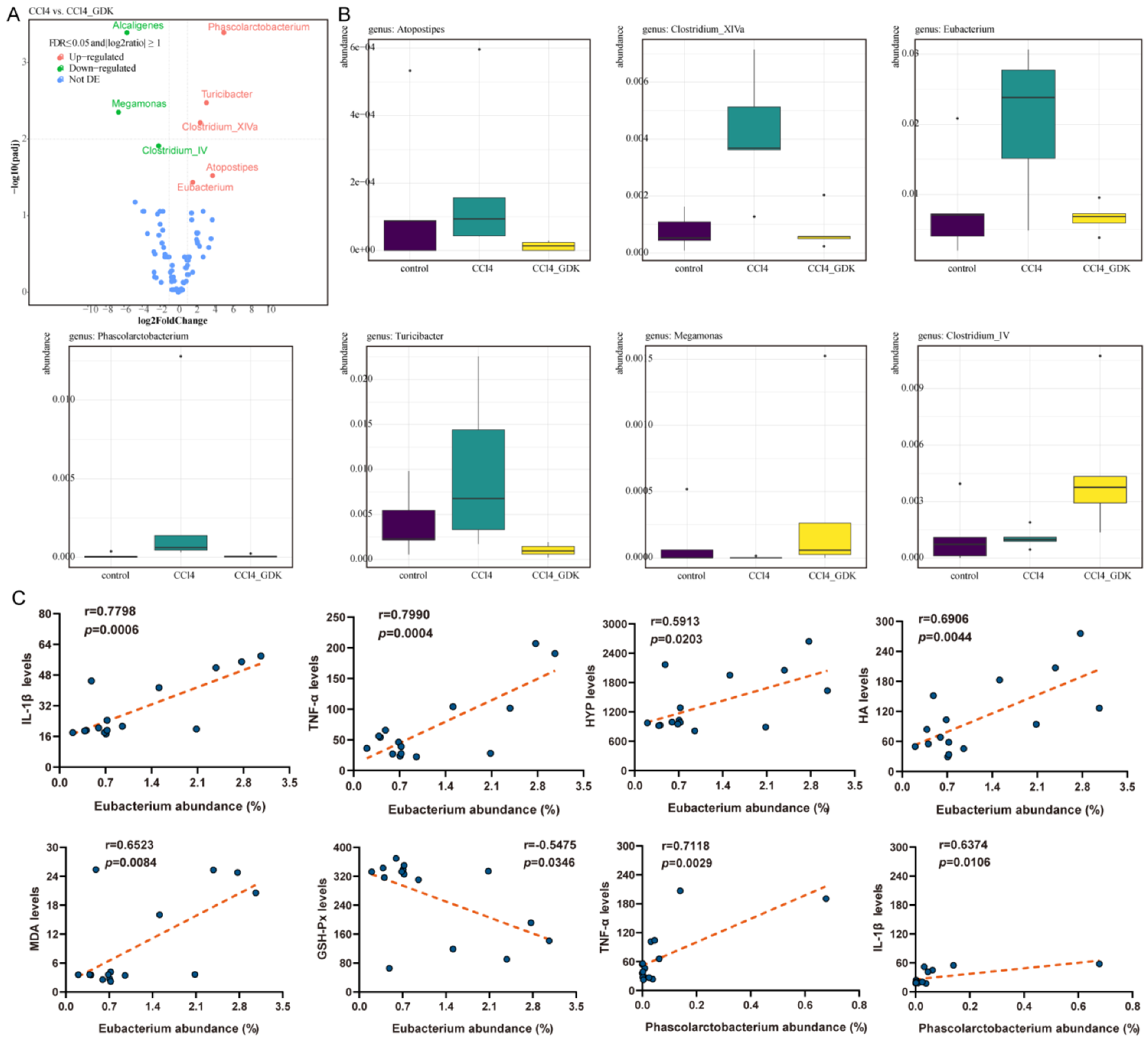
3.6. GDK Restores Intestinal Flora Diversity to Curb Chronic Liver Injury

Compound traditional Chinese medicine often affects the changes in intestinal flora, possibly affecting disease development. However, pertaining to its complexity, the regulation of the intestinal flora has not been revealed. We explored the effect of GDK on the abundance of intestinal flora to further explore the mechanism. First, the species accumulation curve was used to determine the sample size for the detection of diversity (Figure 6A). The Simpson index represents the probability (0–1) that two randomly selected sequences belong to the same classification (species level). The closer the value is to 1, the more uneven the abundance distribution of ZOTUs, showing a more uniform abundance distribution of flora in each group. Analysis of similarity (ANOSIM) was used to investigate whether there were significant differences in community structures between and within groups. The results of ANOSIM based on the Bray–Curtis distance showed marked differences among groups. CCl4 (10%) considerably decreased the abundance of intestinal microflora in mice, which was restored by 2.34 mg/kg GDK (Figure 5B). Euclidean Principal Coordinate Analysis (PCoA) and infinite multi-dimensional calibration (NMDS, Non-Metric Multi-Dimensional Scaling) showed that samples in the 10% CCl4, 2.34 mg/kg GDK, and control groups were closely gathered together, and the coordinate positions of the 2.34 mg/kg GDK and control groups were closer, indicating that their species composition was more similar (Figure 6C). Species diversity analysis showed that CCl4 and GDK treatment had notable effects on the composition of microflora at genus and phylum levels (Figure 6D,E).
To study the similarity between different samples, we used the unweighted pair-group method with arithmetic mean (UPGMA) cluster analysis to construct a cluster tree of the samples. The distance matrix and UPGMA cluster analyses were performed, and the results were combined with the relative abundance of some species of each sample at the gate level (phylum). The samples in the control and 2.34 mg/kg GDK groups were clustered together, and those in the 10% CCl4 group were clustered together. GDK (2.34 mg/kg) restored the change in flora structure caused by CCl4, and the regulation of Firmicutes, Bacteroidetes, Proteobacteria, Romboutsia, Acidobacteria, Campilobacterota, etc., was obvious (Figure 6F,G). We analyzed the differences in considerably changed microflora before and after GDK treatment and found that, compared with the model, 2.34 mg/kg GDK markedly downregulated the abundance of Phascolarctobacterium, Clostridium_IV, and Megamonas and considerably upregulated the abundance of Eubacterium, Atopostipes, Clostridium clusterXlVa, and Turicibacter. The changes in microflora after GDK treatment tended toward the control group (Figure 7A,B). Furthermore, Eubacterium was positively correlated with IL-1β, TNF-α, HYP, HA, and MDA, as well as negatively correlated with GSH-Px. Phascolarctobacterium were positively correlated with IL-1β and TNF-α (Figure 7C).

4. Discussion

Chronic liver injury mainly refers to hepatocyte necrosis and abnormal liver function owing to various reasons. Their incidence is increasing, and they are among the most serious risk factors for metabolic complications and secondary death, posing a heavy burden on the economy and healthcare system [1]. There is no effective treatment for chronic liver injury [39]. Chinese herbal prescriptions have been used for thousands of years to treat liver injury diseases. GDK is derived from the famous prescription “Yinchenhao decoction” for jaundice treatment by Zhongjing Zhang, a medical saint of the Eastern Han Dynasty, and is named Yinshanlian prescription in the Chinese Pharmacopoeia. We previously found that the protective effect of this prescription on CCl4-induced liver injury was mainly through antioxidation, anti-inflammation, and fibrosis inhibition through p38 MAPK and NF-κB signal transduction pathways. In this study, we established toxin-mediated experimental chronic liver injury models. Different doses of GDK reduced the balloon degeneration of hepatocytes and the levels of oxidative stress and inflammation in liver and serum and protected against liver injury, as indicated by reduced levels of serum AST, ALT, γ-GT, TBA, and TBIL. Through comprehensive application of 16S rDNA sequencing analysis, we systematically evaluated the specific effects of GDK on the intestinal flora and the expression levels of proteins related to oxidative stress and ferroptosis.
Ferroptosis plays a crucial regulatory role in liver inflammation and fibrosis [40]. It is a dynamic process involved in iron metabolism and lipid peroxidation [40,41,42]. Iron overload induces membrane lipid peroxidation during ferroptosis because of the Fenton reaction in multiple subcellular organelles [41]. Oxidative damage of membrane lipids is central to the execution of ferroptosis [43,44]. The final product of lipid oxidation, MDA, not only affects the activities of key enzymes in mitochondria but also aggravates membrane damage. GDK can also be used as an antioxidant to remove excess ROS and reduce the content of MDA in serum and liver tissue in chronic liver injury. The main members of antioxidant system include antioxidant enzymes and nonenzymatic antioxidants [45,46]. We then determined the levels of SOD, GSH-Px, CAT, POD, and GST. GDK increased the levels of SOD, GSH-Px, and POD in serum and liver tissue in chronic liver injury models in keeping with the previous study [47]. Interestingly, Serum CAT activity in the model group was significantly higher than that in the control and treated groups, but there was no significant difference in the tissue levels. The serum activity of GST was also very high in the chronic liver injury model group and was significantly decreased after administration of different doses of GDK. However, at the tissue level, the trend was opposite to that in serum. The peak serum GST level in liver disease appeared earlier and was higher than that of AST. Therefore, the serum GST level is a very sensitive index of hepatocyte injury. GSH, a nonenzymatic antioxidant, is a coenzyme of many enzymes, such as GSH-Px, mainly involved in scavenging ROS and in protecting the body from oxidative damage. GDK (2.34 and 4.68 mg/kg) significantly increased the content of serum GSH in mice with chronic liver injury.
Nrf2 is a central player in the regulation of antioxidant molecules in cells [48]. Nrf2 activation in patients with non-alcoholic steatohepatitis correlates with the grade of inflammation. Functional analysis in mice demonstrated that Nrf2 activation in chronic liver disease ameliorates fibrogenesis, initiation, and progression of HCC [49]. It controls cellular antioxidant systems in hepatocytes, playing a key role in protecting against intracellular and environmental stress [7]. We found that Nrf2 expression decreased with fibrosis severity, whereas it was high in normal liver tissue. Activation of the Nrf2 signal transduction pathway activates the antioxidant system. Nrf2 is constantly degraded by Keap1 and is activated by the inhibition of Keap1 [50]. The detection of Keap1 also shows that GDK weakens the inhibition of Nrf2, thus promoting its nuclear translocation, and thereby increases the expression of HO-1 and NQO-1. Interestingly, the expression of HO-1 is not completely consistent with that of Nrf2, which may be related to another function of HO-1 in promoting the production of Fe2+. The Keap1–Nrf2 antioxidant signaling pathway is a negative regulator of ferroptosis and determines the sensitivity to ferroptosis inducers such as erastin, sulfasalazine, and sorafenib, which inhibit xCT. SLC7A11 is a key component of system Xc2, an amino acid antiporter that typically mediates the exchange of extracellular L-cystine and intracellular L-glutamate across the plasma membrane. Suppression of SLC7A11 leads to GSH depletion and increased ferroptosis. Interestingly, SLC7A11 is a transcriptional target of Nrf2 [51,52].
Furthermore, we found that GDK inhibited ferroptosis by activating the Nrf2 pathway, which influences liver inflammation and the fibrotic phenotype in mice with liver injury. Ferroptosis can promote liver fibrosis [17]. We showed that not only the keap1–Nrf2 pathway was inhibited, but the classical NF-κB pathway was also activated, and significant cell death occurred during chronic liver injury. In chronic liver injury, the proliferation of fibers was significant, and there was a tendency to form false lobules [53]. Our research shows that HA are also introduced as characteristic indicators of fibrosis [47]. As expected, the increase in serum HA levels, which is an indicator of fibrosis initiation, significantly decreased with all doses of GDK, indicating that the antagonistic relationship was gradually recovered, thereby reducing the inflammatory response and cell death.
Based on our sequencing results for intestinal flora in mice with chronic liver injury, we found that GDK upregulated the abundance of Turicibacter, which was then found to interact with bile acid (BA) [54]. However, unlike our results, it is reported that the relative abundance of Turicibacter is strongly positively correlated with liver MDA levels [55]. GDK also increases the abundance of Clostridium cluster XlVa, Atopostipes, and Eubacterium. Eubacterium is associated with bile acid regulation [56]. Indeed, Eubacterium spp., along with other genera, such as Roseburia and Clostridium, constitute a major reservoir of BSHs in the gut [57]. Besides, Eubacterium spp. contribute to gut and hepatic health through modulation of bile acid metabolism [57]. Clostridium cluster XlVa, Atopostipes, and Eubacterium are also related to the synthesis of amino acids and short-chain fatty acids [55,57]. The increase in the abundance of these bacteria may play a pivotal role in the recovery of liver function, but their internal causal relationship needs to be further explored. We also found that GDK decreased the abundance of Phascolarctobacterium, Clostridium_IV, and Megamonas flora. Phascolarctobacterium is related to the production of short-chain fatty acids such as acetate and propionate and has a certain detoxification effect [58]. The role of Clostridium_IV and Megamonas in liver diseases has not been reported. Contrarily, by comparing with the microflora related to chronic liver fibrosis or liver cirrhosis queried in the Disbiome database, Phascolarctobacterium and Eubacterium were not only found to be associated with liver cirrhosis but were also verified by our sequencing results. Therefore, we have two propositions to further explore the mechanism in the future. On the one hand, we can select Clostridium_IV and Megamonas, which have not been reported in the study on chronic liver injury, and perform macro-genome sequencing, followed by their isolation, purification, and animal verification to unravel their effect on chronic liver injury. On the other hand, we can also choose Phascolarctobacterium and Eubacterium, which were identified by querying the database and make use of operations such as fecal-oral transplantation to further explore the specific causal relationship between Phascolarctobacterium, Eubacterium, and chronic liver injury.

5. Conclusions

Our findings show that different doses of GDK can alleviate chronic liver injury and 2.34 mg/kg GDK has the best comprehensive effect. The effect of GDK on chronic liver injury may be through the activation of the Keap1/Nrf2 pathway and inhibition of ferroptosis, alleviating liver fibrosis and abnormal functional changes. Additionally, GDK also affects the flora related to the synthesis and metabolism of intestinal bile acids, amino acids, and short-chain fatty acids, but its specific causal relationship needs to be further explored.

Supplementary Materials

The following supporting information can be downloaded at: https://www.mdpi.com/article/10.3390/antiox11112234/s1, Table S1: Ishak scoring system for liver fibrosis, Table S2: Ishak scoring system for liver inflammation, Table S3: The antibodies used for western blotting, and Table S4: Primers for qRT-PCR.

Author Contributions

Y.W.: Conceptualization, Data curation, Formal analysis, Methodology, Writing—original draft, Writing—review & editing. H.W.: Data curation, Methodology, Investigation. Y.Z.: Investigation, Supervision. J.G., X.Z. and X.G.: Writing—review & editing. Z.H.: Conceptualization, Data curation, Funding acquisition, Project administration. All authors have read and agreed to the published version of the manuscript.

Funding

This research was funded by the Key Project at Central Government Level (2060302) and the National Natural Science Foundation of China (No. 32172897).

Institutional Review Board Statement

All experiments involving animals were conducted according to the ethical policies of, and procedures approved by, the Institutional Animal Care and Use Committee of China Agricultural University (No. Aw20801202–2–2).

Informed Consent Statement

Not applicable.

Data Availability Statement

Data is contained within the article and Supplementary Material.

Conflicts of Interest

The authors declare no conflict of interest.

References

  1. Marsh, K.; Tayler, R.; Pollock, L.; Roy, K.; Lakha, F.; Ho, A.; Henderson, D.; Divala, T.; Currie, S.; Yirrell, D.; et al. Investigation into Cases of Hepatitis of Unknown Aetiology among Young Children, Scotland, 1 January 2022 to 12 April 2022. Eurosurveillance 2022, 27, 2200318. [Google Scholar] [CrossRef] [PubMed]
  2. Friedman, S.L. Evolving Challenges in Hepatic Fibrosis. Nat. Rev. Gastroenterol. Hepatol. 2010, 7, 425–436. [Google Scholar] [CrossRef] [PubMed]
  3. Parola, M.; Pinzani, M. Liver Fibrosis: Pathophysiology, Pathogenetic Targets and Clinical Issues. Mol. Asp. Med. 2019, 65, 37–55. [Google Scholar] [CrossRef] [PubMed]
  4. Zou, A.; Magee, N.; Deng, F.; Lehn, S.; Zhong, C.; Zhang, Y. Hepatocyte Nuclear Receptor Shp Suppresses Inflammation and Fibrosis in a Mouse Model of Nonalcoholic Steatohepatitis. J. Biol. Chem. 2018, 293, 8656–8671. [Google Scholar] [CrossRef] [PubMed] [Green Version]
  5. Lin, Y.; Dong, M.Q.; Liu, Z.M.; Xu, M.; Huang, Z.H.; Liu, H.J.; Gao, Y.; Zhou, W.J. A Strategy of Vascular-Targeted Therapy for Liver Fibrosis. Hepatology 2022, 76, 660–675. [Google Scholar] [CrossRef]
  6. Sanchez-Valle, V.; Chavez-Tapia, N.C.; Uribe, M.; Mendez-Sanchez, N. Role of Oxidative Stress and Molecular Changes in Liver Fibrosis: A Review. Curr. Med. Chem. 2012, 19, 4850–4860. [Google Scholar] [CrossRef]
  7. Ke, B.; Shen, X.D.; Zhang, Y.; Ji, H.; Gao, F.; Yue, S.; Kamo, N.; Zhai, Y.; Yamamoto, M.; Busuttil, R.W.; et al. Keap1-Nrf2 Complex in Ischemia-Induced Hepatocellular Damage of Mouse Liver Transplants. J. Hepatol. 2013, 59, 1200–1207. [Google Scholar] [CrossRef] [Green Version]
  8. Wakabayashi, N.; Slocum, S.L.; Skoko, J.J.; Shin, S.; Kensler, T.W. When Nrf2 Talks, Who’s Listening? Antioxid. Redox Signal. 2010, 13, 1649–1663. [Google Scholar] [CrossRef] [Green Version]
  9. McMahon, M.; Itoh, K.; Yamamoto, M.; Hayes, J.D. Keap1-Dependent Proteasomal Degradation of Transcription Factor Nrf2 Contributes to the Negative Regulation of Antioxidant Response Element-Driven Gene Expression. J. Biol. Chem. 2003, 278, 21592–21600. [Google Scholar] [CrossRef] [Green Version]
  10. Nam, E.; Derrick, J.S.; Lee, S.; Kang, J.; Han, J.; Lee, S.J.C.; Chung, S.W.; Lim, M.H. Regulatory Activities of Dopamine and Its Derivatives toward Metal-Free and Metal-Induced Amyloid-Β Aggregation, Oxidative Stress, and Inflammation in Alzheimer’s Disease. ACS Chem. Neurosci. 2018, 9, 2655–2666. [Google Scholar] [CrossRef]
  11. Fan, J.; Chen, Q.; Wei, L.; Zhou, X.; Wang, R.; Zhang, H. Asiatic Acid Ameliorates Ccl4-Induced Liver Fibrosis in Rats: Involvement of Nrf2/Are, Nf-Kappab/Ikappabalpha, and Jak1/Stat3 Signaling Pathways. Drug Des. Dev. Ther. 2018, 12, 3595–3605. [Google Scholar] [CrossRef] [PubMed] [Green Version]
  12. Gordillo-Bastidas, D.; Oceguera-Contreras, E.; Salazar-Montes, A.; Gonzalez-Cuevas, J.; Hernandez-Ortega, L.D.; Armendariz-Borunda, J. Nrf2 and Snail-1 in the Prevention of Experimental Liver Fibrosis by Caffeine. World J. Gastroenterol. 2013, 19, 9020–9033. [Google Scholar] [CrossRef] [PubMed]
  13. Yan, H.; Huang, Z.; Bai, Q.; Sheng, Y.; Hao, Z.; Wang, Z.; Ji, L. Natural Product Andrographolide Alleviated Apap-Induced Liver Fibrosis by Activating Nrf2 Antioxidant Pathway. Toxicology 2018, 396–397, 1–12. [Google Scholar] [CrossRef] [PubMed]
  14. Husain, H.; Latief, U.; Ahmad, R. Pomegranate Action in Curbing the Incidence of Liver Injury Triggered by Diethylnitrosamine by Declining Oxidative Stress Via Nrf2 and Nfkappab Regulation. Sci. Rep. 2018, 8, 8606. [Google Scholar] [CrossRef] [PubMed]
  15. Orru, C.; Szydlowska, M.; Taguchi, K.; Zavattari, P.; Perra, A.; Yamamoto, M.; Columbano, A. Genetic Inactivation of Nrf2 Prevents Clonal Expansion of Initiated Cells in a Nutritional Model of Rat Hepatocarcinogenesis. J. Hepatol. 2018, 69, 635–643. [Google Scholar] [CrossRef]
  16. Yu, M.; Wang, D.; Xu, M.; Liu, Y.; Wang, X.; Liu, J.; Yang, X.; Yao, P.; Yan, H.; Liu, L. Quinocetone-Induced Nrf2/Ho-1 Pathway Suppression Aggravates Hepatocyte Damage of Sprague-Dawley Rats. Food Chem. Toxicol. 2014, 69, 210–219. [Google Scholar] [CrossRef]
  17. Xu, W.; Hellerbrand, C.; Kohler, U.A.; Bugnon, P.; Kan, Y.W.; Werner, S.; Beyer, T.A. The Nrf2 Transcription Factor Protects from Toxin-Induced Liver Injury and Fibrosis. Lab Investig. 2008, 88, 1068–1078. [Google Scholar] [CrossRef] [Green Version]
  18. Duarte, T.L.; Caldas, C.; Santos, A.G.; Silva-Gomes, S.; Santos-Gonçalves, A.; Martins, M.J.; Porto, G.; Lopes, J.M. Genetic Disruption of Nrf2 Promotes the Development of Necroinflammation and Liver Fibrosis in a Mouse Model of Hfe-Hereditary Hemochromatosis. Redox Biol. 2017, 11, 157–169. [Google Scholar] [CrossRef] [Green Version]
  19. Koyama, Y.; Brenner, D.A. Liver Inflammation and Fibrosis. J. Clin. Investig. 2017, 127, 55–64. [Google Scholar] [CrossRef]
  20. Brenner, D.A. Molecular Pathogenesis of Liver Fibrosis. Trans. Am. Clin. Climatol. Assoc. 2009, 120, 361–368. [Google Scholar]
  21. Elsharkawy, A.M.; Mann, D.A. Nuclear Factor-Kappab and the Hepatic Inflammation-Fibrosis-Cancer Axis. Hepatology 2007, 46, 590–597. [Google Scholar] [CrossRef] [PubMed]
  22. Muriel, P. Nf-Kappab in Liver Diseases: A Target for Drug Therapy. J. Appl. Toxicol. 2009, 29, 91–100. [Google Scholar] [CrossRef] [PubMed]
  23. Stewart, A.G.; Thomas, B.; Koff, J. Tgf-Beta: Master Regulator of Inflammation and Fibrosis. Respirology 2018, 23, 1096–1097. [Google Scholar] [CrossRef] [PubMed]
  24. Li, D.; Ke, Y.; Zhan, R.; Liu, C.; Zhao, M.; Zeng, A.; Shi, X.; Ji, L.; Cheng, S.; Pan, B.; et al. Trimethylamine-N-Oxide Promotes Brain Aging and Cognitive Impairment in Mice. Aging Cell 2018, 17, e12768. [Google Scholar] [CrossRef]
  25. Sun, J.; Zhang, J.; Wang, X.; Ji, F.; Ronco, C.; Tian, J.; Yin, Y. Gut-Liver Crosstalk in Sepsis-Induced Liver Injury. Crit. Care 2020, 24, 614. [Google Scholar] [CrossRef]
  26. Loomba, R.; Seguritan, V.; Li, W.; Long, T.; Klitgord, N.; Bhatt, A.; Dulai, P.S.; Caussy, C.; Bettencourt, R.; Highlander, S.K.; et al. Gut Microbiome-Based Metagenomic Signature for Non-Invasive Detection of Advanced Fibrosis in Human Nonalcoholic Fatty Liver Disease. Cell Metab. 2019, 30, 607. [Google Scholar] [CrossRef] [Green Version]
  27. Saeedi, B.J.; Liu, K.H.; Owens, J.A.; Hunter-Chang, S.; Camacho, M.C.; Eboka, R.U.; Chandrasekharan, B.; Baker, N.F.; Darby, T.M.; Robinson, B.S.; et al. Gut-Resident Lactobacilli Activate Hepatic Nrf2 and Protect against Oxidative Liver Injury. Cell Metab. 2020, 31, 956–968.e5. [Google Scholar] [CrossRef]
  28. Roehlen, N.; Crouchet, E.; Baumert, T.F. Liver Fibrosis: Mechanistic Concepts and Therapeutic Perspectives. Cells 2020, 9, 875. [Google Scholar] [CrossRef] [Green Version]
  29. Cai, F.F.; Wu, R.; Song, Y.N.; Xiong, A.Z.; Chen, X.L.; Yang, M.D.; Yang, L.; Hu, Y.; Sun, M.Y.; Su, S.B. Yinchenhao Decoction Alleviates Liver Fibrosis by Regulating Bile Acid Metabolism and Tgf-Beta/Smad/Erk Signalling Pathway. Sci. Rep. 2018, 8, 15367. [Google Scholar] [CrossRef] [Green Version]
  30. Jiang, S.L.; Hu, X.D.; Liu, P. Immunomodulation and Liver Protection of Yinchenhao Decoction against Concanavalin a-Induced Chronic Liver Injury in Mice. J. Integr. Med. 2015, 13, 262–268. [Google Scholar] [CrossRef]
  31. Xu, L.; Xie, T.; Shen, T.; Jian, S. Yinchenhao Decoction for Chronic Hepatitis B: Protocol for a Systematic Review and Meta-Analysis. Medicine 2019, 98, e14648. [Google Scholar] [CrossRef] [PubMed]
  32. Wu, Y.L.; Li, Z.L.; Zhang, X.B.; Liu, H. Yinchenhao Decoction Attenuates Obstructive Jaundice-Induced Liver Injury and Hepatocyte Apoptosis by Suppressing Protein Kinase Rna-Like Endoplasmic Reticulum Kinase-Induced Pathway. World J. Gastroenterol. 2019, 25, 6205–6221. [Google Scholar] [CrossRef] [PubMed]
  33. Zhou, Q.; Hu, H.; Zhao, G.; Liu, P.; Wang, Y.; Zhang, H. Effect and Related Mechanism of Yinchenhao Decoction on Mice with Lithogenic Diet-Induced Cholelithiasis. Exp. Ther. Med. 2021, 21, 316. [Google Scholar] [CrossRef] [PubMed]
  34. Cai, F.F.; Bian, Y.Q.; Wu, R.; Sun, Y.; Chen, X.L.; Yang, M.D.; Zhang, Q.R.; Hu, Y.; Sun, M.Y.; Su, S.B. Yinchenhao Decoction Suppresses Rat Liver Fibrosis Involved in an Apoptosis Regulation Mechanism Based on Network Pharmacology and Transcriptomic Analysis. Biomed. Pharmacother. 2019, 114, 108863. [Google Scholar] [CrossRef] [PubMed]
  35. Xing, P.; Niu, Y.; Mu, R.; Wang, Z.; Xie, D.; Li, H.; Dong, L.; Wang, C. A Pocket-Escaping Design to Prevent the Common Interference with near-Infrared Fluorescent Probes in Vivo. Nat. Commun. 2020, 11, 1573. [Google Scholar] [CrossRef] [Green Version]
  36. Zhu, Z.; Hu, R.; Li, J.; Xing, X.; Chen, J.; Zhou, Q.; Sun, J. Alpinetin Exerts Anti-Inflammatory, Anti-Oxidative and Anti-Angiogenic Effects through Activating the Nrf2 Pathway and Inhibiting Nlrp3 Pathway in Carbon Tetrachloride-Induced Liver Fibrosis. Int. Immunopharmacol. 2021, 96, 107660. [Google Scholar] [CrossRef]
  37. Schindelin, J.; Arganda-Carreras, I.; Frise, E.; Kaynig, V.; Longair, M.; Pietzsch, T.; Preibisch, S.; Rueden, C.; Saalfeld, S.; Schmid, B.; et al. Fiji: An Open-Source Platform for Biological-Image Analysis. Nat. Methods 2012, 9, 676–682. [Google Scholar] [CrossRef] [Green Version]
  38. Wei, Y.Y.; Zhang, Y.N.; Wang, H.; Ga, Y.; Fan, Y.; Wang, Q.; Gu, J.H.; Zhang, X.Y.; Gong, X.H.; Hao, Z.H. Mori Fructus Aqueous Extracts Attenuate Carbon Tetrachloride-Induced Renal Injury Via the Nrf2 Pathway and Intestinal Flora. Ecotoxicol. Environ. Saf. 2022, 245, 114118. [Google Scholar] [CrossRef]
  39. Arroyo, V.; Moreau, R.; Jalan, R. Acute-on-Chronic Liver Failure. N. Engl. J. Med. 2020, 382, 2137–2145. [Google Scholar] [CrossRef]
  40. Capelletti, M.M.; Manceau, H.; Puy, H.; Peoc’h, K. Ferroptosis in Liver Diseases: An Overview. Int. J. Mol. Sci. 2020, 21, 4908. [Google Scholar] [CrossRef]
  41. Yuan, H.; Li, X.; Zhang, X.; Kang, R.; Tang, D. Identification of Acsl4 as a Biomarker and Contributor of Ferroptosis. Biochem. Biophys. Res. Commun. 2016, 478, 1338–1343. [Google Scholar] [CrossRef] [PubMed]
  42. Xie, Y.; Hou, W.; Song, X.; Yu, Y.; Huang, J.; Sun, X.; Kang, R.; Tang, D. Ferroptosis: Process and Function. Cell Death Differ. 2016, 23, 369–379. [Google Scholar] [CrossRef] [PubMed] [Green Version]
  43. Chen, X.; Li, J.; Kang, R.; Klionsky, D.J.; Tang, D. Ferroptosis: Machinery and Regulation. Autophagy 2021, 17, 2054–2081. [Google Scholar] [CrossRef] [PubMed]
  44. Stockwell, B.R.; Jiang, X.; Gu, W. Emerging Mechanisms and Disease Relevance of Ferroptosis. Trends Cell Biol. 2020, 30, 478–490. [Google Scholar] [CrossRef] [PubMed]
  45. Hadian, K.; Stockwell, B.R. Snapshot: Ferroptosis. Cell 2020, 181, 1188–1188.e1. [Google Scholar] [CrossRef] [PubMed]
  46. Liu, M.; Kong, X.Y.; Yao, Y.; Wang, X.A.; Yang, W.; Wu, H.; Li, S.; Ding, J.W.; Yang, J. The Critical Role and Molecular Mechanisms of Ferroptosis in Antioxidant Systems: A Narrative Review. Ann. Transl. Med. 2022, 10, 368. [Google Scholar] [CrossRef] [PubMed]
  47. Wei, Y.Y.; Fan, Y.M.; Yuan, Y.Y.; Ga, Y.; Zhang, Y.N.; Han, J.C.; Hao, Z.H. Protective Effect of Aqueous Extract of Yinshanlian on Acute Liver Injury Induced by Carbon Tetrachloride in Mice. Acta Vet. Zootech. Sin. 2022, 53, 2333–2342. [Google Scholar]
  48. Dodson, M.; Castro-Portuguez, R.; Zhang, D.D. Nrf2 Plays a Critical Role in Mitigating Lipid Peroxidation and Ferroptosis. Redox Biol. 2019, 23, 101107. [Google Scholar] [CrossRef]
  49. Mohs, A.; Otto, T.; Schneider, K.M.; Peltzer, M.; Boekschoten, M.; Holland, C.H.; Hudert, C.A.; Kalveram, L.; Wiegand, S.; Saez-Rodriguez, J.; et al. Hepatocyte-Specific Nrf2 Activation Controls Fibrogenesis and Carcinogenesis in Steatohepatitis. J. Hepatol. 2021, 74, 638–648. [Google Scholar] [CrossRef]
  50. Roh, J.L.; Kim, E.H.; Jang, H.; Shin, D. Nrf2 Inhibition Reverses the Resistance of Cisplatin-Resistant Head and Neck Cancer Cells to Artesunate-Induced Ferroptosis. Redox Biol. 2017, 11, 254–262. [Google Scholar] [CrossRef]
  51. Sun, X.; Ou, Z.; Chen, R.; Niu, X.; Chen, D.; Kang, R.; Tang, D. Activation of the P62-Keap1-Nrf2 Pathway Protects against Ferroptosis in Hepatocellular Carcinoma Cells. Hepatology 2016, 63, 173–184. [Google Scholar] [CrossRef] [PubMed] [Green Version]
  52. Jiang, L.; Kon, N.; Li, T.; Wang, S.J.; Su, T.; Hibshoosh, H.; Baer, R.; Gu, W. Ferroptosis as a P53-Mediated Activity During Tumour Suppression. Nature 2015, 520, 57–62. [Google Scholar] [CrossRef] [PubMed]
  53. Frissen, M.; Liao, L.; Schneider, K.M.; Djudjaj, S.; Haybaeck, J.; Wree, A.; Rolle-Kampczyk, U.; von Bergen, M.; Latz, E.; Boor, P.; et al. Bidirectional Role of Nlrp3 During Acute and Chronic Cholestatic Liver Injury. Hepatology 2021, 73, 1836–1854. [Google Scholar] [CrossRef] [PubMed]
  54. Wu, J.; Zhang, D.; Zhu, B.; Wang, S.; Xu, Y.; Zhang, C.; Yang, H.; Wang, S.; Liu, P.; Qin, L.; et al. Rubus chingii Hu. Unripe Fruits Extract Ameliorates Carbon Tetrachloride-Induced Liver Fibrosis and Improves the Associated Gut Microbiota Imbalance. Chin. Med. 2022, 17, 56. [Google Scholar] [CrossRef] [PubMed]
  55. Li, T.T.; Huang, Z.R.; Jia, R.B.; Lv, X.C.; Zhao, C.; Liu, B. Spirulina Platensis Polysaccharides Attenuate Lipid and Carbohydrate Metabolism Disorder in High-Sucrose and High-Fat Diet-Fed Rats in Association with Intestinal Microbiota. Food Res. Int. 2021, 147, 110530. [Google Scholar] [CrossRef] [PubMed]
  56. Iwaki, M.; Kessoku, T.; Ozaki, A.; Kasai, Y.; Kobayashi, T.; Nogami, A.; Honda, Y.; Ogawa, Y.; Imajo, K.; Yoneda, M.; et al. Gut Microbiota Composition Associated with Hepatic Fibrosis in Non-Obese Patients with Non-Alcoholic Fatty Liver Disease. J. Gastroenterol. Hepatol. 2021, 36, 2275–2284. [Google Scholar] [CrossRef]
  57. Mukherjee, A.; Lordan, C.; Ross, R.P.; Cotter, P.D. Gut Microbes from the Phylogenetically Diverse Genus Eubacterium and Their Various Contributions to Gut Health. Gut Microbes 2020, 12, 1802866. [Google Scholar] [CrossRef]
  58. Tsai, H.Y.; Shih, Y.Y.; Yeh, Y.T.; Huang, C.H.; Liao, C.A.; Hu, C.Y.; Nagabhushanam, K.; Ho, C.T.; Chen, Y.K. Pterostilbene and Its Derivative 3′-Hydroxypterostilbene Ameliorated Nonalcoholic Fatty Liver Disease through Synergistic Modulation of the Gut Microbiota and Sirt1/Ampk Signaling Pathway. J. Agric. Food Chem. 2022, 70, 4966–4980. [Google Scholar] [CrossRef]
Figure 1. Chemical composition identification in GDK by LC-Orbitrap-ESI-MS.
Antioxidants 11 02234 g001
Figure 2. GDK improves hepatic function and structure in CCl4-induced chronic liver injury mice. (A) Representative images of the liver; hematoxylin and eosin (H&E)- and eosinophil-stained images of liver sections in indicated mice. Scar bar, 100 μm (100× and 200×), 50 μm (400×). n = 5 per group. LFS: Long fiber spacing; HL: Hepatic lobule; HS: Hepatic sinusoid; HC: Hepatic cord. Blue arrow: inflammatory cell infiltration. (B) Pathological scores. (C) Serum ALT, AST, LDH, TBIL, TBA, γ-GT, and TP levels in indicated mice. n = 8 samples per group. * p < 0.05, ** p < 0.01, # p < 0.05, and ## p < 0.01.
Antioxidants 11 02234 g002
Figure 3. GDK reduces inflammation in CCl4-induced chronic liver injury mice. (A) Serum IL-6, TNF-α, and IL-1β levels in indicated mice (n = 8 per group). (B,C) TUNEL analysis of the liver samples of indicated mice. Red arrow: Positive area. (D) Western blot analysis of total and nuclear NF-κBp65 in the liver of indicated mice. Lamin B1 and β-actin served as loading controls (n = 3 per group). * p < 0.05, ** p < 0.01, # p < 0.05, and ## p < 0.01.
Antioxidants 11 02234 g003
Figure 4. GDK suppresses fibrosis in CCl4-induced chronic liver injury mice. (A) Masson’s staining of liver sections and quantitative result in indicated mice. Scar bar, 100 μm. Black arrow: Pseudolobule; Yellow arrow: Fibrosis. (B) Serum HYP and liver HA levels in indicated mice (n = 8 samples per group). ** p < 0.01, # p < 0.05, and ## p < 0.01.
Antioxidants 11 02234 g004
Figure 5. GDK attenuates ferroptosis through the antioxidant system in CCl4-induced chronic liver injury mice. (A) Levels of serum and liver antioxidants in indicated mice (n = 8 samples per group). (B) Relative mRNA levels of Keap-1, Hmox-1, Nqo-1, Nfe2l2, Slc7a11, Gpx4, and Acsl4. * p < 0.05, ** p < 0.01, # p < 0.05, and ## p < 0.01. n = 3 per group.
Antioxidants 11 02234 g005
Figure 6. GDK restores intestinal flora diversity to curb chronic liver injury. (A) Species accumulation curves (B) α-diversity upon oral administration of GDK. (C) β-diversity of PCoA and NMDS plots upon GDK administration, as assessed by Euclidean distance at genus levels. (D,E) The relative abundance of bacterial phyla (D) and genera (E). (F) UPGMA clustering tree based on Euclidean distance and information statistics of Top 10 species at the phylum level. (G) Abundance cluster diagram at genus levels (n = 5 per group). R software (version 3.2.3) was used for statistics and visual display of species annotation results. It uses (https://bitbucket.org/nsegata/hclust, accessed on 23 September 2021) to construct heat maps of some species information at the OTU level and different classification levels as well as to carry out cluster analysis among samples and species.
Antioxidants 11 02234 g006
Figure 7. Analysis of the differences in species composition between groups upon GDK treatment. (A) Volcanic map of grouping difference in species composition (genus level). (B) Differential flora among control, CCl4, and GDK groups. (C) Correlation analysis of Eubacterium and Phascolarctobacterium with IL-1β, TNF-α, HYP, HA, γGT, MDA, GSH-Px, GST, and GSH levels based on Pearson’s Correlation Coefficient.
Antioxidants 11 02234 g007
Table 1. Identified bioactive components of GDK with LC-Orbitrap-ESI-MS.
NoRt (min)FormulaMeasured [M-H]−MS/MS (m/z)Tentative IdentificationStructural Formula
12.69C17H24O9371.13366191.05609Eleutheroside BAntioxidants 11 02234 i001
25.89C12H14O8285.0395152.01164;
153.01944;
108.02183;
285.06177
UralenneosideAntioxidants 11 02234 i002
35.92C10H18O4201.11214110.97005;
201.80287
Sebacic AcidAntioxidants 11 02234 i003
46.9C20H20O5339.0036265.00217;
238.98785
LicocoumaroneAntioxidants 11 02234 i004
57.46C20H20O4323.1349323.13495;
186.85599
licoagrochalcone AAntioxidants 11 02234 i005
67.73C25H28O5407.0068248.98572;
317.01288
6,8-DiprenylnaringeninAntioxidants 11 02234 i006
78.01C27H30O15593.1725593.17535NicotiflorinAntioxidants 11 02234 i007
89.46C20H18O6353.0879191.05623;
161.02438;
93.03436
Gancaonin CAntioxidants 11 02234 i008
99.77C26H28O14563.2354101.02442;
563.16235;
149.04539
SchaftosideAntioxidants 11 02234 i009
1010.16C27H30O15593.1309353.06696;
383.07755;
473.10950
Vicenin-2Antioxidants 11 02234 i010
1110.33C26H32O5423.188221.06671;
179.05582
LicoricidinAntioxidants 11 02234 i011
1210.99C21H20O6367.1038191.05617;
173.04538;
193.05045
CurcuminAntioxidants 11 02234 i012
1310.99C21H20O6367.1038191.05617;
149.06084
Gancaonin BAntioxidants 11 02234 i013
1413.71C15H12O2223.0614223.06140;
69.80861
DibenzoylmethaneAntioxidants 11 02234 i014
1514.06C21H22O9417.1402255.06669;
135.00889;
119.05022
neoliquiritinAntioxidants 11 02234 i015
1614.1C21H20O12463.0887301.03516;
243.06598
IsoquercetinAntioxidants 11 02234 i016
1714.13C15H12O4255.0512119.05028;
135.00887;
255.06685
LiquiritigeninAntioxidants 11 02234 i017
1814.24C27H30O16609.1467300.02774;
301.03549;
609.14716
RutinAntioxidants 11 02234 i018
1915.79C21H20O10431.09727269.04592;
431.09910
IsovitexinAntioxidants 11 02234 i019
2016.60 C27H32O14579.17083271.06131;
151.00371
naringinAntioxidants 11 02234 i020
2117.37C20H18O6353.0875191.05620;
353.08804;
192.90018
Gancaonin OAntioxidants 11 02234 i021
2217.4C28H32O16623.1624315.05094;
623.19692;
299.01936
NarcissosideAntioxidants 11 02234 i022
2317.56C15H10O7301.0355301.03574;
272.84845;
255.03066
QuercetinAntioxidants 11 02234 i023
2420.71C24H3004381.1325337.20242;
381.19220
Gancaonin RAntioxidants 11 02234 i024
2521.79C16H12O4267.0663252.04292; 267.06641FormononetinAntioxidants 11 02234 i025
2622.2C15H10O6285.0395285.04072;
154.89865;
284.03243
KaempferolAntioxidants 11 02234 i026
2722.54C15H10O4253.0356107.05025;
94.02987
ChrysophanolAntioxidants 11 02234 i027
2823.45C16H14O5285.0395285.04056;
241.05064;
165.01947
isosakuranetinAntioxidants 11 02234 i028
2923.97C16H32O2255.0512255.06648palmitic acidAntioxidants 11 02234 i029
3026.09C16H12O7315.0514300.02780;
315.05176;
162.83954
IsorhamnetinAntioxidants 11 02234 i030
3128.21C15H12O5271.0614151.00372;
271.06125;
119.05020;
107.01391
naringeninAntioxidants 11 02234 i031
3228.45C25H28O4391.1403391.14026;
202.92906
(S)-4′,7-Dihydroxy-3′,8-diprenylflavanoneAntioxidants 11 02234 i032
3328.83C16H14O4269.0446269.04580;
241.05042
MedicarpinAntioxidants 11 02234 i033
3429.72C42H62O17837.3926837.39126;
193.03549;
661.36050
Licorice saponin G2Antioxidants 11 02234 i034
3530.79C15H12O4255.0665119.05023;
255.06645;
153.01938;
135.00890
IsoliquiritigeninAntioxidants 11 02234 i035
3631.68C42H62O16821.3978821.39753Glycyrrhizic AcidAntioxidants 11 02234 i036
3731.83C42H62O16821.3978351.05722;
821.39856
GlycyrrhizinAntioxidants 11 02234 i037
3832.24C42H64O15807.4183351.05713;
807.41803;
193.03543
Licorice saponin B2Antioxidants 11 02234 i038
3932.93C42H64O16823.4135351.05748;
823.41446;
193.03565
Licorice saponin J2Antioxidants 11 02234 i039
4037.78C17H14O5297.1531297.15314;
166.92424
3-Hydroxy-3′,4′-DimethoxyflavoneAntioxidants 11 02234 i040
4138.55C16H22O4277.1446121.02955;
134.03772;
Di isobutyl phthalateAntioxidants 11 02234 i041
Publisher’s Note: MDPI stays neutral with regard to jurisdictional claims in published maps and institutional affiliations.

