Review

Ageing and Low-Level Chronic Inflammation: The Role of the Biological Clock

1
Department of Experimental and Clinical Medicine, University of Florence, 50134 Florence, Italy
2
Department of Medical Sciences, Division of Internal Medicine and Chronobiology Laboratory, Fondazione IRCCS “Casa Sollievo della Sofferenza”, Opera di Padre Pio da Pietrelcina, 71013 San Giovanni Rotondo, Italy
3
Division of Internal Medicine I, San Giuseppe Hospital, 50053 Empoli, Italy
*
Authors to whom correspondence should be addressed.
Antioxidants 2022, 11(11), 2228; https://doi.org/10.3390/antiox11112228
Submission received: 16 October 2022 / Revised: 2 November 2022 / Accepted: 9 November 2022 / Published: 11 November 2022

Abstract

:
Ageing is a multifactorial physiological manifestation that occurs inexorably and gradually in all forms of life. This process is linked to the decay of homeostasis due to the progressive decrease in the reparative and regenerative capacity of tissues and organs, with reduced physiological reserve in response to stress. Ageing is closely related to oxidative damage and involves immunosenescence and tissue impairment or metabolic imbalances that trigger inflammation and inflammasome formation. One of the main ageing-related alterations is the dysregulation of the immune response, which results in chronic low-level, systemic inflammation, termed “inflammaging”. Genetic and epigenetic changes, as well as environmental factors, promote and/or modulate the mechanisms of ageing at the molecular, cellular, organ, and system levels. Most of these mechanisms are characterized by time-dependent patterns of variation driven by the biological clock. In this review, we describe the involvement of ageing-related processes with inflammation in relation to the functioning of the biological clock and the mechanisms operating this intricate interaction.

Graphical Abstract

1. Introduction

Ageing is related to the gradual waning of the efficiency and aptitude of cells/tissues/organs to signal reparative and regenerative processes in response to internal and external stress, thereby impeding the progress of age-related diseases. This complex process is strictly linked to oxidative damage and involves immunosenescence and tissue impairment triggering inflammation and inflammasome formation. Essentially, ageing is hallmarked by low-level, systemic, chronic inflammation acknowledged as “inflammaging” [1]. Other age-related processes include augmented levels of reactive oxygen species (ROS) and endoplasmic reticulum (ER) stress-mediated unfolded protein response (UPR) [2,3] with disruption in Ca2+ balance and triggering of IRE1α, PERK, and ATF6 downstream signaling pathways, with inflammation and inflammasome formation [4]. The ER plays a key role in cell homeostasis as the principal cell compartment implicated in protein synthesis, folding, modification, and secretion [2,3]. The stress response prompted by disproportionate misfolded or unfolded proteins amassing in the ER lumen is termed UPR. UPR activates various signaling pathways and transcriptional processes to block protein translation, degrade misfolded proteins, and augment the assembly of chaperones required for protein folding, ultimately re-establishing ER homeostasis to avoid cell apoptosis [2,3]. A main protein turnover pathway through which cellular constituents are transported into the lysosomes for degradation and reprocessing is autophagy. This intracellular process preserves cellular homeostasis under stress conditions and its derangement could initiate physiological alterations. The activity of autophagic processes declines during ageing, resulting in the build-up of damaged macromolecules and organelles and aggravation of ageing-associated diseases [5].
The molecular processes involved in ageing are regulated by oscillatory patterns controlled by the circadian clock circuitry and mainly encompass derangement of inflammation and autophagy with immunesenescence. The complex functioning of the immune system is rhythmically ordered on different timescales, with prevalence of roughly 24-h periodicity termed circadian (from the Latin circa, approximately, and dies, within a day), showing a well-arranged time-qualified organization of the levels of humoral factors and cellular effectors with simultaneous or opposing phases of phagocytic, complement, lysozyme and peroxidase activity in innate immunity, and antibody and cytokine production, leukocyte trafficking, proliferation and apoptosis in adaptive immunity [6,7,8,9]. In the peripheral blood of healthy humans, lymphocyte subsets show 24-h rhythmic fluctuations impacting the amplitude and type of immune response, with the predominance of cytotoxic T cells during daytime and T helper cells at nighttime [10]. The temporal patterns originate from circadian variations of bone marrow production, turnover and redistribution of blood cells, as well as cell mobilization and migration to lymphatic system and peripheral tissues, implicating cyto/chemokines, hormones (cortisol, prolactin, growth hormone, thyroid stimulating hormone), sympathetic nerve fibers, and biogenic amine neurotransmitters (epinephrine, melatonin) [11,12,13,14,15].
Studies performed in innate immune system cells and peripheral blood mononuclear cells showed the presence of biological clocks in inflammatory and immune competent cells [16,17]. Furthermore, circadian rhythmicity regulates the transcriptional processes controlling the expression of genes enriching the signaling pathways involved in inflammatory processes, such as the nuclear factor κB (NF-kB) and the NLR3P3 inflammasome pathway [18]. These multifaceted interactions represent a promising target for valuable interventions to thwart the progression of physiological changes leading to a decline in the organism’s adaptive capacity and weakening of biological functions.

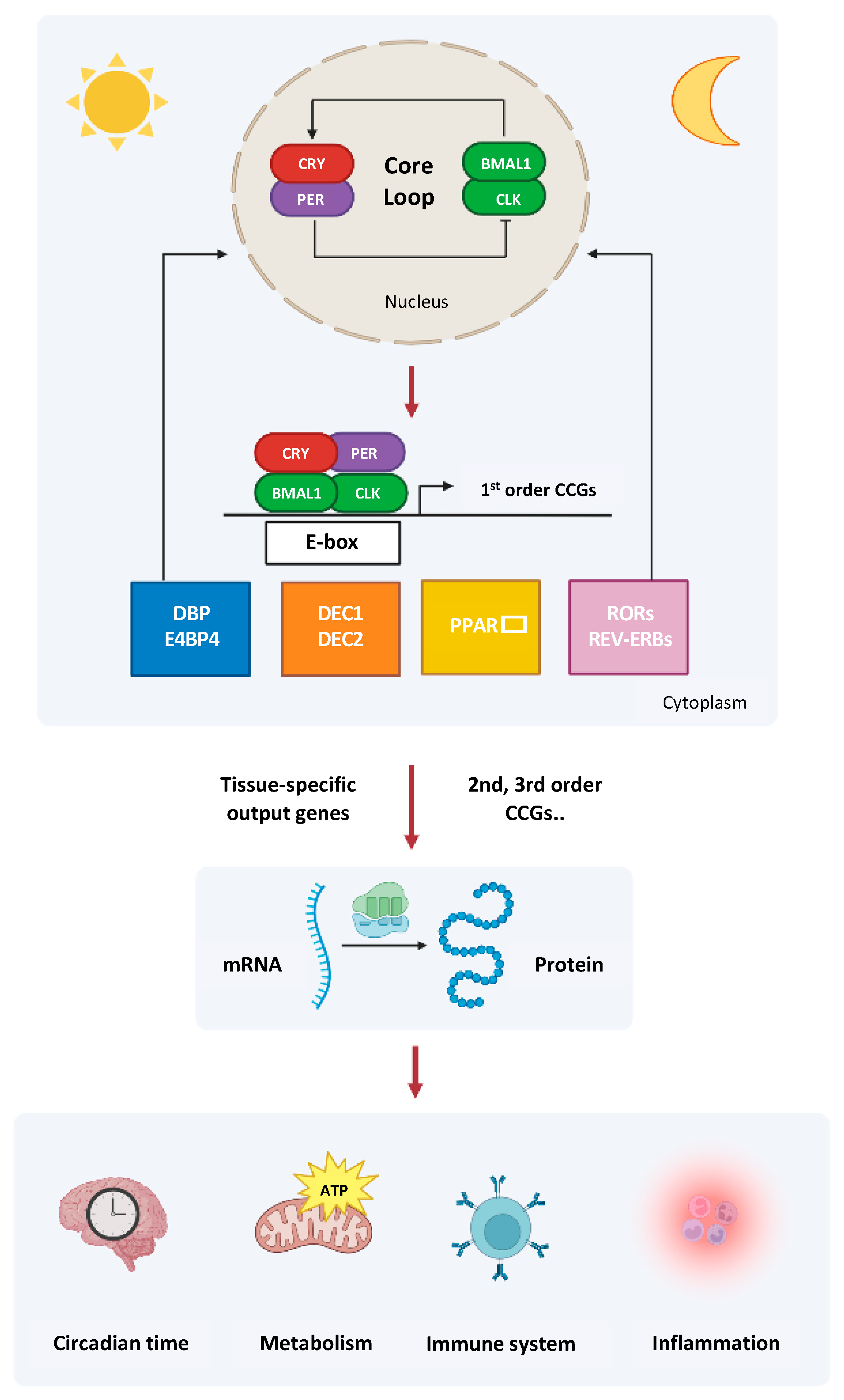
2. The Circadian Clock Circuitry

Physiology and behavior of living beings, scheduled in keeping with sleep/wake, rest/activity, and fasting/feeding cycles, show nychthemeral variations driven by the circadian timing system (CTS), a hierarchical network of biological oscillators comprising a master pacemaker in the suprachiasmatic nuclei (SCN) of anterior hypothalamus [19]. The CTS drives extra-SCN cerebral clocks and self-sustained oscillators in the peripheral tissues through humoral (cortisol, melatonin) or neural (autonomic nervous system fibers) outputs [19]. The SCN are entrained by environmental cues, mainly photic stimuli transmitted by the retino-hypothalamic tract and signaling environmental light-darkness alternation due to Earth’s rotation on its axis to tissues, organs, and organ systems [19].
Biological rhythmicity is generated at the molecular level by way of a group of intertwining genes with their encoded proteins, generating a transcriptional-translational feedback loop (TTFL) revolving with a frequency of 1 cycle in 24 ± 4 h [19]. The TTFL is operated by an activation branch, hard-wired by the Period-Arnt-Single-minded and basic helix-loop-helix (PAS-bHLH) transcription activators CLOCK (circadian locomotor output cycles kaput), and its paralog NPAS2 (neuronal PAS domain protein 2), and BMAL1-2/ARNTL-2 (brain and muscle aryl-hydrocarbon receptor nuclear translocator-like/aryl-hydrocarbon receptor nuclear translocator-like), which heterodimerize and rhythmically bind to E-box (5′-CACGTG-3′) cis-regulatory enhancer sequences of Period (Per1–3) and Cryptochrome (Cry 1–2) genes driving waves of epigenetic modification to promote gene transcription [20,21]. These genes encode PER and CRY proteins, responsible for the inhibitory branch of the feedback loop, accumulate and heterodimerize in the cytoplasm, translocate back to the nucleus, and impede CLOCK/BMAL1-2 transcriptional activity [20,21]. The circadian proteins go through various types of post-translational modifications (PTMs). The key regulators of the clock machinery, the serine/threonine protein kinases casein kinase (CK) 1 δ/ε and CK2, encoded by CSNK1D, CSNK1E, and CSNK2 genes, bind to and phosphorylate multiple circadian substrates [20,21].
Robustness and amplitude of the core TTFL is increased by the nuclear receptors (NRs) REV-ERBs and RORs, whose expression is rhythmically driven by CLOCK/BMAL1 heterodimer. In turn, REV-ERBs and RORs regulate BMAL1 transcription in competition with binding specific ROR response elements (RORE) in BMAL1 promoter and eliciting transcription inhibition and activation, respectively [20,21]. CLOCK/BMAL1 heterodimers bind E-boxes in the second intron of the PPARA gene and activate transcription of PPARA [22], which upon ligand binding, assists BMAL1 expression through a PPARα response element located in the BMAL1 promoter [23]. Furthermore, CLOCK/BMAL1 heterodimers drive the rhythmic expression of first order clock-controlled genes encoding proline and acidic amino acid-rich domain basic leucine zipper (PAR-bZIP) transcription factors, which control the rhythmic expression of thousands of downstream genes [24]. These PAR-bZIP transcription factors comprise DBP (albumin gene D-site binding protein), TEF (thyrotroph embryonic factor), and HLF (hepatic leukemia factor), and the bHLH transcription factors differentially expressed in chondrocytes protein (DEC)1 and DEC2. In addition, the interaction between the opposite oscillatory phase of DBP and REV-ERBs with RORs on RORE at the promoter of Nfil3/E4bp4 gene regulates the rhythmic expression of the nuclear factor interleukin 3 regulated protein (NFIL3, also known as adenoviral E4 protein-binding protein, E4BP4), which plays a key role in immune competent cell development and commitment [24,25].

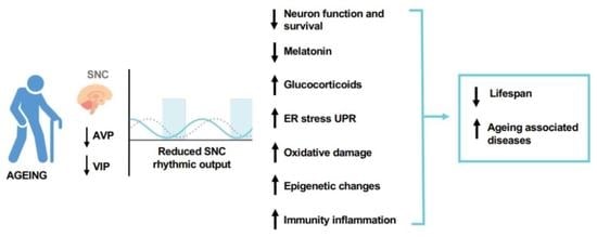
3. The Role of Biological Clock Derangement in the Ageing Process

During the ageing process, progressive remodeling and degeneration of tissues also occur at the level of the cellular elements of brain structures. At the level of the SCN, there is a progressive reduction in the number of neurons expressing vasoactive intestinal peptide and arginine-vasopressin, with weakening of the connectivity in the functional network that ensures the robustness of the oscillation of neuronal and astrocytic cells. With the process of neuronal degeneration associated with aging, the network inside the SCN loses its links and progressively disintegrates, with a reduction in the amplitude of the oscillatory signal and a tendency to shorten the period of oscillation [26]. An important element affected by the ageing process is the pineal gland, which is part of the circadian timing system and whose secretion of melatonin decreases during ageing [27]. Another factor is cortisol, whose secretion by the adrenal glands is not reduced in amplitude but changes in its oscillatory pattern during aging. These alterations determine progressive derangement in the context of the circadian clock circuitry, with alteration of the harmonization of cellular processes and tissue functions in various organs and organ systems [28].
Senescent cells accumulate with aging, modulate their microenvironment through a particular secretory pattern (senescence associated secretory phenotype, SASP), and produce molecules with pro-inflammatory, proapoptotic, and pro-fibrotic activity, such as growth factors, cytokines/chemokines, and extracellular proteases, which not only have an autocrine effect, but also act on neighboring cells (paracrine effect). This bioactive secretome can impact the cell fate by triggering a senescence program and prompting cell-cycle arrest, preventing transmission of DNA damage to daughter cells, and thus preventing potential malignant transformation or, conversely, promoting proliferation, as induced by pro-inflammatory cytokines [29]. SASP is brought on by different signaling pathways, comprising the DNA damage response, stress kinases, inflammation and inflammasome activation, metabolic sensors/pathways, cell survival-associated transcription factors, autophagy, and chromatin remodeling. All these cell processes, primarily genetic modifications based on p16INK4a or p21CIP1/WAF1, are rhythmically driven by the molecular clockwork [30,31]. Lingering senescent cells, which harbor a failing biological clock and drive age-related disorders, pave the way for preventive/therapeutic strategies (senotherapy) that specifically aim to remove senescent cells with senolytics to curb ageing [32,33].
All the processes entailed in ageing are enhanced by the signaling pathways sustaining anabolism, such as growth hormone and insulin/insulin-like growth factor 1 signaling pathways, which activate PI3K-Akt-mTOR signaling pathway enhancing the aging-related processes [34]. Anti-ageing action is carried out by SIRT1-related signaling pathways, acting in part through PGC1alpha, and by AMPK, an important nutrient sensor. AMPK plays an antagonistic role with respect to mTOR signaling and directly modulates the molecular clockwork through phosphorylation of cryptochorme proteins, tagging them for proteasomal degradation [35,36]. Another important player in ageing is mitochondrial dysfunction. Aged and dysfunctional mitochondria must be destroyed and then renewed with processes of mitochondrial biogenesis, for which PGC1alfa and PGC1beta are fundamental. The biological clock is involved in mitochondrial reactive oxygen species (ROS) production and detoxification through the control of nutrient flux, uncoupling mechanism, redox regulation, antioxidant defense, and mitochondrial dynamics. An important mechanism is the excess of oxidizing radicals and decrease in antioxidant mechanisms [37]. The biological clock controls the fundamental processes that counteract damage from oxidizing radicals. Additionally, the role played by the hormone secreted by the pineal gland, melatonin, one of the most powerful molecules with an antioxidant effect is significant [38]. The derangement of the circadian clock circuitry causes a gradual decline in the production of antioxidant molecules in the face of a continuous increase in oxidative damage, as seen in the mouse model with loss of functionality of BMAL1, characterized by increased oxidative stress, impaired expression of several redox defense genes, increased neuronal susceptibility to oxidative damage, synaptic damage, and increased gliosis [39]. When the biological clock is altered, this results in a reduction in life span [40]. Gene mutations, especially in the core circadian genes, have an important impact on the duration of life. In experimental animal models, the knockout of BMAL1 gene results in accelerated ageing processes, increased oxidative damage, reduced body weight, sarcopenia, and altered leukocyte formula [34]. On the other hand, silencing of CLOCK results in reduced lifespan and increased incidence of cataracts [41].

4. The Biological Clock and the Innate Immune System

Inflammation, an adaptive host response also involved in tissue repair, alerts the immune system to sites of infection and tissue damage. Under normal conditions, at the molecular level, it is crucial to rigorously control the expression of genes enriching the signaling pathways that manage inflammatory responses by maintaining repression when external or internal stimuli are absent, and promptly activating their expression in the case of infection or tissue injury. The transcriptional control of a large part of these genes relies on signal-dependent transcription factors comprising members of the NF-κB, activator protein 1 (AP-1, a heterodimer composed of c-Fos/c-Jun family members), and interferon regulatory factor (IRF) families of transcription factors. After activation, NF-κB, AP-1, and IRF elicit the expression of numerous genes that turn on and boost inflammatory processes and support the progression of acquired immunity in different immune competent and inflammatory cells. Many receptor systems for molecules derived from pathogens (PAMPs), endogenous damage-associated molecular patterns (DAMPs), and receptors for cell-derived inducers of inflammation, such as IL1β, TNFα, interferons, can prompt the expression of these transcription factors [42].
The key of the circadian pathways in managing innate immune response was recognized by studying animal models with molecular clockwork changes. A pathophysiological connection between disruption of the molecular clockwork and increased susceptibility to chronic inflammatory diseases was suggested by studies performed in fibroblasts with double knock-out of cryptochrome genes (Cry1−/−Cry2−/−). These studies showed that in a cell-autonomous manner, dual silencing of the cryptochrome (CRY1 and CRY2) proteins remove the inhibitory effect on cyclic adenosine monophosphate (cAMP) production, increase intracellular cAMP levels, and enhance protein kinase A (PKA) signaling, with augmented phosphorylation of p65 at S276 residue and NF-κB signaling activation leading to unremitting increase in the proinflammatory cytokines IL-6 and TNF-α, and considerably augmented expression of inducible nitric oxide (NO) synthase (iNOS) [43]. On the other hand, homozygous Clock mutant mice showed reduced expression of immune-related genes and dampened oscillations of leukocyte number in the peripheral blood [6].
In Per2−/− mice, serum levels of the proinflammatory cytokines interferon IL-1β and (IFN)-γ were significantly reduced after lipopolysaccharide (LPS) challenge, while TNFα, IL-6, and IL-10 production was preserved. Per2−/− mice were more resilient to LPS-induced endotoxic shock with respect to wild-type mice, proposing mPer2 as an essential regulator of natural killer (NK) cell function [44]. Macrophages are key controllers of innate immunity and are responsible for time-based gating of proinflammatory cytokine secretion and systemic immune responses. Mouse macrophages enclose autonomous molecular clockworks driving 24-h rhythmicity in more than 8% of the transcriptome, controlling numerous essential regulators of pathogen recognition and secretion of cytokines, including TNF-alpha and IL-6 upon challenge with bacterial endotoxin at diverse daily intervals [16].
Studies performed on mutant mice and human macrophages showed that REV-ERBα blocks innate immune response to endotoxin driving genes implicated in innate immunity. This circadian gating on inflammatory pathways is lost in REV-ERBα−/− mice [45]. TNF-α interferes with the functioning of the molecular clockwork, mainly thwarting the expression of PER3, DBP, TEF, and HLF, possibly triggering the so-called ‘‘inflammatory clock gene response’’ responsible for weakness in innate immunity activation and inflammatory diseases [46,47]. Innate immune response includes the acute phase response (APR), consisting of prompt gene expression reprogramming and metabolism adjustments upon inflammatory cytokine secretion and acute phase protein (APP) production in the liver, with increased APP levels characteristic of metabolic disorders. APR, along with lipid sensing nuclear receptors (NR) and signal-dependent activation of proinflammatory transcription factors, such as NF-kB, signal transducers, and activators of transcription (STATs) and AP-1 family members backs the interplay among metabolic and inflammatory pathways, sustaining the so-called “metaflammation” process and upholding atherogenesis [48]. Numerous NR involved in metabolic pathways, such as liver X receptor (LXR, binding oxysterols), peroxisome proliferator-activated receptor (PPAR, binding fatty acids), and farnesoid X receptor (FXR, binding bile acids) transcriptionally interfere with the pro-inflammatory signal-dependent initiating transcription factors NF-kB [homo- or heterodimer composed of p50 and p65 (RelA)], signal transducers, and activators of transcription (STATs) and AP-1 family members [49]. Upon ligand binding, these NR suppress inflammatory gene expression with a process called transrepression, which consists in NR–proinflammatory transcription factor complexes exclusion from genomic binding sites [49].
Close interactions among NR and the molecular clockwork influence its entrainment in diverse tissues: PER2 inhibits PPARγ expression and prevents its recruitment to promoters of target genes. Conversely, PPARγ activates BMAL1 transcription. There is a mutual activation of BMAL1 expression by PPARα and PPARα expression by BMAL1, while CLOCK cooperatively activates the 24-h rhythmic expression of PPARα [50]. During the inflammatory response, PPARγ expression can be activated by IL-4 and other immunoregulatory molecules, while it is suppressed by IFN-γ and LPS [51]. In addition, REV-ERBα increases the expression of IL-6 and cyclooxygenase-2 in primary human macrophages in blood vessel wall [52]. REV-ERBα expression is prompted by ligands binding to LXRs, which bind to a specific response element in the human Rev-erba promoter and regulate cholesterol turn-over in macrophages and their role in inflammation and immune response. Moreover, LXRα induces transcriptional expression of TLR4, while REV-ERBα binds as a monomer to a RE overlying the LXR RE in the TLR4 promoter and rhythmically inhibits LXR transactivation of TLR4 expression [53]. Furthermore, PER2 drives the rhythmic expression of the TLR9, which recognizes deoxyribonucleic acid (DNA) leading to circadian fluctuation of cellular activation and cytokine production influencing the immune response [54].

5. The Biological Clock and the Adaptive Immune System

The adaptive immune system operates clear-cut recognition of non-self-elements through the discriminatory expansion of cells primed to target definite antigens and implement immunological memory. Cells crucial for adaptive immune responses are T and B lymphocytes in cooperation with dendritic cells (DCs), which intercept non-self-elements and connect the innate and adaptive responses. All cellular components involved in adaptive immunity are equipped with biological clocks that rhythmically regulate their specific functions [55].
The major protagonists in adaptive immune response are CD4+ T cells (effector or helper T cells) which secrete cytokines and chemokines and assist in antibody production. The differentiation of naïve CD4+ T helper cells (Th) through Th1, Th2, Th17, and induced regulatory T (iTreg) cell lines is managed by the cooperation of a signal transduction web and a transcription factors set (STAT, GATA-3, NF-AT, NF-kB, and AP-1) whose expression is rhythmically driven by the molecular clockwork [56,57]. The transcription factors T-bet, STAT 4, eomesodermin, Hlx, and Runx3 are involved in the differentiation of Th1 cells, which secrete IFNγ, lymphotoxin α (LTα), and IL-2 and mediate immune responses against intracellular pathogens. The transcription factors STAT6, GATA-3, NF-AT, NF-kB, c-Maf, and AP-1 are involved in the differentiation of Th2 cells, which secrete IL-4, IL-5, IL-9, IL-10, IL-13, IL-25, and amphiregulin. Moreover, Th2 cells defense the host against extracellular parasites, including helminths. Furthermore, they are important in the induction and persistence of asthma and other allergic diseases [58].
Th cell differentiation is a specific and dynamic process accompanied by changes in expression patterns of thousands of genes at different stages. Once initiated, T cells develop several mechanisms to reinforce this program until epigenetic modifications are established in certain gene loci that lead to the acquisition of a stable and specific phenotype. The transcription factors DEC1 and DEC2 oscillate with 24-h periodicity in peripheral blood mononuclear cells and determine Th cells fate and function [59]. Faults in T lymphocyte activation and failed turn-over of activated T and B cells hallmark DEC1-deficient mice. On the other hand, DEC2 levels induced by IL-4 signaling gradually increase throughout Th2 cells differentiation upon inducing stimuli and mainly regulates IL-2Rα expression with upregulation of CD25 expression elicited by DEC2 upon STAT6 activation and IL-2 signaling [59]. An additional transcription factor greatly expressed in Th2 cells and in natural killer (NK) cell and NKT cells is NFIL3/E4BP4, which elicits transcription of genes regulating cytokine secretion and effector function [60,61,62].
PPARs play a key role in adaptive immunity. PPARγ activation in CD4+ T cells suppresses Th17 differentiation, impedes IL-4 production, and facilitates inhibition of IL-2 secretion by the T cells [63]. Unliganded PPARα negatively regulates the T-box transcription factor T-bet, a master driver of genetic programs in both the innate and adaptive immune systems, that specifies the Th1 lineage by inhibiting different T-cell fates. T-bet expression is induced by cytokines, such as IFN-γ, IL-12, IL-15, and IL-21 through the respective cytokine receptors. T-bet expression promotes downstream JAK/STATs or PI3K-AKT-mTORC1 signaling pathways by inhibiting p38 mitogen activated protein (MAP) kinase phosphorylation upon T cell activation [64]. The NR RORγt and RORα, along with STAT3, and Interferon regulatory factor–4 (IRF4), play a part in the differentiation process and function of Th17 cells, which secrete IL-17a, IL-17f, IL-21, and IL-22 and manage immune responses against extracellular bacteria and fungi [65,66,67]. Conversely, REV-ERBα negatively regulates Th17 cell development by competing with RORγt and driving the expression of NFIL3, which directly binds and represses the RORγt promoter. This results in modulation of Th17 signature genes [68,69].
RORα is also important for the development of the nuocytes, cytokine-secreting cells involved in type 2 immune responses that share developmental lineage with natural helper cells [70]. Alternatively, RORα is highly expressed in IgA (+) memory B cells and BMAL1 downregulation decreases B-cell development, determining dynamic transcriptional regulation of class-specific memory B cells [71]. In the absence of proinflammatory cytokines, TGF-β with FOXP3 and STAT5 drives differentiation of naïve CD4 T cells into induced regulatory T (iTreg) cells, which are essential in preserving self-tolerance in addition to regulating immune responses [72]. Aryl hydrocarbon receptor (AhR), a transcription factor of the bHLH/PAS family, crosstalks with the circadian clock circuitry and rhythmically mediates the biochemical and toxic effects of environmental xenobiotics and endogenous reactive by-products of normal metabolism through a specific receptor complex. Its activation promotes epigenetic changes modulating the differentiation of Foxp3 (+) Tregs and Th17 cells [73,74,75].

6. Ageing, Inflammation, and the Immune Response

Natural ageing is an unremitting and unrelenting process that commences in initial adulthood and is sustained by structural and functional changes at the cellular, tissue, and organ level. These changes are cumulative, progressive, intrinsic, and deleterious, and are associated with stress tolerance impairment and frailty, morbidity, and mortality in older adults. Other hallmarks of ageing are genomic instability, telomere attrition, mitochondrial dysfunction, autophagy insufficiency, cellular senescence and immunosenescence, chronic inflammation and inflammasome activation [4]. Pattern recognition receptors (PRRs) host sensor proteins largely expressed by innate immune effector cells to identify PAMPs and/or DAMPs. PAMPs are small molecules with conserved motifs connected with LPS from Gram-negative bacteria, while DAMPs are endogenous constituents discharged upon cell stress or death [76]. Based on protein domain homology, PRRs can be categorized as: Toll-like receptors (TLRs), retinoic acid-inducible gene I (RIG-I)-like receptor (RLR), nucleotide-binding oligomerization domain (NOD)-like receptor (NLR), C-type lectin receptors (CLRs), and absent in melanoma-2 (AIM2)-like receptors (ALRs) [77,78]. TLRs are mainly positioned intracellularly and on the cell membrane, while RLRs and NLRs are found in the cytoplasm [77]. Among the human TLR family, TLR3, TLR7, and TLR8 are in the endosomal compartment as endosomal RNA sensors [77]. Upon recognition of DAMPs, TLRs recruit intracytoplasmic toll-interleukin-1 receptor (TIR) domain-containing adaptor proteins, such as MyD88 and TRIF to initiate intracellular signal transduction and increase the secretion of proinflammatory cytokines, chemokines, and type I interferons (IFN) by immune cells [79,80,81].
Inflammasomes are intracellular multiprotein complexes located in the cytoplasm and composed of NLR protein 3 (NLRP3), adaptor apoptosis-associated speck-like (ASC) protein, and procaspasce-1. Upon activation, inflammasome formation prompts caspase-1 expression and upholds discharge of proinflammatory cytokines, IL-1β and IL-18 [82,83]. Assemblage of the NLRP3 inflammasome requires two components: The priming phase and the activation phase. Priming is necessary for NLRP3-mediated gene expression and encompasses post-translational modifications to organize precise inflammasome assembly. Upon inflammasome activation, NLRP3 polymerizes to ASC and recruits procaspase-1 to cleave pro-IL-1β into IL-1β combined with pro-IL-18 [82,83]. The NLRP3 inflammasome represents an essential element of the innate immune system through caspase-1 activation and proinflammatory cytokines IL-1β/IL-18 secretion in response to cellular damage [82,83].
As previously mentioned, the chronic, asymptomatic, low-grade inflammation occurring in the absence of infection during ageing is called “inflammaging” (alias inflammaging, inflammageing) [84]. Inflammaging has harmful effects on health and contributes to biological ageing and the development of age-related chronic diseases, such as atherosclerosis, chronic kidney disease, cardiovascular disease, adult diabetes, and Alzheimer’s disease. The specific causes of inflammaging remain largely unknown, but inflammation may be caused by: (i) Release by damaged cells and accumulation of altered molecules (microRNA, mitochondrial DNA or histones), which are recognized by the cells of the immune system, resulting in the activation and development of inflammation; (ii) increase in the number of senescent cells that release pro-inflammatory molecules into the blood; (iii) chronic stress conditions; (iv) derangement of autophagic processes; (v) alteration of the intestinal microbiota; (vi) immunosenescence, defining the steady waning of immune system function during ageing [85]. Autophagy and microbiome homeostasis are rhythmically regulated by the biological clock and their derangement cause several pathological processes underlying inflammatory, metabolic, degenerative, and neoplastic diseases. An in-depth discussion of the involvement of these processes in pathological conditions and in age-related diseases goes beyond the boundaries of this review; therefore, we refer to exhaustive articles already present in the international scientific literature [86,87,88,89,90,91,92,93].
Innate and adaptive immune response, including humoral and cellular immunity, decays during ageing. Increasing evidence shows that ageing significantly affects all cell compartments of the innate immune system. Numerous neutrophilic functions, for instance, phagocytic capacity, ROS production, and intracellular killing ability, are impaired in the elderly. Similarly, macrophage functions, including phagocytic activity, cytokine and chemokine secretion, antigen presentation, infiltration and wound repair and antibacterial proficiency decline with ageing. Age-related reduction in mast cell and eosinophil and alterations of functional properties have been demonstrated. Conversely, data regarding the influence of ageing on natural killer (NK) and natural killer T (NKT) cells numbers and functions are less evident [94].
Ageing-related changes in adaptive immunity include considerable reduction in the number of naïve lymphocytes formed in the bone marrow and thymus, in addition to the accumulation of functionally incompetent atypical B lymphocytes, named age-associated B cells (ABCs) [95]. The assortment of immune receptor repertoires is seriously restricted in aged B lymphocytes. Moreover, the helper function of naïve CD4+ T cells in B cells antibody production decreases during ageing [96,97]. Similarly, ageing disturbs the B lymphocyte stimulator (BLyS) cytokine family, an essential ligand class for B cell survival and maturation. ABCs exclusively react to TLR7 and TLR9 stimuli rather than B cell antigen receptor stimuli. Consequently, augmented ABCs are inclined to yield low affinity antibodies through the decreased aptitude of antigen recognition sites to identify antigens and activate inflammatory processes [96,97]. Furthermore, the number of naïve T cells that enable antigen presenting cells to communicate with antigen-specific CD8+ T cells is decreased during ageing [98,99].
A schematic representation of the molecular clockwork driving the expression of downstream genes is shown in Figure 1.

7. Endoplasmic Reticulum Stress and UPR during Ageing

Synthesis and folding of membrane and secretory proteins, lipids, and sterols, as well as free calcium storage take place in the ER [100]. Folding is the biophysical process through which the proteins arrange in their precise, three-dimensional outlines necessary for their specific biological function. Ageing is associated with changes in the expression of ER chaperones and folding enzymes, leading to the impairment of proteostasis, and accumulation of unfolded or misfolded proteins that build up in the ER causing ER stress and triggering UPR [100,101]. ER stress reestablishes homeostasis by prompting downstream gene transcription, together with ER overload response (EOR), sterol cascade reaction (SCR), and UPR. EOR is triggered by undue accumulation of correctly folded proteins in the ER and, in addition, prompts various signaling pathways. EOR includes Ca2+ release from the ER, ROS production, and prompting of nuclear factor kappa B (NF-κB)-dependent signaling pathway [102]. In conditions of cellular stress, EOR intervenes to re-establish in the ER proper efficiency in managing protein synthesis and folding, oxidative equilibrium, and Ca2+ flux [103]. SCR is started by cholesterol depletion, causing cleavage of the membrane-bound transcription factor sterol-regulated binding protein (SREBP), which prompts the transcription of genes controlling cholesterol absorption and biosynthesis as well as fatty acid synthesis and absorption [104]. During ageing, numerous factors, such as altered redox or Ca2+ homeostasis and protein glycosylation, could cause ER stress, disrupting correct protein folding and leading to an accumulation of misfolded proteins. Augmented ROS levels elicit pro-inflammatory signals with NF-κB activation in addition to NLRP3 inflammasome triggering and interleukin-1β secretion, hastening neutrophil recruitment, as well as endothelial adhesion molecules and cytokines/chemokines induction, T-helper cells activation, and B cells proliferation [105,106].
Protein quality control in the ER and activation of ER-transmembrane signaling molecules are managed by chaperone proteins, such as glucose-regulated protein 78 (GRP78) [107,108]. GRP78 interacts with the proximal sensors inositol-requiring transmembrane kinase/endoribonuclease 1α (IRE1α), protein kinase R-like endoplasmic reticulum kinase (PERK), and activating transcription factor 6 (ATF6) [109,110,111]. ER stress activates IRE1α, which oligomerizes and autophosphorylates through a kinase domain and upon activation splices and cleaves XBP1 mRNA to yield truncated XBP1 mRNA, encoding XBP1. Activated IRE1α mediates the splicing of an intron from the mRNA of XBP1, triggering a frameshift throughout translation and introducing a new carboxyl domain in the XBP1 protein, which was found to be a functional transcription factor [112]. This transcription factor begins the expression of genes managing synthesis of proteins essential for ER-related molecular chaperoning, protein folding, and degradation and adipogenesis [109,110,111].
Another UPR pathway is PERK, a protein serine/threonine kinase that oligomerizes and autophosphorylates on the kinase domain following ER stress and, in turn, phosphorylates eIF2α to hinder protein translation. Activated PERK enhances ATF4 transcription and translation to increase the level of proteins involved in vital cellular processes, including autophagy, amino acid turnover, protein secretion, redox equilibrium, and apoptosis. Furthermore, ATF4 binds a conserved site in the promoter of the PPP1R15A (GADD34) gene guiding a negative regulatory feedback loop that controls protein translation in response to ER stress [113]. ATF6 is an ER transmembrane protein working as ER-stress sensor/transducer and transcription factor that in cooperation with XBP1s binds cis-acting ER stress response elements (ERSE) and elicits the expression of genes encoding ER chaperones involved in UPR, protein folding, ER-associated protein degradation (ERAD), and mitochondrial biogenesis [109,110,111].
The biological clock drives an ancillary rhythmic activation with 12-h periodicity of the IRE1α pathway in the ER coordinating rhythmic expression of several ER-localized enzymes and UPR constituents involved in hepatic metabolism [114]. The intricate interplay between the circadian pathway and the UPR is operated by several components among which the microRNA miR-211 is crucial. ER stress impedes the transcription of core clock and clock-controlled genes via an ATF4-dependent mechanism [115,116,117].

8. UPR Mediators and Inflammation

IRE1α prompts inflammation via numerous mechanisms, especially through the regulation of inflammatory cytokine production and cellular signaling pathways, mainly through the kinase and endoribonuclease (RNase) activity of its C-terminal cytoplasmic region [118,119]. On the other hand, TLRs, particularly TLR2 and TLR4, can trigger IRE1α in mouse macrophages following stimulation by invading pathogens or endogenous signals, such as damaged cells [120]. After oligomerization, IRE1α RNase cleaves specific mRNAs through a definite process called regulated IRE1-dependent decay (RIDD) [121]. Moreover, IRE1α RNase prompts TNF-α production in macrophages through direct binding to its promoter/enhancer region [122]. Then, the activated IRE1α-TNF receptor-associated factor 2 (TRAF2) complex triggers JNK-AP1 and NF-κB and upholds IL-6 and TNFα production [122]. Furthermore, IRE1α-mediated activation of GSK-3β prevents XBP-1 splicing and transcription of IL-1β and amplifies inflammation [123], confirming that the IRE1 pathway is essential for unrelenting production of pro-inflammatory cytokines.
A pathway essential for inflammatory gene expression in the context of the ER stress-induced inflammatory response is represented by PERK-dependent phosphorylation of eIF2α with translation blockade. PERK-eIF2α-prompted translational block upholds IL-6, MCP-1, and CCL20 transcription independently of ATF4. PERK prompts NF-κB in a NOD1-dependent manner and triggers the JAK1/STAT3 pathway to uphold IL-6 production. Furthermore, the PERK/p38/ERK axis enhances IL-6 and IL-8 secretion [124,125]. The ATF6 family comprises seven members: The ATF6 paralogs (ATF6α and ATF6β) and five CREB3 proteins. ER stress and its specific signaling pathway mediated by ATF6 operates a crucial intracellular mechanism in regulating innate immune cells in vitro and innate immunity in vivo [126].

9. Ageing and NLRP3 Inflammasome Formation

The NLRP3 inflammasome is a cytoplasmic multiprotein complex consisting of the intracellular innate immune receptor NLRP3, the adaptor protein ASC (apoptosis-associated speck-like protein), and the protease caspase 1. NLRP3 inflammasome is initiated in response to extra- and intracellular danger signals, including mitochondrial oxidative stress. Upon activation, the inflammasome triggers an inflammatory response through the caspase-1-dependent activation of the cytokines IL-1β and IL18. The NLRP3 inflammasome recognizes PAMPs or DAMPs and triggers recruitment of the inflammatory protease caspase-1 [127], which modulates the inflammatory response through IL-1β and IL-18 precursors cleavage into active forms [128]. The NLRP3 inflammasome can be activated by various exogenous and endogenous stimuli, including pathogens, LPS, oxidized low-density lipoproteins, lysosomal damage, potassium leakage, ROS production and oxidative stress, Ca2+ ion mobilization and intracellular gradient [128,129]. NLRP3 inflammasome activation may also depend on ER stress as IRE1α activation enhances mitochondrial ROS levels supporting NLRP3 and mitochondria interplay.
Furthermore, ER stress triggers the inflammasome via NLRP3-caspase-2 driven mitochondrial damage and promotes inflammation, embracing cellular stress and innate immunity [130]. Similarly, IRE1α worsens ROS production and triggers the NLRP3 inflammasome to stimulate IL-1β maturation in long-lived, donor-reactive memory B cells (BMEMs) throughout ER stress [131,132]. NLRP3 inflammasome activation is enhanced by mitochondrial ROS production upon Ca2+ release from the ER during stress. Moreover, NLRP3 inflammasome activity is regulated through ROS and mitochondria-associated membrane (MAM) proteins “diaphony” [133]. NLRP3 expression and activation, as well as IL-1β and IL-18 secretion in various tissues and immune/inflammatory cells, particularly macrophages, is controlled by the biological clock, which drives 24-h rhythmic fluctuations of NLRP3 signaling.
Studies performed in peritoneal mouse macrophages in vivo showed that REV-ERBα transcriptionally inhibits expression of the NLRP3 inflammasome components, whose mRNA levels peak throughout the active phase, coinciding with the lowermost intracellular levels of REV-ERBα [134]. On the other hand, decreased Nlrp3 and Il1b mRNA levels and diminished ability to secrete IL-1β was shown in bone marrow-derived macrophages isolated from RORγ-null mice, and numerous presumed ROREs occupied by RORγ were discovered in the Nlrp3 and Il1b genes promoters [135].

10. The Circadian Epigenome and the Epigenetic Clock in Ageing

The functioning of the molecular clockwork and the rhythmic bursts of gene transcription depend on definite and cyclic chromatin changes operated by circadian chromatin remodelers and occurring on a genome-wide scale [136]. The transactivation capacity of the core protein CLOCK on E-box–containing promoters is linked, at least in part, to its histone acetyltransferase (HAT) activity on histone H3 at K9 and K14, with recruitment of other HATs, i.e., CREB binding protein (CBP), p300, P300/CBP-associated factor (PCAF), thus determining chromatin modifications that increase DNA accessibility and enable gene transcription [137]. HATs are counterpoised by a group of histone deacetylases (HDACs)-containing repressive complexes rhythmically recruited to chromatin, such as SIN3A–HDAC1 complex recruited by PER proteins, SIN3B–HDAC1/2 complex recruited by CRY1, and NCoR–HDAC3 complex recruited by the nuclear receptor REV-ERBα [137].
Among HDACs, a peculiar role is played by a class of seven enzymes, communally identified as sirtuins and implicated in many processes linked to metabolism, inflammation, and ageing. The nicotinamide adenine dinucleotide (NAD+)-dependent class III HDACs sirtuin (SIRT) 1 modulates the transcriptional activity of CLOCK:BMAL1 heterodimer in the nucleus [138]. SIRT6 plays a role in the rhythmic mitochondrial function driving acetylation/deacetylation cycles at the level of Complex I. Precisely, circadian oscillations in the activity of nicotinamide phosphoribosyltransferase, the limiting enzyme in the NAD+ salvage pathway encoded by the clock-controlled gene NAMPT, determine 24-h fluctuations of cellular NAD+ content, a crucial nutrient sensor. Moreover, they fuel the cyclical deacetylation of a single subunit of the respiratory chain Complex I and, ultimately, the rhythmic activity of mitochondrial respiration [139,140]. In addition to acetylation, other post-translational chromatin modifications have an impact on the ticking of the biological clock, such as H3K4me3 activation operated by members of the mixed-lineage leukemia (MLL) family histone methyltransferases (HMTs), and the H3K4 histone methyltransferase (HMT) MLL1, which induces cyclic trimethylation of H3K4 and recruitment of CLOCK:BMAL1 in chromatin at definite circadian gene promoters [141]. In addition, the clock-controlled histone-remodeling enzyme MLL3 epigenetically targets and modulates circadian transcription resulting in a whole-genome cycle of activating (H3K4me3) and inhibitory (H3K9me3) chromatin marks [142]. Similarly, the methyltransferase EZH2 determines the repressive mark H3K27me3 at the promoter of Per1 [143]. Moreover, the JumonjiC and ARID domain-containing histone lysine demethylase 1a (JARID1) act as an inhibitor of HDAC1, thus increasing CLOCK:BMAL1 transcriptional activity [144]. The flavin adenine dinucleotide (FAD) dependent demethylase LSD1 acts as circadian chromatin remodeler, controlled by PKCα-mediated 24-h rhythmic phosphorylation [145].
Another important level of epigenetic modification and gene transcription regulation is the transfer of a methyl group from donor S-adenyl-l-methionine (SAM) through a set of DNA-modifying enzymes with covalent addition and methylation at cytosine bases. This process is operated by a conserved and highly regulated family of DNA methyltransferases, among which the canonical DNMT enzymes are DNMT1, mainly involved in maintenance methylation, and DNMT3A and DNMT3B, principally operating de novo methylation. In eukaryotic genomes, common methylation marks occur as methylation of the carbon-5 of cytosine (5 mC), generally present on cytosines preceding a guanine nucleotide (also known as CpG dinucleotide sites) and occur on both DNA strands to maintain the post-replicative DNA methylation patterns [146]. Remarkably, experiments performed in animal models and human cells showed circadian oscillation of global DNA 5mC content and 24-h rhythmic changes in canonical DNMTs mRNA levels and enzymatic activity, suggesting a crucial role played by the biological clock in the control of maintenance and de novo DNA methylation [147,148,149].
Age-associated deregulation of the epigenome characterizes the ageing process, partly due to an alteration in the circadian clock circuitry [150]. Disruption of the biological clock has been associated with numerous diseases and age-related processes at the transcriptional, translational, and post-translational level, as well as with age-specific signatures at the genomic, genetic, and epigenetic levels [151,152]. In all species, tissues, cell types, and epigenetic traits change dynamically throughout life. Specifically, during the ageing process, DNA methylation patterns modify leading to genome-wide hypo-methylation and site-wide hyper-methylation. These epigenetic drifts are generally erratic and non-directional, limiting prediction of both DNA hypo-methylation and hyper-methylation, with different patterns of changes in the methylome among ageing individuals. Nonetheless, there is plausible evidence of ageing-associated differentially methylated regions, through consecutive groups of cytosine-phosphate-guanine (CpG) dinucleotides. These sites could show non-stochastic methylation changes in a constant direction over time and could be related to biological mechanisms strictly associated with the ageing process and longevity. They represent DNA methylation clocks, generally referred to as epigenetic clocks, and confer DNA methylation-based approximations of biological age (Horvath’s clock, Hannum’s clock, DNA PhenoAge, and DNA GrimAge), developed through joint exploitation of mathematical algorithms, mostly based on machine learning. The sets of CpGs are robustly correlated with age, but not convincingly with a decline in physical function. Indeed, additional studies are needed to elucidate whether epigenetic ageing renders the decline in muscle strength with ageing or whether epigenetic clocks simply measure the evolution of ageing [153,154,155,156].

11. “Circadian Medicine”: A New Anti-Ageing Therapy

“Circadian medicine” or “Chronotherapy” is a new model of anti-ageing intervention. It is based on current evidence that circadian rhythms are reduced and more likely to be altered with age, leading to metabolic disorders that reduce longevity [157,158,159]. An alteration of circadian rhythms can also cause a direct disruption of age-related pathways, that oscillate physiologically during the day [160,161]. In light of this evidence, it is hypothesized that there is an optimal time for the administration of drugs that can restore the correct target rhythms, and ultimately improve health and prolong lifespan.
There are many treatments for age-related diseases that are more effective when administered at specific times of day, particularly those for cardiovascular diseases (CVD). One of the most illustrative examples is aspirin, used as a secondary prevention of CVD in humans. A randomized crossover study showed that aspirin reduces blood clotting more effectively when taken at bedtime rather than in the morning [162]. These results were also confirmed by a placebo-controlled study conducted in healthy adults over the age of 65, in which it was observed that those who received aspirin at breakfast, rather than at dinner, experienced an increase in bleeding, CVD risk, and all-cause mortality [163]. Similarly, the efficacy of the statin simvastatin and antihypertensive drugs, such as the Ca2+ channel blocker nifedipine and angiotensin II receptor antagonists, is greater when taken before bedtime [164,165].
Other treatments for age-related diseases act directly on the activity of the internal clock. Polyamines represent a pharmacological intervention that mediates the crosstalk between the circadian clock, metabolic pathways, and lifespan. Indeed, these molecules regulate circadian periodicity via PER2/CRY1175 interactions leading to increased lifespan in mice [166]. Another molecule that enhances clock action is a natural flavonoid called nobiletin (NOB). This flavonoid has been found to reduce body weight gain without altering food intake, stimulate energy expenditure and circadian activity, improve glucose and insulin tolerance, decrease lipids, and improve mitochondrial respiration in mice [167,168]. These findings clearly demonstrated that maintaining a strong circadian organization in the body protects against metabolic perturbations.
Considering the promising evidence obtained to date, future human studies that consider tissue-specific circadian fluctuations in drug treatments to counter age-related diseases are needed. Providing guidance on when and how often treatments should be given is crucial to reduce drug resistance, side effects, and promote healthy ageing.

12. Conclusions

The processes that undermine ageing include immunosenescence, inflammation, and inflammasome formation in the context of ER stress and autophagy derangement. Innate and adaptive immune response is characterized by rhythms with diverse frequency ranges, generally with 24-h periodicity and with coincident or opposing phases in the levels of cellular effectors and humoral factors. Biological clocks ticking in inflammatory/immune cells drive fluctuations of leukocyte trafficking and turn-over, phagocytic and peroxidase activity, as well as cytokine/chemokine secretion and antibody production. The circadian clock circuitry and the immune system intermingle bidirectionally integrating environmental cues with the internal milieu. Signal-dependent transcription factors and nuclear receptors maneuver transcriptional circuits and gene signature determining inflammatory/immune cells’ function and fate through mutual communications linking the molecular clockwork to the rhythmic regulation of inflammatory pathways and immune response. Increasing evidence highlights the role played by changes in the circadian clock circuitry during ageing and the possible nutritional and pharmacological interventions aimed at limiting or delaying these alterations [169,170]. A deeper understanding of the molecular alterations occurring within the interactions between the circadian pathways, the inflammatory process, and the immune response, especially in the context of chronodisruption, would allow us to ascertain and therapeutically address some of the age-related pathophysiological mechanisms involved in inflammatory response and immune system dysregulation during ageing.

Author Contributions

Conceptualization, B.C. and G.M.; writing—original draft preparation, M.D., E.M., S.L., R.T. and F.S.; writing—review and editing, B.C. and G.M.; supervision, B.C., F.S. and G.M. All authors have read and agreed to the published version of the manuscript.

Funding

This research was funded by the “5 × 1000” voluntary contribution and by a grant from the Italian Ministry of Health (1 January 2022–31 December 2024; number RC2022-2024) to G.M.

Institutional Review Board Statement

Not applicable.

Informed Consent Statement

Not applicable.

Data Availability Statement

The data presented in this study are available in the article.

Conflicts of Interest

The authors declare no conflict of interest.

References

  1. Liguori, I.; Russo, G.; Curcio, F.; Bulli, G.; Aran, L.; Della-Morte, D.; Gargiulo, G.; Testa, G.; Cacciatore, F.; Bonaduce, D.; et al. Oxidative stress, aging, and diseases. Clin. Interv. Aging 2018, 13, 757–772. [Google Scholar] [CrossRef] [PubMed] [Green Version]
  2. Adams, C.J.; Kopp, M.C.; Larburu, N.; Nowak, P.R.; Ali, M.M.U. Structure and Molecular Mechanism of ER Stress Signaling by the Unfolded Protein Response Signal Activator IRE1. Front. Mol. Biosci. 2019, 12, 11. [Google Scholar] [CrossRef] [PubMed] [Green Version]
  3. Szegezdi, E.; Logue, S.E.; Gorman, A.M.; Samali, A. Mediators of endoplasmic reticulum stress-induced apoptosis. EMBO Rep. 2006, 7, 880–885. [Google Scholar] [CrossRef] [PubMed] [Green Version]
  4. López-Otín, C.; Blasco, M.A.; Partridge, L.; Serrano, M.; Kroemer, G. The hallmarks of aging. Cell 2013, 153, 1194–1217. [Google Scholar] [CrossRef] [Green Version]
  5. Barbosa, M.C.; Grosso, R.A.; Fader, C.M. Hallmarks of Aging: An Autophagic Perspective. Front. Endocrinol. 2019, 9, 790. [Google Scholar] [CrossRef]
  6. Hergenhan, S.; Holtkamp, S.; Scheiermann, C. Molecular Interactions Between Components of the Circadian Clock and the Immune System. J. Mol. Biol. 2020, 432, 3700–3713. [Google Scholar] [CrossRef]
  7. Logan, R.W.; Sarkar, D.K. Circadian nature of immune function. Mol. Cell Endocrinol. 2012, 349, 82–90. [Google Scholar] [CrossRef]
  8. Man, K.; Loudon, A.; Chawla, A. Immunity around the clock. Science 2016, 354, 999–1003. [Google Scholar] [CrossRef] [Green Version]
  9. Scheiermann, C.; Gibbs, J.; Ince, L.; Loudon, A. Clocking into immunity. Nat. Rev. Immunol. 2018, 18, 423–437. [Google Scholar] [CrossRef]
  10. Mazzoccoli, G.; Sothern, R.B.; De Cata, A.; Giuliani, F.; Fontana, A.; Copetti, M.; Pellegrini, F.; Tarquini, R. A timetable of 24-hour patterns for human lymphocyte subpopulations. J. Biol. Regul. Homeost. Agents 2011, 25, 387–395. [Google Scholar]
  11. Méndez-Ferrer, S.; Chow, A.; Merad, M.; Frenette, P.S. Circadian rhythms influence hematopoietic stem cells. Curr. Opin. Hematol. 2009, 16, 235–242. [Google Scholar] [CrossRef] [PubMed]
  12. Dimitrov, S.; Lange, T.; Born, J. Selective mobilization of cytotoxic leukocytes by epinephrine. J. Immunol. 2010, 184, 503–511. [Google Scholar] [CrossRef] [PubMed] [Green Version]
  13. Ebisawa, T.; Numazawa, K.; Shimada, H.; Izutsu, H.; Sasaki, T.; Kato, N.; Tokunaga, K.; Mori, A.; Honma, K.; Honma, S.; et al. Self-sustained circadian rhythm in cultured human mononuclear cells isolated from peripheral blood. Neurosci. Res. 2010, 66, 223–227. [Google Scholar] [CrossRef] [PubMed]
  14. Mazzoccoli, G.; De Cata, A.; Greco, A.; Carughi, S.; Giuliani, F.; Tarquini, R. Circadian rhythmicity of lymphocyte subpopulations and relationship with neuro-endocrine system. J. Biol. Regul. Homeost. Agents 2010, 24, 341–350. [Google Scholar]
  15. Bollinger, T.; Leutz, A.; Leliavski, A.; Skrum, L.; Kovac, J.; Bonacina, L.; Benedict, C.; Lange, T.; Westermann, J.; Oster, H.; et al. Circadian clocks in mouse and human CD4+ T cells. PLoS ONE 2011, 6, e29801. [Google Scholar] [CrossRef]
  16. Keller, M.; Mazuch, J.; Abraham, U.; Eom, G.D.; Herzog, E.D.; Volk, H.D.; Kramer, A.; Maier, B. A circadian clock in macrophages controls inflammatory immune responses. Proc. Natl. Acad. Sci. USA 2009, 106, 21407–21412. [Google Scholar] [CrossRef] [Green Version]
  17. Mazzoccoli, G.; Sothern, R.B.; Greco, G.; Pazienza, V.; Vinciguerra, M.; Liu, S.; Cai, Y. Time-Related Dynamics of Variation in Core Clock Gene Expression Levels in Tissues Relevant to the Immune System. Int. J. Immunopathol. Pharmacol. 2011, 24, 869–879. [Google Scholar] [CrossRef]
  18. Pourcet, B.; Duez, H. Circadian Control of Inflammasome Pathways: Implications for Circadian Medicine. Front. Immunol. 2020, 11, 1630. [Google Scholar] [CrossRef]
  19. Albrecht, U. Timing to perfection: The biology of central and peripheral circadian clocks. Neuron 2012, 74, 246–260. [Google Scholar] [CrossRef] [Green Version]
  20. Koike, N.; Yoo, S.H.; Huang, H.C.; Kumar, V.; Lee, C.; Kim, T.K.; Takahashi, J.S. Transcriptional architecture and chromatin landscape of the core circadian clock in mammals. Science 2012, 338, 349–354. [Google Scholar] [CrossRef] [Green Version]
  21. Takahashi, J.S. Transcriptional architecture of the mammalian circadian clock. Nat. Rev. Genet. 2017, 18, 164–179. [Google Scholar] [CrossRef] [PubMed] [Green Version]
  22. Oishi, K.; Shirai, H.; Ishida, N. CLOCK is involved in the circadian transactivation of peroxisome-proliferator-activated receptor alpha (PPARalpha) in mice. Biochem. J. 2005, 386, 575–581. [Google Scholar] [CrossRef] [PubMed]
  23. Canaple, L.; Rambaud, J.; Dkhissi-Benyahya, O.; Rayet, B.; Tan, N.S.; Michalik, L.; Delaunay, F.; Wahli, W.; Laudet, V. Reciprocal regulation of brain and muscle Arnt-like protein 1 and peroxisome proliferator-activated receptor alpha defines a novel positive feedback loop in the rodent liver circadian clock. Mol. Endocrinol. 2006, 20, 1715–1727. [Google Scholar] [CrossRef] [PubMed] [Green Version]
  24. Bozek, K.; Relógio, A.; Kielbasa, S.M.; Heine, M.; Dame, C.; Kramer, A.; Herzel, H. Regulation of clock-controlled genes in mammals. PLoS ONE 2009, 4, e4882. [Google Scholar] [CrossRef] [PubMed] [Green Version]
  25. Male, V.; Nisoli, I.; Gascoyne, D.M.; Brady, H.J. E4BP4: An unexpected player in the immune response. Trends Immunol. 2012, 33, 98–102. [Google Scholar] [CrossRef]
  26. Nakamura, T.J.; Takasu, N.N.; Nakamura, W. The suprachiasmatic nucleus: Age-related decline in biological rhythms. J. Physiol. Sci. 2016, 66, 367–374. [Google Scholar] [CrossRef] [PubMed]
  27. Melhuish Beaupre, L.M.; Brown, G.M.; Gonçalves, V.F.; Kennedy, J.L. Melatonin’s neuroprotective role in mitochondria and its potential as a biomarker in aging, cognition and psychiatric disorders. Transl. Psychiatry 2021, 11, 339. [Google Scholar] [CrossRef] [PubMed]
  28. Hood, S.; Amir, S. The aging clock: Circadian rhythms and later life. J. Clin. Investig. 2017, 127, 437–446. [Google Scholar] [CrossRef]
  29. Chou, L.Y.; Ho, C.T.; Hung, S.C. Paracrine Senescence of Mesenchymal Stromal Cells Involves Inflammatory Cytokines and the NF-κB Pathway. Cells 2022, 11, 3324. [Google Scholar] [CrossRef]
  30. Wagner, K.D.; Wagner, N. The Senescence Markers p16INK4A, p14ARF/p19ARF, and p21 in Organ Development and Homeostasis. Cells 2022, 11, 1966. [Google Scholar] [CrossRef]
  31. Kowalska, E.; Ripperger, J.A.; Hoegger, D.C.; Bruegger, P.; Buch, T.; Birchler, T.; Mueller, A.; Albrecht, U.; Contaldo, C.; Brown, S.A. NONO couples the circadian clock to the cell cycle. Proc. Natl. Acad. Sci. USA 2013, 110, 1592–1599. [Google Scholar] [CrossRef] [PubMed] [Green Version]
  32. Hashikawa, K.I.; Katamune, C.; Kusunose, N.; Matsunaga, N.; Koyanagi, S.; Ohdo, S. Dysfunction of the circadian transcriptional factor CLOCK in mice resists chemical carcinogen-induced tumorigenesis. Sci. Rep. 2017, 7, 9995. [Google Scholar] [CrossRef] [PubMed]
  33. Birch, J.; Gil, J. Senescence and the SASP: Many therapeutic avenues. Genes Dev. 2020, 34, 1565–1576. [Google Scholar] [CrossRef] [PubMed]
  34. Khapre, R.V.; Kondratova, A.A.; Patel, S.; Dubrovsky, Y.; Wrobel, M.; Antoch, M.P.; Kondratov, R.V. BMAL1-dependent regulation of the mTOR signaling pathway delays aging. Aging 2014, 6, 48–57. [Google Scholar] [CrossRef] [Green Version]
  35. Imai, S.I.; Guarente, L. It takes two to tango: NAD + and sirtuins in aging / longevity control. NPJ Aging Mech. Dis. 2016, 2, 16017. [Google Scholar] [CrossRef] [Green Version]
  36. Sadria, M.; Layton, A.T. Aging affects circadian clock and metabolism and modulates timing of medication. iScience 2021, 24, 102245. [Google Scholar] [CrossRef]
  37. Mezhnina, V.; Ebeigbe, O.P.; Poe, A.; Kondratov, R.V. Circadian Control of Mitochondria in Reactive Oxygen Species Homeostasis. Antioxid. Redox Signal. 2022, 37, 647–663. [Google Scholar] [CrossRef]
  38. Ferlazzo, N.; Andolina, G.; Cannata, A.; Costanzo, M.G.; Rizzo, V.; Currò, M.; Ientile, R.; Caccamo, D. Is Melatonin the Cornucopia of the 21st Century? Antioxidants 2020, 9, 1088. [Google Scholar] [CrossRef]
  39. Musiek, E.S.; Lim, M.M.; Yang, G.; Bauer, A.Q.; Qi, L.; Lee, Y.; Roh, J.H.; Ortiz-Gonzalez, X.; Dearborn, J.T.; Culver, J.P.; et al. Circadian clock proteins regulate neuronal redox homeostasis and neurodegeneration. J. Clin. Investig. 2013, 123, 5389–5400. [Google Scholar] [CrossRef] [Green Version]
  40. Welz, P.S.; Benitah, S.A. Molecular Connections Between Circadian Clocks and Aging. J. Mol. Biol. 2020, 432, 3661–3679. [Google Scholar] [CrossRef]
  41. Dubrovsky, Y.V.; Samsa, W.E.; Kondratov, R.V. Deficiency of circadian protein CLOCK reduces lifespan and increases age-related cataract development in mice. Aging 2010, 2, 936–944. [Google Scholar] [CrossRef] [PubMed] [Green Version]
  42. Marcello, M.; White, M.R. Spatial and temporal information coding and noise in the NF-κB system. Biochem. Soc. Trans. 2010, 38, 1247–1250. [Google Scholar] [CrossRef] [PubMed]
  43. Narasimamurthy, R.; Hatori, M.; Nayak, S.K.; Liu, F.; Panda, S.; Verma, I.M. Circadian clock protein cryptochrome regulates the expression of proinflammatory cytokines. Proc. Natl. Acad. Sci. USA 2012, 109, 12662–12667. [Google Scholar] [CrossRef] [PubMed] [Green Version]
  44. Liu, J.; Malkani, G.; Shi, X.; Meyer, M.; Cunningham-Runddles, S.; Ma, X.; Sun, Z.S. The circadian clock Period 2 gene regulates gamma interferon production of NK cells in host response to lipopolysaccharide-induced endotoxic shock. Infect. Immun. 2006, 74, 4750–4756. [Google Scholar] [CrossRef] [Green Version]
  45. Gibbs, J.E.; Blaikley, J.; Beesley, S.; Matthews, L.; Simpson, K.D.; Boyce, S.H.; Farrow, S.N.; Else, K.J.; Singh, D.; Ray, D.W.; et al. The nuclear receptor REV-ERBα mediates circadian regulation of innate immunity through selective regulation of inflammatory cytokines. Proc. Natl. Acad. Sci. USA 2012, 109, 582–587. [Google Scholar] [CrossRef] [Green Version]
  46. Cavadini, G.; Petrzilka, S.; Kohler, P.; Jud, C.; Tobler, I.; Birchler, T.; Fontana, A. TNF-alpha suppresses the expression of clock genes by interfering with E-box-mediated transcription. Proc. Natl. Acad. Sci. USA 2007, 104, 12843–12848. [Google Scholar] [CrossRef] [Green Version]
  47. Petrzilka, S.; Taraborrelli, C.; Cavadini, G.; Fontana, A.; Birchler, T. Clock gene modulation by TNF-alpha depends on calcium and p38 MAP kinase signaling. J. Biol. Rhythm. 2009, 24, 283–294. [Google Scholar] [CrossRef]
  48. Venteclef, N.; Jakobsson, T.; Steffensen, K.R.; Treuter, E. Metabolic nuclear receptor signaling and the inflammatory acute phase response. Trends Endocrinol. Metab. 2011, 22, 333–343. [Google Scholar] [CrossRef]
  49. Glass, C.K.; Saijo, K. Nuclear receptor transrepression pathways that regulate inflammation in macrophages and T cells. Nat. Rev. Immunol. 2010, 10, 365–376. [Google Scholar] [CrossRef]
  50. Charoensuksai, P.; Xu, W. PPARs in Rhythmic Metabolic Regulation and Implications in Health and Disease. PPAR Res. 2010, 2010, pii243643. [Google Scholar] [CrossRef] [Green Version]
  51. Chawla, A. Control of macrophage activation and function by PPARs. Circ. Res. 2010, 106, 1559–1569. [Google Scholar] [CrossRef] [PubMed]
  52. Migita, H.; Morser, J.; Kawai, K. Rev-erb alpha upregulates NF-kappaB-responsive genes in vascular smooth muscle cells. FEBS Lett. 2004, 561, 69–74. [Google Scholar] [CrossRef]
  53. Fontaine, C.; Rigamonti, E.; Pourcet, B.; Duez, H.; Duhem, C.; Fruchart, J.C.; Chinetti-Gbaguidi, G.; Staels, B. The nuclear receptor Rev-erbalpha is a liver X receptor (LXR) target gene driving a negative feedback loop on select LXR-induced pathways in human macrophages. Mol. Endocrinol. 2008, 22, 1797–1811. [Google Scholar] [CrossRef] [Green Version]
  54. Silver, A.C.; Arjona, A.; Walker, W.E.; Fikrig, E. The circadian clock controls toll-like receptor 9-mediated innate and adaptive immunity. Immunity 2012, 36, 251–261. [Google Scholar] [CrossRef] [PubMed] [Green Version]
  55. Gray, K.J.; Gibbs, J.E. Adaptive immunity, chronic inflammation and the clock. Semin. Immunopathol. 2022, 44, 209–224. [Google Scholar] [CrossRef] [PubMed]
  56. Spengler, M.L.; Kuropatwinski, K.K.; Comas, M.; Gasparian, A.V.; Fedtsova, N.; Gleiberman, A.S.; Gitlin, I.I.; Artemicheva, N.M.; Deluca, K.A.; Gudkov, A.V.; et al. Core circadian protein CLOCK is a positive regulator of NF-κB-mediated transcription. Proc. Natl. Acad. Sci. USA 2012, 109, E2457–E2465. [Google Scholar] [CrossRef] [Green Version]
  57. Cermakian, N.; Lange, T.; Golombek, D.; Sarkar, D.; Nakao, A.; Shibata, S.; Mazzoccoli, G. Crosstalk between the circadian clock circuitry and the immune system. Chronobiol. Int. 2013, 30, 870–888. [Google Scholar] [CrossRef]
  58. Zhu, J.; Paul, W.E. CD4 T cells: Fates, functions, and faults. Blood 2008, 112, 1557–1569. [Google Scholar] [CrossRef] [Green Version]
  59. Liu, Z.; Li, Z.; Mao, K.; Zou, J.; Wang, Y.; Tao, Z.; Lin, G.; Tian, L.; Ji, Y.; Wu, X.; et al. Dec2 promotes Th2 cell differentiation by enhancing IL-2R signaling. J. Immunol. 2009, 183, 6320–6329. [Google Scholar] [CrossRef] [Green Version]
  60. Kashiwada, M.; Cassel, S.L.; Colgan, J.D.; Rothman, P.B. NFIL3/E4BP4 controls type 2 T helper cell cytokine expression. EMBO J. 2011, 30, 2071–2082. [Google Scholar] [CrossRef] [Green Version]
  61. Kamizono, S.; Duncan, G.S.; Seidel, M.G.; Morimoto, A.; Hamada, K.; Grosveld, G.; Akashi, K.; Lind, E.F.; Haight, J.P.; Ohashi, P.S.; et al. Nfil3/E4bp4 is required for the development and maturation of NK cells in vivo. J. Exp. Med. 2009, 206, 2977–2986. [Google Scholar] [CrossRef] [PubMed] [Green Version]
  62. Gascoyne, D.M.; Long, E.; Veiga-Fernandes, H.; de Boer, J.; Williams, O.; Seddon, B.; Coles, M.; Kioussis, D.; Brady, H.J. The basic leucine zipper transcription factor E4BP4 is essential for natural killer cell development. Nat. Immunol. 2009, 10, 1118–1124. [Google Scholar] [CrossRef] [PubMed]
  63. Klotz, L.; Burgdorf, S.; Dani, I.; Saijo, K.; Flossdorf, J.; Hucke, S.; Alferink, J.; Nowak, N.; Beyer, M.; Mayer, G.; et al. The nuclear receptor PPAR gamma selectively inhibits Th17 differentiation in a T cell-intrinsic fashion and suppresses CNS autoimmunity. J. Exp. Med. 2009, 206, 2079–2089. [Google Scholar] [CrossRef]
  64. Jones, D.C.; Ding, X.; Zhang, T.Y.; Daynes, R.A. Peroxisome proliferator-activated receptor alpha negatively regulates T-bet transcription through suppression of p38 mitogen-activated protein kinase activation. J. Immunol. 2003, 171, 196–203. [Google Scholar] [CrossRef] [PubMed] [Green Version]
  65. Jetten, A.M. Retinoid-related orphan receptors (RORs): Critical roles in development, immunity, circadian rhythm, and cellular metabolism. Nucl. Recept. Signal. 2009, 7, e003. [Google Scholar] [CrossRef] [Green Version]
  66. Solt, L.A.; Kumar, N.; Nuhant, P.; Wang, Y.; Lauer, J.L.; Liu, J.; Istrate, M.A.; Kamenecka, T.M.; Roush, W.R.; Vidović, D.; et al. Suppression of TH17 differentiation and autoimmunity by a synthetic ROR ligand. Nature 2011, 472, 491–494. [Google Scholar] [CrossRef] [Green Version]
  67. Rauen, T.; Juang, Y.T.; Hedrich, C.M.; Kis-Toth, K.; Tsokos, G.C. A novel isoform of the orphan receptor RORγt suppresses IL-17 production in human T cells. Genes Immun. 2012, 13, 346–350. [Google Scholar] [CrossRef] [Green Version]
  68. Yu, X.; Rollins, D.; Ruhn, K.A.; Stubblefeld, J.J.; Green, C.B.; Kashiwada, M.; Rothman, P.B.; Takahashi, J.S.; Hooper, L.V. TH17 cell diferentiation is regulated by the circadian clock. Science 2013, 342, 727–730. [Google Scholar] [CrossRef] [Green Version]
  69. Amir, M.; Chaudhari, S.; Wang, R.; Campbell, S.; Mosure, S.A.; Chopp, L.B.; Lu, Q.; Shang, J.; Pelletier, O.B.; He, Y.; et al. REV-ERBalpha Regulates TH17 Cell Development and Autoimmunity. Cell Rep. 2018, 25, 3733–3749.e8. [Google Scholar] [CrossRef] [Green Version]
  70. Wong, S.H.; Walker, J.A.; Jolin, H.E.; Drynan, L.F.; Hams, E.; Camelo, A.; Barlow, J.L.; Neill, D.R.; Panova, V.; Koch, U.; et al. Transcription factor RORα is critical for nuocyte development. Nat. Immunol. 2012, 13, 229–236. [Google Scholar] [CrossRef] [Green Version]
  71. Wang, N.S.; McHeyzer-Williams, L.J.; Okitsu, S.L.; Burris, T.P.; Reiner, S.L.; McHeyzer-Williams, M.G. Divergent transcriptional programming of class-specific B cell memory by T-bet and RORα. Nat. Immunol. 2012, 13, 604–611. [Google Scholar] [CrossRef] [PubMed]
  72. Ogawa, C.; Tone, Y.; Tsuda, M.; Peter, C.; Waldmann, H.; Tone, M. TGF-β-mediated Foxp3 gene expression is cooperatively regulated by Stat5, Creb, and AP-1 through CNS2. J. Immunol. 2014, 192, 475–483. [Google Scholar] [CrossRef] [PubMed]
  73. Rothhammer, V.; Quintana, F.J. The aryl hydrocarbon receptor: An environmental sensor integrating immune responses in health and disease. Nat. Rev. Immunol. 2019, 19, 184–197. [Google Scholar] [CrossRef] [PubMed]
  74. Singh, N.P.; Singh, U.P.; Singh, B.; Price, R.L.; Nagarkatti, M.; Nagarkatti, P.S. Activation of aryl hydrocarbon receptor (AhR) leads to reciprocal epigenetic regulation of FoxP3 and IL-17 expression and amelioration of experimental colitis. PLoS ONE 2011, 6, e23522. [Google Scholar] [CrossRef] [PubMed] [Green Version]
  75. Anderson, G.; Beischlag, T.V.; Vinciguerra, M.; Mazzoccoli, G. The circadian clock circuitry and the AHR signaling pathway in physiology and pathology. Biochem. Pharmacol. 2013, 85, 1405–1416. [Google Scholar] [CrossRef]
  76. Mogensen, T.H. Pathogen recognition and inflammatory signaling in innate immune defenses. Clin. Microbiol. Rev. 2009, 22, 240–273. [Google Scholar] [CrossRef] [Green Version]
  77. Li, D.; Wu, M. Pattern recognition receptors in health and diseases. Signal Transduct. Target. Ther. 2021, 6, 291. [Google Scholar] [CrossRef]
  78. Rehwinkel, J.; Gack, M.U. RIG-I-like receptors: Their regulation and roles in RNA sensing. Nat. Rev. Immunol. 2020, 20, 537–551. [Google Scholar] [CrossRef]
  79. Uehata, T.; Takeuchi, O. RNA Recognition and Immunity-Innate Immune Sensing and Its Posttranscriptional Regulation Mechanisms. Cells 2020, 9, 1701. [Google Scholar] [CrossRef]
  80. Kawasaki, T.; Kawai, T. Toll-like receptor signaling pathways. Front. Immunol. 2014, 5, 461. [Google Scholar] [CrossRef] [Green Version]
  81. Kawai, T.; Akira, S. The role of pattern-recognition receptors in innate immunity: Update on Toll-like receptors. Nat. Immunol. 2010, 11, 373–384. [Google Scholar] [CrossRef] [PubMed]
  82. Franchi, L.; Eigenbrod, T.; Muñoz-Planillo, R.; Nuñez, G. The inflammasome: A caspase-1-activation platform that regulates immune responses and disease pathogenesis. Nat. Immunol. 2009, 10, 241–247. [Google Scholar] [CrossRef] [PubMed] [Green Version]
  83. Vande Walle, L.; Lamkanfi, M. Inflammasomes: Caspase-1-activating platforms with critical roles in host defense. Front. Microbiol. 2011, 2, 3. [Google Scholar] [CrossRef] [PubMed] [Green Version]
  84. Ferrucci, L.; Fabbri, E. Inflammageing: Chronic inflammation in ageing, cardiovascular disease, and frailty. Nat. Rev. Cardiol. 2018, 15, 505–522. [Google Scholar] [CrossRef] [PubMed]
  85. Franceschi, C.; Bonafè, M.; Valensin, S.; Olivieri, F.; De Luca, M.; Ottaviani, E.; De Benedictis, G. Inflamm-aging. An evolutionary perspective on immunosenescence. Ann. N. Y. Acad. Sci. 2000, 908, 244–254. [Google Scholar] [CrossRef] [PubMed]
  86. Rubinsztein, D.C.; Mariño, G.; Kroemer, G. Autophagy and Aging. Cell 2011, 5, 682–695. [Google Scholar] [CrossRef] [Green Version]
  87. Ma, D.; Li, S.; Molusky, M.M.; Lin, J.D. Circadian autophagy rhythm: A link between clock and metabolism? Trends Endocrinol. Metab. 2012, 23, 319–325. [Google Scholar] [CrossRef] [Green Version]
  88. Pastore, N.; Ballabio, A. Keeping the autophagy tempo. Autophagy 2019, 15, 1854–1856. [Google Scholar] [CrossRef]
  89. Maffei, V.J.; Kim, S.; Blanchard, E.; Luo, M.; Jazwinski, S.M.; Taylor, C.M.; Welsh, D.A. Biological Aging and the Human Gut Microbiota. J. Gerontol. A Biol. Sci. Med. Sci. 2017, 72, 1474–1482. [Google Scholar] [CrossRef] [Green Version]
  90. Ratiner, K.; Abdeen, S.K.; Goldenberg, K.; Elinav, E. Utilization of Host and Microbiome Features in Determination of Biological Aging. Microorganisms 2022, 10, 668. [Google Scholar] [CrossRef]
  91. Bishehsari, F.; Voigt, R.M.; Keshavarzian, A. Circadian rhythms and the gut microbiota: From the metabolic syndrome to cancer. Nat. Rev. Endocrinol. 2020, 16, 731–739. [Google Scholar] [CrossRef] [PubMed]
  92. Liang, X.; Bushman, F.D.; FitzGerald, G.A. Rhythmicity of the intestinal microbiota is regulated by gender and the host circadian clock. Proc. Natl. Acad. Sci. USA 2015, 112, 10479–10484. [Google Scholar] [CrossRef] [PubMed]
  93. Choi, H.; Rao, M.C.; Chang, E.B. Gut microbiota as a transducer of dietary cues to regulate host circadian rhythms and metabolism. Nat. Rev. Gastroenterol. Hepatol. 2021, 18, 679–689. [Google Scholar] [CrossRef] [PubMed]
  94. Gomez, C.R.; Nomellini, V.; Faunce, D.E.; Kovacs, E.J. Innate immunity and aging. Exp. Gerontol. 2008, 43, 718–728. [Google Scholar] [CrossRef] [PubMed] [Green Version]
  95. Mahbub, S.; Brubaker, A.L.; Kovacs, E.J. Aging of the Innate Immune System: An Update. Curr. Immunol. Rev. 2011, 7, 104–115. [Google Scholar] [CrossRef]
  96. Ma, S.; Wang, C.; Mao, X.; Hao, Y. B Cell Dysfunction Associated With Aging and Autoimmune Diseases. Front. Immunol. 2019, 10, 318. [Google Scholar] [CrossRef] [Green Version]
  97. Weng, N.P. Aging of the immune system: How much can the adaptive immune system adapt? Immunity 2006, 24, 495–499. [Google Scholar] [CrossRef] [Green Version]
  98. Sprent, J.; Surh, C.D. Normal T cell homeostasis: The conversion of naive cells into memory-phenotype cells. Nat. Immunol. 2011, 12, 478–484. [Google Scholar] [CrossRef]
  99. Lazuardi, L.; Jenewein, B.; Wolf, A.M.; Pfister, G.; Tzankov, A.; Grubeck-Loebenstein, B. Age-related loss of naïve T cells and dysregulation of T-cell/B-cell interactions in human lymph nodes. Immunology 2005, 114, 37–43. [Google Scholar] [CrossRef]
  100. Lin, J.H.; Walter, P.; Yen, T.S. Endoplasmic reticulum stress in disease pathogenesis. Annu. Rev. Pathol. 2008, 3, 399–425. [Google Scholar] [CrossRef]
  101. Zhang, K.; Kaufman, R.J. Protein folding in the endoplasmic reticulum and the unfolded protein response. Handb. Exp. Pharmacol. 2006, 172, 69–91. [Google Scholar]
  102. Schmitz, M.L.; Shaban, M.S.; Albert, B.V.; Gökçen, A.; Kracht, M. The Crosstalk of Endoplasmic Reticulum (ER) Stress Pathways with NF-κB: Complex Mechanisms Relevant for Cancer, Inflammation and Infection. Biomedicines 2018, 6, 58. [Google Scholar] [CrossRef] [PubMed] [Green Version]
  103. Ni, L.; Yuan, C.; Wu, X. Endoplasmic Reticulum Stress in Diabetic Nephrology: Regulation, Pathological Role, and Therapeutic Potential. Oxidative Med. Cell. Longev. 2021, 2021, 7277966. [Google Scholar] [CrossRef]
  104. Shao, W.; Espenshade, P.J. Sterol regulatory element-binding protein (SREBP) cleavage regulates Golgi-to-endoplasmic reticulum recycling of SREBP cleavage-activating protein (SCAP). J. Biol. Chem. 2014, 289, 7547–7557. [Google Scholar] [CrossRef] [PubMed] [Green Version]
  105. Menu, P.; Mayor, A.; Zhou, R.; Tardivel, A.; Ichijo, H.; Mori, K.; Tschopp, J. ER stress activates the NLRP3 inflammasome via an UPR-independent pathway. Cell Death Dis. 2012, 3, e261. [Google Scholar] [CrossRef] [PubMed] [Green Version]
  106. Li, W.; Cao, T.; Luo, C.; Cai, J.; Zhou, X.; Xiao, X.; Shuangquan, L. Crosstalk between ER stress, NLRP3 inflammasome, and inflammation. Appl. Microbiol. Biotechnol. 2020, 104, 6129–6140. [Google Scholar] [CrossRef] [PubMed]
  107. Wang, M.; Wey, S.; Zhang, Y.; Ye, R.; Lee, A.S. Role of the unfolded protein response regulator GRP78/BiP in development, cancer, and neurological disorders. Antioxid. Redox Signal. 2009, 11, 2307–2316. [Google Scholar] [CrossRef] [Green Version]
  108. Ibrahim, I.M.; Abdelmalek, D.H.; Elfiky, A.A. GRP78: A cell’s response to stress. Life Sci. 2019, 226, 156–163. [Google Scholar] [CrossRef]
  109. Oakes, S.A.; Papa, F.R. The role of endoplasmic reticulum stress in human pathology. Annu. Rev. Pathol. 2015, 10, 173–194. [Google Scholar] [CrossRef] [Green Version]
  110. Hetz, C.; Papa, F.R. The Unfolded Protein Response and Cell Fate Control. Mol. Cell 2018, 69, 169–181. [Google Scholar] [CrossRef] [Green Version]
  111. Hetz, C.; Zhang, K.; Kaufman, R.J. Mechanisms, regulation and functions of the unfolded protein response. Nat. Rev. Mol. Cell Biol. 2020, 21, 421–438. [Google Scholar] [CrossRef] [PubMed]
  112. Coelho, D.S.; Domingos, P.M. Physiological roles of regulated Ire1 dependent decay. Front. Genet. 2014, 5, 76. [Google Scholar] [CrossRef] [PubMed] [Green Version]
  113. Gambardella, G.; Staiano, L.; Moretti, M.N.; De Cegli, R.; Fagnocchi, L.; Di Tullio, G.; Polletti, S.; Braccia, C.; Armirotti, A.; Zippo, A.; et al. GADD34 is a modulator of autophagy during starvation. Sci. Adv. 2020, 6, eabb0205. [Google Scholar] [CrossRef] [PubMed]
  114. Cretenet, G.; Le Clech, M.; Gachon, F. Circadian clock-coordinated 12 Hr period rhythmic activation of the IRE1alpha pathway controls lipid metabolism in mouse liver. Cell Metab. 2010, 11, 47–57. [Google Scholar] [CrossRef] [Green Version]
  115. Bu, Y.; Yoshida, A.; Chitnis, N.; Altman, B.J.; Tameire, F.; Oran, A.; Gennaro, V.; Armeson, K.E.; McMahon, S.B.; Wertheim, G.B.; et al. A PERK-miR-211 axis suppresses circadian regulators and protein synthesis to promote cancer cell survival. Nat. Cell Biol. 2018, 20, 104–115. [Google Scholar] [CrossRef] [PubMed]
  116. Milev, N.B.; Gatfield, D. Circadian Clocks and UPR: New Twists as the Story Unfolds. Dev. Cell 2018, 44, 7–9. [Google Scholar] [CrossRef] [PubMed] [Green Version]
  117. Gao, L.; Chen, H.; Li, C.; Xiao, Y.; Yang, D.; Zhang, M.; Zhou, D.; Liu, W.; Wang, A.; Jin, Y. ER stress activation impairs the expression of circadian clock and clock-controlled genes in NIH3T3 cells via an ATF4-dependent mechanism. Cell Signal. 2019, 57, 89–101. [Google Scholar] [CrossRef]
  118. So, J.S. Roles of Endoplasmic Reticulum Stress in Immune Responses. Mol. Cells 2018, 41, 705–716. [Google Scholar] [CrossRef]
  119. Garg, A.D.; Kaczmarek, A.; Krysko, O.; Vandenabeele, P.; Krysko, D.V.; Agostinis, P. ER stress-induced inflammation: Does it aid or impede disease progression? Trends Mol. Med. 2012, 18, 589–598. [Google Scholar] [CrossRef]
  120. Martinon, F.; Chen, X.; Lee, A.H.; Glimcher, L.H. TLR activation of the transcription factor XBP1 regulates innate immune responses in macrophages. Nat. Immunol. 2010, 11, 411–418. [Google Scholar] [CrossRef] [Green Version]
  121. Lencer, W.I.; DeLuca, H.; Grey, M.J.; Cho, J.A. Innate immunity at mucosal surfaces: The IRE1-RIDD-RIG-I pathway. Trends Immunol. 2015, 36, 401–409. [Google Scholar] [CrossRef] [PubMed] [Green Version]
  122. Junjappa, R.P.; Patil, P.; Bhattarai, K.R.; Kim, H.R.; Chae, H.J. IRE1α Implications in Endoplasmic Reticulum Stress-Mediated Development and Pathogenesis of Autoimmune Diseases. Front. Immunol. 2018, 9, 1289. [Google Scholar] [CrossRef] [PubMed] [Green Version]
  123. Kim, S.; Joe, Y.; Kim, H.J.; Kim, Y.S.; Jeong, S.O.; Pae, H.O.; Ryter, S.W.; Surh, Y.J.; Chung, H.T. Endoplasmic reticulum stress-induced IRE1α activation mediates cross-talk of GSK-3β and XBP-1 to regulate inflammatory cytokine production. J. Immunol. 2015, 194, 4498–4506. [Google Scholar] [CrossRef] [PubMed]
  124. Rutkowski, D.T.; Hegde, R.S. Regulation of basal cellular physiology by the homeostatic unfolded protein response. J. Cell Biol. 2010, 189, 783–794. [Google Scholar] [CrossRef] [Green Version]
  125. Meares, G.P.; Liu, Y.; Rajbhandari, R.; Qin, H.; Nozell, S.E.; Mobley, J.A.; Corbett, J.A.; Benveniste, E.N. PERK-dependent activation of JAK1 and STAT3 contributes to endoplasmic reticulum stress-induced inflammation. Mol. Cell Biol. 2014, 34, 3911–3925. [Google Scholar] [CrossRef] [Green Version]
  126. Rao, J.; Yue, S.; Fu, Y.; Zhu, J.; Wang, X.; Busuttil, R.W.; Kupiec-Weglinski, J.W.; Lu, L.; Zhai, Y. ATF6 mediates a pro-inflammatory synergy between ER stress and TLR activation in the pathogenesis of liver ischemia-reperfusion injury. Am. J. Transplant. 2014, 14, 1552–1561. [Google Scholar] [CrossRef] [Green Version]
  127. Kim, Y.G.; Kim, S.M.; Kim, K.P.; Lee, S.H.; Moon, J.Y. The Role of Inflammasome-Dependent and Inflammasome-Independent NLRP3 in the Kidney. Cells 2019, 8, 1389. [Google Scholar] [CrossRef] [Green Version]
  128. He, Y.; Hara, H.; Núñez, G. Mechanism and Regulation of NLRP3 Inflammasome Activation. Trends Biochem. Sci. 2016, 41, 1012–1021. [Google Scholar] [CrossRef] [Green Version]
  129. Murakami, T.; Ockinger, J.; Yu, J.; Byles, V.; McColl, A.; Hofer, A.M.; Horng, T. Critical role for calcium mobilization in activation of the NLRP3 inflammasome. Proc. Natl. Acad. Sci. USA 2012, 109, 11282–11287. [Google Scholar] [CrossRef] [Green Version]
  130. Bronner, D.N.; Abuaita, B.H.; Chen, X.; Fitzgerald, K.A.; Nuñez, G.; He, Y.; Yin, X.M.; O’Riordan, M.X. Endoplasmic Reticulum Stress Activates the Inflammasome via NLRP3- and Caspase-2-Driven Mitochondrial Damage. Immunity 2015, 43, 451–462. [Google Scholar] [CrossRef] [Green Version]
  131. Tufanli, O.; Telkoparan Akillilar, P.; Acosta-Alvear, D.; Kocaturk, B.; Onat, U.I.; Hamid, S.M.; Cimen, I.; Walter, P.; Weber, C.E. Targeting IRE1 with small molecules counteracts progression of atherosclerosis. Proc. Natl. Acad. Sci. USA 2017, 114, E1395–E1404. [Google Scholar] [CrossRef] [PubMed] [Green Version]
  132. Zhou, Y.; Murugan, D.D.; Khan, H.; Huang, Y.; Cheang, W.S. Roles and Therapeutic Implications of Endoplasmic Reticulum Stress and Oxidative Stress in Cardiovascular Diseases. Antioxidants 2021, 10, 1167. [Google Scholar] [CrossRef] [PubMed]
  133. Thoudam, T.; Jeon, J.H.; Ha, C.M.; Lee, I.K. Role of Mitochondria-Associated Endoplasmic Reticulum Membrane in Inflammation-Mediated Metabolic Diseases. Mediat. Inflamm. 2016, 2016, 1851420. [Google Scholar] [CrossRef] [PubMed]
  134. Pourcet, B.; Zecchin, M.; Ferri, L.; Beauchamp, J.; Sitaula, S.; Billon, C.; Delhaye, S.; Vanhoutte, J.; Mayeuf-Louchart, A.; Thorel, Q.; et al. Nuclear Receptor Subfamily 1 Group D Member 1 Regulates Circadian Activity of NLRP3 Inflammasome to Reduce the Severity of Fulminant Hepatitis in Mice. Gastroenterology 2018, 154, 1449–1464.e20. [Google Scholar] [CrossRef]
  135. Billon, C.; Murray, M.H.; Avdagic, A.; Burris, T.P. RORγ regulates the NLRP3 inflammasome. J. Biol. Chem. 2019, 294, 10–19. [Google Scholar] [CrossRef] [PubMed] [Green Version]
  136. Masri, S.; Sassone-Corsi, P. Plasticity and specificity of the circadian epigenome. Nat. Neurosci. 2010, 13, 1324–1329. [Google Scholar] [CrossRef] [PubMed]
  137. Masri, S.; Orozco-Solis, R.; Aguilar-Arnal, L.; Cervantes, M.; Sassone-Corsi, P. Coupling circadian rhythms of metabolism and chromatin remodelling. Diabetes Obes. Metab. 2015, 17 (Suppl. S1), 17–22. [Google Scholar] [CrossRef] [Green Version]
  138. Nakahata, Y.; Kaluzova, M.; Grimaldi, B.; Sahar, S.; Hirayama, J.; Chen, D.; Guarente, L.P.; Sassone-Corsi, P. The NAD+ −dependent deacetylase SIRT1 modulates CLOCK-mediated chromatin remodeling and circadian control. Cell 2008, 134, 329–340. [Google Scholar] [CrossRef] [Green Version]
  139. Cela, O.; Scrima, R.; Pazienza, V.; Merla, G.; Benegiamo, G.; Augello, B.; Fugetto, S.; Menga, M.; Rubino, R.; Fuhr, L.; et al. Clock genes-dependent acetylation of complex I sets rhythmic activity of mitochondrial OxPhos. Biochim. Biophys. Acta 2016, 1863, 596–606. [Google Scholar] [CrossRef]
  140. de Goede, P.; Wefers, J.; Brombacher, E.C.; Schrauwen, P.; Kalsbeek, A. Circadian rhythms in mitochondrial respiration. J. Mol. Endocrinol. 2018, 60, R115–R130. [Google Scholar] [CrossRef] [Green Version]
  141. Katada, S.; Sassone-Corsi, P. The histone methyltransferase MLL1 permits the oscillation of circadian gene expression. Nat. Struct. Mol. Biol. 2010, 17, 1414–1421. [Google Scholar] [CrossRef] [PubMed]
  142. Valekunja, U.K.; Edgar, R.S.; Oklejewicz, M.; van der Horst, G.T.; O’Neill, J.S.; Tamanini, F.; Turner, D.J.; Reddy, A.B. Histone methyltransferase MLL3 contributes to genome-scale circadian transcription. Proc. Natl. Acad. Sci. USA 2013, 110, 1554–1559. [Google Scholar] [CrossRef] [PubMed] [Green Version]
  143. Etchegaray, J.P.; Yang, X.; DeBruyne, J.P.; Peters, A.H.F.M.; Weaver, D.R.; Jenuwein, T.; Reppert, S.M. The polycomb group protein EZH2 is required for mammalian circadian clock function. J. Biol. Chem. 2006, 281, 21209–21215. [Google Scholar] [CrossRef] [PubMed]
  144. Di Tacchio, L.; Le, H.D.; Vollmers, C.; Hatori, M.; Witcher, M.; Secombe, J.; Panda, S. Histone lysine demethylase JARID1a activates CLOCK-BMAL1 and influences the circadian clock. Science 2011, 333, 1881–1885. [Google Scholar] [CrossRef] [Green Version]
  145. Nam, H.J.; Boo, K.; Kim, D.; Han, D.; Choe, H.K.; Kim, C.R.; Sun, W.; Kim, H.; Kim, K.; Lee, H.; et al. Phosphorylation of LSD1 by PKCalpha is crucial for circadian rhythmicity and phase resetting. Mol. Cell 2014, 53, 791–805. [Google Scholar] [CrossRef] [Green Version]
  146. Lyko, F. The DNA methyltransferase family: A versatile toolkit for epigenetic regulation. Nat. Rev. Genet. 2018, 19, 81–92. [Google Scholar] [CrossRef]
  147. Maekawa, F.; Shimba, S.; Takumi, S.; Sano, T.; Suzuki, T.; Bao, J.; Ohwada, M.; Ehara, T.; Ogawa, Y.; Nohara, K. Diurnal expression of Dnmt3b mRNA in mouse liver is regulated by feeding and hepatic clockwork. Epigenetics 2012, 7, 1046–1056. [Google Scholar] [CrossRef] [Green Version]
  148. Xia, L.; Ma, S.; Zhang, Y.; Wang, T.; Zhou, M.; Wang, Z.; Zhang, J. Daily variation in global and local DNA methylation in mouse livers. PLoS ONE 2015, 10, e0118101. [Google Scholar] [CrossRef] [Green Version]
  149. Pazienza, V.; Tavano, F.; Francavilla, M.; Fontana, A.; Pellegrini, F.; Benegiamo, G.; Corbo, V.; di Mola, F.F.; Di Sebastiano, P.; Andriulli, A.; et al. Time-Qualified Patterns of Variation of PPARγ, DNMT1, and DNMT3B Expression in Pancreatic Cancer Cell Lines. PPAR Res. 2012, 2012, 890875. [Google Scholar] [CrossRef] [Green Version]
  150. Chen, C.; Zhou, M.; Ge, Y.; Wang, X. SIRT1 and aging related signaling pathways. Mech. Ageing Dev. 2020, 187, 111215. [Google Scholar] [CrossRef]
  151. Poole, J.; Ray, D. The role of circadian clock genes in critical illness: The potential role of translational clock gene therapies to target inflammation, mitochondrial function and muscle mass in ICU. J. Biol. Rhythm. 2022, 37, 385–402. [Google Scholar] [CrossRef] [PubMed]
  152. Orozco-Solis, R.; Sassone-Corsi, P. Circadian clock: Linking epigenetics to aging. Curr. Opin. Genet. Dev. 2014, 26, 66–72. [Google Scholar] [CrossRef] [PubMed] [Green Version]
  153. Duan, R.; Fu, Q.; Sun, Y.; Li, Q. Epigenetic clock: A promising biomarker and practical tool in aging. Res. Aging Rev. 2022, 81, 101743. [Google Scholar] [CrossRef] [PubMed]
  154. Galow, A.M.; Peleg, S. How to slow down the tick of time: Age-associated epigenetic alterations and related interventions to prolong lifespan. Cells 2022, 11, 468. [Google Scholar] [CrossRef]
  155. Sillanpää, E.; Laakkonen, E.K.; Vaara, E.; Rantanen, T.; Kovanen, V.; Sipilä, S.; Kaprio, J.; Ollikainen, M. Biological clocks and physical functioning in monozygotic female twins. BMC Geriatr. 2018, 18, 83. [Google Scholar] [CrossRef] [PubMed] [Green Version]
  156. Milicic, L.; Vacher, M.; Porter, T.; Doré, V.; Burnham, S.C.; Bourgeat, P.; Shishegar, R.; Doecke, J.; Armstrong, N.J.; Tankard, R.; et al. Alzheimer’s Disease Neuroimaging Initiative (ADNI); Australian Imaging Biomarkers and Lifestyle (AIBL) Study. Comprehensive analysis of epigenetic clocks reveals associations between disproportionate biological ageing and hippocampal volume. Geroscience 2022, 44, 1807–1823. [Google Scholar] [CrossRef]
  157. Chang, H.C.; Guarente, L. SIRT1 mediates central circadian control in the SCN by a mechanism that decays with aging. Cell 2013, 153, 1448–1460. [Google Scholar] [CrossRef] [Green Version]
  158. Sato, S.; Solanas, G.; Peixoto, F.O.; Bee, L.; Symeonidi, A.; Schmidt, M.S.; Brenner, C.; Masri, S.; Aznar Benitah, S.; Sassone-Corsi, P. Circadian Reprogramming in the Liver Identifies Metabolic Pathways of Aging. Cell 2017, 170, 664–677.e11. [Google Scholar] [CrossRef]
  159. Solanas, G.; Peixoto, F.O.; Perdiguero, E.; Jardì, M.; Ruiz-Bonilla, V.; Datta, D.; Symeonidi, A.; Castellanos, A.; Welz, P.; Caballero, J.M.; et al. Aged Stem Cells Reprogram Their Daily Rhythmic Functions to Adapt to Stress. Cell 2017, 170, 678–692.e20. [Google Scholar] [CrossRef] [Green Version]
  160. Marcheva, B.; Ramsey, K.M.; Buhr, E.D.; Kobayashi, Y.; Su, H.; Ko, C.H.; Ivanova, G.; Omura, C.; Mo, S.; Vitaterna, M.H.; et al. Disruption of the clock components CLOCK and BMAL1 leads to hypoinsulinaemia and diabetes. Nature 2010, 466, 627–631. [Google Scholar] [CrossRef] [Green Version]
  161. Morris, C.J.; Purvis, T.E.; Hu, K.; Scheer, F.A. Circadian misalignment increases cardiovascular disease risk factors in humans. Proc. Natl. Acad. Sci. USA 2016, 113, E1402–E1411. [Google Scholar] [CrossRef] [PubMed] [Green Version]
  162. Bonten, T.N.; Snoep, J.D.; Assendelft, W.J.; Zwaginga, J.J.; Eikenboom, J.; Huisman, M.V.; Rosendaal, F.R.; Bom, J.G. Time-dependent effects of aspirin on blood pressure and morning platelet reactivity: A randomized cross-over trial. Hypertension 2015, 65, 743–750. [Google Scholar] [CrossRef] [PubMed]
  163. McNeil, J.J.; Nelson, M.R.; Woods, R.L.; Lockery, J.E.; Wolfe, R.; Reid, C.M.; Kirpach, B.; Shah, R.C.; Ives, D.G.; Storey, E.; et al. Effect of Aspirin on All-Cause Mortality in the Healthy Elderly. N. Engl. J. Med. 2018, 379, 1519–1528. [Google Scholar] [CrossRef] [PubMed]
  164. Hermida, R.C.; Ayala, D.E.; Mojón, A.; Fernández, J.R. Chronotherapy with nifedipine GITS in hypertensive patients: Improved efficacy and safety with bedtime dosing. Am. J. Hypertens. 2008, 21, 948–954. [Google Scholar] [CrossRef] [PubMed] [Green Version]
  165. Greco, C.M.; Sassone-Corsi, P. Personalized medicine and circadian rhythms: Opportunities for modern society. J. Exp. Med. 2020, 217, e20200702. [Google Scholar] [CrossRef] [PubMed]
  166. Minois, N.; Carmona-Gutierrez, D.; Madeo, F. Polyamines in aging and disease. Aging 2011, 3, 716–732. [Google Scholar] [CrossRef] [PubMed] [Green Version]
  167. He, B.; Nohara, K.; Park, N.; Park, Y.; Guillory, B.; Zhao, Z.; Garcia, J.M.; Koike, N.; Lee, C.; Takahashi, J.S.; et al. The Small Molecule Nobiletin Targets the Molecular Oscillator to Enhance Circadian Rhythms and Protect against Metabolic Syndrome. Cell Metab. 2016, 23, 610–621. [Google Scholar] [CrossRef] [Green Version]
  168. Nohara, K.; Mallampalli, V.; Nemkov, T.; Wirianto, M.; Yang, J.; Ye, Y.; Sun, Y.; Han, L.; Esser, K.A.; Meleykovskaya, E.; et al. Nobiletin fortifies mitochondrial respiration in skeletal muscle to promote healthy aging against metabolic challenge. Nat. Commun. 2019, 10, 3923. [Google Scholar] [CrossRef] [Green Version]
  169. Acosta-Rodríguez, V.A.; Rijo-Ferreira, F.; Green, C.B.; Takahashi, J.S. Importance of circadian timing for aging and longevity. Nat. Commun. 2021, 12, 2862. [Google Scholar] [CrossRef]
  170. Lotti, S.; Pagliai, G.; Colombini, B.; Sofi, F.; Dinu, M. Chronotype Differences in Energy Intake, Cardiometabolic Risk Parameters, Cancer, and Depression: A Systematic Review with Meta-Analysis of Observational Studies. Adv. Nutr. 2022, 13, 269–281. [Google Scholar] [CrossRef]
Figure 1. Schematic representation of the molecular clockwork driving the expression of thousands of downstream genes through transcription factors encoded by first order clock-controlled genes (CCGs). The second and third order specific output genes manage cell processes and tissue functions crucial for body homeostasis along with chronotype and taking part in ageing-related derangements.
Antioxidants 11 02228 g001
Publisher’s Note: MDPI stays neutral with regard to jurisdictional claims in published maps and institutional affiliations.

