Article

Phytochemical Composition and Biological Activities of Arctium minus (Hill) Bernh.: A Potential Candidate as Antioxidant, Enzyme Inhibitor, and Cytotoxic Agent

by
Selen İlgün
1,
Gökçe Şeker Karatoprak
2,
Derya Çiçek Polat
3,
Esra Köngül Şafak
2,
Gülsüm Yıldız
4,
Esra Küpeli Akkol
5,* and
Eduardo Sobarzo-Sánchez
6,7,*
1
Department of Pharmaceutical Botany, Faculty of Pharmacy, Erciyes University, Kayseri 38039, Turkey
2
Department of Pharmacognosy, Faculty of Pharmacy, Erciyes University, Kayseri 38039, Turkey
3
Department of Pharmaceutical Botany, Faculty of Pharmacy, Ankara University, Ankara 06560, Turkey
4
Department of Pharmacognosy, Faculty of Pharmacy, Van Yüzüncü Yıl University, Van 65080, Turkey
5
Department of Pharmacognosy, Faculty of Pharmacy, Gazi University, Ankara 06330, Turkey
6
Instituto de Investigación y Postgrado, Facultad de Ciencias de la Salud, Universidad Central de Chile, Santiago 1783, Chile
7
Department of Organic Chemistry, Faculty of Pharmacy, University of Santiago de Compostela, Santiago de Compostela 15782, Spain
*
Authors to whom correspondence should be addressed.
Antioxidants 2022, 11(10), 1852; https://doi.org/10.3390/antiox11101852
Submission received: 23 August 2022 / Revised: 13 September 2022 / Accepted: 15 September 2022 / Published: 20 September 2022
(This article belongs to the Collection Advances in Antioxidant Ingredients from Natural Products)

Abstract

:
Arctium minus (Hill) Bernh. (Asteraceae), which has a wide distribution area in Turkey, is a medicinally important plant. Eighty percent methanol extracts of the leaf, flower head, and root parts of A. minus were prepared and their sub-fractions were obtained. Spectrophotometric and chromatographic (high-performance liquid chromatography) techniques were used to assess the phytochemical composition. The extracts were evaluated for antioxidant activity by diphenyl-2-picrylhydrazil radical (DPPH), 2,2′-Azino-bis 3-ethylbenzothiazoline-6-sulfonic acid (ABTS●+) radical scavenging, and β-carotene linoleic acid bleaching assays. Furthermore, the extracts were subjected to α-amylase, α-glucosidase, lipoxygenase, and tyrosinase enzyme inhibition tests. The cytotoxic effects of extracts were investigated on MCF-7 and MDA-MB-231 breast cancer cell lines. The richest extract in terms of phenolic compounds was identified as the ethyl acetate sub-fraction of the root extract (364.37 ± 7.18 mgGAE/gextact). Furthermore, chlorogenic acid (8.855 ± 0.175%) and rutin (8.359 ± 0.125%) were identified as the primary components in the leaves’ ethyl acetate sub-fraction. According to all methods, it was observed that the extracts with the highest antioxidant activity were the flower and leaf ethyl acetate fractions. Additionally, ABTS radical scavenging activity of roots’ ethyl acetate sub-fraction (2.51 ± 0.09 mmol/L Trolox) was observed to be as effective as that of flower and leaf ethyl acetate fractions at 0.5 mg/mL. In the β-carotene linoleic acid bleaching assay, leaves’ methanol extract showed the highest antioxidant capacity (1422.47 ± 76.85) at 30 min. The enzyme activity data showed that α-glucosidase enzyme inhibition of leaf dichloromethane extract was moderately high, with an 87.12 ± 8.06% inhibition value. Lipoxygenase enzyme inhibition was weakly detected in all sub-fractions. Leaf methanol extract, leaf butanol, and root ethyl acetate sub-fractions showed 99% tyrosinase enzyme inhibition. Finally, it was discovered that dichloromethane extracts of leaves, roots, and flowers had high cytotoxic effects on the MDA-MB-231 cell line, with IC50 values of 21.39 ± 2.43, 13.41 ± 2.37, and 10.80 ± 1.26 µg/mL, respectively. The evaluation of the plant extracts in terms of several bioactivity tests revealed extremely positive outcomes. The data of this study, in which all parts of the plant were investigated in detail for the first time, offer promising results for future research.

1. Introduction

Humans have throughout history successfully developed various methods of using herbs to treat illness. As a result, herbal treatment applications based on traditional methods are widely used throughout the world. Medicinal plants and their chemical components with anti-inflammatory, free radical scavenging, and anti-diabetic properties have important therapeutic roles in various health-related complications. In this context, plant extracts obtained by traditional methods or using modern techniques are utilized in clinical applications due to their chemical content, and their therapeutic effects have been investigated [1,2].
Free radicals are molecules produced in the form of reactive oxygen species (ROS) in biological systems. They cause destructive and irreversible degeneration of components such as lipids, proteins, and DNA, which are the basic elements of the cell [3]. There are many factors contributing to the gradual accumulation of ROS in cells. Inflammation is an organism’s physiological response to a variety of stimuli, including microbial infections, physical damage, ultraviolet light, malignantly transformed cancer cells, and immune reactions; enormous amounts of ROS are produced during inflammation [4]. Long-term inflammatory and oxidative reactions cause chronic inflammation [5]. Chronic inflammation is a main cause of aging and serious diseases such as asthma, arthritis, inflammatory bowel diseases, bronchitis, pancreatitis, liver fibrosis, cardiovascular diseases, neurodegenerative disorders, and cancer [5]. Experimental and clinical studies have shown that chronic inflammation leads to the development of cancer. In addition, oxidative enzymes are known to play a key role in inflammation, and their relationship with cancer is being investigated in greater depth [6]. Lipoxygenases are a group of oxidative enzymes involved in the regulation of inflammatory responses. Lipoxygenases and their catalysis products have been reported to be associated with carcinogenic processes such as tumor cell proliferation, differentiation, and apoptosis [6]. Diabetes is a chronic metabolic disease characterized by high blood sugar levels [7,8]. It has been reported that there is a strong link between hyperglycemia, oxidative stress, inflammation, and the development and progression of type 2 diabetes. High blood glucose levels cause the overproduction of ROS by the electron transport chain of the mitochondria [9]. Inhibition of α-glucosidase and α-amylase and regulation of oxidative stress in the digestive system are antidiabetic mechanisms of action in the treatment of diabetes [8]. When exposed to ultraviolet radiation, human skin produces an abundance of ROS, which activates a variety of biological responses. Increasing ROS levels activate α-melanocyte-stimulating hormone in the epidermis, activating tyrosinase, and finally stimulating melanocytes to produce melanin [10]. This results in melanogenesis, referring to the over-synthesis of melanin pigments. Melanogenesis can increase tumor growth or induce tumor progression, and can also increase the risk of cancer and Parkinson’s disease [11]. Melanogenesis and antioxidant defense mechanisms have a complex and fascinating interaction. This relationship is associated with ROS scavenging activity According to the synergistic effect in the relationship, tyrosinase inhibitors increase the effectiveness of antioxidants when they are active. The resulting free radicals are cleared and melanin production is reduced. [10]. Therefore, antioxidant metabolites work to stop the overproduction or to effect the elimination of reactive oxygen species before they harm vital cellular components, preventing oxidative damage to cellular components. For this reason, research and discovery of natural antioxidant components are of great importance in the fight against diseases caused by reactive oxygen derivatives [12].
Arctium (Asteraceae), a Mediterranean genus with species thought to have high nutritional and medicinal value, consists of 17 species, three of which (A. lappa L., A.tomentosum Mill, and Arctium minus (Hill) Bernh.) are widely distributed. These biennial herbaceous species are defined as global invasive plants that can spread all over the world (especially in temperate regions of Europe and Asia, and occasionally in subtropical and tropical regions) [13,14,15,16].
Arctium minus is also known as “lesser burdock”, “petite bardane”, and “cibourroche”. This species’ leaves have traditionally been utilized to cure rheumatic pains, fever, sunstroke, wounds, general infections, skin and body inflammations, alopecia, and bladder diseases [17,18,19]. Furthermore, information on its traditional application to the patient’s body with vinegar or milk to stimulate perspiration has been noted [20]. Snake and scorpion bites are sometimes treated with an infusion of the roots and leaves [21]. Basal leaves and stems may be eaten raw or boiled due to their bitter taste, they are also used to stimulate liver functions and increase appetite [22]. Lignans, fatty acids, acetylenic compounds, phytosterols, polysaccharides, caffeoylquinic acid derivatives, flavonoids, terpenes, terpenoids, volatile compounds, and fatty acids are all found in Arctium species [16]. In addition to its anti-inflammatory properties, it also possesses anti-cancer, anti-diabetic, antioxidant, hepatoprotective, gastroprotective, antibacterial, antiviral, and anti-allergic properties [23].
Considering the studies conducted with A. minus, it is remarkable that the biological activities and chemical composition of the species have not been adequately studied in different organs of the plant. For this reason, the scientifically diverse biological activities of this plant, which has historically been used in the treatment of various diseases, are described in this paper for the first time. Radical scavenging and β-carotene linoleic acid bleaching assays, α-amylase, α-glucosidase, lipoxygenase, and tyrosinase enzyme inhibition tests, and MTT ((3-(4,5-Dimethylthiazol-2-yl)-2,5-Diphenyltetrazolium Bromide) assay in breast cancer cell lines were used to assess the biological activity of A. minus root, leaf, and flower heads. The phytochemical composition was carried out with HPLC analysis. The results can serve as a roadmap for future research on A. minus and provide useful information to better understand its traditional usage.

2. Materials and Methods

2.1. Plant Material

A. minus was collected from Kayseri-Sarığolan (39°04′58.2″ N 35°58′36.3″ E) in 2019. After the herbarium samples were prepared, the collected and identified plants were documented and preserved at Ankara University Faculty of Pharmacy Herbarium, with the code AEF 30946.

2.2. Extraction Procedure

The plants were dried under suitable conditions after the flower heads, leaves, and roots had been separated. Each part of the plant was extracted by treatment with 80% methanol for three days. The extracts were lyophilized after the methanol was evaporated under vacuum. The powdered extracts were first dispersed with water to be fractionated and were then subjected to liquid–liquid extraction with dichloromethane, ethyl acetate, and n-butanol, respectively. All prepared sub-fractions and the leftover water sub-fraction were lyophilized after being withdrawn from their solvents.

2.3. Chemical Analysis

2.3.1. Total Content of Phenolic Compounds

Spectrophotometric methods were employed to assess the total phenol and flavonoid content of plant extracts. The extracts were mixed with distilled water (3.95 mL), Folin-Ciocalteu reagent (250 µL), and 20% Na2CO3 (750 µL) and kept at 25 °C for 2 h before being measured at 760 nm. Gallic acid was used as a standard, and three measurements were taken in parallel [24].

2.3.2. Total Content of Flavonoid Compounds

To determine the total flavonoid content, 4 mL of distilled water and 0.3 mL of 5% NaNO2 were added to the concentration-adjusted extracts. After 5 min, 0.3 mL of 10% AlCl36H2O was added. Then, by adding 2 mL of 1M NaOH, the total volume was made up to 10 mL with distilled water. Samples were read at 510 nm using a Shimadzu Spectrophotometer UV 1800, (Washington, USA). Catechin was used as the reference substance [25].

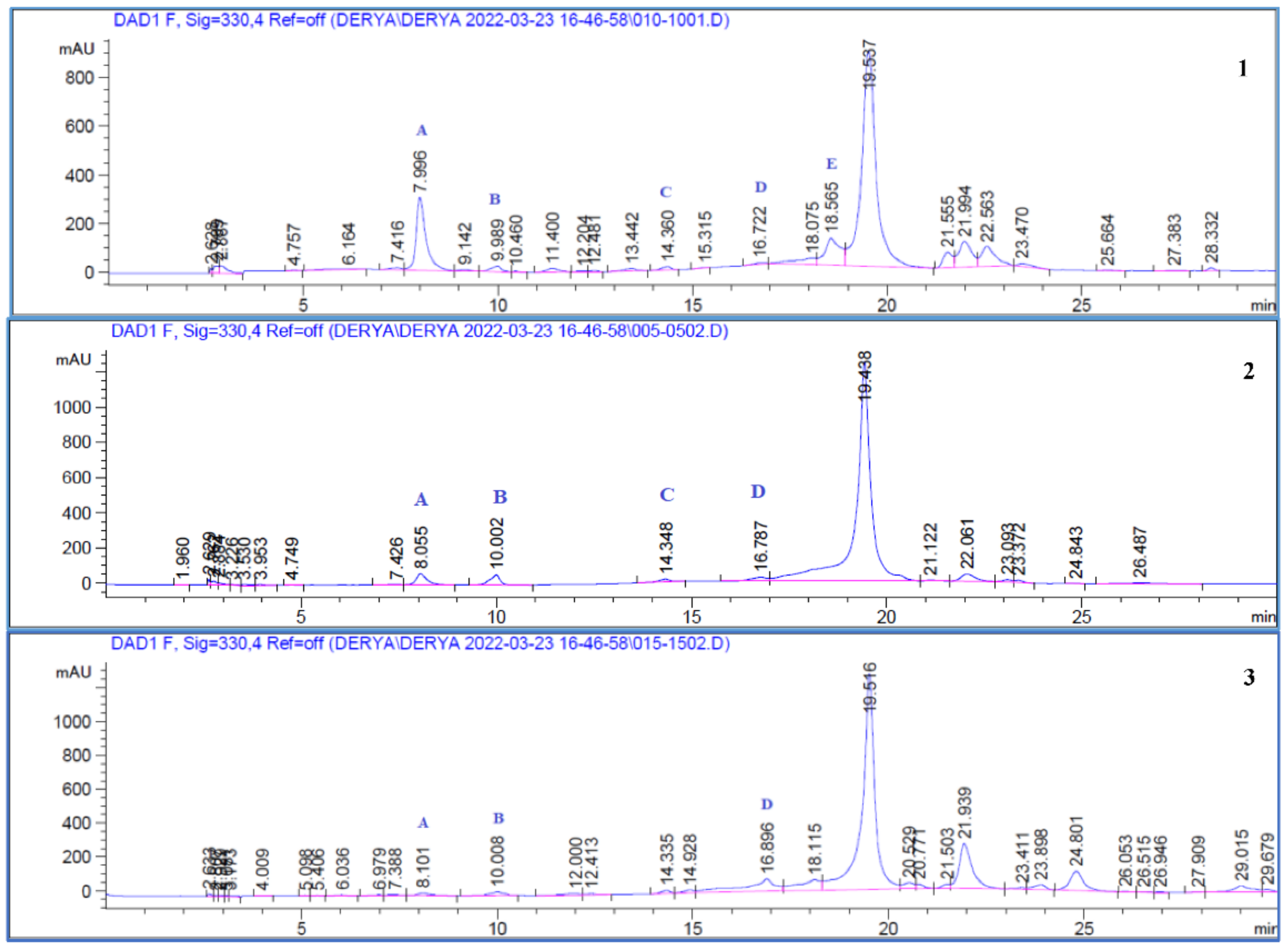
2.3.3. Analysis of Phenolic Acids and Flavonoids by HPLC

For HPLC analysis (Agilent 1100 series with diode array detector, New York, USA), all fractions were prepared at a concentration of 4 mg/mL and stock solutions of each standard (caffeic acid, chlorogenic acid, coumaric acid, ferulic acid, and rutin) were prepared with methanol (500 µg/mL). A Waters Spherisorb® (Philadelphia, PA, USA) C18 column (25 cm × 4.6 mm, 5 µm) was used for analysis. A gradient system with a mobile phase of 0.01% formic acid (A) and acetonitrile (B), a flow rate of 1 mL/min, a column temperature of 40 °C, and a wavelength of 330 nm was used for the analysis. For the calibration curve, five different concentrations of standards were injected in triplicate. The calibration curve equation and the coefficient of correlations were determined. Accuracy, precision, limit of detection (LOD), limit of quantitation (LOQ), and recovery values were calculated for method validation [26,27]. The precision of the method (intra-day and inter-day variation) was carried out, and differences were expressed by relative standard deviation (RSD). For analysis of LOD and LOQ values, 10 injections of standards were made and signal–noise values were calculated. The LOD signal–noise value was 3:1, while the LOQ signal–noise value was 10:1. For recovery analysis, three different known concentrations of the standard were added to the sample and the recovery percentage was calculated. Minor changes were made in flow rate, column temperature, mobile phase and wavelength for the robustness analysis and it was seen that they did not affect the analysis.

2.4. Antioxidant Activity

2.4.1. DPPH Radical Scavenging Activity

The DPPH radical scavenging effects of the samples were investigated using the Gyamfi et al. method [28]. The extracts were mixed with Tris-HCl buffer (50 nM, pH 7.4) and DPPH solution, prepared in 0.1 mM methanol. As a positive control, synthetic antioxidants such as standard antioxidants BHA and BHT (Butylated Hydroxy Anisol, Butylated Hydroxy Toluene) were used. After 30 min of room temperature incubation in the dark, the absorption spectra of the samples were recorded at 517 nm. The process was repeated three times in parallel, and inhibition % calculations were made using the following equation.
Inhibition % = [(Abscontrol − Abssample)/Abscontrol] × 100

2.4.2. ABTS●+ Radical Scavenging Activity

The samples’ ABTS●+ radical scavenging effects were defined according to the method in the literature [29]. By keeping an aqueous solution of ABTS and K2S2O8 (2.45 mM, final concentration) in the dark for 12–16 h, an ABTS●+ radical (7 mM) was obtained. Its absorbance was adjusted to 0.700 (±0.030) at 734 nm. The reaction kinetics were measured and recorded at 734 nm at 1-min periods for 30 min using 990 µL of the prepared radical solution and 10 µL of the extract samples. Trolox equivalents were calculated as percentages of inhibition measured against concentration (TEAC). BHA and BHT were used as positive controls. Experiments were repeated three times in parallel, and mean values were calculated

2.4.3. β-Carotene/Linoleic Acid Bleaching Inhibition Assay

The antioxidant activity of the extracts was determined according to the β-carotene bleaching method [30]. For this, 5 mg of β-carotene was dissolved in 25 mL of chloroform. An aliquot of the β-carotene solution was added to the vial containing linoleic acid (40 mg) and Tween 20 (400 mg). After the chloroform evaporated, distilled water (50 mL) was added slowly. BHA and BHT were used as positive controls. Blanks of the control and the samples were also prepared without β-carotene. The prepared emulsion and samples were subjected to thermal autoxidation by being kept at a constant temperature. The bleaching rate of β-carotene was monitored by measuring absorbance (470 nm) at 15-min intervals. The results of experiments were calculated as antioxidant activity capacity (AAC) using the following equation:
AAC% = [1 − (Abs0sample − Abs90sample)/(Abs0control − Abs90control)] × 100

2.5. Enzyme Inhibitory Activity

2.5.1. Lipoxygenase Inhibitory Activity

The lipoxygenase inhibitory activity of the samples was tested spectrophotometrically using a Cayman LOX Inhibitor Screening Assay Kit. Absorbance was recorded at 490 nm. Nordihydroguaiaretic acid (NDGA) was used as a positive control.

2.5.2. Tyrosinase Inhibitory Activity

Tyrosinase inhibition assay was applied by making certain modifications to the method described by Chang et al. [31]. Kojic acid was used as a standard material. Kinetic readings were taken with a microplate reader at 30-s intervals to determine the linear change in absorbance at 475 nm. A 96-well microplate was used for measurements in all experiments. All samples were studied at concentrations of 100 µg/mL and 500 µg/mL. All enzyme assay measurements were repeated three times, and the results were expressed as Mean ± Standard Error Mean. IC50 values were calculated for samples showing more than 50% inhibition. Inhibition % calculations were carried out using Equation (1).

2.5.3. α-Glucosidase Inhibitory Activity

The α-glucosidase enzyme inhibitory activity assay was performed according to the method reported by Liu et al. [32]. In this process, 50 µL of 2 U/mL α-glucosidase solution was mixed with 1000 µL of phosphate buffer and 200 µL of extract/acarbose. After incubation for 10 min at 37 °C, 5 mM of 50 µL of p-nitrophenyl-α-D-glucopyranoside (pNPG) was added and the mixture was incubated again at 37 °C for 20 min. Then, 2000 µL of 0.2 M sodium carbonate and 4700 µL of distilled water were added to stop the reaction, and absorbances were measured at 405 nm using a spectrophotometer. Inhibition % calculations were made using Equation (1).

2.5.4. α-Amylase Inhibitory Activity

The inhibitory effects of the extracts on the α-amylase enzyme were investigated by the modified Sigma-Aldrich method. In a test tube, varying concentrations of 40 µL of extract/acarbose, 160 µL of 20 mM phosphate buffer (pH 6.9, containing 6.7 mM sodium chloride), and 200 µL of α-amylase enzyme solution (EC3.2.1.1, type VI, Sigma; 20 units/mL) were mixed. After incubating at 25 °C for 5 min, 400 µL of starch solution (0.5% w/v) was added as a substrate and incubated at 25 °C for another 3 min. At the end of the incubation period, 200 μL of dinitro salicylic acid reagent were added to the test tubes, which were kept in a water bath at 85 °C for 15 min. At the end of the period, all tubes were removed from the water bath. After adding 4000 μL of distilled water, their absorbance at 540 nm wavelength was measured with a spectrophotometer. Inhibition % calculations were made using Equation (1).

2.6. Cytotoxic Activity

2.6.1. Cell Cultures

Cells were purchased from American Type Culture Collection (Manassas, VA, USA). MDA-MB-231 (ATCC HTB-26 Human Breast Cancer Cell Series), and MCF-7 (ATCC CCL-222, Human Breast Cancer Cell Series) were grown in Dulbecco’s Modified Eagle’s Medium (DMEM) containing 1% penicillin/streptomycin and 10% fetal bovine serum (FBS) (Gibco Invitrogen, Grand Island, NY, USA). Cell cultures were kept at 37 °C in 5% CO2 and 95% air.

2.6.2. Determination of Cell Viability with MTT Cytotoxicity Assay

At 24 h before the study, the cells in the flask were counted and inoculated into a 96-well microplate with 1 × 10⁴ cells in 100 μL per well. After 24 h, the media on the cells that adhered to the plate were discarded. The stock solution of the extracts was prepared at 1 mg/mL. Then, the extracts were prepared by dilution in the medium at concentrations of 7.81 µg/mL, 15.6 µg/mL, 31.25 µg/mL, 62.5 µg/mL, 125 µg/mL, 250µg/mL, 500 µg/mL, 1000 µg/mL, and 100 µL of each were added to wells. At the end of 48 h, the media in the wells were emptied. The wells were filled with 100 µL of MTT solution diluted 1/10 with medium from a 5 mg/mL (in PBS) MTT stock. The plates were kept in an incubator with CO2 at 37 °C for 4 h. At the end of 4 h, 100 μL of DMSO was added to each well and formazan crystals formed by MTT were dissolved. At the end of 10 min, each well was read at 540 nm wavelength using a microplate reader [33].
% Viability = [(Abssample × 100)/Abscontrol]

2.7. Statistical Analysis

Analysis was executed in triplicate and the mean values were calculated. All the data are presented as the mean ± standard deviation (SD), relative standard deviation (RSD), linear regression analysis. Calculations were performed using the Microsoft Excel program.
The Levene test was used to evaluate variance homogeneity. One-way analysis of variance was used for comparisons between more than two groups. The Dunnett T3 test and Tukey’s test were used for multiple comparisons. The data were evaluated with SPSS Version 11.0 statistic software package. The significance level was set at p <0.05.

3. Results and Discussion

3.1. Chemical Analysis

The quantities of total phenol and flavonoids in the A. minus sub-fractions were calculated. Table 1 shows the total phenol and flavonoid determination results for the extracts. The ethyl acetate fraction of root (Arc R EtOAc) was found to have the highest levels of flavonoids (158.56 ± 12.87 mgCA/gextract) and total phenols (364.37 ± 7.18 mgGAE/gextract).
In a study investigating the biological activities of water and ethanol extracts of A. minus leaves, the total phenol content of the water extract was calculated as 58.93 ± 2.72 mg/g, and the total phenol content of the ethanol extract was calculated as 58.93 ± 2.72 mg/g [18]. In another study, the total phenol and flavonoid contents of the ethanol extract of A. lappa roots were found to be 19.35 mg/g and 12.29 mg/g, respectively. When compared with the results of this study, the total phenol content values of the water sub-extracts, which showed the lowest content in the leaf and root, were determined as 120.01 ± 2.82 mgGAE/gextract and 89.47 ± 2.13 mgGAE/gextract, respectively; it was interpreted that they contained higher levels than the other studied species described in the literature [34].
The quantities of chlorogenic acid, caffeic acid, coumaric acid, ferulic acid, and rutin in the plant extracts were calculated (Figure 1 and Figure 2) and the results are shown in Table 1. Calibration values, precision data, and statistical data from the recovery assays are shown in Table 2, Table 3 and Table 4.
The amounts of chlorogenic acid (8.855 ± 0.175%) and rutin (8.359 ± 0.125%) in the ethyl acetate sub-fraction of the leaf were found to be higher than those of the other compounds. Likewise, it was determined that the leaf butanol extract (Arc L BuOH) was rich in chlorogenic acid content (8.608 ± 0.292%). Coumaric acid was not detected in any extract except flower and leaf ethyl acetate extract (Arc F EtOAc and Arc L EtOAc). Rutin was also recorded detected only in leaf sub-fractions (excluding Arc L Aqua extracts).
Studies on the biologically active components of Arctium species have generally focused on A. lappa species, and have revealed that the biological activities of A. lappa are due to lignans, arctiin, arctigenin, and polysaccharides. It has also been noted that these compounds show antitumor, antibacterial, antiviral, hepatoprotective, and antiurolytic activities in combination with polyphenols (flavonoids and polyphenolcarboxylic acids) [35,36]. Studies on the phytochemical content of A. minus have been limited. For this reason, this research is the first detailed study on the determination of the phenolic content of plant parts from the A. minus species collected from Turkey.
The literature states that rutin and isoquercetin are the two major components in ethanolic extracts of A. minus leaves [16]. Rutin was found to be the major component in the leaf ethyl acetate extract (Arc L EtOAc) and was calculated at 8.359 ± 0.125%. It has also been noted that caffeoylquinic acid derivatives can be found as major active ingredients in Arctium species, and these contribute to the plants’ extraordinary antioxidant properties [16]. In our study, chlorogenic acid was identified by the literature as the major component especially in leaf ethyl acetate and butanol sub-fractions. Studies indicate that Arctium species are cross-pollinating, and this creates significant differences in chemical content within and between species. In particular, A. minus and A. lappa had high variability in terms of flavonoids and hydroxycinnamic acids. A. lappa was particularly rich in hydroxycinnamic acids, while A. minus contained high levels of specific hydroxycinnamic compounds along with various flavonoid compounds. For this reason, it is of great importance to perform a content analysis of A. minus species [37].

3.2. Antioxidant Activity

The antioxidant capacities of the extracts were measured by DPPH, ABTS, and β-carotene/linoleic acid assay. Antioxidant activity was evaluated by comparing extracts with synthetic antioxidants BHT and BHA, and the results are given in Table 5.
The DPPH scavenging activation test is a widely used spectrophotometric methods to evaluate antioxidant capacity. DPPH is employed to represent the radicals in our body and the radical scavenging effects of the extracts are evaluated [38]. In the present study, EC50 values of flower, root, and leaf methanol extracts and their sub-fractions were compared, and the significance of antioxidant activity was evaluated with positive controls. In particular, the activity of ethyl acetate sub-fractions of plant parts was found to be statistically significant with positive controls for BHT and BHA (p < 0.01). The lowest EC50 values were observed in the dichloromethane sub-fractions of the plant. In addition, leaf and flower butanol sub-fractions showed high antioxidant activity (Arc L BuoH: 0.048 ± 0.001 mg/mL and Arc F BuOH: 0.043 ± 0.003 mg/mL), while the root butanol sub-fraction showed low activity (Arc R BuOH: 1.126 ± 0.019) (Table 5). In a study by Erdemoğlu et al., the DPPH radical scavenging effects of A. minus water and ethanol extracts were evaluated. The IC50 values of the extracts were calculated as 5.33 ± 0.62 and 7.18 ± 0.25 mg/mL for the water and ethanol extracts, respectively [18]. Conspicuously, for all the plant parts analyzed in the current study, lower IC50 values were calculated in water and alcohol extracts. This may be due to the chemical content of the plants studied, as well as differences in the extraction methods and experimental methods applied to the plants. In a study by Chui et al. on A. lappa leaves, it was determined that the plant is rich in morin and quercetin 3-O-rhamnoside compounds, and the antioxidant activity (DPPH: 2025.33 ± 84.15 μmol Trolox/g, ABTS: 159.14 ± 5.28 μmol Trolox/g) of the leaf flavonoids of the plant was quite high when tested in vitro [39]. A. lappa root extracts were examined in another study, and it was discovered that the plant’s ethyl acetate fraction had the highest levels of total phenolics, total flavonoids, caffeic acid derivatives, and phenolic acids (mostly chlorogenic, caffeic acid, and p-coumaric acids). In the hexane fraction, only triterpenes were discovered. Additionally, the ethyl acetate fraction displayed the best antioxidant activity due to its high polyphenol concentration. While the DPPH radical scavenging activity of the root ethyl acetate extract was calculated as 308.3 ± 6.1 mM Trolox/g extract, the activity in the hexane and chloroform extracts was calculated as 8.9 ± 0.1 mM Trolox/g extract and 41.1 ± 0.3 mM Trolox/g extract, respectively [40]. Similarly, the DPPH radical scavenging effect of ethyl acetate extract was higher in A. minus root extract (EC50: 0.042 ± 0.00 mg/mL), while the apolar dichloromethane extract showed a lower radical scavenging activity (EC50: 0.630 ± 0.056 mg/mL).
ABTS radical scavenging activity assay is another widely used method, in which ABTS is used as an oxidant and radical reduction is observed relative to absorbance before and after the addition of antioxidants to the study samples. Results from the ABTS test are given as Trolox-equivalent antioxidant capacity (TEAC) [41]. The antioxidant activities of extracts and sub-fractions of A. minus were evaluated by kinetic measurement at 0.5 mg/mL and 0.25 mg/mL concentrations for 30 min. The antioxidant activities of Arc R EtOAc and Arc F EtOAc extracts at 0.25 mg/mL concentration were calculated as 2.289 ± 0.014 mmol/L Trolox and 2.512 ± 0.14 mmol/L Trolox, respectively. According to the results, it was noted that these extracts were more effective than the positive controls BHT and BHA. In addition, at both concentrations it was found that the extracts exhibiting the lowest activity had water and dichloromethane sub-fractions belonging to the leaf, flower, and root. In a study investigating the ABTS radical scavenging effects of teas prepared from A. lappa roots by steaming, drying, and roasting methods, inhibition % was determined as 75.13 ± 1.962, 62.10 ± 1.10, and 85.66 ± 2.77, respectively [42]. In our study, the ABTS radical scavenging activity of the root water extract Arc R Aqua was calculated as 1.199 ± 0.08 mmol/L Trolox at 0.5 mg/mL concentration. In another study, the antioxidant capacities of 70% methanol extracts prepared from the roots, leaves, and seeds of A. lappa were measured, and the TEAC values of the extracts were found in the range of 67.39–1.63 μmol Trolox equivalent/100 g dry weight [43].
The β-carotene linoleic acid bleaching assay is a popular model for studying the oxidation of unsaturated fatty acids [44]. The change in color of β-carotene in reaction with the linoleic acid-free radical is the basis of the β-carotene/linoleic acid test. At high temperatures, this radical is formed by removing the hydrogen atom between the two double bonds of linoleic acid. Antioxidants can prevent β-carotene degradation by reacting with the free-radical linoleate or any other radical formed [45]. According to the results, no statistically significant difference was observed between Arc L MeOH, Arc R EtOAc, and Arc F EtOAc extracts, and they were found to be as protective as positive control BHT and BHA at 30 min. These extracts preserved the color and absorbance of β-carotene longer than the other extracts. When the 90-minute absorbance was evaluated, it was determined that Arc R Aqua, Arc R MeOH, Arc F Aqua, and Arc L CH2Cl2 were statistically significant among themselves, and their protective effects were quite low. At 60 and 90 min, BHT and BHA were found to be more protective than any other extract. For the first time, the antioxidant activities of extracts of the Arctium plant were determined by this method.
As a result, the free-radical-scavenging activity of different parts of the plant was evaluated by different methods. It was revealed that the active components of the plant were concentrated in different parts of their tissues and demonstrated significantly different free radical scavenging activity.

3.3. Enzyme Inhibitory Activity

Natural processes in an organism cooperate to prevent oxidative damage to cellular components, by enabling the formation or removal of reactive oxygen species by enzymes and antioxidant metabolites [46]. Hence, it is critical to evaluate the antioxidant and enzyme inhibition activities of natural compounds. Lipoxygenase plays a role in the synthesis of inflammation markers [47]. Overproduction of lipoxygenase metabolites has been associated with inflammatory diseases including cancer, myocardial rupture, post-ischemic inflammation, allergic asthma, skin diseases, metabolic syndrome, and neurodegenerative diseases such as Alzheimer’s [48]. Tyrosinase is one of the key enzymes in the biosynthetic pathway of melanin [49]. Overactivity of the tyrosinase enzyme leads to excessive production of melanin, which causes hyperpigmentation of the skin. Excessive accumulation of epidermal pigmentation may cause certain dermatological disorders associated with freckles, melasma, blemishes, and senile lentigines [50]. Furthermore, tyrosinase is linked to Parkinson’s disease and other neurodegenerative diseases [51,52].
Results of the lipoxygenase and tyrosinase enzyme inhibition activity analysis of extracts and standards are given in Table 6. According to the results, Arc F Aqua, Arc L BuOH, Arc R BuOH, Arc K Aqua, and Arc L Aqua showed weak inhibition against the Lipoxygenase enzyme, while other samples showed no inhibitory activity. In a study evaluating the antioxidant and anti-lipoxygenase activity of A. lappa and A. tometosum species, it was noted that the extracts were characterized by strong antioxidant properties but showed weak enzyme inhibition activity [53]. Similarly, weak lipoxygenase enzymeinhibition activity was observed in A. minus extract and its fractions.
Arc L EtOAc (99.65%) was demonstrated the most effective inhibition of the tyrosinase enzyme at 500 µg/mL concentration. The IC50 value of Arc L EtOAc (93.00 µg/mL) was found to be approximately three times the IC50 of kojic acid (30.00 µg/mL). The IC50 values of other effective fractions, Arc F BuOH, Arc R EtOAc, and Arc L MeOH were calculated as 176.67, 218.75, and 401.67 µg/mL, respectively. As a result, it was determined that Arc F BuOH, Arc R EtOAc, and Arc MeOH extracts, which are thought to be rich in phenolic compounds, have a high antioxidant capacity and in parallel show high tyrosinase enzyme inhibition activity. In a study evaluating the tyrosinase enzyme inhibition activity of methanol extract and sub-fractions of Arctium lappa roots, the highest activity was observed in the ethyl acetate fraction (IC50: 1.326 ± 0.158 mg/mL) [54]. In our data analysis, the IC50 value of the root ethyl acetate extract was calculated as 218.75 ± 0.83 µg/mL. However, it was observed that the leaf ethyl acetate extract (IC50: 93.00 ± 1.54 µg/mL) was more active than the root ethyl acetate extract.
Inhibition of α-glucosidase and α-amylase in the digestive system, and regulation of oxidative stress, are very important as antidiabetic mechanisms of action in the treatment of diabetes [8]. A strong link has been reported between hyperglycemia, oxidative stress caused by hyperglycemia, inflammation, and the development and progression of type 2 diabetes [9]. As inhibitors of carbohydrate hydrolyzing enzymes, α-glucosidase and α-amylase offer an effective strategy to regulate or prevent hyperglycemia by controlling starch degradation [55].
The inhibitory activity of extracts prepared from different parts of A. minus on α-amylase and α-glucosidase was investigated in this study for the first time, and the results are given in Table 6. Among all extracts, only the leaf extracts (excluding leaf ethyl acetate extract) showed α-amylase inhibition activity at a concentration of 1 mg/mL. In the α- glucosidase inhibition assay, the dichloromethane extract of the A. minus leaf had the highest enzyme inhibition activity, with 87.12% inhibition, compared with the other extracts and with acarbose at a concentration of 1 mg/mL. Nickavar and Yousefian found that crude hydroalcoholic (70% ethanol) extract of A. lappa root caused a 35.06 ± 0.38% reduction in α-amylase activity at a dose of 2.304 mg/mL [56]. In another study, the effects of the A. lappa methanol, ethyl acetate, and water extracts on α-glucosidase enzyme inhibition activity were investigated, and it was reported that ethyl acetate extract caused 77.5%, 50.3%, 43.1% inhibition in a dose-dependent manner at 0.2, 0.1 and 0.002 mg/mL doses, respectively [57].
Synthetic α-glucosidase inhibitors have been developed, such as acarbose, miglitol, and voglibose, but these compounds have been associated with certain serious side effects, including gastrointestinal problems [58]. Therefore, other studies have focused on the identification of natural α-glucosidase inhibitors. Recent research has shown that fructoioigosaccharides and bioactive polyphenols from burdock, especially chlorogenic acid, exhibit antidiabetic effects [59]. In addition, evidence from in vitro experimental studies indicates that phenolic compounds inhibit α-amylase and α-glucosidase activities. Chui et al. investigated the antihyperglycemic effects of the flavonoids of A. lappa leaves and reported promising results (α-amylase IC50 value of 92.01 μg mL−1 and α-glucosidase IC50 value of 29.49 μg mL−1) [60]. Interestingly, in our study it was observed that Arc L EtOAc extract, which had the highest total phenol and flavonoid content, did not show any activity.

3.4. Cytotoxic Activity

Breast cancer remains the main cause of death in women worldwide. Therefore, it is very important to identify alternative or new drug targets and to better understand the metabolic adaptations of various breast cancer subtypes. Breast cancer is classified as hormone-sensitive ER/PR+, representing early benign tumor status, or aggressive ER/PR-, representing late-stage metastasis. In this study, MCF-7 cells with ER/PR+ and MDA-MB-231 cells with ER/PR- were selected as models for these two types of breast cancer [61].
The effects of A. minus extracts on MCF-7 and MDA-MB-231 cell lines were observed using the MTT method. The results are given in Table 7. When all extracts were tested for cytotoxicity, the dichloromethane fractions of the roots, leaves, and flowers were found to be the most effective, particularly in the MDA-MB-231 cell line. The IC50 value of Arc F CH2Cl2 extract was calculated as 10.80 ± 1.26 µg/mL. The IC50 values of Arc R CH2Cl2 and Arc L CH2Cl2 extracts were 13.41 ± 2.37 and 21.39 ± 2.43 µg/mL, respectively, in the MDA-MB-231 cell line. Similarly, flower, root, and leaf dichloromethane extracts were found to be more toxic than other extracts in the MCF-7 cell line. The extracts with the weakest activity were found to be Arc L Aqua, Arc R MeOH, and Arc R Aqua (IC50 > 1000). Interestingly, Arc L EtOAc, Arc F EtOAc, and Arc R EtOAc extracts were found ineffective in terms of cytotoxic activity, despite having very high antioxidant activity.
Previously, the cytotoxic effects of A. lappa have been reported in the literature. Ghafari et al. evaluated the apoptotic and necrotic effects of extracts obtained from A. lappa roots, using MCF-7 and MDA-MB-231 cell lines, and reported that the extracts had a high antiproliferative effect even at 10 µg/mL concentration [62]. In other studies, it was discovered that aqueous extracts of A. lappa roots had no cytotoxic effect on the MCF-7 cell line, whereas the root dichloromethane extract of A. lappa had a strong cytotoxic effect [63,64]. According to our research, it was determined that water sub-extracts of A. minus showed a weak cytotoxic effect, and dichloromethane sub-extracts had a very significant cytotoxic effect compared with other extracts.
Arctigenin, which is isolated from A. lappa and is one of the major components of Arctium species, has been proven in studies to have strong anticancer effects [65]. Generally, petroleum ether, dichloromethane, and butanol extracts of the plant have been reported to be rich in this compound [66]. These data may explain the stronger cytotoxic effect of A. minus dichloromethane extracts compared with that of other extracts. In addition, other research discovered that the arctigenin compound significantly reduced cell proliferation when applied in combination with chlorogenic acid and cinnamaldehyde to MCF-7 and MDA-MB-231 cells [67]. These results also indicate that the compounds in the extracts can exhibit stronger biological activities by providing synergistic effects together.

4. Conclusions

In the present study, the biological activities of the methanol extract prepared from the leaves, roots, and flowers of A. minus were investigated in detail, as were the dichloromethane, ethyl acetate, butanol, and water sub-fractions of these extracts. To the best of our knowledge, this is the first detailed bioactivity assessment study to examine different parts of A. minus plants. Antioxidant, cytotoxic, α-amylase, α-glucosidase, lipoxygenase, and tyrosinase enzyme inhibition activities of 15 different extracts were evaluated, and phytochemical analyses were carried out. It was determined that the extracts with high antioxidant effects were rich in phenolic content. In addition, it was observed that the root, flower, and leaf ethyl acetate extracts of the plant showed high antioxidant activity. The plant’s sub-fractions of dichloromethane were found to be moderately efficient against the two primary kinds of breast cancer, MCF7 (ER/PR-) and MDA-MB-231 (ER/PR+). The extract with the highest α-glucosidase enzyme inhibitory activity was identified as Arc L CH2Cl2. Arc R EtOAc and Arc L EtOAc sub-fractions each showed strong tyrosinase enzyme inhibition activity.
This research provides thorough information that might serve as a scientific foundation for using Arctium minus as a source of bioactive chemicals with diverse roles in the nutraceutical and pharmaceutical sectors. Further study should be undertaken in the future to address a variety of concerns including bioavailability, formula ability, and toxicity characteristics of the extracts examined.

Author Contributions

Conceptualization: E.K.A. and G.Ş.K. and S.İ.; methodology: G.Ş.K., S.İ. and E.K.A.; validation: G.Ş.K. and D.Ç.P.; formal analysis: G.Ş.K., G.Y. and D.Ç.P.; investigation: S.İ. and E.K.Ş., G.Y. and D.Ç.P.; data curation: G.Ş.K. and E.K.A.; writing—original draft preparation: S.İ., E.K.A. and E.S.-S.; writing—review and editing: E.K.A. All authors have read and agreed to the published version of the manuscript.

Funding

This research was supported by Project no: TKB-2020-10129 from Erciyes University Research Council.

Institutional Review Board Statement

Not applicable.

Informed Consent Statement

Not applicable.

Data Availability Statement

The data presented in this study are available in the article.

Acknowledgments

E.S.-S. thanks Proyecto Interno UCEN (CIP2020036) for financial support.

Conflicts of Interest

The authors declare no conflict of interest.

References

  1. Awan, A.A.; Akhtar, T.; Ahmed, M.J.; Murtaza, G. Quantitative ethnobotany of medicinal plants uses in the Jhelum valley, Azad Kashmir, Pakistan. Acta Ecol. Sin. 2021, 41, 88–96. [Google Scholar] [CrossRef]
  2. Rehman, N.U.; Shah, M.; Ullah, S.; Khan, M.; Khan, A.; Ullah, O.; Al-Harrasi, A. Enzymes Inhibition and Antioxidant Potential of Medicinal Plants Growing in Oman. Biomed. Res. Int. 2022, 2022, 7880387. [Google Scholar] [CrossRef] [PubMed]
  3. Lopaczynski, W.; Steven, H.Z. Antioxidants, programmed cell death, and cancer. Nutr Res. 2001, 21, 295–307. [Google Scholar] [CrossRef]
  4. Gautam, R.; Jachak, S.M. Recent developments in anti-inflammatory natural products. Med. Res. Rev. 2009, 29, 767–820. [Google Scholar] [CrossRef]
  5. Dvorakova, M.; Landa, P. Anti-inflammatory activity of natural stilbenoids: A review. Pharmacol. Res. 2017, 124, 126–145. [Google Scholar] [CrossRef]
  6. Wisastra, R.; Dekker, F.J. Inflammation, cancer and oxidative lipoxygenase activity are intimately linked. Cancers 2014, 6, 1500–1521. [Google Scholar] [CrossRef]
  7. Deshpande, A.D.; Harris-Hayes, M.; Schootman, M. Epidemiology of diabetes and diabetes-related complications. Phys. Ther. 2008, 88, 1254–1264. [Google Scholar] [CrossRef]
  8. Yeung, A.W.K.; Tzvetkov, N.T.; Durazzo, A.; Lucarini, M.; Souto, E.B.; Santini, A.; Horbańczuk, J.O. Natural products in diabetes research: Quantitative literature analysis. Nat. Prod. Res. 2021, 35, 5813–5827. [Google Scholar] [CrossRef]
  9. Piconi, L.; Quagliaro, L.; Ceriello, A. Oxidative stress in diabetes. Clin. Chem. Lab Med. 2003, 41, 1144–1149. [Google Scholar] [CrossRef]
  10. Wang, Y.; Hao, M.M.; Sun, Y.; Wang, L.F.; Wang, H.; Zhang, Y.J.; Yang, Z. Synergistic promotion on tyrosinase inhibition by antioxidants. Molecules 2018, 23, 106. [Google Scholar] [CrossRef] [Green Version]
  11. Sepehri, N.; Iraji, A.; Yavari, A.; Asgari, M.S.; Zamani, S.; Hosseini, S.; Khoshneviszadeh, M. The natural-based optimization of kojic acid conjugated to different thio-quinazolinones as potential anti-melanogenesis agents with tyrosinase inhibitory activity. Bioorg. Med. Chem. 2021, 36, 116044. [Google Scholar] [CrossRef]
  12. Sharma, P.; Jha, A.B.; Dubey, R.S.; Pessarakli, M. Reactive oxygen species, oxidative damage, and antioxidative defense mechanism in plants under stressful conditions. J. Bot. 2012, 2012, 26. [Google Scholar] [CrossRef]
  13. ThePlantList. Available online: http://www.theplantlist.org/Arctium (accessed on 21 May 2022).
  14. Yu, J.; Ye, M.; Li, K.; Wang, F.; Shi, X.; Pan, C.; Liu, W. Fragments of a pectin from Arctium lappa L: Molecular properties and intestinal regulation activity. J. Funct. Foods. 2022, 88, 104900. [Google Scholar] [CrossRef]
  15. Mink, J.N.; Holmes, W.C.; Singhurst, J.R.; Treuer-Kuehn, A. Arctium minus (asteraceae) historical review, ecological consequences, and addition to Texas flora. J. Bot. Res. Inst. Tex. 2018, 12, 713–720. [Google Scholar] [CrossRef]
  16. Wang, D.; Bădărau, A.S.; Swamy, M.K.; Shaw, S.; Maggi, F.; Da Silva, L.E.; Atanasov, A.G. Arctium species secondary metabolites chemodiversity and bioactivities. Front. Plant Sci. 2019, 10, 834. [Google Scholar] [CrossRef]
  17. De Souza, G.C.; Haas, A.P.S.; Von Poser, G.L.; Schapoval, E.E.S.; Elisabetsky, E. Ethnopharmacological studies of antimicrobial remedies in the south of Brazil. J. Ethnopharmacol. 2004, 90, 135–143. [Google Scholar] [CrossRef]
  18. Erdemoglu, N.; Turan, N.N.; Akkol, E.K.; Sener, B.; Abacıoglu, N. Estimation of anti-inflammatory, antinociceptive and antioxidant activities on Arctium minus (Hill) Bernh. ssp. minus. J. Ethnopharmacol. 2009, 121, 318–323. [Google Scholar] [CrossRef]
  19. Neves, J.M.; Matos, C.; Moutinho, C.; Queiroz, G.; Gomes, L.R. Ethnopharmacological notes about ancient uses of medicinal plants in Tras-os-Montes (northern of Portugal). J. Ethnopharmacol. 2009, 124, 270–283. [Google Scholar] [CrossRef]
  20. Sezik, E.; Yesilada, E.; Honda, G.; Takaishi, Y.; Takeda, Y.; Tanaka, T. Traditional medicine in Turkey X. folk medicine in central anatolia. J. Ethnopharmacol. 2001, 75, 95–115. [Google Scholar] [CrossRef]
  21. Mosaddegh, M.; Naghibi, F.; Moazzeni, H.; Pirani, A.; Esmaeili, S. Ethnobotanical survey of herbal remedies traditionally used in Kohghiluyeh va Boyer Ahmad province of Iran. J. Ethnopharmacol. 2012, 141, 80–95. [Google Scholar] [CrossRef]
  22. Tardio, J.; Pascual, H. Morales RWild food plants traditionally used in the province of Madrid, central Spain. Econ. Bot. 2005, 59, 122–136. [Google Scholar] [CrossRef]
  23. Stefanov, S.M.; Fetzer, D.E.; de Souza, A.R.C.; Corazza, M.L.; Hamerski, F.; Yankov, D.S.; Stateva, R.P. Valorization by compressed fluids of Arctium lappa seeds and roots as a sustainable source of valuable compounds. J. CO2 Util. 2022, 56, 101821. [Google Scholar] [CrossRef]
  24. Singleton, V.L.; Orthofer, R.; Lamuela-Raventós, R.M. Analysis of Total Phenols and other Oxidation Substrates and Antioxidants by Means of Folin-Ciocalteu Reagent. In Methods in Enzymology; Academic Press: Cambridge, MA, USA, 1999; Volume 299, pp. 152–178. [Google Scholar]
  25. Zhishen, J.; Mengcheng, T.; Jianming, W. The determination of flavonoid contents in mulberry and their scavenging effects on superoxide radicals. Food Chem. 1999, 64, 555–559. [Google Scholar]
  26. ICH Expert Working Group. ICH Guideline Q2(R1) Validation of analytical procedures: Text and methodology. BMJ (Clinical research ed.) 2005.333(7574). In Proceedings of the International Conference on Harmonisation of Technical Requirements for Registration of Pharmaceuticals for Human Use, San Diego, CA, USA, 2 June 2014. [Google Scholar]
  27. Çiçek, P.D.; Coskun, M. Quantitative determination by HPLC-DAD of icariin, epimedin A, epimedin B, and epimedin C in Epimedium (Berberidaceae) species growing in Turkey. NatProd Commun. 2016, 11, 1665–1666. [Google Scholar]
  28. Gyamfi, M.A.; Yonamine, M.; Aniya, Y. Free-radical scavenging action of medicinal herbs from Ghana, Thonningia sanguinea on experimentally ınduced liver ınjuries. Gen Pharmacol. 1999, 32, 661–667. [Google Scholar] [CrossRef]
  29. Re, R.; Pellegrini, N.; Proteggente, A. Antioxidant activity applying an improved ABTS radical cation decolorization assay. Free Radic. Biol. Med. 1999, 26, 1231–1237. [Google Scholar] [CrossRef]
  30. Pizzale, L.; Bortolomeazzi, R.; Vichi, S.; Überegger, E.; Conte, L.S. Antioxidant activity of Sage (Salvia officinalis and S. fruticosa) and Oregano (Origanum onites and O. indercedens) extracts related to their phenolic compound content. J. Agr. Food Chem. 2002, 82, 645–1651. [Google Scholar]
  31. Chang, T.S.; Ding, H.Y.; Tai, S.S.K.; Wu, C.Y. Mushroom tyrosinase inhibitory effects of isoflavones isolated from soygerm koji fermented with Aspergillus oryzae BCRC 32288. Food Chem. 2007, 105, 1430–1438. [Google Scholar] [CrossRef]
  32. Liu, S.; Li, D.; Huang, B.; Chen, Y.; Lu, X.; Wang, Y. Inhibition of pancreatic lipase, α-glucosidase, α-amylase, and hypolipidemic effects of the total flavonoids from Nelumbo nucifera leaves. J. Ethnopharmacol. 2003, 149, 263–269. [Google Scholar] [CrossRef]
  33. Karatoprak, G.Ş.; Yücel, Ç.; Göger, F.; Sobarzo-Sánchez, E.; Küpeli, A.E. Potential Antioxidant and Enzyme Inhibitory Effects of Nanoliposomal Formulation Prepared from Salvia aramiensis Rech. f. Extract. Antioxidants 2020, 9, 293. [Google Scholar] [CrossRef]
  34. Yazdanian, M.; Rostamzadeh, P.; Alam, M.; Abbasi, K.; Tahmasebi, E.; Tebyaniyan, H.; Kahnamoei, M.B. Evaluation of antimicrobial and cytotoxic effects of Echinacea and Arctium extracts and Zataria essential oil. AMB Express 2022, 12, 75. [Google Scholar] [CrossRef] [PubMed]
  35. Jingvi, L.; Yi-Zhong, C.; Ricky, N.S.W. Comparative Analysis of Caffeoylquinic Acids and Lignans in Roots and Seeds among Various Burdock (Arctium lappa) Genotypes with High Antioxidant Activity. J. Agric. Food Chem. 2012, 60, 4067–4075. [Google Scholar]
  36. Chan, Y.S.; Cheng, L.N.; Wu, J.H. A review of the pharmacological effects of Arctium lappa (burdock). Inflammopharmacology 2011, 19, 245–254. [Google Scholar] [CrossRef] [PubMed]
  37. Robbins, L.R. Natural Variability in Phenolic and Sesquiterpene Constituents among Burdock (Arctium lappa L. and Arctium minus L.) Leaves for Potential Medicinal Interests. Master’s Thesis, The Ohio State University, Columbus, OH, USA, 2013. [Google Scholar]
  38. Ömer, I.; Yildiz, I.; Genc, N. A new potentiometric PVC membrane sensor for the determination of DPPH radical scavenging activity of plant extracts. Food Chem. 2022, 373, 131420. [Google Scholar]
  39. Cui, J.; Zong, W.; Zhao, N.; Yuan, R. Burdock (Arctium lappa L.) leaf flavonoids rich in morin and quercetin 3-O-rhamnoside ameliorate lipopolysaccharide-induced inflammation and oxidative stress in RAW264. 7 cells. Food Sci. Nutr. 2022, 10, 2718–2726. [Google Scholar] [CrossRef]
  40. Petkova, N.; Hambarlyiska, I.; Tumbarski, Y.; Vrancheva, R.; Raeva, M.; Ivanov, I. Phytochemical composition and antimicrobial properties of burdock (Arctium lappa L.) roots extracts. Biointerface Res. Appl. Chem. 2022, 12, 2826–2842. [Google Scholar]
  41. Takatsuka, M.; Goto, S.; Kobayashi, K.; Otsuka, Y.; Shimada, Y. Evaluation of pure antioxidative capacity of antioxidants: ESR spectroscopy of stable radicals by DPPH and ABTS assays with singular value decomposition. Food Biosci. 2022, 48, 101714. [Google Scholar] [CrossRef]
  42. Yu-Ri, K.; Youn, K.w. Physicochemical of burdock (Arctium lappa L) tea depending on steaming and roasting treatment. Korean J. Food Preserv. 2014, 21, 646–651. [Google Scholar]
  43. Ferracane, R.; Graziani, G.; Gallo, M.; Fogliano, V.; Ritieni, A. Metabolic profile of the bioactive compounds of burdock (Arctium lappa) seeds, roots and leaves. J. Pharm. Biomed. Anal. 2010, 51, 399–404. [Google Scholar] [CrossRef]
  44. Şeker Karatoprak, G.; İlgün, S.; Koşar, M. Phenolic Composition, Anti-Inflammatory, Antioxidant, and Antimicrobial Activities of Alchemilla mollis (Buser) Rothm. Chem. Biodivers. 2017, 14, e1700150. [Google Scholar] [CrossRef]
  45. Mraihi, F.; Journi, M.; Chérif, J.K.; Sokmen, M.; Sokmen, A.; Trabelsi-Ayadi, M. Phenolic contents and antioxidant potential of Crataegus fruits grown in Tunisia as determined by DPPH, FRAP, and β-carotene/linoleic acid assay. J. Chem. 2013, 2013, 378264. [Google Scholar] [CrossRef]
  46. Shoaib, M.; Shah, A.; Wadood, S.; Ali, N.; Shah, I.; Naveed Umar, M.; Akhtar, S. In vitro enzyme inhibition potentials and antioxidant activity of synthetic flavone derivatives. J. Chem. 2015, 2015, 516878. [Google Scholar] [CrossRef]
  47. Butovich, I.A.; Lukyanova, S.M. Inhibition of lipoxygenases and cyclooxygenases by linoleyl hydroxamic acid: Comparative in vitro studies. J. Lipid Res. 2008, 49, 1284–1294. [Google Scholar] [CrossRef]
  48. Saçan, O.; Turhan, E.Y. Lipoxygenase inhibitory activities of some plant extracts and chemical compounds. Eur. J. Biol. 2014, 73, 47–52. [Google Scholar]
  49. Rijken, F.; Kiekens, R.C.M.; Bruijnzeel, P.L.B. Skin-infiltrating neutrophils following exposure to solar-simulated radiation could play an important role in photoageing of human skin. Br J Dermatol. 2005, 152, 321–328. [Google Scholar] [CrossRef]
  50. Khan, K.M.; Maharvi, G.M.; Khan, M.T.H.; Shaikh, A.J.; Perveen, S.; Begum, S.; Choudhary, M.I. Tetraketones: A new class of tyrosinase inhibitors. Bioorg Med Chem. 2006, 14, 344–351. [Google Scholar] [CrossRef]
  51. Xu, Y.; Stokes, A.H.; Roskoski Jr, R.; Vrana, K.E. Dopamine, in the presence of tyrosinase, covalently modifies and inactivates tyrosine hydroxylase. J. Neurosci. Res. 1998, 54, 691–697. [Google Scholar] [CrossRef]
  52. Asanuma, M.; Miyazaki, I.; Ogawa, N. Dopamine-or L-DOPA-induced neurotoxicity: The role of dopamine quinone formation and tyrosinase in a model of Parkinson’s disease. Neurotox. Res. 2003, 5, 165–176. [Google Scholar] [CrossRef]
  53. Skowrońska, W.; Granica, S.; Dziedzic, M.; Kurkowiak, J.; Ziaja, M.; Bazylko, A. Arctium lappa and Arctium tomentosum, Sources of Arctii radix: Comparison of Anti-Lipoxygenase and Antioxidant Activity as well as the Chemical Composition of Extracts from Aerial Parts and from Roots. Plants 2021, 10, 78. [Google Scholar] [CrossRef]
  54. Im, D.Y.; Lee, K.I. Antioxidative activity and tyrosinase inhibitory activity of the extract and fractions from Arctium lappa roots and analysis of phenolic compounds. Korean J. Pharmacogn. 2014, 45, 141–146. [Google Scholar]
  55. Bakr, S.A. Screening of antidiabetic and antioxidant activities of medicinal plants. J. Integr. Med. 2015, 13, 297–305. [Google Scholar]
  56. Nickavar, B.; Yousefian, N. Evaluation of α-amylase inhibitory activities of selected antidiabetic medicinal plants. J. Für Verbrauch. Und Lebensm. 2011, 6, 191–195. [Google Scholar] [CrossRef]
  57. Miyazawa, M.; Yagi, N.; Taguchi, K. Inhibitory compounds of α-glucosidase activity from Arctium lappa L. J. Oleo Sci. 2005, 54, 589–594. [Google Scholar] [CrossRef]
  58. Dirir, A.M.; Daou, M.; Yousef, A.F.; Yousef, L.F. A review of alpha-glucosidase inhibitors from plants as potential candidates for the treatment of type-2 diabetes. Phytochem. Rev. 2022, 21, 1049–1079. [Google Scholar] [CrossRef]
  59. Mondal, S.C.; Eun, J.B. Mechanistic insights on burdock (Arctium lappa L.) extract effects on diabetes mellitus. Food Sci. Biotechnol. 2022, 31, 999–1008. [Google Scholar] [CrossRef]
  60. Cui, J.; Zeng, S.; Zhang, C. Anti-hyperglycaemic effects of Burdock (Arctium lappa L.) leaf flavonoids through inhibiting α-amylase and α-glucosidase. Int. J. Food Sci. 2022, 57, 541–551. [Google Scholar]
  61. Shinde, A.; Jung, H.; Lee, H.; Singh, K.; Roy, M.; Gohel, D.; Singh, R. TNF-α differentially modulates subunit levels of respiratory electron transport complexes of ER/PR+ ve/− ve breast cancer cells to regulate mitochondrial complex activity and tumorigenic potential. Cancer Metab. 2021, 9, 19. [Google Scholar] [CrossRef]
  62. Ghafari, F.F.; Rajabi, M.R.; Mazoochi, T.; Taghizadeh, M.; Nikzad, H.; Atlasi, M.A.; Taherian, A. Comparing apoptosis and necrosis effects of Arctium lappa root extract and doxorubicin on MCF7 and MDA-MB-231 cell lines. Asian Pac. J. Cancer Prev. 2017, 18, 795. [Google Scholar]
  63. Nazmi, S.A.; Nourazarian, A.; Bahhaj, R.; Khakikhatibi, F. The anticancer effect of Arctium lappa and Glycyrrhiza glabra on HT-29 colon cancer and MCF-7 breast cancer cell lines. Cancer 2018, 6, 7. [Google Scholar]
  64. Predes, F.S.; Ruiz, A.L.; Carvalho, J.E.; Foglio, M.A.; Dolder, H. Antioxidative and in vitro antiproliferative activity of Arctium lappa root extracts. BMC Complement Altern. Med. 2011, 11, 25. [Google Scholar] [CrossRef]
  65. He, Y.; Fan, Q.; Cai, T.; Huang, W.; Xie, X.; Wen, Y.; Shi, Z. Molecular mechanisms of the action of Arctigenin in cancer. Biomed. Pharmacother. 2018, 108, 403–407. [Google Scholar] [CrossRef] [PubMed]
  66. Tabrea, I.; Pirvu, L.; Băbeanu, N.; Cornea, C.P.; Radu, N. Arctium lappa—A Potential Source of Bioactive Compounds with Pharmaceutical Applications. Sci. Bull. Ser. F. Biotechnol. 2022, 25, 158–169. [Google Scholar]
  67. Schuster, C.; Wolpert, N.; Moustaid-Moussa, N.; Gollahon, L.S. Combinatorial Effects of the Natural Products Arctigenin, Chlorogenic Acid, and Cinnamaldehyde Commit Oxidation Assassination on Breast Cancer Cells. Antioxidants 2022, 11, 591. [Google Scholar] [CrossRef] [PubMed]
Figure 1. HPLC chromatograms of the standards, A: Chlorogenic acid, B: Caffeic acid, C: Coumaric acid, D: Ferulic acid, E: Rutin, F: Hyperoside, and G: Rosmarinic acid.
Antioxidants 11 01852 g001
Figure 2. HPLC chromatograms of ethyl acetate fractions of Arctium minus extracts: 1. Ethyl acetate fractions of leaves, 2. ethyl acetate fractions of flower heads, 3. ethyl acetate fractions of roots. Peaks: A—Chlorogenic acid, B—Caffeic acid, C—Coumaric acid, D—Ferulic acid, and E—Rutin.
Antioxidants 11 01852 g002
Table 1. Total phenol and flavonoid content and quantitative determination of caffeic acid, chlorogenic acid, coumaric acid, ferulic acid, and rutin in fractions of Arctium minus (n = 3).
ExtractsTotal Phenol
[mgGAE/gextact]
Total
Flavonoid
[mgCA/gextract]
Chlorogenic Acid (% ± SD *)Caffeic Acid
(% ± SD *)
Coumaric Acid (% ± SD *)Ferulic Acid
(% ± SD *)
Rutin
(% ± SD *)
Arc L MeOH165.68 ± 11.2244.41 ± 2.232.904 ± 0.127ND *ND *ND *0.241 ± 0.023
Arc L CH2Cl2131.74 ± 4.6544.32 ± 0.642.512 ± 0.0050.035 ± 0.0002ND *0.002 ± 0.0012.621 ± 0.076
Arc L EtOAc318.09 ± 3.25128.99 ± 14.008.855 ± 0.1750.095 ± 0.0040.016 ± 0.0040.011 ± 0.0058.359 ± 0.125
Arc L BuOH208.26 ± 7.3465.53 ± 8.898.608 ± 0.292ND *ND *ND *3.607 ± 0.059
Arc L Aqua120.01 ± 2.8233.76 ± 1.291.500 ± 0.007ND *ND *ND *ND *
Arc F MeOH156.73 ± 7.4243.55 ± 3.941.049 ± 0.0810.029 ± 0.0005ND *0.017 ± 0.010ND *
Arc F CH2Cl2164.75 ± 2.4447.58 ± 0.640.256 ± 0.0030.052 ± 0.0002ND *0.014 ± 0.004ND *
Arc F EtOAc240.03 ± 5.95115.85 ± 16.041.858 ± 0.0830.181 ± 0.0070.013 ± 0.0130.206 ± 0.189ND *
Arc F BuOH194.99 ± 3.7463.65 ± 7.595.001 ± 0.2600.027 ± 0.001ND *0.011 ± 0.005ND
Arc F Aqua99.65 ± 2.8224.92 ± 1.570.299 ± 0.005ND *ND *ND *ND *
Arc R MeOH130.50 ± 11.2630.50 ± 1.430.275 ± 0.0890.023 ± 0.0001ND *ND *ND *
Arc R CH2Cl2142.54 ± 2.4452.31 ± 8.630.132 ± 0.0020.016 ± 0.0006ND *0.016 ± 0.002ND *
Arc R EtOAc364.37 ± 7.18158.56 ± 12.870.519±0.0210.102±0.005ND *1.001±0.113ND *
Arc R BuOH173.75± 5.9550.51±5.410.506±0.0150.030±0.0004ND *ND *ND *
Arc R Aqua89.47±2.1323.88±0.140.162±0.0030.021±0.0002ND *ND *ND *
Values are given as mean ± standard deviation, * SD: Standard Deviation, ND: not detected, n = 3. Arc L MeOH = methanol extract of A. minus leaf, Arc L CH2Cl2 = dichloromethane extract of A. minus leaf, Arc L EtOAc = ethyl acetate extract of A. minus leaf, Arc L BuOH = butanol extract of A. minus leaf, Arc L Aqua = water extracts of A. minus leaf, Arc F MeOH = methanol extract of A. minus flower head, Arc F CH2Cl2 = dichloromethane extract of A. minus flower head, Arc F EtOAc = ethyl acetate extract of A. minus flower head, Arc F BuOH = butanol extract of A. minus flower head, Arc F Aqua = water extracts of A. minus flower head, Arc R MeOH = methanol extract of A. minus root, Arc R CH2Cl2 = dichloromethane extract of A. minus root, Arc R EtOAc = ethyl acetate extract of A. minus root, Arc R BuOH = butanol extract of A. minus root, Arc R Aqua = water extract of A. minus root.
Table 2. Calibration values for standards.
StandardsCalibration Range
(μg/mL)
Linear EquationCorrelation Factor (r2 ± SD *)LOD
(μg/mL)
LOQ
(μg/mL)
Caffeic acid5–200y = 145.14x − 85.4130.997 ± 0.0030.0270.091
Chlorogenic acid5–200y = 16.82x − 48.1550.993 ± 0.0040.1210.403
Coumaric acid5–200y = 109.33x + 178.30.999 ± 0.00010.1350.450
Ferulic acid5–200y = 87.646x + 38.140.999 ± 0.00010.0680.227
Rutin5–200y = 9.7686x + 157.040.991 ± 0.0010.0760.255
* SD: Standard Deviation.
Table 3. Precision data of the method.
StandardsAmount (μg/mL)Intra-Day Precision (RSD *%)Inter-Day Precision (RSD *%)
Caffeic acid5
50
200
2.017
1.798
0.514
2.204
1.816
0.515
Chlorogenic acid5
50
200
2.755
0.245
0.910
1.135
0.209
0.871
Coumaric acid5
50
200
0.443
0.861
1.048
0.497
0.870
1.051
Ferulic acid5
50
200
0.908
0.974
1.042
0.971
0.980
1.044
Rutin5
50
200
0.730
3.422
0.428
0.676
3.398
0.427
* RSD: Relative standard deviation.
Table 4. Recovery assay statistical data (n = 3).
StandardsConcentration in Sample (mg/mL)Amount Spiked (mg/mL)Mean Amount Found in Mixture (mg/mL)Mean Recovery (% ± SD *)
Caffeic acid0.0030.0015
0.003
0.006
0.0025
0.003
0.0045
101.357 ± 1.498
100.613 ± 0.787
100.351 ± 0.321
Chlorogenic acid0.30.15
0.3
0.6
0.225
0.3
0.45
96.307 ± 3.589
101.337 ± 1.990
100.658 ± 1.704
Coumaric acid0.00080.0004
0.0008
0.0016
0.0006
0.0008
0.0012
99.127 ± 2.538
102.453 ± 2.826
104.788 ± 2.317
Ferulic acid0.00050.00025
0.0005
0.001
0.000375
0.0005
0.00075
103.252 ± 3.524
99.314 ± 2.409
101.117 ± 1.512
Rutin0.30.15
0.3
0.6
0.225
0.3
0.45
98.659 ± 3.617
101.840 ± 2.378
100.243 ± 0.225
* SD: Standard deviation.
Table 5. Antioxidant activities of extracts from A. minus.
ExtractsDPPHABTSβ-Carotene
EC50 (mg/mL)TEAC
(mmol/L Trolox)
AAC
Arc L MeOH0.060 ± 0.004 a,b1.546 ± 0.16 c,d,e (0.5 mg/mL)
0.923 ± 0.09 1,2 (0.25 mg/mL)
1422,47 ± 76.85 d (30. min)
911.53 ± 50.63 4 (60. min)
667.11 ± 69.89 v (90. min)
Arc L CH2Cl21.196 ± 0.004 c1.08 ± 0.07 a,b (0.5 mg/mL)
0.852 ± 0.03 1,2 (0.25 mg/mL)
1161.86 ± 38.64 a,b,c (30. min)
571.55 ± 36.02 1,2 (60. min)
314.58 ± 49.65 ı (90 min)
Arc L EtOAc0.019 ± 0.001 a2.041 ± 0,12 g (0.5 mg/mL)
1.425 ± 0.05 3 (0.25 mg/mL)
1193.86 ± 91.66 a,b,c,d (30. min)
666.53 ± 67.40 1,2,3 (60. min)
538.95 ± 84.11 ıı,ııı,ıv (90. min)
Arc L BuOH0.048 ± 0.001 a,b1.54 ± 0.1 c,d,e (0.5 mg/mL)
1.099 ± 0.08 1,2,3 (0.25 mg/mL)
1204.12 ± 49.73 a,b,c,d (30. dk)
635.32 ± 54.49 1,2,3 (60. min)
487.95 ± 42.69 ı,ıı,ııı (90. min)
Arc L Aqua0.383 ± 0.014 d1.526 ± 0.14 c,d,e (0.5 mg/mL)
1.131 ± 0.08 1,2,3 (0.25 mg/mL)
1232.36 ± 101.37 a,b,c,d (30. min)
795.13 ± 46.93 3,4 (60. min)
581.38 ± 86.34 ııı,ıv,v (90. min)
Arc F MeOH0.088 ± 0.004 b1.718 ± 0.1 e,f (0.5 mg/mL)
1.513 ± 0.06 3 (0.25 mg/mL)
1350.89 ± 73.55 b,c,d (30. min)
807.67 ± 58.41 4 (60. min)
627.37 ± 76.94 v (90. min)
Arc F CH2Cl21.019 ± 0.014 e1.641 ± 0.09 d,e,f (0.5 mg/mL)
1.469 ± 0.08 3 (0.25 mg/mL)
1324.03 ± 88.61 b,c,d (30. min)
849.81 ± 68.04 2,3,4 (60. min)
681.21 ± 35.80 ıı,ııı,ıv (90 min)
Arc F EtOAc0.005 ± 0.001 a2.575 ± 0.1 h (0.5 mg/mL)
2.512 ± 0.1 4 (0.25 mg/mL)
1397.47 ± 88.61 c,d (30. min)
849.81 ± 68.04 4 (60. min)
681.21 ± 35.80 v (90. min)
Arc F BuOH0.043 ± 0.003 a,b1.373 ± 0.05 b,c,d (0.5 mg/mL)
0.857 ± 0.02 1,2 (0.25 mg/mL)
1223.33 ± 105.85 a,b,c,d (30. min)
648.42 ± 69.39 1,2,3 (60. min)
441.45 ± 29.94 ı,ıı (90. min)
Arc F Aqua0.383 ± 0.014 d1.040 ± 0.06 a (0.5 mg/mL)
0.77 ± 0.02 1 (0.25 mg/mL)
1158.45 ± 84.29 a,b,c,d (30. min)
596.40 ± 66.81 1,2 (60. min)
378.75 ± 31.43 ı (90. min)
Arc R MeOH0.454 ± 0.018 f1.327 ± 0.08 a,b (0.5 mg/mL)
0.950 ± 0.031,2 (0.25 mg/mL)
1076.80 ± 111.41 a,b (30. min)
562.84 ± 71.79 1 (60. min)
357.83 ± 29.98 ı (90. min)
Arc R CH2Cl20.630 ± 0.056 g1.349 ± 0.07 a,b,c (0.5 mg/mL)
1.181 ± 0.06 1,2,3 (0.25 mg/mL)
1177.45 ± 131.27 a,b,c,d (30. min)
638.01 ± 57.17 1,2,3 (60. min)
430.19 ± 84.33 ı,ıı (90. min)
Arc R EtOAc0.042 ± 0.00 a,b2.51 ± 0.09 h (0.5 mg/mL)
2.289 ± 0.014 (0.25 mg/mL)
1350.77 ± 138.64 b,c,d (30. min)
853.87 ± 87.73 4 (60. min)
657.57 ± 102.74 ıv,v (90. min)
Arc R BuOH1.126 ± 0.019 h1.737 ± 0.06 f g (0.5 mg/mL)
1.249 ± 0.04 2,3(0.25 mg/mL)
1112.40 ± 133.77 a,b,c (30. min)
592.89 ± 82.68 1,2 (60. min)
468.84 ± 69.53 ı,ıı (90. min)
Arc R Aqua0.711 ± 0.049 ı1.199 ± 0.08 a (0.5 mg/mL)
0.845 ± 0.02 1,2 (0.25 mg/mL)
986.98 ± 189.82 a,d (30. min)
590.26 ± 44.39 1,2 (60. min)
380.08 ± 22.10 ı (90. min)
BHA0.007 ± 0.001 a2.34 ± 0.17 h (0.5 mg/mL)
1.23 ± 0.06 2,3 (0.25 mg/mL)
1353.47 ± 45.57 b,c,d (30. min)
1201.41 ± 35.54 5 (60. min)
1085.59 ± 65.75 (90. min)
BHT0.008 ± 0.001 a1.89 ± 0.08 f,g (0.5 mg/mL)
1.23 ± 0.06 2,3 (0.25 mg/mL)
1479.26 ± 130.89 d (30. min)
1213.43 ± 55.55 5 (60. min)
1148.99 ± 42.38 (90. min)
Values expressed as mean  ±  standard error (n  =  3), statistical analysis by Tukey comparison test. Bars with the same lower case letters (a–g), numbers (1–4), and symbols (ı-vı) are not significantly (p  >  0.05) different. Arc L MeOH = methanol extract of A. minus leaf, Arc L CH2Cl2 = dichloromethane extract of A. minus leaf, Arc L EtOAc = ethyl acetate extract of A. minus leaf, Arc L BuOH = butanol extract of A. minus leaf, Arc L Aqua = water extracts of A. minus leaf, Arc F MeOH = methanol extract of A. minus flower head, Arc F CH2Cl2 = dichloromethane extract of A. minus flower head, Arc F EtOAc = ethyl acetate extract of A. minus flower head, Arc F BuOH = butanol extract of A. minus flower head, Arc F Aqua = water extracts of A. minus flower head, Arc R MeOH = methanol extract of A. minus root, Arc R CH2Cl2 = dichloromethane extract of A. minus root, Arc R EtOAc = ethyl acetate extract of A. minus root, Arc R BuOH = butanol extract of A. minus root, Arc R Aqua = water extract of A. minus root.
Table 6. Enzyme inhibitory activities of the extracts from A. minus.
Test Materialα-Glucosidaseα-AmylaseLipoxygenaseTyrosinaseIC50 (µg/mL) ± SE. *
Inhibition % ± SE *
1 mg/mL1 mg/mL100 µg/mL500 µg/mL100 µg/mL500 µg/mL
Arc L MeOH3.32 ± 9.80 b12.65 ± 6.40 a--19.61 ± 0.45 e99.53 ± 0.2 a401.67 ± 1.69
Arc L CH2Cl287.12 ± 8.06 a28.84 ± 5.57 b--27.99 ± 0.39 c28.05 ± 0.3 g
Arc L EtOAc----57.78 ± 0.51 a99.65 ± 0.3 a93.00 ± 1.54
Arc L BuOH24.49 ± 15.92 c30.50 ± 8.35 b-2.92 ± 0.27 c19.46 ± 1.05 e50.76 ± 0.7 d
Arc L Aqua15.51 ± 6.96 c5.74 ± 5.95 a-6.67 ± 0.27 a6.35 ± 0.59 h7.12 ± 0.02 i
Arc F MeOH--- 13.32 ± 0.51 f,g20.10 ± 0.8 h
Arc F CH2Cl221.68 ± 3.12 c----29.33 ± 0.9 g
Arc F EtOAc40.69 ± 6.90 d-----
Arc F BuOH6.40 ± 4.45 b---23.50 ± 1.04 d86.68 ± 0.0 c176.67 ± 0.47
Arc F Aqua13.32 ± 2.22 c--3.59 ± 0.44 b,c--
Arc R MeOH -- 14.89 ± 0.12 f34.18 ± 0.5 f
Arc R CH2Cl268.01 ± 7.02 a---28.38 ± 1.13 c52.39 ± 0.5 d
Arc R EtOAc36.11 ± 10.68 d---43.26 ± 0.79 b99.40 ± 0.3 a218.75 ± 0.83
Arc R BuOH---4.77 ± 0.44 b10.90 ± 0.64 g34.08 ± 0.6 f
Arc R Aqua30.40 ± 8.50 d------
Acarbose 79.91 ± 3.11 a
(1 mg/mL)
78.4 ± 3.67 c
(0.1 mg/mL)
NDGA
(20 µg/mL)
99.19 ± 0.58
Kojic acid
(500 µg/mL)
95.98 ± 0.3 b30.00 ± 0.05
* Values expressed as mean  ±  standard error (n  =  3), Data are presented as mean values ± 95% confidence interval. Values within a column with different superscripts differ significantly (p < 0.05). Arc L MeOH = methanol extract of A. minus leaf, Arc L CH2Cl2 = dichloromethane extract of A. minus leaf, Arc L EtOAc = ethyl acetate extract of A. minus leaf, Arc L BuOH = butanol extract of A. minus leaf, Arc L Aqua = water extracts of A. minus leaf, Arc F MeOH = methanol extract of A. minus flower head, Arc F CH2Cl2 = dichloromethane extract of A. minus flower head, Arc F EtOAc = ethyl acetate extract of A. minus flower head, Arc F BuOH = butanol extract of A. minus flower head, Arc F Aqua = water extracts of A. minus flower head, Arc R MeOH = methanol extract of A. minus root, Arc R CH2Cl2 = dichloromethane extract of A. minus root, Arc R EtOAc = ethyl acetate extract of A. minus root, Arc R BuOH = utanol extract of A. minus root, Arc R Aqua = water extract of A. minus root.
Table 7. IC50 values (µg/mL) of extracts in breast cancer cell lines.
IC50 (µg/mL)
ExtractsMCF-7MDA-MB-217
Arc L MeOH>50043.87 ± 3.40 d
Arc L CH2 Cl264.90 ± 6.83 e21.39 ± 2.43 b
Arc L EtOAc>12530.05 ± 4.44 b,c
Arc L BuOH>500>125
Arc L Aqua>1000>125
Arc F MeOH>25071.68 ± 3.11 f
Arc F CH2Cl239.65 ± 3.21 c10.80 ± 1.26 a
Arc F EtOAc>125>125
Arc F BuOH41.67 ± 3.76 c,d27.75 ± 8.95 b
Arc F Aqua>125>125
Arc R MeOH>1000>250
Arc R CH2Cl246.72 ± 0.98 d13.41 ± 2.37 a
Arc R EtOAc>25075.69 ± 2.43 f
Arc R BuOH>1000>250
Arc R Aqua>1000>1000
Values (µg/mL) given as mean ± standard error (n = 3). Bars with the same lower case letters (a–f) are not significantly (p  >  0.05) different. Arc L MeOH = methanol extract of A. minus leaf, Arc L CH2Cl2 = dichloromethane extract of A. minus leaf, Arc L EtOAc = ethyl acetate extract of A. minus leaf, Arc L BuOH = butanol extract of A. minus leaf, Arc L Aqua = water extracts of A. minus leaf, Arc F MeOH = methanol extract of A. minus flower head, Arc F CH2Cl2 = dichloromethane extract of A. minus flower head, Arc F EtOAc = ethyl acetate extract of A. minus flower head, Arc F BuOH = butanol extract of A. minus flower head, Arc F Aqua = water extracts of A. minus flower head, Arc R MeOH = methanol extract of A. minus root, Arc R CH2Cl2 = dichloromethane extract of A. minus root, Arc R EtOAc = ethyl acetate extract of A. minus root, Arc R BuOH = butanol extract of A. minus root, Arc R Aqua = water extract of A. minus root.
Publisher’s Note: MDPI stays neutral with regard to jurisdictional claims in published maps and institutional affiliations.

