Review

Glutathione in the Nervous System as a Potential Therapeutic Target to Control the Development and Progression of Amyotrophic Lateral Sclerosis

1
Department of Medical Biotechnology, Soonchunhyang University, Asan 31538, Korea
2
Department of Medical Sciences, Soonchunhyang University, Asan 31538, Korea
Antioxidants 2021, 10(7), 1011; https://doi.org/10.3390/antiox10071011
Submission received: 2 June 2021 / Revised: 19 June 2021 / Accepted: 21 June 2021 / Published: 23 June 2021
(This article belongs to the Special Issue Glutathione in Health and Disease)

Abstract

:
Amyotrophic lateral sclerosis (ALS) is a rare neurological disorder that affects the motor neurons responsible for regulating muscle movement. However, the molecular pathogenic mechanisms of ALS remain poorly understood. A deficiency in the antioxidant tripeptide glutathione (GSH) in the nervous system appears to be involved in several neurodegenerative diseases characterized by the loss of neuronal cells. Impaired antioxidant defense systems, and the accumulation of oxidative damage due to increased dysfunction in GSH homeostasis are known to be involved in the development and progression of ALS. Aberrant GSH metabolism and redox status following oxidative damage are also associated with various cellular organelles, including the mitochondria and nucleus, and are crucial factors in neuronal toxicity induced by ALS. In this review, we provide an overview of the implications of imbalanced GSH homeostasis and its molecular characteristics in various experimental models of ALS.

1. Introduction

Amyotrophic lateral sclerosis (ALS), also known as Lou Gehrig’s disease, is a progressive neurodegenerative disease characterized by the selective loss of motor neurons in the spinal cord and motor cortex [1,2]. Muscle weakness and degradation at 2–5 years after the onset of symptoms can result in fatal muscle dystrophy, paralysis, and death [3,4]. Most ALS cases (90–95%) occur sporadically with no clearly related risk factors, while approximately 5–10% of ALS cases are considered to be hereditary, attributed to various mutations in specific genes [1]. Pathogenic mutations in genes encoding Cu/Zn-superoxide dismutase 1 (SOD1), chromosome 9 open reading frame 72 (C9orf72), optineurin (OPTN), p97/valosin-containing protein (VCP), TAR DNA-binding protein (TDP-43), fused in sarcoma (FUS), Ewing sarcoma breakpoint region 1 (EWSR1), and TATA box-binding protein-associated factor 15 (TAF15), have been linked to the sporadic and familial forms of ALS [5,6,7,8,9]. There are only two FDA-approved drugs for ALS, and both extend patient lifespan by a few months [10,11]. Therefore, there is an urgent demand for the development of therapeutics for ALS.
SOD1, the first discovered ALS-linked gene, codes for an important enzyme in the defense mechanism against oxidative stress. Mutations in the SOD1 gene account for approximately 20% of familial ALS cases [12]. Previous studies have suggested that mutant SOD1 forms insoluble aggregates in the mitochondria and induces mitochondrial defects that lead to cell death [13]. Recently, dominant mutations in specific genes coding for different RNA-binding proteins with prion-like domains, including TDP-43, FUS, EWSR1, and TAF15, have been found in sporadic and familial ALS cases [14,15,16,17]. Several studies have discovered mutations and functions of RNA-binding proteins in the pathogenesis of ALS. The expression of wild-type or mutant human TDP-43 in mice has been found to result in the degeneration of motor neurons [18]. There is evidence of ALS onset as a result of the presence of cytoplasmic inclusions in TDP-43 mutants in familial and sporadic ALS patients [3]. Furthermore, FUS is involved in RNA metabolism, including RNA processing, splicing, transport, and translation, by regulating cellular localization and degradation [9,15]. FUS is also localized in the nucleus and has been identified as a component of cytoplasmic inclusions in patients with familial ALS [19]. ALS-linked mutations in FUS lead to the mislocalization of FUS to the cytoplasm from the nucleus [20,21]. A recent study found a missense mutation in the EWSR1 gene in patients with ALS [17]. Similar to other FET proteins, the mutant EWSR1 protein is mislocalized in the cytoplasm of the spinal cord [22]. The overexpression of EWSR1, namely EWSR1G511A and EWSR1P552L, in neurons leads to neuronal dysfunction in Drosophila [17]. In addition, mutations in the TAF15 gene have been implicated in the pathogenesis of familial and sporadic ALS, and the TAF15 protein also forms prion-like aggregates in the cytoplasm of spinal cord neurons of rats, leading to increased neuronal toxicity [16,23,24]. The property of cytoplasmic aggregate formation in several ALS-linked proteins has been suggested to play a critical role in the development and progression of ALS. Therefore, the clearance and degradation of cytoplasmic aggregates in motor neurons could be a therapeutic strategy for ALS. However, despite efforts by several research groups, the pathogenic mechanisms underlying neurodegeneration through gene mutations and the screening of potential therapeutics remain poorly understood.
Oxidative stress arises from an imbalance between the production of reactive oxygen species (ROS) and the antioxidant system, which removes oxidative damage in cells and neurons. Therefore, maintaining this balance is critical in the pathogenesis of neurodegenerative diseases, including ALS. Numerous pathological studies have found evidence of increased oxidative stress in ALS. Previous studies have revealed increased oxidative damage to proteins in the tissue of ALS patients postmortem. Increased levels of protein carbonyl were observed in the spinal cord and motor cortex of patients with sporadic ALS [25]. Furthermore, several biomarkers for antioxidant defense and ROS damage are altered in the peripheral tissues and cerebrospinal fluid of patients with ALS [26,27]. The activity and expression of SOD, catalase, glutathione reductase (GR), and glutathione transferase (GST) are also reduced in the cerebrospinal fluid or peripheral blood mononuclear cells of patients with familial or sporadic ALS [28,29,30]. The reduced form of glutathione (GSH), a tripeptide, is well-known to non-enzymatically react with ROS to scavenge free radicals. A reduction in the GSH/oxidized glutathione (GSSG) ratio and GSH levels in cerebrospinal fluid has also been observed in patients with ALS [31]. Thus, the antioxidant defense mechanism against oxidative stress, which includes reactive species scavengers, is an important system in ALS, and the dysregulation of GSH homeostasis is believed to contribute to the development and progression of ALS. Although uncertainties remain regarding the mechanisms underlying the association between GSH and the dysfunction of neuronal cells and the specific functions of GSH that are critical for inducing neuronal toxicity, such as ROS production and protein aggregation in ALS pathogenesis, dysfunctional GSH metabolism, GSH-related enzymatic systems, such as GR and GST, and an imbalanced redox status are increasingly postulated to be crucial to the development and progression of ALS.
In this review, we provide an update on recent advances in our understanding of the relationship between GSH and the progression of ALS and an overview of the pathophysiological role of GSH deficiency in the brain. Specifically, we focus on evidence of aberrant GSH metabolism in various ALS models and summarize experimental studies supporting GSH redox imbalance as the cause of ALS both in vitro and in vivo. To our knowledge, this review is the first to focus on the functions of GSH in ALS conditions and its role as a potential therapeutic agent in ALS pathogenesis.

2. The Roles of GSH as an Antioxidant in the Nervous System

GSH is found at different levels in all cells. It accounts for approximately 95% of the total non-protein thiol groups in cells and is ubiquitously distributed in the body. High levels of GSH have been found in the nervous system [32]. Many studies have suggested that an intensive metabolic exchange of GSH occurs between astrocytes and neurons, whose interactions appear to be critical for neuronal GSH homeostasis and the protection of neurons in the brain against ROS and oxidative damage.

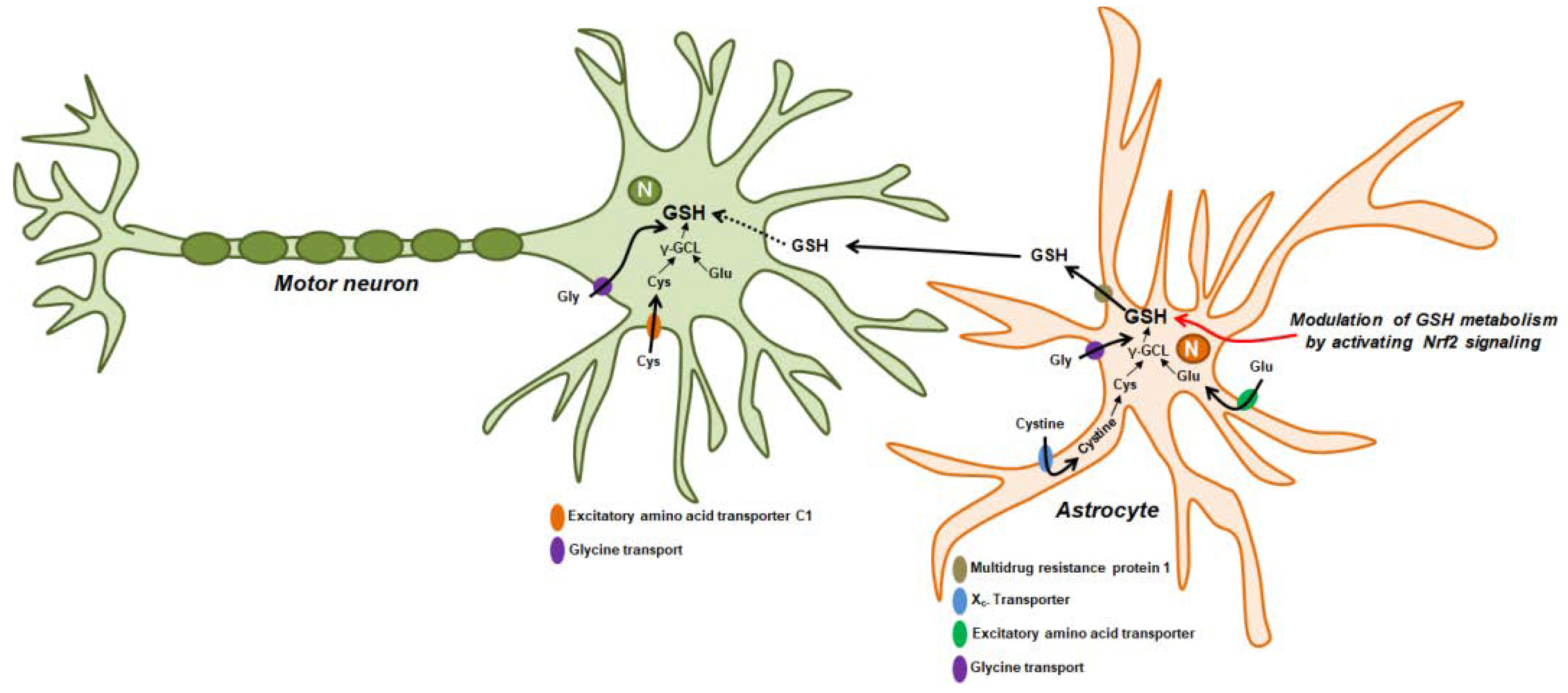
GSH Synthesis and Cellular Distribution in Neurons

GSH is a tripeptide composed of glutamate, cysteine, and glycine. It is the most abundant thiol molecule found in tissues, including the brain, with a concentration of approximately 1–10 mM in the latter compared to approximately 2–3 mM in neurons, which is higher than that in the blood or cerebrospinal fluid (approximately 4 μM) [33,34,35,36]. GSH is synthesized in the cytosol by consecutive ATP-dependent reactions catalyzed by two enzymes, namely γ-glutamate-cysteine ligase (γ-GCL) and GSH synthetase (GS) [37,38]. γ-GCL mediates the first step of GSH synthesis, an ATP-dependent enzymatic process, with glutamate and cysteine to form the dipeptide, γ-glutamyl cysteine. γ-GCL is composed of a catalytic subunit (GCLC) and a modulatory subunit (GCLM), and is a rate-limiting enzyme in GSH synthesis [39]. Conditional GCLC knockout mice in whole neuronal cells displayed GSH depletion and neuronal cell death [40]. GCLM knockout mice exhibited reduced GSH levels and abnormal behavior phenotypes [41]. GS in the last step of GSH synthesis mediates the formation of GSH, which combines with glycine to form γ-glutamyl cysteine [42].
GSH is oxidized to glutathione disulfide (GSSG) by the reaction of glutathione peroxidase (GPx) coupled with the reduction of hydrogen oxide or hydroperoxides. GSH is regenerated from GSSG by GR, which uses NADPH as an electron donor [43]. Therefore, the reaction catalyzed by GPx and GR mediates GSH recycling. GSH is consumed by the extracellular release of GSH from cells via the generation of GSH-conjugates in the cytosol or oxidation to form GSSG. These processes induce a reduction in intracellular GSH. Thus, to maintain the intracellular levels of GSH, the synthesis, and recycling of GSH and the inhibition of GSH release must be induced. The depletion or redox imbalance of GSH (decreased GSH/GSSG ratio) has been reported to be involved in various neuronal dysfunction and neurodegenerative diseases, including autism, Alzheimer’s disease, Parkinson’s disease, and Huntington’s disease [44,45,46,47].
Although GSH is synthesized exclusively in the cytosol, it is present in the most important cellular organelles, namely the mitochondria, endoplasmic reticulum (ER), peroxisome, and nucleus [48]. These results suggest that GSH has specific functions in different cell compartments. Mitochondrial GSH represents 10–15% of the cellular GSH [49]. As the mitochondria lack the enzymes involved in de novo GSH synthesis, the maintenance of mitochondrial GSH levels depends on its uptake from the cytosol via carrier-mediated transporter systems. Many studies have shown that mitochondrial protection systems against free radicals and ROS, such as GSH, are critical for protecting neuronal cells from oxidative stress in the mitochondria. Moreover, mitochondrial GSH is important for protecting the organelles from ROS generated via the oxidative phosphorylation system. Muyderman et al. found that the selective depletion of mitochondrial GSH in astrocytes significantly increased hydrogen peroxide-induced apoptotic cell death and provided evidence for the crucial role played by mitochondrial GSH in preserving cell viability [50]. Wüllner et al. investigated the effects of acute GSH depletion and reduced mitochondrial GSH in relation to mitochondrial dysfunction in the cerebellar granule neurons of rats [51]. They found that the depletion of neuronal mitochondrial GSH led to a significant increase in ROS production and cell death in the nervous system. Furthermore, Wilkins et al. investigated the mechanism of mitochondrial GSH transport in the brain [52]. They showed that the dysfunction of mitochondrial GSH transporters, such as dicarboxylate and 2-oxoglutarate carriers, could result in an increased susceptibility of neurons to oxidative stress. These results demonstrate that the maintenance of mitochondrial GSH via sustained mitochondrial GSH transport is critical for protecting neurons from oxidative stress. Feng et al. also showed a reduction in mitochondrial GSH in the brain and spinal cord of GCLC-deficient mice [40]. Therefore, reduced mitochondrial GSH levels might be associated with mitochondrial defects in the nervous system.
The cytosol in cells is maintained in a reduced state to stabilize the free thiol groups. However, the ER environment is more oxidized than the cytosol to promote disulfide bond formation [48,53]. The redox environment in the ER influences the activity of various enzymes, including protein disulfide isomerase (PDI), which is responsible for the formation of disulfide bonds [53]. Previous studies have reported that the maintenance of redox status by regulating the GSH:GSSG ratio in the ER is considerably more oxidized than that in the cytosol [54]. Measuring the GSH levels in the ER revealed that the GSH:GSSG ratio in the ER is between 1:1 and 3:1 [55]. Furthermore, several studies have suggested the role of ER GSH in the formation of protein disulfide bonds; GSH is suggested to act as a net reductant in the ER by maintaining ER oxidoreductases, including PDI and endoplasmic reticulum oxidation 1 (ERO1) in a reduced state or by directly reducing non-native disulfide bonds in folding proteins [56,57,58]. Tsunoda et al. revealed that the selective depletion of ER GSH by expressing a cytosolic GSH-degrading enzyme, ChaC1, in the ER did not alter protein folding or ER stress response [59]. Although this study suggests the existence of an alternative electron donor that maintains the redox status in the ER, this result does not exclude the importance of the role of GSH in protein folding in the ER. However, direct evidence for the role of ER GSH in neuronal cells has yet to be found. However, as misfolded and aggregated proteins in the ER are one of the causes of neurodegenerative diseases, there is a need to determine the precise role of GSH in the ER of neurons.
GSH is present in the peroxisome—it is transported from the cytosol to the peroxisome by diffusion across the peroxisomal membrane [60,61]. Furthermore, GSSG is thought to be exported to the cytosol through a peroxisomal glutathione transporter, Opt2, and is subsequently reduced to GSH by cytosolic GR in an NADPH-dependent manner [62]. Catalase, peroxidase, and GSH are major components of the peroxisomal antioxidant system. GSH peroxidase (GPx) in the peroxisome requires GSH as a cellular reductant to reduce hydrogen peroxide to water [63].
GSH also plays a critical role in the nucleus. Previous studies have shown that the distribution of GSH to the nucleus is a critical factor in cell proliferation [64]. In plants, GSH depletion blocks the transition from G1 to S phase in the cell cycle of the root [65]. Moreover, GSH recruitment into the nucleus can regulate chromatin structure and condensation, which controls gene expression [66]. Therefore, the recruitment and translocation of GSH from the cytosol to the nucleus during the cell cycle has a great influence on cellular redox homeostasis and gene expression. Jeong et al. investigated GSH levels in living mammalian cells using a fluorescent real-time thiol tracer (FreSHtracer) [67]. They found that GSH levels were markedly higher in the nucleus than in the cytosol, and GSH was required for the maintenance of stem cell functions. Miller et al. also investigated the precise localization of GSH in the mouse central nervous system and found that GSH is synthesized in neurons and diffuses into the nucleus to protect DNA from oxidative stress [68]. However, the molecular functions of GSH in the nucleus of neurons and the mechanisms of GSH transport to the nucleus in neurons are not yet clearly understood and remain a topic of debate.

3. Evidence for the Dysfunction of GSH Metabolism in ALS

3.1. GSH Redox Imbalance in Cellular Models of ALS: In Vitro Studies

Extensive research has been conducted to investigate GSH metabolism and redox imbalance in experimental cellular models of ALS. As a result, an aberrant GSH redox balance, including GSH depletion in the nervous system, has been identified as a pathogenic mechanism of ALS in various in vitro models (Table 1). Lee et al. found that the levels of GSH and relative enzymes were decreased in familial ALS models [69]. They also examined the levels of GSH and GSSG in NT-2 cells (human teratocarcinoma cells) and SK-N-MC cells (human neuroblastoma cells) expressing human wild-type SOD1 or SOD1 mutants, including G37R and G85R, which are associated with fALS [69]. The GSH levels were found to be significantly reduced in NT-2 cells expressing SOD1G37R or SOD1G85R. Furthermore, The GSH levels were significantly decreased in SK-N-MC cells expressing SOD1G37R or SOD1G85R. However, the levels of GSSG in both mutants of the SOD1-expressing cells were increased. The activity of Gpx and protein carbonylation, a marker of protein damage induced by oxidative stress, was increased in cells transfected with the SOD1 mutants [69]. Rizzardini et al. showed that GSH depletion induced by ethacrynic acid impairs mitochondrial functions, including ROS production and membrane potential, in NSC-34 mouse motor neurons [70]. To investigate the effects of GSH depletion in motor neurons, NSC-34 cells were treated with ethacrynic acid, a GSH-depleting agent that directly conjugates GSH. The decreased GSH levels led to increased ROS, loss of mitochondrial membrane potential, and apoptosis. Furthermore, these researchers developed a cellular model of fALS by transfecting NSC-34 cells with human wild-type SOD1 or mutant SOD1G94A. SOD1G94A-expressing cells are more sensitive to mitochondrial dysfunction, including decreased mitochondrial membrane potential induced by ethacrynic acid, a GSH-depleting agent [71]. These results indicate that GSH depletion may be a suitable indicator in studies of the pathogenic mechanisms of oxidative stress-induced toxicity in motor neurons, suggesting that oxidative stress coupled with mitochondrial damage induces the development of disease onset similar to ALS.
Chi et al. investigated the effect of cellular GSH alterations on the cell death of motor neurons [72]. Their results showed that the treatment of NSC-34 cells with ethacrynic acid or L-buthionine sulfoximine significantly reduced GSH production and was accompanied by increased ROS generation. GSH depletion by ethacrynic acid enhanced the expression of the oxidative stress markers AP-1, c-Jun, c-Fos, and HO-1. Moreover, GSH depletion promoted apoptotic cell death by increasing cytochrome c release and capase-3 activation in NSC-34 cells [72]. Collectively, these results strongly suggest that decreased cellular GSH production and availability leads to increased intracellular oxidative stress and apoptosis, and promotes oxidative stress propagation and motor neuronal degeneration. Muyderman et al. also investigated the role of mitochondrial GSH in NSC-34 cells with human SOD1G93A. Interestingly, cells stably expressing SOD1G93A showed significantly decreased mitochondrial GSH levels [73]. Furthermore, the treatment of SOD1G93A-expressing cells with ethacrynic acid resulted in increased apoptotic cell death and decreased the mitochondrial GSH pool. Such finding suggests that SOD1G93A regulates mitochondrial oxidative stress by inducing the selective loss of the GSH pool in mitochondria.
Glutamate, an excitatory neurotransmitter in the mammalian nervous system, is a substrate for glutamate cysteine ligase together with cysteine in an ATP-dependent reaction [74]. This is the rate-limiting step in the synthesis of GSH, the main antioxidant in the central nervous system [74]. Abnormalities in both glutamate and glutamine levels were identified in the nervous tissues of a mouse model of fALS [75]. As a result, Cantoni’s group investigated the relationship between GSH synthesis and glutamine metabolism in a cellular model of fALS [76]. They found that both GSH and glutamate levels were significantly decreased in SOD1G93A-expressing NSC-34 cells cultured with the standard concentrations of glucose and glutamine, as well as alterations in the metabolic pathways involving glutamine/glutamate [76]. These results indicate that the decrease in GSH caused by SOD1G93A expression is due to mitochondrial dysfunction associated with the reduction of the flux of glucose-derived pyruvate.
GSH production is regulated by the Nrf2 signaling pathway. This signaling pathway is impaired in various SOD1 models of fALS [77], and may therefore play a critical role in ALS pathogenesis. Moujalled et al. studied the GSH content in primary astrocytes from TDP-43Q331K-expressing mice and found a significant impairment in total GSH induction in response to sodium arsenite treatment in the astrocytes of TDP-43Q331K-expressing mice [78]. This result suggested that the mutant form of TDP-43 impairs the production of GSH by regulating the Nrf2 antioxidant signaling pathway. Moreover, Muyderman’s group showed that GSH is depleted, resulting in increased ROS levels in TDP-43A315T-expressing NSC-34 cells [79]. The depletion of GSH in TDP-43 A315T-expressing cells or the loss of TDP-43 function resulted in increased intracellular ROS production, cell death, and cytosolic mislocalization of TDP-43, whereas protection against mutant TDP-43-mediated cytotoxicity was restored by increasing intracellular GSH levels by treatment with GSH monoethyl ester [79]. Collectively, these results suggest that oxidative stress is a critical factor in TDP-43-associated ALS pathogenesis and may result from the loss of GSH production. Therefore, novel therapeutics that restore GSH content in motor neurons may be beneficial in the prevention of TDP-induced neurotoxicity.

3.2. GSH Redox Imbalance in Animal Models of ALS: In Vivo Studies

Several in vitro studies have implicated GSH redox imbalance as a critical mediator of increased ROS generation and apoptosis in motor neurons and astrocytes. Mutations in ALS-causing genes have been found to lead to GSH depletion and neuronal toxicity. Therefore, there is a need to determine the precise role of GSH depletion and novel regulators of ALS pathogenesis. In particular, the novel regulatory mechanisms of GSH redox imbalance, including GSH depletion, dysfunction of GSH metabolism, and GSH transport, associated with ROS production in in vivo ALS models, need to be explored. Consistent with in vitro cellular studies, in vivo studies using various ALS animal models have provided evidence that the expression of ALS-causing genes activates various pathways and regulators that lead to a GSH redox imbalance. Astrocytes may play a critical role in the survival of motor neurons in ALS (Figure 1) [80]. According to previous studies, increased levels of GSH caused by activated astrocytes and released GSH from astrocytes improve the antioxidant status of co-cultured neurons [81,82,83,84]. Vargas et al. showed that GSH production in spinal cord astrocytes from SOD1G93A-expressing rats can prevent motor neuron apoptosis induced by nitric oxide [85]. Further, these researchers found that the neurotoxic effects of SOD1G93A expression in astrocytes can be counteracted by increased GSH levels induced by the activation of the Nrf2 signaling pathway [85]. They also revealed that Nrf2 activation in astrocytes protects against neuronal toxicity in SOD1G93A transgenic mice [86]. Nrf2 overexpression in astrocytes increased the survival rate and delayed neuromuscular denervation in SOD1G93A transgenic mice. Furthermore, an increased GSH content was observed in the spinal cord and cerebellum tissues of SOD1G93A-coexpressing mice with Nrf2 [86]. Such a finding indicates that the activation of the Nrf2 signaling pathway can restore motor neuronal toxicity by increasing GSH synthesis and release in astrocytes (Figure 1).
Both Liu’s and Linseman’s groups studied the levels of GSH and GSSG in whole blood and the spinal cord lumbar region of SOD1G93A-expressing mice during disease progression [72,87]. As a result, decreased extracellular GSH levels were observed in the whole blood of SOD1G93A-expressing mice. Moreover, intracellular GSH levels in motor neurons were found to be significantly reduced during disease progression in the mice [87]. However, the GSSG levels were significantly increased in SOD1G93A-expressing mice [72]. Therefore, reduced GSH may contribute to motor neuron cell death by inducing the nuclear translocation of apoptosis-inducing factor (AIF) in SOD1G93A transgenic mice. Vargas et al. examined the effect of decreased GSH in ALS models with the knockout of a modifier subunit of glutamate-cysteine ligase (GCLM) [88]. Decreased GSH was found to reduce survival and neurons in the ventral horn and spinal cord tissues of SOD1G93A-expressing mice with GCLM knockout. Moreover, increased ROS production and mitochondrial dysfunction were also observed in SOD1G93A-expressing mice with GCLM knockout [88]. The researchers also investigated the effects of GCLM loss in SOD1WT-expressing mice [89]. A reduction of approximately 70% in the total GSH content was observed in the brain cortex, brainstem, and spinal cord in the GCLM knockout of SOD1WT-expressing mice. Interestingly, the loss of GCLM resulted in a decreased survival, increased motor neuron loss, and accelerated muscle denervation in SOD1WT-expressing mice [89]. A reduction in GSH synthesis caused by the loss of GCLM may contribute to disease development and progression by modulating mitochondrial function in SOD1WT and SOD1G93A-induced mouse models of ALS.
The cystine/glutamate antiporter (system XC-) is an important factor for GSH synthesis, tasked with transporting cystine into the cell to facilitate the release of glutamate into the extracellular space [90,91]. Cytoplasmic cystine is reduced to cysteine and is used in GSH synthesis in the brain [92,93]. Albano et al. revealed increased cystine uptake in the spinal cord of SOD1G93A transgenic mice [94]. Therefore, it is possible that enhanced system XC- activity could be a protective mechanism for oxidative damage in motor neurons by maintaining intracellular GSH levels.
Treatment with urate has been found to suppress oxidative stress-induced toxicity in vitro [95]. Moreover, Serum uric acid levels were found to be reduced in patients with ALS [96,97]. Thus, decreased urate levels may be associated with the development and progression of neurodegenerative diseases, including ALS. Zhang et al. showed that treatment with urate increased the levels of GSH in the SOD1G85R-expressing Drosophila model of fALS by upregulating the Akt signaling pathway and catalytic subunit of glutamate-cysteine ligase (GCLC) [98]. This finding indicates that urate plays a neuroprotective role in motor neuronal damage by activating GSH synthesis.
Glucocorticoids are cholesterol-derived steroid hormones secreted exclusively by the adrenal gland and are critical regulators of homeostasis under basal and stress conditions [99,100]. Glucocorticoids are involved in excessive ROS production, which is associated with various neurodegenerative diseases, including TDP-43-induced proteinopathies [101,102]. Caccamo et al. found that dexamethasone, a synthetic glucocorticoid, increased the susceptibility to TDP-43-induced neurotoxicity in a mouse model [103]. Dexamethasone treatment was also found to exacerbate memory deficits and impair autophagy activation in the C-terminal fragment of TDP-43 and TDP-25-expressing mice. By examining the alteration of cellular redox status following dexamethasone treatment, the researchers found that the GSH/GSSG ratio was significantly decreased in dexamethasone-treated TDP-25-expressing mice [103]. The data indicate that there is a correlation between the decrease in the GSH/GSSG ratio and TDP-25-induced neurotoxicity in the brains after treatment with dexamethasone. Finally, accumulating evidence suggests that further research on the dysregulation of GSH metabolism and GSH-related enzymes induced by neurotoxic conditions could facilitate the development of a potential therapeutic strategy for ALS.
Table 1. Alteration of GSH content in various experimental models of ALS.
Cell Lines and AnimalsExperimental ALS ModelsPhenotypes Associated with Oxidative StressGSH StatusReference
NT-2
SK-N-MC
Human SOD1WT
Human SOD1G37R
Human SOD1G85R
Increased protein carbonyl
Increased 8-OHG
Increased lipid peroxidation
Decrease GSH
Increased GSSG
[69]
NSC-34Ethacrynic acid treatmentDecreased mitochondrial membrane potential
Increased ROS generation
Increased apoptosis
Decreased GSH[70]
NSC-34Human SOD1WT
Human SOD1G93A
Decreased cell viability
Increased ROS generation
Decreased mitochondrial membrane potential
Increased mitochondrial toxicity
-[71]
NSC-34Ethacrynic acid treatmentIncreased ROS generation
Increased oxidative response gene expression
Increased apoptosis
Decreased GSH[72]
NSC-34Human SOD1G93ADecreased cell proliferation
Decreased cell viability
Increased apoptosis
Decreased mitochondrial GSH[73]
NSC-34Human SOD1WT
Human SOD1G93A
Increased mitochondrial dysfunctionDecreased GSH[76]
NSC-34Human TDP-43M337VIncreased gene expression of Nrf2 signaling pathwayDecreased GSH[78]
NSC-34Human TDP-43WT
Human TDP-43A315T
Increased ROS generation
Decreased cell viability
Increased cell death
Decreased GSH[79]
NSC-34Human SOD1G93ARestored cell viability by treating urateIncreased GSH by treating urate[98]
Rat astrocytesHuman SOD1G93ARestored cell survival by activating Nrf2Increased GSH by activating Nrf2[85]
Mouse motor neuronsHuman SOD1G93AIncreased apoptosisDecrease GSH
Increased GSSG
[72]
Mouse astrocytesHuman SOD1G93AExtended survival by expressing Nrf2
Delayed muscle denervation by expressing Nrf2
Increased GSH secretion by expressing Nrf2[86]
Mouse astrocytesHuman SOD1G93A
Human SOD1H46R/H48Q
Decreased cell survival by GCLM knockout
Increased motor neuron loss by GCLM knockout
Increased oxidative stress by GCLM knockout
Decreased complex IV activity by GCLM knockout
Decrease GSH by GCLM knockout[88]
MouseHuman SOD1G93AIncreased cystine uptake by cystine/glutamate antiporter-[94]
FlyHuman SOD1WT
Human SOD1G85R
Extended survival by treatment with urate
Improved motor defect by treatment with urate
Enhanced antioxidant enzyme activity by treatment with urate
Decreased ROS level by treatment with urate
-[98]
MouseHuman TDP-25Increased memory deficit by treatment with dexamethasoneDecreased GSH/GSSG ratio by treatment with dexamethasone[103]
MouseHuman SOD1G93ADecreased survivalDecreased GSH in whole blood and spinal cord[87]
MouseHuman SOD1WTDecreased cell survival by GCLM knockout
Increased motor neuron loss by GCLM knockout
Decrease GSH by GCLM knockout[89]
Figure 1. Astrocyte-mediated regulation of GSH biosynthesis and transport to motor neurons. The biosynthesis and release of GSH are associated with the Nrf2 signaling pathway in astrocytes [83]. Astrocytes take up cysteine, glutamate, and glycine through various transporters; thereafter, GSH is released from astrocytes via multidrug resistance protein 1 and transported to the motor neurons [104]. Cysteine and glutamate uptake are mediated by the Xc- transporter and excitatory amino acid transporter in astrocytes [91,105]. Glycine is transported by astrocytic glycine transport type 1 [106]. Intracellular GSH is synthetized from these amino acids in astrocyte [107]. Extracellular GSH may be taken up by motor neurons directly or indirectly [108]. Motor neurons also take up three amino acids for GSH synthesis. Increased GSH synthesis in astrocytes may exert a protective effect against motor neuronal toxicity under oxidative stress and in ALS.
Antioxidants 10 01011 g001

3.3. GSH Redox Imbalance in ALS Patients

Previous studies have identified binding sites for GSH in the synaptic membranes of the brain and spinal cord [109,110]. In 1993, Lanius et al. examined GSH binding in the spinal cord of sALS patients using a radioactively labeled GSH, [35S]-GSH, and found that [35S]-GSH binding in the spinal cord was increased in patients with sALS [111]. Similarly, Babu et al. observed an imbalance in the antioxidant system in the erythrocytes of patients with sALS [28]. In fact, they found that GSH levels were significantly decreased in the erythrocytes of patients with sALS. Intracellular GSH recycling is catalyzed by GR using NADPH as a reducing agent to convert GSSG to two molecules of GSH [112]. GR activity was also found to be reduced in sALS patients compared to that in control patients [28]. Another group found that erythrocyte GPx activity was significantly impaired in patients with sALS, which remained low during disease progression [29]. GPx catalyzes the detoxification of hydrogen peroxide by utilizing GSH as a major source of protection against oxidative damage in the nervous system [113]. Thus, it can be hypothesized that an antioxidant imbalance, including the depletion of GSH and GSH-related enzymes, could be a contributing factor to the development and progression of ALS.
Weiduschat et al. investigated the in vivo levels of GSH in the motor cortex region of patients with ALS using the J-edited spin-echo difference magnetic resonance spectroscopy (MRS) technique [114]. Consistent with this study, other groups have reported a decrease in GSH levels in the motor cortex and corticospinal tract in patients with ALS compared to healthy controls [115,116]. The GSH levels in the corticospinal tract were more strongly correlated with disease progression than those in the motor cortex. Decreased GSH levels in the motor cortex of patients with ALS are likely to be a manifestation of clinical changes and pathogenic processes specific to ALS. Yang et al. also performed a Mendelian randomization analysis to identify ALS-associated metabolites, and detected 18 metabolites, including γ-glutamyl amino acids, that may exert causal effects on the development and progression of ALS [117]. The γ-glutamyl cycle is responsible for GSH biosynthesis [118]. Therefore, oxidative damage caused by the dysfunction of GSH metabolism may be a critical risk factor associated with the pathogenesis of ALS.

4. Clinical Trials in ALS

In 1998, Schiffer’s group studied the effect of GSH treatment on the rate of progression of ALS in patients and found that GSH treatment did not have a significant effect on ALS progression [119]. Some cysteine-containing molecules, such as N-acetylcysteine (NAC) and procysteine, are used to increase GSH levels. Brown’s group studied the pharmacokinetic properties of procysteine, a cysteine prodrug that increases intracellular GSH levels in patients with ALS [120]. However, the subcutaneous infusion of NAC did not reduce disease progression in patients with ALS [121]. Of note, the GSH levels in the cerebrospinal fluid of patients with ALS are markedly reduced with aging. Although the oral administration of cysteine-containing molecules did not have statistically beneficial effects on ALS patients, cysteine-containing molecules may be a valuable therapeutic option given its ability to increase GSH content in the cerebrospinal fluid.

5. Conclusions

GSH exerts important functions in the central nervous system as an antioxidant, enzyme cofactor, redox buffer, and neuromodulator. GSH deficiency and the dysfunction of GSH metabolism are common to several neurodegenerative diseases, including Alzheimer’s disease and Parkinson’s disease. Although accumulating evidence suggests that aberrant GSH homeostasis is linked to the development and progression of ALS, the precise role and mechanism of redox imbalance in neuronal cells in this disease remains to be determined. In this review, we first summarized the experimental evidence supporting the role of GSH in the development and progression of ALS. Many studies on ALS have focused on the decreased GSH content and imbalanced GSH redox status in the nervous system, which result in ALS progression. Furthermore, several GSH-related enzymes that target oxidative stress and regulate GSH homeostasis have also been studied. The mechanism of ALS progression may involve decreased GSH, increased GSSG, and an impaired GSH-associated antioxidant system, suggesting that an imbalance in redox status beyond its physiological limit may be detrimental to motor neuronal functions and survival that regulate disease initiation and conditions. Although several studies provide indirect or direct evidence that the dysregulation of GSH homeostasis and metabolism in the nervous system are associated with the pathogenesis of ALS, it remains unclear whether reduced GSH is a causative factor in ALS or whether various mutations in ALS-causing genes are responsible for the impaired GSH redox status observed in ALS. In addition, the molecular mechanisms and pathways related to the synthesis, transport, and degradation of GSH in neurons and astrocytes are complex, and our understanding of the imbalanced GSH redox status in the brain of ALS patients remains incomplete. Therefore, future studies should aim to intensively investigate whether treatments that increase the GSH/GSSG ratio in motor neurons and astrocytes by activating GSH biosynthesis have clinical efficacy in the treatment of ALS. In addition, there is a need to study and develop effective pharmacological agents that may enhance GSH functions to reduce oxidative stress and damage cellular organelles, including the mitochondria, in motor neurons.

Author Contributions

Conceptualization, K.K.; formal analysis, K.K.; writing—original draft preparation, K.K.; writing—review and editing, K.K.; project administration, K.K.; funding acquisition, K.K. The author has read and agreed to the published version of the manuscript.

Funding

This work was supported by the Basic Science Research Program through the National Research Foundation of Korea (NRF), funded by the Ministry of Science and ICT (MSIT) (NRF-2017R1C1B1008825; NRF-2019R1F1A1045639) and by the Soonchunhyang University Research Fund.

Conflicts of Interest

The authors declare no conflict of interest.

References

  1. Cleveland, D.W.; Rothstein, J.D. From Charcot to Lou Gehrig: Deciphering selective motor neuron death in ALS. Nat. Rev. Neurosci. 2001, 2, 806–819. [Google Scholar] [CrossRef]
  2. Rowland, L.P.; Shneider, N.A. Amyotrophic lateral sclerosis. N. Engl. J. Med. 2001, 344, 1688–1700. [Google Scholar] [CrossRef]
  3. Arai, T.; Hasegawa, M.; Akiyama, H.; Ikeda, K.; Nonaka, T.; Mori, H.; Mann, D.; Tsuchiya, K.; Yoshida, M.; Hashizume, Y.; et al. TDP-43 is a component of ubiquitin-positive tau-negative inclusions in frontotemporal lobar degeneration and amyotrophic lateral sclerosis. Biochem. Biophys. Res. Commun. 2006, 351, 602–611. [Google Scholar] [CrossRef]
  4. Neumann, M.; Sampathu, D.M.; Kwong, L.K.; Truax, A.C.; Micsenyi, M.C.; Chou, T.T.; Bruce, J.; Schuck, T.; Grossman, M.; Clark, C.M.; et al. Ubiquitinated TDP-43 in frontotemporal lobar degeneration and amyotrophic lateral sclerosis. Science 2006, 314, 130–133. [Google Scholar] [CrossRef] [Green Version]
  5. DeJesus-Hernandez, M.; Mackenzie, I.R.; Boeve, B.F.; Boxer, A.L.; Baker, M.; Rutherford, N.J.; Nicholson, A.M.; Finch, N.A.; Flynn, H.; Adamson, J.; et al. Expanded GGGGCC hexanucleotide repeat in noncoding region of C9ORF72 causes chromosome 9p-linked FTD and ALS. Neuron 2011, 72, 245–256. [Google Scholar] [CrossRef] [Green Version]
  6. Deng, H.X.; Chen, W.; Hong, S.T.; Boycott, K.M.; Gorrie, G.H.; Siddique, N.; Yang, Y.; Fecto, F.; Shi, Y.; Zhai, H.; et al. Mutations in UBQLN2 cause dominant X-linked juvenile and adult-onset ALS and ALS/dementia. Nature 2011, 477, 211–215. [Google Scholar] [CrossRef] [Green Version]
  7. Fecto, F.; Siddique, T. Making connections: Pathology and genetics link amyotrophic lateral sclerosis with frontotemporal lobe dementia. J. Mol. Neurosci. 2011, 45, 663–675. [Google Scholar] [CrossRef]
  8. Ince, P.G.; Highley, J.R.; Kirby, J.; Wharton, S.B.; Takahashi, H.; Strong, M.J.; Shaw, P.J. Molecular pathology and genetic advances in amyotrophic lateral sclerosis: An emerging molecular pathway and the significance of glial pathology. Acta Neuropathol. 2011, 122, 657–671. [Google Scholar] [CrossRef]
  9. Kapeli, K.; Martinez, F.J.; Yeo, G.W. Genetic mutations in RNA-binding proteins and their roles in ALS. Hum. Genet. 2017. [Google Scholar] [CrossRef] [Green Version]
  10. Costa, J.; Gomes, C.; de Carvalho, M. Diagnosis, pathogenesis and therapeutic targets in amyotrophic lateral sclerosis. CNS Neurol. Disord. Drug Targets 2010, 9, 764–778. [Google Scholar] [CrossRef]
  11. Bhandari, R.; Kuhad, A.; Kuhad, A. Edaravone: A new hope for deadly amyotrophic lateral sclerosis. Drugs Today 2018, 54, 349–360. [Google Scholar] [CrossRef]
  12. Rosen, D.R. Mutations in Cu/Zn superoxide dismutase gene are associated with familial amyotrophic lateral sclerosis. Nature 1993, 364, 362. [Google Scholar] [CrossRef]
  13. Pasinelli, P.; Belford, M.E.; Lennon, N.; Bacskai, B.J.; Hyman, B.T.; Trotti, D.; Brown, R.H., Jr. Amyotrophic lateral sclerosis-associated SOD1 mutant proteins bind and aggregate with Bcl-2 in spinal cord mitochondria. Neuron 2004, 43, 19–30. [Google Scholar] [CrossRef] [Green Version]
  14. Gitler, A.D.; Shorter, J. RNA-binding proteins with prion-like domains in ALS and FTLD-U. Prion 2011, 5, 179–187. [Google Scholar] [CrossRef] [Green Version]
  15. Lagier-Tourenne, C.; Cleveland, D.W. Rethinking ALS: The FUS about TDP-43. Cell 2009, 136, 1001–1004. [Google Scholar] [CrossRef] [Green Version]
  16. Ticozzi, N.; Vance, C.; Leclerc, A.L.; Keagle, P.; Glass, J.D.; McKenna-Yasek, D.; Sapp, P.C.; Silani, V.; Bosco, D.A.; Shaw, C.E.; et al. Mutational analysis reveals the FUS homolog TAF15 as a candidate gene for familial amyotrophic lateral sclerosis. Am. J. Med. Genet. B Neuropsychiatr. Genet. 2011, 156B, 285–290. [Google Scholar] [CrossRef]
  17. Couthouis, J.; Hart, M.P.; Erion, R.; King, O.D.; Diaz, Z.; Nakaya, T.; Ibrahim, F.; Kim, H.J.; Mojsilovic-Petrovic, J.; Panossian, S.; et al. Evaluating the role of the FUS/TLS-related gene EWSR1 in amyotrophic lateral sclerosis. Hum. Mol. Genet. 2012, 21, 2899–2911. [Google Scholar] [CrossRef]
  18. Suk, T.R.; Rousseaux, M.W.C. The role of TDP-43 mislocalization in amyotrophic lateral sclerosis. Mol. Neurodegener. 2020, 15, 45. [Google Scholar] [CrossRef]
  19. Mackenzie, I.R.; Rademakers, R.; Neumann, M. TDP-43 and FUS in amyotrophic lateral sclerosis and frontotemporal dementia. Lancet Neurol. 2010, 9, 995–1007. [Google Scholar] [CrossRef]
  20. Dormann, D.; Haass, C. TDP-43 and FUS: A nuclear affair. Trends Neurosci. 2011, 34, 339–348. [Google Scholar] [CrossRef]
  21. Dormann, D.; Rodde, R.; Edbauer, D.; Bentmann, E.; Fischer, I.; Hruscha, A.; Than, M.E.; Mackenzie, I.R.; Capell, A.; Schmid, B.; et al. ALS-associated fused in sarcoma (FUS) mutations disrupt Transportin-mediated nuclear import. EMBO J. 2010, 29, 2841–2857. [Google Scholar] [CrossRef] [Green Version]
  22. Lee, J.; Nguyen, P.T.; Shim, H.S.; Hyeon, S.J.; Im, H.; Choi, M.H.; Chung, S.; Kowall, N.W.; Lee, S.B.; Ryu, H. EWSR1, a multifunctional protein, regulates cellular function and aging via genetic and epigenetic pathways. Biochim. Biophys. Acta Mol. Basis Dis. 2019, 1865, 1938–1945. [Google Scholar] [CrossRef]
  23. Neumann, M.; Bentmann, E.; Dormann, D.; Jawaid, A.; DeJesus-Hernandez, M.; Ansorge, O.; Roeber, S.; Kretzschmar, H.A.; Munoz, D.G.; Kusaka, H.; et al. FET proteins TAF15 and EWS are selective markers that distinguish FTLD with FUS pathology from amyotrophic lateral sclerosis with FUS mutations. Brain 2011, 134, 2595–2609. [Google Scholar] [CrossRef] [Green Version]
  24. Couthouis, J.; Hart, M.P.; Shorter, J.; DeJesus-Hernandez, M.; Erion, R.; Oristano, R.; Liu, A.X.; Ramos, D.; Jethava, N.; Hosangadi, D.; et al. A yeast functional screen predicts new candidate ALS disease genes. Proc. Natl. Acad. Sci. USA 2011, 108, 20881–20890. [Google Scholar] [CrossRef] [Green Version]
  25. Ferrante, R.J.; Browne, S.E.; Shinobu, L.A.; Bowling, A.C.; Baik, M.J.; MacGarvey, U.; Kowall, N.W.; Brown, R.H., Jr.; Beal, M.F. Evidence of increased oxidative damage in both sporadic and familial amyotrophic lateral sclerosis. J. Neurochem. 1997, 69, 2064–2074. [Google Scholar] [CrossRef]
  26. Niedzielska, E.; Smaga, I.; Gawlik, M.; Moniczewski, A.; Stankowicz, P.; Pera, J.; Filip, M. Oxidative Stress in Neurodegenerative Diseases. Mol. Neurobiol. 2016, 53, 4094–4125. [Google Scholar] [CrossRef] [Green Version]
  27. Mitsumoto, H.; Santella, R.M.; Liu, X.; Bogdanov, M.; Zipprich, J.; Wu, H.C.; Mahata, J.; Kilty, M.; Bednarz, K.; Bell, D.; et al. Oxidative stress biomarkers in sporadic ALS. Amyotroph. Lateral Scler. 2008, 9, 177–183. [Google Scholar] [CrossRef]
  28. Babu, G.N.; Kumar, A.; Chandra, R.; Puri, S.K.; Singh, R.L.; Kalita, J.; Misra, U.K. Oxidant-antioxidant imbalance in the erythrocytes of sporadic amyotrophic lateral sclerosis patients correlates with the progression of disease. Neurochem. Int. 2008, 52, 1284–1289. [Google Scholar] [CrossRef]
  29. Cova, E.; Bongioanni, P.; Cereda, C.; Metelli, M.R.; Salvaneschi, L.; Bernuzzi, S.; Guareschi, S.; Rossi, B.; Ceroni, M. Time course of oxidant markers and antioxidant defenses in subgroups of amyotrophic lateral sclerosis patients. Neurochem. Int. 2010, 56, 687–693. [Google Scholar] [CrossRef]
  30. Kuzma, M.; Jamrozik, Z.; Baranczyk-Kuzma, A. Activity and expression of glutathione S-transferase pi in patients with amyotrophic lateral sclerosis. Clin. Chim. Acta 2006, 364, 217–221. [Google Scholar] [CrossRef]
  31. Tohgi, H.; Abe, T.; Yamazaki, K.; Murata, T.; Ishizaki, E.; Isobe, C. Increase in oxidized NO products and reduction in oxidized glutathione in cerebrospinal fluid from patients with sporadic form of amyotrophic lateral sclerosis. Neurosci. Lett. 1999, 260, 204–206. [Google Scholar] [CrossRef]
  32. Kang, Y.; Viswanath, V.; Jha, N.; Qiao, X.; Mo, J.Q.; Andersen, J.K. Brain gamma-glutamyl cysteine synthetase (GCS) mRNA expression patterns correlate with regional-specific enzyme activities and glutathione levels. J. Neurosci. Res. 1999, 58, 436–441. [Google Scholar] [CrossRef]
  33. Pastore, A.; Federici, G.; Bertini, E.; Piemonte, F. Analysis of glutathione: Implication in redox and detoxification. Clin. Chim. Acta 2003, 333, 19–39. [Google Scholar] [CrossRef]
  34. Wu, G.; Fang, Y.Z.; Yang, S.; Lupton, J.R.; Turner, N.D. Glutathione metabolism and its implications for health. J. Nutr. 2004, 134, 489–492. [Google Scholar] [CrossRef] [Green Version]
  35. Dringen, R. Metabolism and functions of glutathione in brain. Prog. Neurobiol. 2000, 62, 649–671. [Google Scholar] [CrossRef]
  36. Cooper, A.J.; Kristal, B.S. Multiple roles of glutathione in the central nervous system. Biol. Chem. 1997, 378, 793–802. [Google Scholar] [PubMed]
  37. Chakravarthi, S.; Jessop, C.E.; Bulleid, N.J. The role of glutathione in disulphide bond formation and endoplasmic-reticulum-generated oxidative stress. EMBO Rep. 2006, 7, 271–275. [Google Scholar] [CrossRef] [Green Version]
  38. Bjorklund, G.; Peana, M.; Maes, M.; Dadar, M.; Severin, B. The glutathione system in Parkinson’s disease and its progression. Neurosci. Biobehav. Rev. 2021, 120, 470–478. [Google Scholar] [CrossRef]
  39. Toroser, D.; Yarian, C.S.; Orr, W.C.; Sohal, R.S. Mechanisms of gamma-glutamylcysteine ligase regulation. Biochim. Biophys. Acta 2006, 1760, 233–244. [Google Scholar] [CrossRef] [Green Version]
  40. Feng, W.; Rosca, M.; Fan, Y.; Hu, Y.; Feng, P.; Lee, H.G.; Monnier, V.M.; Fan, X. Gclc deficiency in mouse CNS causes mitochondrial damage and neurodegeneration. Hum. Mol. Genet. 2017, 26, 1376–1390. [Google Scholar] [CrossRef]
  41. Chen, Y.; Curran, C.P.; Nebert, D.W.; Patel, K.V.; Williams, M.T.; Vorhees, C.V. Effect of chronic glutathione deficiency on the behavioral phenotype of Gclm-/- knockout mice. Neurotoxicol. Teratol. 2012, 34, 450–457. [Google Scholar] [CrossRef] [Green Version]
  42. Lu, S.C. Glutathione synthesis. Biochim. Biophys. Acta 2013, 1830, 3143–3153. [Google Scholar] [CrossRef] [Green Version]
  43. Schafer, F.Q.; Buettner, G.R. Redox environment of the cell as viewed through the redox state of the glutathione disulfide/glutathione couple. Free Radic. Biol. Med. 2001, 30, 1191–1212. [Google Scholar] [CrossRef]
  44. Gu, F.; Chauhan, V.; Chauhan, A. Impaired synthesis and antioxidant defense of glutathione in the cerebellum of autistic subjects: Alterations in the activities and protein expression of glutathione-related enzymes. Free Radic. Biol. Med. 2013, 65, 488–496. [Google Scholar] [CrossRef]
  45. Saharan, S.; Mandal, P.K. The emerging role of glutathione in Alzheimer’s disease. J. Alzheimer’s Dis. 2014, 40, 519–529. [Google Scholar] [CrossRef]
  46. Smeyne, M.; Smeyne, R.J. Glutathione metabolism and Parkinson’s disease. Free Radic. Biol. Med. 2013, 62, 13–25. [Google Scholar] [CrossRef] [Green Version]
  47. Mason, R.P.; Casu, M.; Butler, N.; Breda, C.; Campesan, S.; Clapp, J.; Green, E.W.; Dhulkhed, D.; Kyriacou, C.P.; Giorgini, F. Glutathione peroxidase activity is neuroprotective in models of Huntington’s disease. Nat. Genet. 2013, 45, 1249–1254. [Google Scholar] [CrossRef] [Green Version]
  48. Oestreicher, J.; Morgan, B. Glutathione: Subcellular distribution and membrane transport. Biochem. Cell Biol. 2019, 97, 270–289. [Google Scholar] [CrossRef] [Green Version]
  49. Fernandez-Checa, J.C.; Kaplowitz, N.; Garcia-Ruiz, C.; Colell, A.; Miranda, M.; Mari, M.; Ardite, E.; Morales, A. GSH transport in mitochondria: Defense against TNF-induced oxidative stress and alcohol-induced defect. Am. J. Physiol. 1997, 273, G7–G17. [Google Scholar] [CrossRef]
  50. Muyderman, H.; Wadey, A.L.; Nilsson, M.; Sims, N.R. Mitochondrial glutathione protects against cell death induced by oxidative and nitrative stress in astrocytes. J. Neurochem. 2007, 102, 1369–1382. [Google Scholar] [CrossRef]
  51. Wullner, U.; Seyfried, J.; Groscurth, P.; Beinroth, S.; Winter, S.; Gleichmann, M.; Heneka, M.; Loschmann, P.; Schulz, J.B.; Weller, M.; et al. Glutathione depletion and neuronal cell death: The role of reactive oxygen intermediates and mitochondrial function. Brain Res. 1999, 826, 53–62. [Google Scholar] [CrossRef]
  52. Wilkins, H.M.; Kirchhof, D.; Manning, E.; Joseph, J.W.; Linseman, D.A. Mitochondrial glutathione transport is a key determinant of neuronal susceptibility to oxidative and nitrosative stress. J. Biol. Chem. 2013, 288, 5091–5101. [Google Scholar] [CrossRef] [Green Version]
  53. Go, Y.M.; Jones, D.P. Redox compartmentalization in eukaryotic cells. Biochim. Biophys. Acta 2008, 1780, 1273–1290. [Google Scholar] [CrossRef] [Green Version]
  54. Hwang, C.; Sinskey, A.J.; Lodish, H.F. Oxidized redox state of glutathione in the endoplasmic reticulum. Science 1992, 257, 1496–1502. [Google Scholar] [CrossRef]
  55. Bass, R.; Ruddock, L.W.; Klappa, P.; Freedman, R.B. A major fraction of endoplasmic reticulum-located glutathione is present as mixed disulfides with protein. J. Biol. Chem. 2004, 279, 5257–5262. [Google Scholar] [CrossRef] [Green Version]
  56. Jessop, C.E.; Bulleid, N.J. Glutathione directly reduces an oxidoreductase in the endoplasmic reticulum of mammalian cells. J. Biol. Chem. 2004, 279, 55341–55347. [Google Scholar] [CrossRef] [Green Version]
  57. Molteni, S.N.; Fassio, A.; Ciriolo, M.R.; Filomeni, G.; Pasqualetto, E.; Fagioli, C.; Sitia, R. Glutathione limits Ero1-dependent oxidation in the endoplasmic reticulum. J. Biol. Chem. 2004, 279, 32667–32673. [Google Scholar] [CrossRef] [Green Version]
  58. Ponsero, A.J.; Igbaria, A.; Darch, M.A.; Miled, S.; Outten, C.E.; Winther, J.R.; Palais, G.; D’Autreaux, B.; Delaunay-Moisan, A.; Toledano, M.B. Endoplasmic Reticulum Transport of Glutathione by Sec61 is Regulated by Ero1 and Bip. Mol. Cell 2017, 67, 962–973. [Google Scholar] [CrossRef] [Green Version]
  59. Tsunoda, S.; Avezov, E.; Zyryanova, A.; Konno, T.; Mendes-Silva, L.; Pinho Melo, E.; Harding, H.P.; Ron, D. Intact protein folding in the glutathione-depleted endoplasmic reticulum implicates alternative protein thiol reductants. Elife 2014, 3, e03421. [Google Scholar] [CrossRef]
  60. Horiguchi, H.; Yurimoto, H.; Kato, N.; Sakai, Y. Antioxidant system within yeast peroxisome. Biochemical and physiological characterization of CbPmp20 in the methylotrophic yeast Candida boidinii. J. Biol. Chem. 2001, 276, 14279–14288. [Google Scholar] [CrossRef] [Green Version]
  61. Antonenkov, V.D.; Hiltunen, J.K. Transfer of metabolites across the peroxisomal membrane. Biochim. Biophys. Acta 2012, 1822, 1374–1386. [Google Scholar] [CrossRef] [Green Version]
  62. Elbaz-Alon, Y.; Morgan, B.; Clancy, A.; Amoako, T.N.; Zalckvar, E.; Dick, T.P.; Schwappach, B.; Schuldiner, M. The yeast oligopeptide transporter Opt2 is localized to peroxisomes and affects glutathione redox homeostasis. FEMS Yeast Res. 2014, 14, 1055–1067. [Google Scholar] [CrossRef] [Green Version]
  63. Ohdate, T.; Inoue, Y. Involvement of glutathione peroxidase 1 in growth and peroxisome formation in Saccharomyces cerevisiae in oleic acid medium. Biochim. Biophys. Acta 2012, 1821, 1295–1305. [Google Scholar] [CrossRef] [Green Version]
  64. Markovic, J.; Borras, C.; Ortega, A.; Sastre, J.; Vina, J.; Pallardo, F.V. Glutathione is recruited into the nucleus in early phases of cell proliferation. J. Biol. Chem. 2007, 282, 20416–20424. [Google Scholar] [CrossRef] [Green Version]
  65. Reichheld, J.P.; Khafif, M.; Riondet, C.; Droux, M.; Bonnard, G.; Meyer, Y. Inactivation of thioredoxin reductases reveals a complex interplay between thioredoxin and glutathione pathways in Arabidopsis development. Plant Cell 2007, 19, 1851–1865. [Google Scholar] [CrossRef] [Green Version]
  66. Bellomo, G.; Palladini, G.; Vairetti, M. Intranuclear distribution, function and fate of glutathione and glutathione-S-conjugate in living rat hepatocytes studied by fluorescence microscopy. Microsc. Res. Tech. 1997, 36, 243–252. [Google Scholar] [CrossRef]
  67. Jeong, E.M.; Yoon, J.H.; Lim, J.; Shin, J.W.; Cho, A.Y.; Heo, J.; Lee, K.B.; Lee, J.H.; Lee, W.J.; Kim, H.J.; et al. Real-Time Monitoring of Glutathione in Living Cells Reveals that High Glutathione Levels are Required to Maintain Stem Cell Function. Stem Cell Rep. 2018, 10, 600–614. [Google Scholar] [CrossRef] [Green Version]
  68. Miller, V.M.; Lawrence, D.A.; Mondal, T.K.; Seegal, R.F. Reduced glutathione is highly expressed in white matter and neurons in the unperturbed mouse brain—Implications for oxidative stress associated with neurodegeneration. Brain Res. 2009, 1276, 22–30. [Google Scholar] [CrossRef] [Green Version]
  69. Lee, M.; Hyun, D.; Jenner, P.; Halliwell, B. Effect of overexpression of wild-type and mutant Cu/Zn-superoxide dismutases on oxidative damage and antioxidant defences: Relevance to Down’s syndrome and familial amyotrophic lateral sclerosis. J. Neurochem. 2001, 76, 957–965. [Google Scholar] [CrossRef] [Green Version]
  70. Rizzardini, M.; Lupi, M.; Bernasconi, S.; Mangolini, A.; Cantoni, L. Mitochondrial dysfunction and death in motor neurons exposed to the glutathione-depleting agent ethacrynic acid. J. Neurol. Sci. 2003, 207, 51–58. [Google Scholar] [CrossRef]
  71. Rizzardini, M.; Mangolini, A.; Lupi, M.; Ubezio, P.; Bendotti, C.; Cantoni, L. Low levels of ALS-linked Cu/Zn superoxide dismutase increase the production of reactive oxygen species and cause mitochondrial damage and death in motor neuron-like cells. J. Neurol. Sci. 2005, 232, 95–103. [Google Scholar] [CrossRef]
  72. Chi, L.; Ke, Y.; Luo, C.; Gozal, D.; Liu, R. Depletion of reduced glutathione enhances motor neuron degeneration in vitro and in vivo. Neuroscience 2007, 144, 991–1003. [Google Scholar] [CrossRef] [Green Version]
  73. Muyderman, H.; Hutson, P.G.; Matusica, D.; Rogers, M.L.; Rush, R.A. The human G93A-superoxide dismutase-1 mutation, mitochondrial glutathione and apoptotic cell death. Neurochem. Res. 2009, 34, 1847–1856. [Google Scholar] [CrossRef]
  74. Chen, Y.; Shertzer, H.G.; Schneider, S.N.; Nebert, D.W.; Dalton, T.P. Glutamate cysteine ligase catalysis: Dependence on ATP and modifier subunit for regulation of tissue glutathione levels. J. Biol. Chem. 2005, 280, 33766–33774. [Google Scholar] [CrossRef] [Green Version]
  75. Choi, J.K.; Kustermann, E.; Dedeoglu, A.; Jenkins, B.G. Magnetic resonance spectroscopy of regional brain metabolite markers in FALS mice and the effects of dietary creatine supplementation. Eur. J. Neurosci. 2009, 30, 2143–2150. [Google Scholar] [CrossRef] [Green Version]
  76. D’Alessandro, G.; Calcagno, E.; Tartari, S.; Rizzardini, M.; Invernizzi, R.W.; Cantoni, L. Glutamate and glutathione interplay in a motor neuronal model of amyotrophic lateral sclerosis reveals altered energy metabolism. Neurobiol. Dis. 2011, 43, 346–355. [Google Scholar] [CrossRef]
  77. Sarlette, A.; Krampfl, K.; Grothe, C.; Neuhoff, N.; Dengler, R.; Petri, S. Nuclear erythroid 2-related factor 2-antioxidative response element signaling pathway in motor cortex and spinal cord in amyotrophic lateral sclerosis. J. Neuropathol. Exp. Neurol. 2008, 67, 1055–1062. [Google Scholar] [CrossRef]
  78. Moujalled, D.; Grubman, A.; Acevedo, K.; Yang, S.; Ke, Y.D.; Moujalled, D.M.; Duncan, C.; Caragounis, A.; Perera, N.D.; Turner, B.J.; et al. TDP-43 mutations causing amyotrophic lateral sclerosis are associated with altered expression of RNA-binding protein hnRNP K and affect the Nrf2 antioxidant pathway. Hum. Mol. Genet. 2017, 26, 1732–1746. [Google Scholar] [CrossRef]
  79. Chen, T.; Turner, B.J.; Beart, P.M.; Sheehan-Hennessy, L.; Elekwachi, C.; Muyderman, H. Glutathione monoethyl ester prevents TDP-43 pathology in motor neuronal NSC-34 cells. Neurochem. Int. 2018, 112, 278–287. [Google Scholar] [CrossRef] [Green Version]
  80. Phatnani, H.; Maniatis, T. Astrocytes in neurodegenerative disease. Cold Spring Harb. Perspect. Biol. 2015, 7. [Google Scholar] [CrossRef] [Green Version]
  81. Dringen, R.; Pfeiffer, B.; Hamprecht, B. Synthesis of the antioxidant glutathione in neurons: Supply by astrocytes of CysGly as precursor for neuronal glutathione. J. Neurosci. 1999, 19, 562–569. [Google Scholar] [CrossRef]
  82. Dringen, R.; Gutterer, J.M.; Hirrlinger, J. Glutathione metabolism in brain metabolic interaction between astrocytes and neurons in the defense against reactive oxygen species. Eur. J. Biochem. 2000, 267, 4912–4916. [Google Scholar] [CrossRef]
  83. Shih, A.Y.; Johnson, D.A.; Wong, G.; Kraft, A.D.; Jiang, L.; Erb, H.; Johnson, J.A.; Murphy, T.H. Coordinate regulation of glutathione biosynthesis and release by Nrf2-expressing glia potently protects neurons from oxidative stress. J. Neurosci. 2003, 23, 3394–3406. [Google Scholar] [CrossRef]
  84. Kraft, A.D.; Johnson, D.A.; Johnson, J.A. Nuclear factor E2-related factor 2-dependent antioxidant response element activation by tert-butylhydroquinone and sulforaphane occurring preferentially in astrocytes conditions neurons against oxidative insult. J. Neurosci. 2004, 24, 1101–1112. [Google Scholar] [CrossRef] [Green Version]
  85. Vargas, M.R.; Pehar, M.; Cassina, P.; Beckman, J.S.; Barbeito, L. Increased glutathione biosynthesis by Nrf2 activation in astrocytes prevents p75NTR-dependent motor neuron apoptosis. J. Neurochem. 2006, 97, 687–696. [Google Scholar] [CrossRef]
  86. Vargas, M.R.; Johnson, D.A.; Sirkis, D.W.; Messing, A.; Johnson, J.A. Nrf2 activation in astrocytes protects against neurodegeneration in mouse models of familial amyotrophic lateral sclerosis. J. Neurosci. 2008, 28, 13574–13581. [Google Scholar] [CrossRef]
  87. Ross, E.K.; Winter, A.N.; Wilkins, H.M.; Sumner, W.A.; Duval, N.; Patterson, D.; Linseman, D.A. A Cystine-Rich Whey Supplement (Immunocal((R))) Delays Disease Onset and Prevents Spinal Cord Glutathione Depletion in the hSOD1(G93A) Mouse Model of Amyotrophic Lateral Sclerosis. Antioxidants 2014, 3, 843–865. [Google Scholar] [CrossRef] [Green Version]
  88. Vargas, M.R.; Johnson, D.A.; Johnson, J.A. Decreased glutathione accelerates neurological deficit and mitochondrial pathology in familial ALS-linked hSOD1(G93A) mice model. Neurobiol. Dis. 2011, 43, 543–551. [Google Scholar] [CrossRef] [Green Version]
  89. Killoy, K.M.; Harlan, B.A.; Pehar, M.; Helke, K.L.; Johnson, J.A.; Vargas, M.R. Decreased glutathione levels cause overt motor neuron degeneration in hSOD1(WT) over-expressing mice. Exp. Neurol. 2018, 302, 129–135. [Google Scholar] [CrossRef]
  90. Bannai, S. Exchange of cystine and glutamate across plasma membrane of human fibroblasts. J. Biol. Chem. 1986, 261, 2256–2263. [Google Scholar] [CrossRef]
  91. Lewerenz, J.; Hewett, S.J.; Huang, Y.; Lambros, M.; Gout, P.W.; Kalivas, P.W.; Massie, A.; Smolders, I.; Methner, A.; Pergande, M.; et al. The cystine/glutamate antiporter system x(c)(-) in health and disease: From molecular mechanisms to novel therapeutic opportunities. Antioxid. Redox Signal. 2013, 18, 522–555. [Google Scholar] [CrossRef] [Green Version]
  92. Stipanuk, M.H.; Dominy, J.E., Jr.; Lee, J.I.; Coloso, R.M. Mammalian cysteine metabolism: New insights into regulation of cysteine metabolism. J. Nutr. 2006, 136, 1652S–1659S. [Google Scholar] [CrossRef]
  93. Sato, H.; Tamba, M.; Okuno, S.; Sato, K.; Keino-Masu, K.; Masu, M.; Bannai, S. Distribution of cystine/glutamate exchange transporter, system x(c)-, in the mouse brain. J. Neurosci. 2002, 22, 8028–8033. [Google Scholar] [CrossRef] [Green Version]
  94. Albano, R.; Liu, X.; Lobner, D. Regulation of system x(c)- in the SOD1-G93A mouse model of ALS. Exp. Neurol. 2013, 250, 69–73. [Google Scholar] [CrossRef]
  95. Sautin, Y.Y.; Johnson, R.J. Uric acid: The oxidant-antioxidant paradox. Nucleosides Nucleotides Nucleic Acids 2008, 27, 608–619. [Google Scholar] [CrossRef] [Green Version]
  96. Keizman, D.; Ish-Shalom, M.; Berliner, S.; Maimon, N.; Vered, Y.; Artamonov, I.; Tsehori, J.; Nefussy, B.; Drory, V.E. Low uric acid levels in serum of patients with ALS: Further evidence for oxidative stress? J. Neurol. Sci. 2009, 285, 95–99. [Google Scholar] [CrossRef]
  97. Paganoni, S.; Nicholson, K.; Chan, J.; Shui, A.; Schoenfeld, D.; Sherman, A.; Berry, J.; Cudkowicz, M.; Atassi, N.; Pooled Resource Open-Access, A.L.S.C.T.C. Urate levels predict survival in amyotrophic lateral sclerosis: Analysis of the expanded Pooled Resource Open-Access ALS clinical trials database. Muscle Nerve 2018, 57, 430–434. [Google Scholar] [CrossRef]
  98. Zhang, C.; Yang, Y.; Liang, W.; Wang, T.; Wang, S.; Wang, X.; Wang, Y.; Jiang, H.; Feng, H. Neuroprotection by urate on the mutant hSOD1-related cellular and Drosophila models of amyotrophic lateral sclerosis: Implication for GSH synthesis via activating Akt/GSK3beta/Nrf2/GCLC pathways. Brain Res. Bull. 2019, 146, 287–301. [Google Scholar] [CrossRef]
  99. Sapolsky, R.M.; Romero, L.M.; Munck, A.U. How do glucocorticoids influence stress responses? Integrating permissive, suppressive, stimulatory, and preparative actions. Endocr. Rev. 2000, 21, 55–89. [Google Scholar] [CrossRef] [Green Version]
  100. Srinivasan, S.; Shariff, M.; Bartlett, S.E. The role of the glucocorticoids in developing resilience to stress and addiction. Front. Psychiatry 2013, 4, 68. [Google Scholar] [CrossRef] [Green Version]
  101. McIntosh, L.J.; Sapolsky, R.M. Glucocorticoids increase the accumulation of reactive oxygen species and enhance adriamycin-induced toxicity in neuronal culture. Exp. Neurol. 1996, 141, 201–206. [Google Scholar] [CrossRef]
  102. Behl, C.; Lezoualc’h, F.; Trapp, T.; Widmann, M.; Skutella, T.; Holsboer, F. Glucocorticoids enhance oxidative stress-induced cell death in hippocampal neurons in vitro. Endocrinology 1997, 138, 101–106. [Google Scholar] [CrossRef]
  103. Caccamo, A.; Medina, D.X.; Oddo, S. Glucocorticoids exacerbate cognitive deficits in TDP-25 transgenic mice via a glutathione-mediated mechanism: Implications for aging, stress and TDP-43 proteinopathies. J. Neurosci. 2013, 33, 906–913. [Google Scholar] [CrossRef]
  104. Hirrlinger, J.; Konig, J.; Keppler, D.; Lindenau, J.; Schulz, J.B.; Dringen, R. The multidrug resistance protein MRP1 mediates the release of glutathione disulfide from rat astrocytes during oxidative stress. J. Neurochem. 2001, 76, 627–636. [Google Scholar] [CrossRef] [Green Version]
  105. Kim, K.; Lee, S.G.; Kegelman, T.P.; Su, Z.Z.; Das, S.K.; Dash, R.; Dasgupta, S.; Barral, P.M.; Hedvat, M.; Diaz, P.; et al. Role of excitatory amino acid transporter-2 (EAAT2) and glutamate in neurodegeneration: Opportunities for developing novel therapeutics. J. Cell Physiol. 2011, 226, 2484–2493. [Google Scholar] [CrossRef] [Green Version]
  106. Shibasaki, K.; Hosoi, N.; Kaneko, R.; Tominaga, M.; Yamada, K. Glycine release from astrocytes via functional reversal of GlyT1. J. Neurochem. 2017, 140, 395–403. [Google Scholar] [CrossRef]
  107. Kranich, O.; Hamprecht, B.; Dringen, R. Different preferences in the utilization of amino acids for glutathione synthesis in cultured neurons and astroglial cells derived from rat brain. Neurosci. Lett. 1996, 219, 211–214. [Google Scholar] [CrossRef]
  108. Wang, X.F.; Cynader, M.S. Astrocytes provide cysteine to neurons by releasing glutathione. J. Neurochem. 2000, 74, 1434–1442. [Google Scholar] [CrossRef]
  109. Ogita, K.; Yoneda, Y. Selective potentiation by L-cysteine of apparent binding activity of [3H]glutathione in synaptic membranes of rat brain. Biochem. Pharmacol. 1989, 38, 1499–1505. [Google Scholar] [CrossRef]
  110. Ogita, K.; Yoneda, Y. Possible presence of [3H]glutathione (GSH) binding sites in synaptic membranes from rat brain. Neurosci. Res. 1987, 4, 486–496. [Google Scholar] [CrossRef]
  111. Lanius, R.A.; Krieger, C.; Wagey, R.; Shaw, C.A. Increased [35S]glutathione binding sites in spinal cords from patients with sporadic amyotrophic lateral sclerosis. Neurosci. Lett. 1993, 163, 89–92. [Google Scholar] [CrossRef]
  112. Couto, N.; Wood, J.; Barber, J. The role of glutathione reductase and related enzymes on cellular redox homoeostasis network. Free Radic. Biol. Med. 2016, 95, 27–42. [Google Scholar] [CrossRef]
  113. McLean, C.W.; Mirochnitchenko, O.; Claus, C.P.; Noble-Haeusslein, L.J.; Ferriero, D.M. Overexpression of glutathione peroxidase protects immature murine neurons from oxidative stress. Dev. Neurosci. 2005, 27, 169–175. [Google Scholar] [CrossRef]
  114. Weiduschat, N.; Mao, X.; Hupf, J.; Armstrong, N.; Kang, G.; Lange, D.J.; Mitsumoto, H.; Shungu, D.C. Motor cortex glutathione deficit in ALS measured in vivo with the J-editing technique. Neurosci. Lett. 2014, 570, 102–107. [Google Scholar] [CrossRef]
  115. Weerasekera, A.; Peeters, R.; Sima, D.; Dresselaers, T.; Sunaert, S.; de Vocht, J.; Claeys, K.; van Huffel, S.; van Damme, P.; Himmelreich, U. Motor cortex metabolite alterations in amyotrophic lateral sclerosis assessed in vivo using edited and non-edited magnetic resonance spectroscopy. Brain Res. 2019, 1718, 22–31. [Google Scholar] [CrossRef]
  116. Andronesi, O.C.; Nicholson, K.; Jafari-Khouzani, K.; Bogner, W.; Wang, J.; Chan, J.; Macklin, E.A.; Levine-Weinberg, M.; Breen, C.; Schwarzschild, M.A.; et al. Imaging Neurochemistry and Brain Structure Tracks Clinical Decline and Mechanisms of ALS in Patients. Front. Neurol. 2020, 11, 590573. [Google Scholar] [CrossRef]
  117. Yang, L.; Lv, X.; Du, H.; Wu, D.; Wang, M. Causal effects of serum metabolites on amyotrophic lateral sclerosis: A Mendelian randomization study. Prog. Neuropsychopharmacol. Biol. Psychiatry 2020, 97, 109771. [Google Scholar] [CrossRef]
  118. Bachhawat, A.K.; Yadav, S. The glutathione cycle: Glutathione metabolism beyond the gamma-glutamyl cycle. IUBMB Life 2018, 70, 585–592. [Google Scholar] [CrossRef] [Green Version]
  119. Chio, A.; Cucatto, A.; Terreni, A.A.; Schiffer, D. Reduced glutathione in amyotrophic lateral sclerosis: An open, crossover, randomized trial. Ital. J. Neurol. Sci. 1998, 19, 363–366. [Google Scholar] [CrossRef]
  120. Cudkowicz, M.E.; Sexton, P.M.; Ellis, T.; Hayden, D.L.; Gwilt, P.R.; Whalen, J.; Brown, R.H., Jr. The pharmacokinetics and pharmaco-dynamics of Procysteine in amyotrophic lateral sclerosis. Neurology 1999, 52, 1492–1494. [Google Scholar] [CrossRef]
  121. Louwerse, E.S.; Weverling, G.J.; Bossuyt, P.M.; Meyjes, F.E.; de Jong, J.M. Randomized, double-blind, controlled trial of acetylcysteine in amyotrophic lateral sclerosis. Arch. Neurol. 1995, 52, 559–564. [Google Scholar] [CrossRef]
Publisher’s Note: MDPI stays neutral with regard to jurisdictional claims in published maps and institutional affiliations.

