Next Issue
Volume 10, July
Previous Issue
Volume 10, May


Antioxidants, Volume 10, Issue 6 (June 2021) – 172 articles

Cover Story (view full-size image): 
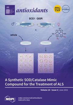
The amphipolar iron(III) corrole 1-Fe has emerged as a promising therapeutic agent for oxidative-stress-related diseases. It decomposes reactive oxygen and nitrogen species in a highly catalytic and effective manner, acting as a synthetic SOD/catalase/thioredoxin enzyme, decomposing superoxide anion radical, hydrogen peroxide and peroxynitrite, respectively. 1-Fe displays a robust efficacy in murine models of diabetes, neuropathy, and atherosclerosis, and further research has described its potential as a therapeutic agent for Parkinson’s and Alzheimer’s diseases. In this study, we disclose the efficacy of 1-Fe in a G93R-mutated SOD1 ALS zebrafish model. The displayed results disclose a substantial improvement in locomotor activity for ALS fish treated by 1-Fe relative to their non-treated counterparts. View this paper.
  • Issues are regarded as officially published after their release is announced to the table of contents alert mailing list.
  • You may sign up for e-mail alerts to receive table of contents of newly released issues.
  • PDF is the official format for papers published in both, html and pdf forms. To view the papers in pdf format, click on the "PDF Full-text" link, and use the free Adobe Reader to open them.
Order results
Result details
Section
Select all
Export citation of selected articles as:
15 pages, 3012 KiB  
Article
Clinical Performance of Paraoxonase-1-Related Variables and Novel Markers of Inflammation in Coronavirus Disease-19. A Machine Learning Approach
by Elisabet Rodríguez-Tomàs, Simona Iftimie, Helena Castañé, Gerard Baiges-Gaya, Anna Hernández-Aguilera, María González-Viñas, Antoni Castro, Jordi Camps and Jorge Joven
Antioxidants 2021, 10(6), 991; https://doi.org/10.3390/antiox10060991 - 21 Jun 2021
Cited by 14 | Viewed by 2790
Abstract
SARS-CoV-2 infection produces a response of the innate immune system causing oxidative stress and a strong inflammatory reaction termed ‘cytokine storm’ that is one of the leading causes of death. Paraoxonase-1 (PON1) protects against oxidative stress by hydrolyzing lipoperoxides. Alterations in PON1 activity [...] Read more.
SARS-CoV-2 infection produces a response of the innate immune system causing oxidative stress and a strong inflammatory reaction termed ‘cytokine storm’ that is one of the leading causes of death. Paraoxonase-1 (PON1) protects against oxidative stress by hydrolyzing lipoperoxides. Alterations in PON1 activity have been associated with pro-inflammatory mediators such as the chemokine (C-C motif) ligand 2 (CCL2), and the glycoprotein galectin-3. We aimed to investigate the alterations in the circulating levels of PON1, CCL2, and galectin-3 in 126 patients with COVID-19 and their interactions with clinical variables and analytical parameters. A machine learning approach was used to identify predictive markers of the disease. For comparisons, we recruited 45 COVID-19 negative patients and 50 healthy individuals. Our approach identified a synergy between oxidative stress, inflammation, and fibrogenesis in positive patients that is not observed in negative patients. PON1 activity was the parameter with the greatest power to discriminate between patients who were either positive or negative for COVID-19, while their levels of CCL2 and galectin-3 were similar. We suggest that the measurement of serum PON1 activity may be a useful marker for the diagnosis of COVID-19. Full article
(This article belongs to the Section Health Outcomes of Antioxidants and Oxidative Stress)
Show Figures

Graphical abstract

12 pages, 1400 KiB  
Article
GABA Regulates Phenolics Accumulation in Soybean Sprouts under NaCl Stress
by Yunyun Zhao, Chong Xie, Pei Wang, Zhenxin Gu and Runqiang Yang
Antioxidants 2021, 10(6), 990; https://doi.org/10.3390/antiox10060990 - 21 Jun 2021
Cited by 19 | Viewed by 2897
Abstract
NaCl stress causes oxidative stress in plants; γ-aminobutyric acid (GABA) could alleviate such abiotic stress by enhancing the synthesis of phenolics, but the underlying mechanism is not clear. We investigated the effects of GABA on phenolics accumulation in soybean sprouts under NaCl stress [...] Read more.
NaCl stress causes oxidative stress in plants; γ-aminobutyric acid (GABA) could alleviate such abiotic stress by enhancing the synthesis of phenolics, but the underlying mechanism is not clear. We investigated the effects of GABA on phenolics accumulation in soybean sprouts under NaCl stress by measuring changes in the content of physiological biochemicals and phenolic substances, in the activity and gene expression of key enzymes, and in antioxidant capacity. GABA reduced the oxidative damage in soybean sprouts caused by NaCl stress and enhanced the content of total phenolics, phenolic acids, and isoflavones by 16.58%, 22.47%, and 3.75%, respectively. It also increased the activities and expression of phenylalanine ammonia lyase, cinnamic acid 4-hydroxylase, and 4-coumarate coenzyme A ligase. Furthermore, GABA increased the activity of antioxidant enzymes and the antioxidant capacity. These events were inhibited by 3-mercaptopropionate (an inhibitor for GABA synthesis), indicating that GABA mediated phenolics accumulation and antioxidant system enhancement in soybean sprouts under NaCl stress. Full article
(This article belongs to the Special Issue Antioxidant and Biological Properties of Plant Extracts)
Show Figures

Graphical abstract

18 pages, 1534 KiB  
Article
Glycoside Hydrolases and Non-Enzymatic Glycation Inhibitory Potential of Viburnum opulus L. Fruit—In Vitro Studies
by Dominika Kajszczak, Agnieszka Kowalska-Baron and Anna Podsędek
Antioxidants 2021, 10(6), 989; https://doi.org/10.3390/antiox10060989 - 21 Jun 2021
Cited by 6 | Viewed by 2408
Abstract
Phytochemicals of various origins are of great interest for their antidiabetic potential. In the present study, the inhibitory effects against carbohydrate digestive enzymes and non-enzymatic glycation, antioxidant capacity, and phenolic compounds composition of Viburnum opulus L. fruits have been studied. Crude extract (CE), [...] Read more.
Phytochemicals of various origins are of great interest for their antidiabetic potential. In the present study, the inhibitory effects against carbohydrate digestive enzymes and non-enzymatic glycation, antioxidant capacity, and phenolic compounds composition of Viburnum opulus L. fruits have been studied. Crude extract (CE), purified extract (PE), and ethyl acetate (PEAF) and water (PEWF) fractions of PE were used in enzymatic assays to evaluate their inhibitory potential against α-amylase with potato and rice starch as substrate, α-glucosidase using maltose and sucrose as substrate, the antioxidant capacity (ABTS, ORAC and FRAP assays), antiglycation (BSA-fructose and BSA-glucose model) properties. Among four tested samples, PEAF not only had the highest content of total phenolics, but also possessed the strongest α-glucosidase inhibition, antiglycation and antioxidant activities. UPLC analysis revealed that this fraction contained mainly chlorogenic acid, proanthocyanidin oligomers and flavalignans. Contrary, the anti-amylase activity of V. opulus fruits probably occurs due to the presence of proanthocyanidin polymers and chlorogenic acids, especially dicaffeoylquinic acids present in PEWF. All V. opulus samples have an uncompetitive and mixed type inhibition against α-amylase and α-glucosidase, respectively. Considering strong anti-glucosidase, antioxidant and antiglycation activities, V. opulus fruits may find promising applications in nutraceuticals and functional foods with antidiabetic activity. Full article
(This article belongs to the Special Issue Antioxidants in Fruits and Vegetables)
Show Figures

Figure 1

18 pages, 2753 KiB  
Article
Plasmatic Oxidative and Metabonomic Profile of Patients with Different Degrees of Biliary Acute Pancreatitis Severity
by Pedro Silva-Vaz, Ivana Jarak, Luís Rato, Pedro F. Oliveira, Sara Morgado-Nunes, Aida Paulino, Miguel Castelo-Branco, Maria Filomena Botelho, José Guilherme Tralhão, Marco G. Alves and Ana Margarida Abrantes
Antioxidants 2021, 10(6), 988; https://doi.org/10.3390/antiox10060988 - 21 Jun 2021
Cited by 7 | Viewed by 2412
Abstract
Acute pancreatitis (AP) is an inflammatory process of the pancreas with variable involvement of the pancreatic and peripancreatic tissues and remote organ systems. The main goal of this study was to evaluate the inflammatory biomarkers, oxidative stress (OS), and plasma metabolome of patients [...] Read more.
Acute pancreatitis (AP) is an inflammatory process of the pancreas with variable involvement of the pancreatic and peripancreatic tissues and remote organ systems. The main goal of this study was to evaluate the inflammatory biomarkers, oxidative stress (OS), and plasma metabolome of patients with different degrees of biliary AP severity to improve its prognosis. Twenty-nine patients with biliary AP and 11 healthy controls were enrolled in this study. We analyzed several inflammatory biomarkers, multifactorial scores, reactive oxygen species (ROS), antioxidants defenses, and the plasma metabolome of biliary AP and healthy controls. Hepcidin (1.00), CRP (0.94), and SIRI (0.87) were the most accurate serological biomarkers of AP severity. OS played a pivotal role in the initial phase of AP, with significant changes in ROS and antioxidant defenses relating to AP severity. Phenylalanine (p < 0.05), threonine (p < 0.05), and lipids (p < 0.01) showed significant changes in AP severity. The role of hepcidin and SIRI were confirmed as new prognostic biomarkers of biliary AP. OS appears to have a role in the onset and progression of the AP process. Overall, this study identified several metabolites that may predict the onset and progression of biliary AP severity, constituting the first metabonomic study in the field of biliary AP. Full article
(This article belongs to the Special Issue Antioxidants, Oxidative Stress and Non-communicable Diseases)
Show Figures

Figure 1

14 pages, 2261 KiB  
Article
A New Geniposidic Acid Derivative Exerts Antiaging Effects through Antioxidative Stress and Autophagy Induction
by Ying Wang, Yanjun Pan, Yanan Liu, Dejene Disasa, Matsuura Akira, Lan Xiang and Jianhua Qi
Antioxidants 2021, 10(6), 987; https://doi.org/10.3390/antiox10060987 - 21 Jun 2021
Cited by 11 | Viewed by 2535
Abstract
Two compounds that can prolong the replicative lifespan of yeast, geniposidic acid (Compound 1) and geniposide (Compound 2), were isolated from Gardenia jasminoides Ellis. Compared with Compound 1, Compound 2 was different at C11 and showed better bioactivity. On this [...] Read more.
Two compounds that can prolong the replicative lifespan of yeast, geniposidic acid (Compound 1) and geniposide (Compound 2), were isolated from Gardenia jasminoides Ellis. Compared with Compound 1, Compound 2 was different at C11 and showed better bioactivity. On this basis, seven new geniposidic derivatives (39) were synthesized. Geniposidic 4-isoamyl ester (8, GENI), which remarkably prolonged the replicative and chronological lifespans of K6001 yeast at 1 µM, was used as the lead compound. Autophagy and antioxidative stress were examined to clarify the antiaging mechanism of GENI. GENI increased the enzymes activities and gene expression levels of superoxide dismutase (SOD) and reduced the contents of reactive oxygen species (ROS) and malondialdehyde (MDA) to improve the survival rate of yeast under oxidative stress. In addition, GENI did not extend the replicative lifespan of ∆sod1, ∆sod2, ∆uth1, ∆skn7, ∆cat, and ∆gpx mutants with K6001 background. The free green fluorescent protein (GFP) signal from the cleavage of GFP-Atg8 was increased by GENI. The protein level of free GFP showed a considerable increase and was time-dependent. Furthermore, GENI failed to extend the replicative lifespans of ∆atg32 and ∆atg2 yeast mutants. These results indicated that antioxidative stress and autophagy induction were involved in the antiaging effect of GENI. Full article
(This article belongs to the Section Natural and Synthetic Antioxidants)
Show Figures

Graphical abstract

11 pages, 1036 KiB  
Perspective
Opportunities for Ferroptosis in Cancer Therapy
by Kenji M. Fujihara, Bonnie Z. Zhang and Nicholas J. Clemons
Antioxidants 2021, 10(6), 986; https://doi.org/10.3390/antiox10060986 - 21 Jun 2021
Cited by 15 | Viewed by 3753
Abstract
A critical hallmark of cancer cells is their ability to evade programmed apoptotic cell death. Consequently, resistance to anti-cancer therapeutics is a hurdle often observed in the clinic. Ferroptosis, a non-apoptotic form of cell death distinguished by toxic lipid peroxidation and iron accumulation, [...] Read more.
A critical hallmark of cancer cells is their ability to evade programmed apoptotic cell death. Consequently, resistance to anti-cancer therapeutics is a hurdle often observed in the clinic. Ferroptosis, a non-apoptotic form of cell death distinguished by toxic lipid peroxidation and iron accumulation, has garnered substantial attention as an alternative therapeutic strategy to selectively destroy tumours. Although there is a plethora of research outlining the molecular mechanisms of ferroptosis, these findings are yet to be translated into clinical compounds inducing ferroptosis. In this perspective, we elaborate on how ferroptosis can be leveraged in the clinic. We discuss a therapeutic window for compounds inducing ferroptosis, the subset of tumour types that are most sensitive to ferroptosis, conventional therapeutics that induce ferroptosis, and potential strategies for lowering the threshold for ferroptosis. Full article
(This article belongs to the Special Issue Paradox Role of Oxidative Stress in Cancer: State of the Art)
Show Figures

Figure 1

29 pages, 1408 KiB  
Review
The Implication of Reactive Oxygen Species and Antioxidants in Knee Osteoarthritis
by Nicoleta Bianca Tudorachi, Eugenia Eftimie Totu, Adrian Fifere, Valeriu Ardeleanu, Veronica Mocanu, Cornelia Mircea, Ibrahim Isildak, Katarina Smilkov and Elena Mihaela Cărăuşu
Antioxidants 2021, 10(6), 985; https://doi.org/10.3390/antiox10060985 - 21 Jun 2021
Cited by 52 | Viewed by 6029
Abstract
Knee osteoarthritis (KOA) is a chronic multifactorial pathology and a current and essential challenge for public health, with a negative impact on the geriatric patient’s quality of life. The pathophysiology is not fully known; therefore, no specific treatment has been found to date. [...] Read more.
Knee osteoarthritis (KOA) is a chronic multifactorial pathology and a current and essential challenge for public health, with a negative impact on the geriatric patient’s quality of life. The pathophysiology is not fully known; therefore, no specific treatment has been found to date. The increase in the number of newly diagnosed cases of KOA is worrying, and it is essential to reduce the risk factors and detect those with a protective role in this context. The destructive effects of free radicals consist of the acceleration of chondrosenescence and apoptosis. Among other risk factors, the influence of redox imbalance on the homeostasis of the osteoarticular system is highlighted. The evolution of KOA can be correlated with oxidative stress markers or antioxidant status. These factors reveal the importance of maintaining a redox balance for the joints and the whole body’s health, emphasizing the importance of an individualized therapeutic approach based on antioxidant effects. This paper aims to present an updated picture of the implications of reactive oxygen species (ROS) in KOA from pathophysiological and biochemical perspectives, focusing on antioxidant systems that could establish the premises for appropriate treatment to restore the redox balance and improve the condition of patients with KOA. Full article
(This article belongs to the Special Issue Biological Activity of Mammalian Metabolites of Antioxidants)
Show Figures

Figure 1

14 pages, 2139 KiB  
Article
Nerolidol Attenuates Oxidative Stress, Inflammation, and Apoptosis by Modulating Nrf2/MAPK Signaling Pathways in Doxorubicin-Induced Acute Cardiotoxicity in Rats
by Seenipandi Arunachalam, M. F. Nagoor Meeran, Sheikh Azimullah, Charu Sharma, Sameer N. Goyal and Shreesh Ojha
Antioxidants 2021, 10(6), 984; https://doi.org/10.3390/antiox10060984 - 21 Jun 2021
Cited by 36 | Viewed by 3709
Abstract
The clinical usage of doxorubicin (DOX), a potent anthracycline antineoplastic drug, is often limited by its cardiotoxic effects. Thus, for improving usage of DOX, the aim of this study was to assess the cardioprotective effects of nerolidol (NERO) in a rat model of [...] Read more.
The clinical usage of doxorubicin (DOX), a potent anthracycline antineoplastic drug, is often limited by its cardiotoxic effects. Thus, for improving usage of DOX, the aim of this study was to assess the cardioprotective effects of nerolidol (NERO) in a rat model of DOX-induced acute cardiotoxicity and examine underlying molecular mechanisms that contribute to these effects. To induce acute cardiotoxicity male albino Wistar rats were injected with single dose intraperitoneal DOX (12.5 mg/kg). The rats were treated with NERO (50 mg/kg, orally) for five days. DOX-injected rats showed elevated levels of cardiac marker enzymes and enhanced oxidative stress markers along with altered Nrf2/Keap1/HO-1 signaling pathways. DOX administration also induced the activation of NF-κB/MAPK signaling and increased the levels and expression of pro-inflammatory cytokines (TNF-α, IL-6, and IL-1β) as well as expression of inflammatory mediators (iNOS and COX-2) in the heart. DOX also triggered DNA damage and apoptotic cell death in the myocardium. Additionally, histological studies revealed structural alterations of the myocardium. NERO treatment exhibited protection against the deleterious results of DOX on myocardium, as evidenced by the restoration of altered biochemical parameters, mitigated oxidative stress, inflammation, and apoptosis. The findings of the present study demonstrate that NERO provides cardioprotective effects against DOX-induced acute cardiotoxicity attributed to its potent antioxidant, anti-inflammatory, and antiapoptotic activities through modulating cellular signaling pathways. Full article
(This article belongs to the Special Issue Role of Natural Antioxidants in Free Radical Biology and Human Health)
Show Figures

Figure 1

22 pages, 4420 KiB  
Article
Vitamin K2 Modulates Organelle Damage and Tauopathy Induced by Streptozotocin and Menadione in SH-SY5Y Cells
by Shruti Shandilya, Kavindra Kumar Kesari and Janne Ruokolainen
Antioxidants 2021, 10(6), 983; https://doi.org/10.3390/antiox10060983 - 20 Jun 2021
Cited by 8 | Viewed by 4071
Abstract
Vitamin K2, known for its antioxidative and anti-inflammatory properties, can act as a potent neuroprotective molecule. Despite its action against mitochondrial dysfunction, the mechanism underlying the links between the protective effects of vitamin K2 and endoplasmic reticulum (ER) stress along with basal levels [...] Read more.
Vitamin K2, known for its antioxidative and anti-inflammatory properties, can act as a potent neuroprotective molecule. Despite its action against mitochondrial dysfunction, the mechanism underlying the links between the protective effects of vitamin K2 and endoplasmic reticulum (ER) stress along with basal levels of total tau protein and amyloid-beta 42 (Aβ42) has not been elucidated yet. To understand the neuroprotective effect of vitamin K2 during metabolic complications, SH-SY5Y cells were treated with streptozotocin for 24 h and menadione for 2 h in a dose-dependent manner, followed by post-treatment of vitamin K2 for 5 h. The modulating effects of vitamin K2 on cell viability, lactate dehydrogenase release, reactive oxygen species (ROS), mitochondrial membrane potential, ER stress marker (CHOP), an indicator of unfolded protein response (UPR), inositol requiring enzyme 1 (p-IRE1α), glycogen synthase kinase 3 (GSK3α/β), total tau and Aβ42 were studied. Results showed that vitamin K2 significantly reduces neuronal cell death by inhibiting cytotoxicity and ROS levels and helps in the retainment of mitochondrial membrane potential. Moreover, vitamin K2 significantly decreased the expression of CHOP protein along with the levels and the nuclear localization of p-IRE1α, thus showing its significant role in inhibiting chronic ER stress-mediated UPR and eventually cell death. In addition, vitamin K2 significantly down-regulated the expression of GSK3α/β together with the levels of total tau protein, with a petite effect on secreted Aβ42 levels. These results suggested that vitamin K2 alleviated mitochondrial damage, ER stress and tauopathy-mediated neuronal cell death, which highlights its role as new antioxidative therapeutics targeting related cellular processes. Full article
Show Figures

Figure 1

13 pages, 1273 KiB  
Article
Green Tea Extract Enhances the Oxidative Stability of DHA-Rich Oil
by Caroline Waingeh Nain, Gwennaëlle Berdal, Phan Thi Phuong Thao, Eric Mignolet, Marine Buchet, Melissa Page and Yvan Larondelle
Antioxidants 2021, 10(6), 982; https://doi.org/10.3390/antiox10060982 - 19 Jun 2021
Cited by 16 | Viewed by 3710
Abstract
Docosahexaenoic acid (DHA) is one of the most important omega-3 polyunsaturated fatty acids, with proven health-promoting properties. However, oils with a very high content in DHA (DHAO) are extremely susceptible to oxidation, which affects shelf stability and limits incorporation in food products. Green [...] Read more.
Docosahexaenoic acid (DHA) is one of the most important omega-3 polyunsaturated fatty acids, with proven health-promoting properties. However, oils with a very high content in DHA (DHAO) are extremely susceptible to oxidation, which affects shelf stability and limits incorporation in food products. Green tea extracts (GTE) are potential candidates for the protection of these oils, but their use in such oils has not been previously reported. This study investigated the effect of GTE (160 ppm, 400 ppm, 1000 ppm) and α-tocopherol (80 ppm, 200 ppm, 500 ppm) on the oxidative stability of a DHAO over a 9-week storage at 30 °C. The oxidative status was monitored during storage by the measurement of peroxide value (PV) and p-anisidine value (p-AV). Changes in eicosapentaenoic acid (EPA) and DHA content, as well as in catechins and tocopherol contents, were also evaluated. The addition of GTE enhanced the oxidative stability of DHAO by reducing the formation of peroxides and secondary oxidation products, whereas α-tocopherol had no significant effect on the PV of oil during storage but led to a significantly higher p-AV. The EPA and DHA content of DHAO was stable in GTE-supplemented samples whereas a decrease was observed in the control and α-tocopherol-supplemented samples. GTE also delayed the degradation of tocopherols initially present in the oil, while catechins resulting from the addition of GTE decreased progressively during the storage period. Full article
(This article belongs to the Special Issue Antioxidant and Biological Properties of Plant Extracts)
Show Figures

Figure 1

11 pages, 2551 KiB  
Article
Sigma 1 Receptor Co-Localizes with NRF2 in Retinal Photoreceptor Cells
by Shannon R. Barwick, Mevish S. Siddiq, Jing Wang, Haiyan Xiao, Brendan Marshall, Elizabeth Perry and Sylvia B. Smith
Antioxidants 2021, 10(6), 981; https://doi.org/10.3390/antiox10060981 - 19 Jun 2021
Cited by 9 | Viewed by 2686
Abstract
Sigma 1 receptor (Sig1R), a modulator of cell survival, has emerged as a novel target for retinal degenerative disease. Studies have shown that activation of Sig1R, using the high affinity ligand (+)-pentazocine ((+)-PTZ), improves cone function in a severe retinopathy model. The rescue [...] Read more.
Sigma 1 receptor (Sig1R), a modulator of cell survival, has emerged as a novel target for retinal degenerative disease. Studies have shown that activation of Sig1R, using the high affinity ligand (+)-pentazocine ((+)-PTZ), improves cone function in a severe retinopathy model. The rescue is accompanied by normalization of levels of NRF2, a key transcription factor that regulates the antioxidant response. The interaction of Sig1R with a number of proteins has been investigated; whether it interacts with NRF2, however, is not known. We used co-immunoprecipitation (co-IP), proximity ligation assay (PLA), and electron microscopy (EM) immunodetection methods to investigate this question in the 661W cone photoreceptor cell line. For co-IP experiments, immune complexes were precipitated by protein A/G agarose beads and immunodetected using anti-NRF2 antibody. For PLA, cells were incubated with anti-Sig1R polyclonal and anti-NRF2 monoclonal antibodies, then subsequently with (−)-mouse and (+)-rabbit PLA probes. For EM analysis, immuno-EM gold labeling was performed using nanogold-enhanced labeling with anti-NRF2 and anti-Sig1R antibodies, and data were confirmed using colloidal gold labeling. The co-IP experiment suggested that NRF2 was bound in a complex with Sig1R. The PLA assays detected abundant orange fluorescence in cones, indicating that Sig1R and NRF2 were within 40 nm of each other. EM immunodetection confirmed co-localization of Sig1R with NRF2 in cells and in mouse retinal tissue. This study is the first to report co-localization of Sig1R-NRF2 and supports earlier studies implicating modulation of NRF2 as a mechanism by which Sig1R mediates retinal neuroprotection. Full article
(This article belongs to the Special Issue Oxidative Stress in Neurons)
Show Figures

Figure 1

14 pages, 4757 KiB  
Article
Newborns of Mothers with Venous Disease during Pregnancy Show Increased Levels of Lipid Peroxidation and Markers of Oxidative Stress and Hypoxia in the Umbilical Cord
by Miguel A. Ortega, Lara Sánchez-Trujillo, Coral Bravo, Oscar Fraile-Martinez, Cielo García-Montero, Miguel A. Saez, Miguel A. Alvarez-Mon, Felipe Sainz, Melchor Alvarez-Mon, Julia Bujan, Juan A. De Leon-Luis and Natalio García-Honduvilla
Antioxidants 2021, 10(6), 980; https://doi.org/10.3390/antiox10060980 - 18 Jun 2021
Cited by 14 | Viewed by 2138
Abstract
Chronic venous disease (CVD) encompasses a set of disorders of the venous system that have a high prevalence in Western societies and are associated with significant sociohealth costs. Pregnancy is a period in which different hormonal and haemodynamic changes occur that lead to [...] Read more.
Chronic venous disease (CVD) encompasses a set of disorders of the venous system that have a high prevalence in Western societies and are associated with significant sociohealth costs. Pregnancy is a period in which different hormonal and haemodynamic changes occur that lead to significant changes in the cardiovascular system, increasing the risk of developing venous problems, especially during the third trimester of gestation. In turn, CVD involves a series of local and systemic alterations that can have negative repercussions in pregnancy. In this context, the role of oxidative stress in the pathophysiology of this condition has been shown to significantly affect other vascular structures during pregnancy, such as the placenta. However, the effects of oxidative stress on the umbilical cord in women with CVD have not yet been fully elucidated. Thus, the objective of this study was to analyse the gene and protein expression of the enzymes NOX-1, NOX-2 and iNOS, which are involved in the production of reactive oxygen and nitrogen species, respectively. Similarly, the presence of hypoxia-inducible factor 1-alpha (HIF-1α) in the umbilical cord in women with CVD was compared to that of pregnant control women, and the levels of the lipid peroxidation marker malonyldialdehyde (MDA) in cord tissue and blood was also analysed. Our results support a significant increase in the enzymes NOX-1, NOX-2 and iNOS and HIF-1α and MDA in the umbilical cord tissue and blood of women with CVD. For the first time, our work demonstrates an increase in oxidative stress and cellular damage in the umbilical cords of pregnant women who develop this condition, deepening the understanding of the consequences of CVD during pregnancy. Full article
(This article belongs to the Special Issue Oxidative Stress in Newborns and Children)
Show Figures

Figure 1

11 pages, 966 KiB  
Article
Electron Donor–Acceptor Capacity of Selected Pharmaceuticals against COVID-19
by Ana Martínez
Antioxidants 2021, 10(6), 979; https://doi.org/10.3390/antiox10060979 - 18 Jun 2021
Cited by 8 | Viewed by 3722
Abstract
More than a year ago, the first case of infection by a new coronavirus was identified, which subsequently produced a pandemic causing human deaths throughout the world. Much research has been published on this virus, and discoveries indicate that oxidative stress contributes to [...] Read more.
More than a year ago, the first case of infection by a new coronavirus was identified, which subsequently produced a pandemic causing human deaths throughout the world. Much research has been published on this virus, and discoveries indicate that oxidative stress contributes to the possibility of getting sick from the new SARS-CoV-2. It follows that free radical scavengers may be useful for the treatment of coronavirus 19 disease (COVID-19). This report investigates the antioxidant properties of nine antivirals, two anticancer molecules, one antibiotic, one antioxidant found in orange juice (Hesperidin), one anthelmintic and one antiparasitic (Ivermectin). A molecule that is apt for scavenging free radicals can be either an electron donor or electron acceptor. The results I present here show Valrubicin as the best electron acceptor (an anticancer drug with three F atoms in its structure) and elbasvir as the best electron donor (antiviral for chronic hepatitis C). Most antiviral drugs are good electron donors, meaning that they are molecules capable of reduzing other molecules. Ivermectin and Molnupiravir are two powerful COVID-19 drugs that are not good electron acceptors, and the fact that they are not as effective oxidants as other molecules may be an advantage. Electron acceptor molecules oxidize other molecules and affect the conditions necessary for viral infection, such as the replication and spread of the virus, but they may also oxidize molecules that are essential for life. This means that the weapons used to defend us from COVID-19 may also harm us. This study posits the idea that oxide reduction balance may help explain the toxicity or efficacy of these drugs. These results represent a further advance on the road towards understanding the action mechanisms of drugs used as possible treatments for COVID-19. Looking ahead, clinical studies are needed to define the importance of antioxidants in treating COVID-19. Full article
(This article belongs to the Special Issue ROS and COVID)
Show Figures

Figure 1

13 pages, 2406 KiB  
Article
Nonaqueous Polyethylene Glycol as a Safer Alternative to Ethanolic Propolis Extracts with Comparable Antioxidant and Antimicrobial Activity
by Jelena Šuran, Ivica Cepanec, Tomislav Mašek, Kristina Starčević, Ivana Tlak Gajger, Mihaela Vranješ, Božo Radić, Saša Radić, Ivan Kosalec and Josipa Vlainić
Antioxidants 2021, 10(6), 978; https://doi.org/10.3390/antiox10060978 - 18 Jun 2021
Cited by 15 | Viewed by 3166
Abstract
We compared the chemical composition, antioxidant and antimicrobial activity of two propolis extracts: one obtained with nonaqueous polyethylene glycol, PEG 400 (PgEP), and the other obtained with ethanol (EEP). We analyzed the total phenolic content (TPC) and the concentrations of ten markers [...] Read more.
We compared the chemical composition, antioxidant and antimicrobial activity of two propolis extracts: one obtained with nonaqueous polyethylene glycol, PEG 400 (PgEP), and the other obtained with ethanol (EEP). We analyzed the total phenolic content (TPC) and the concentrations of ten markers of propolis antioxidant activity with HPLC-UV: caffeic acid, p-coumaric acid, trans-ferulic acid, trans-cinnamic acid, kaempferol, apigenin, pinocembrin, chrysin, CAPE, and galangin. Antioxidant activity was tested using DPPH and FRAP assay, and antimicrobial activity was assessed through minimum inhibitory concentrations (MICs) and minimum biofilm eradication concentration (MBEC) determination. Maceration gave the yield of propolis of 25.2 ± 0.08% in EEP, and 21.5 ± 0.24% in PgEP. All ten markers of antioxidant activity were found in both extracts, with all marker concentrations, except kaempferol, higher in EEP. There was no significant difference between the TPC and antioxidant activity of the PgEP and the EEP extract; TPC of PgEP was 16.78 ± 0.23 mg/mL, while EEP had TPC of 15.92 ± 0.78 mg/mL. Both extracts had antimicrobial activity against most investigated pathogens and Staphylococcus aureus, Acinetobacter baumannii, and Escherichia coli biofilms. EEP was more effective against all tested susceptible pathogens, except E. coli, possibly due to higher content of kaempferol in PgEP relative to other polyphenols. Nonaqueous PEG 400 could be used for propolis extraction. It gives extracts with comparable concentrations of antioxidants and has a good antioxidant and antimicrobial activity. It is a safe excipient, convenient for pediatric and veterinary formulations. Full article
(This article belongs to the Special Issue Bee Products as a Source of Natural Antioxidants)
Show Figures

Figure 1

18 pages, 366 KiB  
Review
Peroxiredoxins—The Underrated Actors during Virus-Induced Oxidative Stress
by Inna L. Karpenko, Vladimir T. Valuev-Elliston, Olga N. Ivanova, Olga A. Smirnova and Alexander V. Ivanov
Antioxidants 2021, 10(6), 977; https://doi.org/10.3390/antiox10060977 - 18 Jun 2021
Cited by 14 | Viewed by 3132
Abstract
Enhanced production of reactive oxygen species (ROS) triggered by various stimuli, including viral infections, has attributed much attention in the past years. It has been shown that different viruses that cause acute or chronic diseases induce oxidative stress in infected cells and dysregulate [...] Read more.
Enhanced production of reactive oxygen species (ROS) triggered by various stimuli, including viral infections, has attributed much attention in the past years. It has been shown that different viruses that cause acute or chronic diseases induce oxidative stress in infected cells and dysregulate antioxidant its antioxidant capacity. However, most studies focused on catalase and superoxide dismutases, whereas a family of peroxiredoxins (Prdx), the most effective peroxide scavengers, were given little or no attention. In the current review, we demonstrate that peroxiredoxins scavenge hydrogen and organic peroxides at their physiological concentrations at various cell compartments, unlike many other antioxidant enzymes, and discuss their recycling. We also provide data on the regulation of their expression by various transcription factors, as they can be compared with the imprint of viruses on transcriptional machinery. Next, we discuss the involvement of peroxiredoxins in transferring signals from ROS on specific proteins by promoting the oxidation of target cysteine groups, as well as briefly demonstrate evidence of nonenzymatic, chaperone, functions of Prdx. Finally, we give an account of the current state of research of peroxiredoxins for various viruses. These data clearly show that Prdx have not been given proper attention despite all the achievements in general redox biology. Full article
(This article belongs to the Special Issue Modifications of Cysteine Proteins Redox Status in Cell Signalling)
18 pages, 3769 KiB  
Article
Inhibition of Oxidative Stress and ALOX12 and NF-κB Pathways Contribute to the Protective Effect of Baicalein on Carbon Tetrachloride-Induced Acute Liver Injury
by Chongshan Dai, Hui Li, Yang Wang, Shusheng Tang, Tony Velkov and Jianzhong Shen
Antioxidants 2021, 10(6), 976; https://doi.org/10.3390/antiox10060976 - 18 Jun 2021
Cited by 55 | Viewed by 4688
Abstract
This study investigates the protective effect of baicalein on carbon tetrachloride (CCl4)-induced acute liver injury and the underlying molecular mechanisms. Mice were orally administrated baicalein at 25 and 100 mg/kg/day for 7 consecutive days or ferrostatin-1 (Fer-1) at 10 mg/kg was [...] Read more.
This study investigates the protective effect of baicalein on carbon tetrachloride (CCl4)-induced acute liver injury and the underlying molecular mechanisms. Mice were orally administrated baicalein at 25 and 100 mg/kg/day for 7 consecutive days or ferrostatin-1 (Fer-1) at 10 mg/kg was i.p. injected in mice at 2 and 24 h prior to CCl4 injection or the vehicle. Our results showed that baicalein or Fer-1 supplementation significantly attenuated CCl4 exposure-induced elevations of serum alanine aminotransferase and aspartate aminotransferase, and malondialdehyde levels in the liver tissues and unregulated glutathione levels. Baicalein treatment inhibited the nuclear factor kappa-B (NF-κB) pathway, activated the erythroid 2-related factor 2 (Nrf2)/heme oxygenase 1 (HO-1) pathway in liver tissues, and markedly improved CCl4-induced apoptosis, inflammation and ferroptosis in liver tissues exposed with CCl4. In vitro, baicalein treatment improved CCl4 -induced decreases of cell viabilities and knockdown of Nrf2 and arachidonate 12-lipoxygenase (ALOX12) genes partly abolished the protective effect of baicalein on CCl4 -induced cytotoxicity in HepG2 cells. In conclusion, our results reveal that baicalein supplementation ameliorates CCl4-induced acute liver injury in mice by upregulating the antioxidant defense pathways and downregulating oxidative stress, apoptosis, inflammation and ferroptosis, which involved the activation of Nrf2 pathway and the inhibition of ALOX12 and NF-κB pathways. Full article
(This article belongs to the Special Issue Effect of Antioxidant Therapy on Oxidative Stress in Vivo 2021)
Show Figures

Figure 1

23 pages, 2296 KiB  
Article
Cytoprotective Effects of Fish Protein Hydrolysates against H2O2-Induced Oxidative Stress and Mycotoxins in Caco-2/TC7 Cells
by Mercedes Taroncher, Yelko Rodríguez-Carrasco, Tone Aspevik, Katerina Kousoulaki, Francisco J. Barba and María-José Ruiz
Antioxidants 2021, 10(6), 975; https://doi.org/10.3390/antiox10060975 - 18 Jun 2021
Cited by 8 | Viewed by 3992
Abstract
Many studies report the potent antioxidant capacity for fish protein hydrolysates, including radical scavenging activity and inhibition ability on lipid peroxidation (LPO). In this study, the in vitro cytotoxicity of protein hydrolysates from different salmon, mackerel, and herring side streams fractions was evaluated [...] Read more.
Many studies report the potent antioxidant capacity for fish protein hydrolysates, including radical scavenging activity and inhibition ability on lipid peroxidation (LPO). In this study, the in vitro cytotoxicity of protein hydrolysates from different salmon, mackerel, and herring side streams fractions was evaluated in the concentration range from 1 to 1:32 dilution, using cloned human colon adenocarcinoma cells TC7 (Caco-2/TC7) by MTT and PT assays. The protein hydrolysates’ antioxidant capacity and oxidative stress effects were evaluated by LPO and reactive oxygen species (ROS) generation, respectively. The antioxidant capacity for pure and bioavailable hydrolysate fraction was also evaluated and compared. Additionally, mycotoxin levels were determined in the fish protein hydrolysates, and their cytoprotective effect against T-2 toxin was evaluated. Both hydrolysates and their bioavailable fraction induced similar cell viability rates. The highest cytoprotective effect was obtained for the salmon viscera protein hydrolysate (HSV), which increased the cell viability by 51.2%. ROS accumulation induced by H2O2 and LPO was suppressed by all pure hydrolysates. The cytoprotective effect of hydrolysates was observed against T-2. Moreover, the different fish fraction protein hydrolysates contain variable nutrients and unique bioactive peptide composition showing variable bioactivity, which could be a useful tool in developing dietary supplements with different target functional properties. Full article
Show Figures

Graphical abstract

23 pages, 3661 KiB  
Article
New Insights on NETosis Induced by Entamoeba histolytica: Dependence on ROS from Amoebas and Extracellular MPO Activity
by César Díaz-Godínez, Joshue Fabián Jorge-Rosas, Mario Néquiz, Santiago Martínez-Calvillo, Juan P. Laclette, Carlos Rosales and Julio C. Carrero
Antioxidants 2021, 10(6), 974; https://doi.org/10.3390/antiox10060974 - 18 Jun 2021
Cited by 9 | Viewed by 3388
Abstract
NETosis is a neutrophil process involving sequential steps from pathogen detection to the release of DNA harboring antimicrobial proteins, including the central generation of NADPH oxidase dependent or independent ROS. Previously, we reported that NETosis triggered by Entamoeba histolytica trophozoites is independent of [...] Read more.
NETosis is a neutrophil process involving sequential steps from pathogen detection to the release of DNA harboring antimicrobial proteins, including the central generation of NADPH oxidase dependent or independent ROS. Previously, we reported that NETosis triggered by Entamoeba histolytica trophozoites is independent of NADPH oxidase activity in neutrophils, but dependent on the viability of the parasites and no ROS source was identified. Here, we explored the possibility that E. histolytica trophozoites serve as the ROS source for NETosis. NET quantitation was performed using SYTOX® Green assay in the presence of selective inhibitors and scavengers. We observed that respiratory burst in neutrophils was inhibited by trophozoites in a dose dependent manner. Mitochondrial ROS was not also necessary, as the mitochondrial scavenger mitoTEMPO did not affect the process. Surprisingly, ROS-deficient amoebas obtained by pre-treatment with pyrocatechol were less likely to induce NETs. Additionally, we detected the presence of MPO on the cell surface of trophozoites after the interaction with neutrophils and found that luminol and isoluminol, intracellular and extracellular scavengers for MPO derived ROS reduced the amount of NET triggered by amoebas. These data suggest that ROS generated by trophozoites and processed by the extracellular MPO during the contact with neutrophils are required for E. histolytica induced NETosis. Full article
(This article belongs to the Special Issue Oxidative Stress in Parasites)
Show Figures

Graphical abstract

28 pages, 1792 KiB  
Review
NADPH Oxidases: Redox Regulators of Stem Cell Fate and Function
by Tullia Maraldi, Cristina Angeloni, Cecilia Prata and Silvana Hrelia
Antioxidants 2021, 10(6), 973; https://doi.org/10.3390/antiox10060973 - 17 Jun 2021
Cited by 30 | Viewed by 3946
Abstract
One of the major sources of reactive oxygen species (ROS) generated within stem cells is the nicotinamide adenine dinucleotide phosphate (NADPH) oxidase family of enzymes (NOXs), which are critical determinants of the redox state beside antioxidant defense mechanisms. This balance is involved in [...] Read more.
One of the major sources of reactive oxygen species (ROS) generated within stem cells is the nicotinamide adenine dinucleotide phosphate (NADPH) oxidase family of enzymes (NOXs), which are critical determinants of the redox state beside antioxidant defense mechanisms. This balance is involved in another one that regulates stem cell fate: indeed, self-renewal, proliferation, and differentiation are decisive steps for stem cells during embryo development, adult tissue renovation, and cell therapy application. Ex vivo culture-expanded stem cells are being investigated for tissue repair and immune modulation, but events such as aging, senescence, and oxidative stress reduce their ex vivo proliferation, which is crucial for their clinical applications. Here, we review the role of NOX-derived ROS in stem cell biology and functions, focusing on positive and negative effects triggered by the activity of different NOX isoforms. We report recent findings on downstream molecular targets of NOX-ROS signaling that can modulate stem cell homeostasis and lineage commitment and discuss the implications in ex vivo expansion and in vivo engraftment, function, and longevity. This review highlights the role of NOX as a pivotal regulator of several stem cell populations, and we conclude that these aspects have important implications in the clinical utility of stem cells, but further studies on the effects of pharmacological modulation of NOX in human stem cells are imperative. Full article
Show Figures

Figure 1

5 pages, 212 KiB  
Editorial
Plant Antioxidants for Food Safety and Quality: Exploring New Trends of Research
by Martina Loi and Costantino Paciolla
Antioxidants 2021, 10(6), 972; https://doi.org/10.3390/antiox10060972 - 17 Jun 2021
Cited by 11 | Viewed by 2631
Abstract
Antioxidants are an heterogeneous group of compounds able to counteract cell oxidation by acting as reducing agents, as free radical scavengers, and quenchers of radical species and other pro-oxidants, such as metals [...] Full article
(This article belongs to the Special Issue Plant Antioxidants for Food Safety and Quality)
33 pages, 1582 KiB  
Review
Use of Antioxidants for the Neuro-Therapeutic Management of COVID-19
by Noemí Cárdenas-Rodríguez, Cindy Bandala, América Vanoye-Carlo, Iván Ignacio-Mejía, Saúl Gómez-Manzo, Estefani Yaquelin Hernández-Cruz, José Pedraza-Chaverri, Liliana Carmona-Aparicio and Beatriz Hernández-Ochoa
Antioxidants 2021, 10(6), 971; https://doi.org/10.3390/antiox10060971 - 17 Jun 2021
Cited by 23 | Viewed by 6302
Abstract
Coronavirus Disease 2019 (COVID-19), caused by Severe Acute Respiratory Syndrome Coronavirus 2 (SARS-CoV-2), is an emergent infectious disease that has caused millions of deaths throughout the world. COVID-19 infection’s main symptoms are fever, cough, fatigue, and neurological manifestations such as headache, myalgias, anosmia, [...] Read more.
Coronavirus Disease 2019 (COVID-19), caused by Severe Acute Respiratory Syndrome Coronavirus 2 (SARS-CoV-2), is an emergent infectious disease that has caused millions of deaths throughout the world. COVID-19 infection’s main symptoms are fever, cough, fatigue, and neurological manifestations such as headache, myalgias, anosmia, ageusia, impaired consciousness, seizures, and even neuromuscular junctions’ disorders. In addition, it is known that this disease causes a series of systemic complications such as adverse respiratory distress syndrome, cardiac injury, acute kidney injury, and liver dysfunction. Due to the neurological symptoms associated with COVID-19, damage in the central nervous system has been suggested as well as the neuroinvasive potential of SARS-CoV-2. It is known that CoV infections are associated with an inflammation process related to the imbalance of the antioxidant system; cellular changes caused by oxidative stress contribute to brain tissue damage. Although anti-COVID-19 vaccines are under development, there is no specific treatment for COVID-19 and its clinical manifestations and complications; only supportive treatments with immunomodulators, anti-vascular endothelial growth factors, modulating drugs, statins, or nutritional supplements have been used. In the present work, we analyzed the potential of antioxidants as adjuvants for the treatment of COVID-19 and specifically their possible role in preventing or decreasing the neurological manifestations and neurological complications present in the disease. Full article
(This article belongs to the Section Health Outcomes of Antioxidants and Oxidative Stress)
Show Figures

Graphical abstract

15 pages, 2680 KiB  
Article
Effects of Green Lettuce Leaf Extract on Sleep Disturbance Control in Oxidative Stress-Induced Invertebrate and Vertebrate Models
by Kyungae Jo, Singeun Kim, Yejin Ahn and Hyung Joo Suh
Antioxidants 2021, 10(6), 970; https://doi.org/10.3390/antiox10060970 - 17 Jun 2021
Cited by 11 | Viewed by 3565
Abstract
This study investigated the effect of ethanol-extracted green lettuce leaf (GLE) on sleep behavior in physical stress-induced invertebrate and vertebrate models. In Drosophila melanogaster, the group that experienced vibration stress showed decreased sleep time compared to the no-vibration-stress control group, but the [...] Read more.
This study investigated the effect of ethanol-extracted green lettuce leaf (GLE) on sleep behavior in physical stress-induced invertebrate and vertebrate models. In Drosophila melanogaster, the group that experienced vibration stress showed decreased sleep time compared to the no-vibration-stress control group, but the GLE treatment group recovered this lost sleep time. The GLE group also recovered the gene expression of downregulated superoxide dismutase induced by vibration stress conditions. According to electroencephalography analysis of rats, non-rapid eye movement (NREM) sleep significantly decreased with a decrease in sleep time for the group in which immobilization stress was induced. In the GLE group (120 mg/kg), the change in sleep pattern caused by stress was restored, and NREM sleep increased by 68.8%, improving overall sleep quality. In addition, GLE upregulated the expression levels of oxidation-related factors and γ-aminobutyric acid (GABAA) receptor. Quercetin-3-glucuronide (Q3G) was evaluated as a sleep-promoting active substance contained in GLE using the pentobarbital-induced sleep test and showed the effect of prolonged sleep time. Q3G inhibited [3H]-flumazenil binding in a concentration-dependent manner with GLE. Taken together, the results indicate that GLE effectively binds to the GABAA receptor to promote sleep, demonstrating the potential of Q3G as an active substance. Full article
Show Figures

Graphical abstract

24 pages, 2763 KiB  
Article
A Novel Strategy to Produce a Soluble and Bioactive Wheat Bran Ingredient Rich in Ferulic Acid
by Ana Belén Martín-Diana, Irene Tomé-Sánchez, María Jesús García-Casas, Cristina Martínez-Villaluenga, Juana Frías and Daniel Rico
Antioxidants 2021, 10(6), 969; https://doi.org/10.3390/antiox10060969 - 16 Jun 2021
Cited by 23 | Viewed by 3100
Abstract
Wheat bran (WB) is a byproduct from the milling industry that contains bioactive compounds beneficial to human health. The aim of this work was on the one hand, increasing extractability of antioxidant and anti-inflammatory compounds (specifically ferulic acid, FA), through enzymatic hydrolysis combined [...] Read more.
Wheat bran (WB) is a byproduct from the milling industry that contains bioactive compounds beneficial to human health. The aim of this work was on the one hand, increasing extractability of antioxidant and anti-inflammatory compounds (specifically ferulic acid, FA), through enzymatic hydrolysis combined with hydrothermal treatment (HT) and high hydrostatic pressure (HHP). On the other hand, enhancing the stability of final ingredient applying spray-drying (SPD) and microencapsulation (MEC). The use of HT increased FA, total phenolics (TP), and antioxidant capacity (AC) in WB hydrolysates, regardless the HT duration. However, the HT tested (30 min, HT30) produced a loss in anti-inflammatory activity (AIA). The combination of HT (15 min, HT15) with HHP increased AIA of the WB. SPD enhanced the TP yield in WB with no significant effect of inlet temperature (up to 140 °C) on phenolic profile mainly composed of trans-FA and smaller amounts of cis-FA and apigenin diglucosides. SPD caused a temperature-dependent increase in AC (160 °C > 140 °C > 130 °C). SPD inlet temperatures affected total solids yield (from 22 to 36%), with the highest values at 140 °C. The use of HHP in combination with HT resulted in >2-fold increase in total solids yield. Full article
Show Figures

Figure 1

12 pages, 5902 KiB  
Article
Ozone-Based Eye Drops Activity on Ocular Epithelial Cells and Potential Pathogens Infecting the Front of the Eye
by Roman Paduch, Teresa Urbanik-Sypniewska, Jolanta Kutkowska, Tomasz Chorągiewicz, Anna Matysik-Woźniak, Sandrine Zweifel, Aleksandra Czarnek-Chudzik, Wojciech Załuska, Robert Rejdak and Mario Damiano Toro
Antioxidants 2021, 10(6), 968; https://doi.org/10.3390/antiox10060968 - 16 Jun 2021
Cited by 7 | Viewed by 3401
Abstract
Confirmation of the biological effectiveness of new ophthalmic preparations introduced in the market is an important element in maintaining the safety of using this type of medications. This study aimed to investigate the activity of Ozodrop® on human corneal and conjunctival epithelial [...] Read more.
Confirmation of the biological effectiveness of new ophthalmic preparations introduced in the market is an important element in maintaining the safety of using this type of medications. This study aimed to investigate the activity of Ozodrop® on human corneal and conjunctival epithelial cells, as well as its antibacterial and antifungal activity. Cytotoxicity analyses of ocular surface epithelial cells were performed in vitro by MTT (3-(4,5-Dimethylthiazol-2-yl)-2,5-Diphenyltetrazolium Bromide) and Neutral Red uptake assays. The level of nitric oxide released by the cells was assessed by the Griess method. The reduction of the DPPH (2,2-diphenyl-1-picrylhydrazyl) free radical by the tested formulation was analyzed. Microbiological tests were also performed. It was found that the Ozodrop® preparation exhibited biological activity, but was less active than the reference antibiotics and the anti-yeast agent. The cytotoxic activity of the Ozodrop® formulation was dependent on the time of cell exposure to it. No toxic effect was observed in the short-term, for up to 3 h. It appeared after 24 h of exposure of the cells to the preparation. The drops showed antioxidant activity in the specified concentration range. They also stimulated the release of nitric oxide, mainly by corneal epithelial cells. The Ozodrop® formulation exhibits biological activity that can be considered useful in the treatment of infections in the front part of the eye. Full article
Show Figures

Figure 1

34 pages, 1740 KiB  
Review
N-Acetylcysteine (NAC): Impacts on Human Health
by Micaely Cristina dos Santos Tenório, Nayara Gomes Graciliano, Fabiana Andréa Moura, Alane Cabral Menezes de Oliveira and Marília Oliveira Fonseca Goulart
Antioxidants 2021, 10(6), 967; https://doi.org/10.3390/antiox10060967 - 16 Jun 2021
Cited by 141 | Viewed by 50021
Abstract
N-acetylcysteine (NAC) is a medicine widely used to treat paracetamol overdose and as a mucolytic compound. It has a well-established safety profile, and its toxicity is uncommon and dependent on the route of administration and high dosages. Its remarkable antioxidant and anti-inflammatory [...] Read more.
N-acetylcysteine (NAC) is a medicine widely used to treat paracetamol overdose and as a mucolytic compound. It has a well-established safety profile, and its toxicity is uncommon and dependent on the route of administration and high dosages. Its remarkable antioxidant and anti-inflammatory capacity is the biochemical basis used to treat several diseases related to oxidative stress and inflammation. The primary role of NAC as an antioxidant stems from its ability to increase the intracellular concentration of glutathione (GSH), which is the most crucial biothiol responsible for cellular redox imbalance. As an anti-inflammatory compound, NAC can reduce levels of tumor necrosis factor-alpha (TNF-α) and interleukins (IL-6 and IL-1β) by suppressing the activity of nuclear factor kappa B (NF-κB). Despite NAC’s relevant therapeutic potential, in several experimental studies, its effectiveness in clinical trials, addressing different pathological conditions, is still limited. Thus, the purpose of this chapter is to provide an overview of the medicinal effects and applications of NAC to human health based on current therapeutic evidence. Full article
Show Figures

Graphical abstract

19 pages, 5969 KiB  
Article
HO-1 Modulates Aerobic Glycolysis through LDH in Prostate Cancer Cells
by Florencia Cascardo, Nicolás Anselmino, Alejandra Páez, Estefanía Labanca, Pablo Sanchis, Valeria Antico-Arciuch, Nora Navone, Geraldine Gueron, Elba Vázquez and Javier Cotignola
Antioxidants 2021, 10(6), 966; https://doi.org/10.3390/antiox10060966 - 16 Jun 2021
Cited by 10 | Viewed by 3605
Abstract
Prostate cancer (PCa) is the second most diagnosed malignancy and the fifth leading cause of cancer associated death in men worldwide. Dysregulation of cellular energetics has become a hallmark of cancer, evidenced by numerous connections between signaling pathways that include oncoproteins and key [...] Read more.
Prostate cancer (PCa) is the second most diagnosed malignancy and the fifth leading cause of cancer associated death in men worldwide. Dysregulation of cellular energetics has become a hallmark of cancer, evidenced by numerous connections between signaling pathways that include oncoproteins and key metabolic enzymes. We previously showed that heme oxygenase 1 (HO-1), a cellular homeostatic regulator counteracting oxidative and inflammatory damage, exhibits anti-tumoral activity in PCa cells, inhibiting cell proliferation, migration, tumor growth and angiogenesis. The aim of this study was to assess the role of HO-1 on the metabolic signature of PCa. After HO-1 pharmacological induction with hemin, PC3 and C4-2B cells exhibited a significantly impaired cellular metabolic rate, reflected by glucose uptake, ATP production, lactate dehydrogenase (LDH) activity and extracellular lactate levels. Further, we undertook a bioinformatics approach to assess the clinical significance of LDHA, LDHB and HMOX1 in PCa, identifying that high LDHA or low LDHB expression was associated with reduced relapse free survival (RFS). Interestingly, the shortest RFS was observed for PCa patients with low HMOX1 and high LDHA, while an improved prognosis was observed for those with high HMOX1 and LDHB. Thus, HO-1 induction causes a shift in the cellular metabolic profile of PCa, leading to a less aggressive phenotype of the disease. Full article
(This article belongs to the Special Issue Role of Heme Oxygenase in Human Disease)
Show Figures

Figure 1

13 pages, 3485 KiB  
Article
RedEfish: Generation of the Polycistronic mScarlet: GSG-T2A: Ttpa Zebrafish Line
by Brian Head, Jane La Du, Carrie Barton, Jie Zhang, Carmen Wong, Emily Ho, Robyn L. Tanguay and Maret G. Traber
Antioxidants 2021, 10(6), 965; https://doi.org/10.3390/antiox10060965 - 16 Jun 2021
Cited by 1 | Viewed by 3560
Abstract
The vitamin E regulatory protein, the alpha-tocopherol transfer protein (Ttpa), is necessary for zebrafish embryo development. To evaluate zebrafish embryo Ttpa function, we generated a fluorescent-tagged zebrafish transgenic line using CRISPR-Cas9 technology. One-cell stage embryos (from Casper (colorless) zebrafish adults) were injected the [...] Read more.
The vitamin E regulatory protein, the alpha-tocopherol transfer protein (Ttpa), is necessary for zebrafish embryo development. To evaluate zebrafish embryo Ttpa function, we generated a fluorescent-tagged zebrafish transgenic line using CRISPR-Cas9 technology. One-cell stage embryos (from Casper (colorless) zebrafish adults) were injected the mScarlet coding sequence in combination with cas9 protein complexed to single guide RNA molecule targeting 5′ of the ttpa genomic region. Embryos were genotyped for proper insertion of the mScarlet coding sequence, raised to adulthood and successively in-crossed to produce the homozygote RedEfish (mScarlet: GSG-T2A: Ttpa). RedEfish were characterized by in vivo fluorescence detection at 1, 7 and 14 days post-fertilization (dpf). Fluorescent color was detectable in RedEfish embryos at 1 dpf; it was distributed throughout the developing brain, posterior tailbud and yolk sac. At 7 dpf, the RedEfish was identifiable by fluorescence in olfactory pits, gill arches, pectoral fins, posterior tail region and residual yolk sac. Subsequently (14 dpf), the mScarlet protein was found in olfactory pits, distributed throughout the digestive tract, along the lateral line and especially in caudal vertebrae. No adverse morphological outcomes or developmental delays were observed. The RedEfish will be a powerful model to study Ttpa function during embryo development. Full article
(This article belongs to the Special Issue Role of Natural Antioxidants in Free Radical Biology and Human Health)
Show Figures

Figure 1

21 pages, 2887 KiB  
Article
Interventricular Differences of Signaling Pathways-Mediated Regulation of Cardiomyocyte Function in Response to High Oxidative Stress in the Post-Ischemic Failing Rat Heart
by Árpád Kovács, Melissa Herwig, Heidi Budde, Simin Delalat, Detmar Kolijn, Beáta Bódi, Roua Hassoun, Melina Tangos, Saltanat Zhazykbayeva, Ágnes Balogh, Dániel Czuriga, Sophie Van Linthout, Carsten Tschöpe, Naranjan S. Dhalla, Andreas Mügge, Attila Tóth, Zoltán Papp, Judit Barta and Nazha Hamdani
Antioxidants 2021, 10(6), 964; https://doi.org/10.3390/antiox10060964 - 16 Jun 2021
Cited by 5 | Viewed by 2823
Abstract
Standard heart failure (HF) therapies have failed to improve cardiac function or survival in HF patients with right ventricular (RV) dysfunction suggesting a divergence in the molecular mechanisms of RV vs. left ventricular (LV) failure. Here we aimed to investigate interventricular differences in [...] Read more.
Standard heart failure (HF) therapies have failed to improve cardiac function or survival in HF patients with right ventricular (RV) dysfunction suggesting a divergence in the molecular mechanisms of RV vs. left ventricular (LV) failure. Here we aimed to investigate interventricular differences in sarcomeric regulation and function in experimental myocardial infarction (MI)-induced HF with reduced LV ejection fraction (HFrEF). MI was induced by LAD ligation in Sprague–Dawley male rats. Sham-operated animals served as controls. Eight weeks after intervention, post-ischemic HFrEF and Sham animals were euthanized. Heart tissue samples were deep-frozen stored (n = 3–5 heart/group) for ELISA, kinase activity assays, passive stiffness and Ca2+-sensitivity measurements on isolated cardiomyocytes, phospho-specific Western blot, and PAGE of contractile proteins, as well as for collagen gene expressions. Markers of oxidative stress and inflammation showed interventricular differences in post-ischemic rats: TGF-β1, lipid peroxidation, and 3-nitrotyrosine levels were higher in the LV than RV, while hydrogen peroxide, VCAM-1, TNFα, and TGF-β1 were increased in both ventricles. In addition, nitric oxide (NO) level was significantly decreased, while FN-1 level was significantly increased only in the LV, but both were unchanged in RV. CaMKII activity showed an 81.6% increase in the LV, in contrast to a 38.6% decrease in the RV of HFrEF rats. Cardiomyocyte passive stiffness was higher in the HFrEF compared to the Sham group as evident from significantly steeper Fpassive vs. sarcomere length relationships. In vitro treatment with CaMKIIδ, however, restored cardiomyocyte passive stiffness only in the HFrEF RV, but had no effect in the HFrEF LV. PKG activity was lower in both ventricles in the HFrEF compared to the Sham group. In vitro PKG administration decreased HFrEF cardiomyocyte passive stiffness; however, the effect was more pronounced in the HFrEF LV than HFrEF RV. In line with this, we observed distinct changes of titin site-specific phosphorylation in the RV vs. LV of post-ischemic rats, which may explain divergent cardiomyocyte stiffness modulation observed. Finally, Ca2+-sensitivity of RV cardiomyocytes was unchanged, while LV cardiomyocytes showed increased Ca2+-sensitivity in the HFrEF group. This could be explained by decreased Ser-282 phosphorylation of cMyBP-C by 44.5% in the RV, but without any alteration in the LV, while Ser-23/24 phosphorylation of cTnI was decreased in both ventricles in the HFrEF vs. the Sham group. Our data pointed to distinct signaling pathways-mediated phosphorylations of sarcomeric proteins for the RV and LV of the post-ischemic failing rat heart. These results implicate divergent responses for oxidative stress and open a new avenue in targeting the RV independently of the LV. Full article
(This article belongs to the Special Issue Oxidative Stress in Cardiovascular Diseases)
Show Figures

Graphical abstract

12 pages, 2556 KiB  
Article
Preharvest Treatment with Oxalic Acid Improves Postharvest Storage of Lemon Fruit by Stimulation of the Antioxidant System and Phenolic Content
by Vicente Serna-Escolano, María José Giménez, Salvador Castillo, Juan Miguel Valverde, Domingo Martínez-Romero, Fabián Guillén, María Serrano, Daniel Valero and Pedro Javier Zapata
Antioxidants 2021, 10(6), 963; https://doi.org/10.3390/antiox10060963 - 15 Jun 2021
Cited by 18 | Viewed by 3140
Abstract
Lemon trees (Citrus limon (L.) Burm. F) were treated monthly with oxalic acid (OA) at 0.1, 0.5, and 1 mM from initial fruit growth on the tree until harvest in2019. The experiment was repeated in 2020, with the application of OA 1 [...] Read more.
Lemon trees (Citrus limon (L.) Burm. F) were treated monthly with oxalic acid (OA) at 0.1, 0.5, and 1 mM from initial fruit growth on the tree until harvest in2019. The experiment was repeated in 2020, with the application of OA 1 mM (according to the best results of 2019). In both years, fruit from OA-treated trees and the controls were stored for 35 days at 10 °C. Results showed that all treatments reduced weight loss (WL) and maintained higher firmness, total soluble solids (TSS), and total acidity (TA) than in the controls. Meanwhile, colour (hue angle) did not show significant differences. The activity of antioxidant enzymes, catalase (CAT), ascorbate peroxidase (APX), and peroxidase (POD) in the flavedo of the fruit from the OA-treated trees was higher than in the controls at harvest and after 35 days of storage. Similarly, the total phenolic content (TPC) in the flavedo and juice of the fruit from the OA-treated trees were higher than in the controls. The increase in the activity of the antioxidant enzymes and TPC started with the first preharvest OA treatment and were maintained during fruit development on the tree until harvest. Preharvest OA treatments enhanced the antioxidant system of the lemon fruits, reducing the postharvest incidence of decay. Thus, OA could be a useful tool to increase the quality and functional properties of lemon fruits. Full article
Show Figures

Figure 1

11 pages, 2105 KiB  
Article
Effects of Vitamin B12 Deficiency on Amyloid-β Toxicity in Caenorhabditis elegans
by Arif Andra, Shoko Tanigawa, Tomohiro Bito, Atsushi Ishihara, Fumio Watanabe and Yukinori Yabuta
Antioxidants 2021, 10(6), 962; https://doi.org/10.3390/antiox10060962 - 15 Jun 2021
Cited by 12 | Viewed by 3756
Abstract
High homocysteine (Hcy) levels, mainly caused by vitamin B12 deficiency, have been reported to induce amyloid-β (Aβ) formation and tau hyperphosphorylation in mouse models of Alzheimer’s disease. However, the relationship between B12 deficiency and Aβ aggregation is poorly understood, as is [...] Read more.
High homocysteine (Hcy) levels, mainly caused by vitamin B12 deficiency, have been reported to induce amyloid-β (Aβ) formation and tau hyperphosphorylation in mouse models of Alzheimer’s disease. However, the relationship between B12 deficiency and Aβ aggregation is poorly understood, as is the associated mechanism. In the current study, we used the transgenic C. elegans strain GMC101, which expresses human Aβ1–42 peptides in muscle cells, to investigate the effects of B12 deficiency on Aβ aggregation–associated paralysis. C. elegans GMC101 was grown on nematode growth medium with or without B12 supplementation or with 2-O-α-D-glucopyranosyl-L-ascorbic acid (AsA-2G) supplementation. The worms were age-synchronized by hypochlorite bleaching and incubated at 20 °C. After the worms reached the young adult stage, the temperature was increased to 25 °C to induce Aβ production. Worms lacking B12 supplementation exhibited paralysis faster and more severely than those that received it. Furthermore, supplementing B12-deficient growth medium with AsA-2G rescued the paralysis phenotype. However, AsA-2G had no effect on the aggregation of Aβ peptides. Our results indicated that B12 supplementation lowered Hcy levels and alleviated Aβ toxicity, suggesting that oxidative stress caused by elevated Hcy levels is an important factor in Aβ toxicity. Full article
(This article belongs to the Section Health Outcomes of Antioxidants and Oxidative Stress)
Show Figures

Figure 1

Previous Issue
Next Issue
Back to TopTop