Next Issue
Volume 9, June
Previous Issue
Volume 9, April


Brain Sci., Volume 9, Issue 5 (May 2019) – 29 articles

Cover Story (view full-size image): 
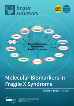
Fragile X syndrome (FXS) is a neurodevelopmental disorder characterized by a wide range of intellectual ability, autism and to various degrees of behavior and social difficulties. Over the past decades, significant advancements have been made in characterizing the molecular and cellular underpinnings of FXS, leading to a better diagnosis. Further, extensive research and preclinical studies have been conducted to develop molecular signatures for target drug development and pharmacologic treatments. However, the lack of feasible biomarkers limits our ability to monitor disease severity and to evaluate the clinical benefit of pharmaceutical interventions. This review highlights the efforts made in establishing potential molecular biomarkers in FXS by using in vivo and in vitro models and the promising stories of the ones that have proven their worth in current human clinical trials. View this paper
  • Issues are regarded as officially published after their release is announced to the table of contents alert mailing list.
  • You may sign up for e-mail alerts to receive table of contents of newly released issues.
  • PDF is the official format for papers published in both, html and pdf forms. To view the papers in pdf format, click on the "PDF Full-text" link, and use the free Adobe Reader to open them.
Order results
Result details
Section
Select all
Export citation of selected articles as:
17 pages, 7423 KiB  
Article
Feasibility of Non-Gaussian Diffusion Metrics in Chronic Disorders of Consciousness
by Elena I. Kremneva, Liudmila A. Legostaeva, Sofya N. Morozova, Dmitry V. Sergeev, Dmitry O. Sinitsyn, Elizaveta G. Iazeva, Aleksandr S. Suslin, Natalia A. Suponeva, Marina V. Krotenkova, Michael A. Piradov and Ivan I. Maximov
Brain Sci. 2019, 9(5), 123; https://doi.org/10.3390/brainsci9050123 - 27 May 2019
Cited by 6 | Viewed by 3992
Abstract
Diagnostic accuracy of different chronic disorders of consciousness (DOC) can be affected by the false negative errors in up to 40% cases. In the present study, we aimed to investigate the feasibility of a non-Gaussian diffusion approach in chronic DOC and to estimate [...] Read more.
Diagnostic accuracy of different chronic disorders of consciousness (DOC) can be affected by the false negative errors in up to 40% cases. In the present study, we aimed to investigate the feasibility of a non-Gaussian diffusion approach in chronic DOC and to estimate a sensitivity of diffusion kurtosis imaging (DKI) metrics for the differentiation of vegetative state/unresponsive wakefulness syndrome (VS/UWS) and minimally conscious state (MCS) from a healthy brain state. We acquired diffusion MRI data from 18 patients in chronic DOC (11 VS/UWS, 7 MCS) and 14 healthy controls. A quantitative comparison of the diffusion metrics for grey (GM) and white (WM) matter between the controls and patient group showed a significant (p < 0.05) difference in supratentorial WM and GM for all evaluated diffusion metrics, as well as for brainstem, corpus callosum, and thalamus. An intra-subject VS/UWS and MCS group comparison showed only kurtosis metrics and fractional anisotropy differences using tract-based spatial statistics, owing mainly to macrostructural differences on most severely lesioned hemispheres. As a result, we demonstrated an ability of DKI metrics to localise and detect changes in both WM and GM and showed their capability in order to distinguish patients with a different level of consciousness. Full article
(This article belongs to the Section Neurotechnology and Neuroimaging)
Show Figures

Figure 1

13 pages, 3847 KiB  
Article
Characterizing Fatigue-Related White Matter Changes in MS: A Proton Magnetic Resonance Spectroscopy Study
by Kalyan Yarraguntla, Fen Bao, Samuel Lichtman-Mikol, Sara Razmjou, Carla Santiago-Martinez, Navid Seraji-Bozorgzad, Shitiz Sriwastava and Evanthia Bernitsas
Brain Sci. 2019, 9(5), 122; https://doi.org/10.3390/brainsci9050122 - 27 May 2019
Cited by 8 | Viewed by 3675
Abstract
Few cross-sectional studies have investigated the correlation between neurochemical changes and multiple sclerosis (MS) fatigue, but little is known on the fatigue-related white matter differences between time points. We aim to investigate the longitudinal neurometabolite profile of white matter in MS fatigue. Forty-eight [...] Read more.
Few cross-sectional studies have investigated the correlation between neurochemical changes and multiple sclerosis (MS) fatigue, but little is known on the fatigue-related white matter differences between time points. We aim to investigate the longitudinal neurometabolite profile of white matter in MS fatigue. Forty-eight relapsing remitting multiple sclerosis (RRMS) patients with an expanded disability status scale (EDSS) ≤ 4 underwent high field 1H-multivoxel magnetic resonance spectroscopy (MRS) at baseline and year 1. Fatigue severity was evaluated by the fatigue severity scale (FSS). Patients were divided into low (LF, FSS ≤ 3), moderate (MF, FSS = 3.1–5), and high fatigue (HF, FSS ≥ 5.1) groups. In a two-way analysis of variance (ANOVA), we observed a decline in the ratio of the sum of N-acetylaspartate (NAA) and N-acetylaspartylglutamate (NAAG) to the sum of creatine (Cr) and phosphocreatine (PCr) in the right anterior quadrant (RAQ) and left anterior quadrant (LAQ) of the MRS grid in the HF group at baseline and year 1. This decline was significant when compared with the LF group (p = 0.018 and 0.020). In a one-way ANOVA, the fatigue group effect was significant and the ratio difference in the right posterior quadrant (RPQ) and left posterior quadrant (LPQ) of the HF group was also significant (p = 0.012 and 0.04). Neurochemical changes in the bilateral frontal white matter and possibly parietooccipital areas were noted in the HF group at two different time points. Our findings may shed some light on the pathology of MS fatigue. Full article
(This article belongs to the Special Issue Fatigue in Multiple Sclerosis: Current Concepts and Future Challenges)
Show Figures

Figure 1

8 pages, 772 KiB  
Communication
Modulating Observation-Execution-Related Motor Cortex Activity by Cathodal Transcranial Direct Current Stimulation
by Fengxue Qi, Michael A. Nitsche and Volker R. Zschorlich
Brain Sci. 2019, 9(5), 121; https://doi.org/10.3390/brainsci9050121 - 26 May 2019
Cited by 6 | Viewed by 3714
Abstract
The aim of this randomized sham-controlled study was to examine the impact of cathodal transcranial direct current stimulation (ctDCS) of the primary motor cortex (M1) during movement observation on subsequent execution-related motor cortex activity. Thirty healthy participants received sham or real ctDCS (1 [...] Read more.
The aim of this randomized sham-controlled study was to examine the impact of cathodal transcranial direct current stimulation (ctDCS) of the primary motor cortex (M1) during movement observation on subsequent execution-related motor cortex activity. Thirty healthy participants received sham or real ctDCS (1 mA) over the left M1 for 10 minutes, respectively. The participants observed a video showing repeated button pressing tasks of the right hand during the sham or real ctDCS, followed by performance of these tasks by the right hand. Motor-evoked potentials (MEP) were recorded from the resting right first dorsal interosseous muscle before movement observation during the sham or real ctDCS, immediately after observation of actions, and after subsequent movement execution. The results of the ANOVA showed a significant main effect on the group (F1,28 = 4.60, p = 0.041) and a significant interaction between time and the group (F2,56 = 5.34, p = 0.008). As revealed by respective post hoc tests, ctDCS induced a significant reduction of MEP amplitudes in connection with movement observation (p = 0.026, Cohen’s d = 0.861) and after subsequent movement execution (p = 0.018, Cohen’s d = 0.914) in comparison with the sham stimulation. It is concluded that ctDCS during movement observation was effective in terms of modulating motor cortex excitability. Moreover, it subsequently influenced execution-related motor cortex activity. This indicates a possible application for rehabilitative treatment in syndromes with pathologically enhanced cortical activity. Full article
Show Figures

Figure 1

11 pages, 741 KiB  
Article
Omega-3 Long-Chain Polyunsaturated Fatty Acids Intake in Children with Attention Deficit and Hyperactivity Disorder
by Milagros Fuentes-Albero, María Isabel Martínez-Martínez and Omar Cauli
Brain Sci. 2019, 9(5), 120; https://doi.org/10.3390/brainsci9050120 - 23 May 2019
Cited by 19 | Viewed by 8119
Abstract
Omega-3 long-chain polyunsaturated fatty acids (LC-PUFA) play a central role in neuronal growth and in the development of the human brain, and a deficiency of these substances has been reported in children with attention deficit hyperactive disorder (ADHD). In this regard, supplementation with [...] Read more.
Omega-3 long-chain polyunsaturated fatty acids (LC-PUFA) play a central role in neuronal growth and in the development of the human brain, and a deficiency of these substances has been reported in children with attention deficit hyperactive disorder (ADHD). In this regard, supplementation with omega-3 polyunsaturated fatty acids is used as adjuvant therapy in ADHD. Seafood, particularly fish, and some types of nuts are the main dietary sources of such fatty acids in the Spanish diet. In order to assess the effect of the intake of common foods containing high amounts of omega-3 polyunsaturated fatty acids, a food frequency questionnaire was administered to parents of children with ADHD (N = 48) and to parents of normally developing children (control group) (N = 87), and the intake of dietary omega-3 LC-PUFA, such as eicosapentaenoic acid (EPA) and docosahexaenoic acid (DHA), was estimated. Children with ADHD consumed fatty fish, lean fish, mollusks, crustaceans, and chicken eggs significantly less often (p < 0.05) than children in the control group. The estimated daily omega-3 LC-PUFA intake (EPA + DHA) was significantly below that recommended by the public health agencies in both groups, and was significantly lower in children with ADHD (p < 0.05, Cohen’s d = 0.45) compared to normally developing children. Dietary intervention to increase the consumption of fish and seafood is strongly advised and it is especially warranted in children with ADHD, since it could contribute to improve the symptoms of ADHD. Full article
Show Figures

Figure 1

20 pages, 1328 KiB  
Article
Target Size Manipulations Affect Error-Processing Duration and Success Perceptions but not Behavioural Indices of Learning
by Nicole T. Ong, Jamie Hawke and Nicola J. Hodges
Brain Sci. 2019, 9(5), 119; https://doi.org/10.3390/brainsci9050119 - 23 May 2019
Cited by 11 | Viewed by 3385
Abstract
We evaluated if and how success perceptions, through target size manipulations, impact processes related to motor learning. This work was based on recent literature suggesting that expectations and self-efficacy exert a direct impact on learning. We measured arousal, kinematics, learner expectancies, motivation, and [...] Read more.
We evaluated if and how success perceptions, through target size manipulations, impact processes related to motor learning. This work was based on recent literature suggesting that expectations and self-efficacy exert a direct impact on learning. We measured arousal, kinematics, learner expectancies, motivation, and outcomes in a dart-throwing task. Novices (n = 29) were assigned to either a “Large-target” (horizontal target, 10-cm high) or “Small-target” (2-cm high) group for practice (t = 90), and both groups completed 24-h retention tests. The Small-target group took longer to plan and process feedback in the pre-throw and post-throw periods, respectively, and showed larger joint amplitudes early in practice compared to the Large-target group. As predicted, the Large-target group made more hits and had heightened outcome expectancies compared to the Small-target group. Surprisingly, only the Large-target group performed better than they expected. Despite the Large-target group having more target hits, enhanced expectancies, and more unexpected success, this group did not outperform the Small-target group on behavioural indices of performance and learning. This research questions assumptions and results related to success-related manipulations for task performance and mechanisms related to target size manipulations. Full article
Show Figures

Figure 1

19 pages, 2024 KiB  
Article
Impaired Fear Extinction Recall in Serotonin Transporter Knockout Rats Is Transiently Alleviated during Adolescence
by Pieter Schipper, Paola Brivio, David de Leest, Leonie Madder, Beenish Asrar, Federica Rebuglio, Michel M. M. Verheij, Tamas Kozicz, Marco A. Riva, Francesca Calabrese, Marloes J. A. G. Henckens and Judith R. Homberg
Brain Sci. 2019, 9(5), 118; https://doi.org/10.3390/brainsci9050118 - 22 May 2019
Cited by 11 | Viewed by 4599
Abstract
Adolescence is a developmental phase characterized by emotional turmoil and coincides with the emergence of affective disorders. Inherited serotonin transporter (5-HTT) downregulation in humans increases sensitivity to these disorders. To reveal whether and how 5-HTT gene variance affects fear-driven behavior in adolescence, we [...] Read more.
Adolescence is a developmental phase characterized by emotional turmoil and coincides with the emergence of affective disorders. Inherited serotonin transporter (5-HTT) downregulation in humans increases sensitivity to these disorders. To reveal whether and how 5-HTT gene variance affects fear-driven behavior in adolescence, we tested wildtype and serotonin transporter knockout (5-HTT−/−) rats of preadolescent, adolescent, and adult age for cued fear extinction and extinction recall. To analyze neural circuit function, we quantified inhibitory synaptic contacts and, through RT-PCR, the expression of c-Fos, brain-derived neurotrophic factor (BDNF), and NDMA receptor subunits, in the medial prefrontal cortex (mPFC) and amygdala. Remarkably, the impaired recall of conditioned fear that characterizes preadolescent and adult 5-HTT−/− rats was transiently normalized during adolescence. This did not relate to altered inhibitory neurotransmission, since mPFC inhibitory immunoreactivity was reduced in 5-HTT−/− rats across all ages and unaffected in the amygdala. Rather, since mPFC (but not amygdala) c-Fos expression and NMDA receptor subunit 1 expression were reduced in 5-HTT−/− rats during adolescence, and since PFC c-Fos correlated negatively with fear extinction recall, the temporary normalization of fear extinction during adolescence could relate to altered plasticity in the developing mPFC. Full article
Show Figures

Figure 1

15 pages, 1679 KiB  
Article
Stability of Estimated Premorbid Cognitive Ability over Time after Minor Stroke and Its Relationship with Post-Stroke Cognitive Ability
by Caroline A. McHutchison, Francesca M. Chappell, Stephen Makin, Kirsten Shuler, Joanna M. Wardlaw and Vera Cvoro
Brain Sci. 2019, 9(5), 117; https://doi.org/10.3390/brainsci9050117 - 22 May 2019
Cited by 9 | Viewed by 4500
Abstract
Considering premorbid or “peak” adult intelligence (IQ) is important when examining post-stroke cognition. The stability of estimated premorbid IQ and its relationship to current cognitive ability in stroke is unknown. We investigated changes in estimated premorbid IQ and current cognitive ability up to [...] Read more.
Considering premorbid or “peak” adult intelligence (IQ) is important when examining post-stroke cognition. The stability of estimated premorbid IQ and its relationship to current cognitive ability in stroke is unknown. We investigated changes in estimated premorbid IQ and current cognitive ability up to three years post-stroke. Minor stroke patients (NIHSS < 8) were assessed at one to three months, one and three years’ post-stroke. The National Adult Reading Test (NART) and Addenbrooke’s Cognitive Examination-Revised (ACE-R) were used to estimate premorbid IQ (NART IQ) and current cognitive ability respectively at each time-point. Baseline demographics, vascular and stroke characteristics were included. Of the 264 patients recruited (mean age 66), 158 (60%), 151 (57%), and 153 (58%) completed cognitive testing at each time-point respectively. NART IQ initially increased (mean difference (MD) = 1.32, 95% CI = 0.54 to 2.13, p < 0.001) before decreasing (MD = −4.269, 95% CI = −5.12 to −3.41, p < 0.001). ACE-R scores initially remained stable (MD = 0.29, 95% CI = −0.49 to 1.07, p > 0.05) before decreasing (MD = −1.05, 95% CI = −2.08 to −0.01, p < 0.05). Adjusting for baseline variables did not change the relationship between NART IQ and ACE-R with time. Increases in NART IQ were associated with more education. For ACE-R, older age was associated with declines, and higher NART IQ and more education was associated with increases. Across 3 years, we observed fluctuations in estimated premorbid IQ and minor changes in current cognitive ability. Future research should aim to identify variables associated with these changes. However, studies of post-stroke cognition should account for premorbid IQ. Full article
Show Figures

Figure 1

20 pages, 2768 KiB  
Article
Joint Modulation of Facial Expression Processing by Contextual Congruency and Task Demands
by Luis Aguado, Karisa B. Parkington, Teresa Dieguez-Risco, José A. Hinojosa and Roxane J. Itier
Brain Sci. 2019, 9(5), 116; https://doi.org/10.3390/brainsci9050116 - 17 May 2019
Cited by 22 | Viewed by 4819
Abstract
Faces showing expressions of happiness or anger were presented together with sentences that described happiness-inducing or anger-inducing situations. Two main variables were manipulated: (i) congruency between contexts and expressions (congruent/incongruent) and (ii) the task assigned to the participant, discriminating the emotion shown by [...] Read more.
Faces showing expressions of happiness or anger were presented together with sentences that described happiness-inducing or anger-inducing situations. Two main variables were manipulated: (i) congruency between contexts and expressions (congruent/incongruent) and (ii) the task assigned to the participant, discriminating the emotion shown by the target face (emotion task) or judging whether the expression shown by the face was congruent or not with the context (congruency task). Behavioral and electrophysiological results (event-related potentials (ERP)) showed that processing facial expressions was jointly influenced by congruency and task demands. ERP results revealed task effects at frontal sites, with larger positive amplitudes between 250–450 ms in the congruency task, reflecting the higher cognitive effort required by this task. Effects of congruency appeared at latencies and locations corresponding to the early posterior negativity (EPN) and late positive potential (LPP) components that have previously been found to be sensitive to emotion and affective congruency. The magnitude and spatial distribution of the congruency effects varied depending on the task and the target expression. These results are discussed in terms of the modulatory role of context on facial expression processing and the different mechanisms underlying the processing of expressions of positive and negative emotions. Full article
(This article belongs to the Special Issue Perceptual and Affective Mechanisms in Facial Expression Recognition)
Show Figures

Figure 1

16 pages, 2416 KiB  
Article
Epilepsy Detection by Using Scalogram Based Convolutional Neural Network from EEG Signals
by Ömer Türk and Mehmet Siraç Özerdem
Brain Sci. 2019, 9(5), 115; https://doi.org/10.3390/brainsci9050115 - 17 May 2019
Cited by 125 | Viewed by 9860
Abstract
The studies implemented with Electroencephalogram (EEG) signals are progressing very rapidly and brain computer interfaces (BCI) and disease determinations are carried out at certain success rates thanks to new methods developed in this field. The effective use of these signals, especially in disease [...] Read more.
The studies implemented with Electroencephalogram (EEG) signals are progressing very rapidly and brain computer interfaces (BCI) and disease determinations are carried out at certain success rates thanks to new methods developed in this field. The effective use of these signals, especially in disease detection, is very important in terms of both time and cost. Currently, in general, EEG studies are used in addition to conventional methods as well as deep learning networks that have recently achieved great success. The most important reason for this is that in conventional methods, increasing classification accuracy is based on too many human efforts as EEG is being processed, obtaining the features is the most important step. This stage is based on both the time-consuming and the investigation of many feature methods. Therefore, there is a need for methods that do not require human effort in this area and can learn the features themselves. Based on that, two-dimensional (2D) frequency-time scalograms were obtained in this study by applying Continuous Wavelet Transform to EEG records containing five different classes. Convolutional Neural Network structure was used to learn the properties of these scalogram images and the classification performance of the structure was compared with the studies in the literature. In order to compare the performance of the proposed method, the data set of the University of Bonn was used. The data set consists of five EEG records containing healthy and epilepsy disease which are labeled as A, B, C, D, and E. In the study, A-E and B-E data sets were classified as 99.50%, A-D and B-D data sets were classified as 100% in binary classifications, A-D-E data sets were 99.00% in triple classification, A-C-D-E data sets were 90.50%, B-C-D-E data sets were 91.50% in quaternary classification, and A-B-C-D-E data sets were in the fifth class classification with an accuracy of 93.60%. Full article
(This article belongs to the Special Issue Advances in EEG/ MEG Source Imaging )
Show Figures

Figure 1

16 pages, 1107 KiB  
Article
Evaluation of Postnatal Sedation in Full-Term Infants
by Jean Carmela Solodiuk, Russell William Jennings and Dusica Bajic
Brain Sci. 2019, 9(5), 114; https://doi.org/10.3390/brainsci9050114 - 17 May 2019
Cited by 14 | Viewed by 4891
Abstract
Prolonged sedation in infants leads to a high incidence of physical dependence. We inquired: (1) “How long does it take to develop physical dependence to sedation in previously naïve full-term infants without known history of neurologic impairment?” and (2) “What is the relationship [...] Read more.
Prolonged sedation in infants leads to a high incidence of physical dependence. We inquired: (1) “How long does it take to develop physical dependence to sedation in previously naïve full-term infants without known history of neurologic impairment?” and (2) “What is the relationship between length of sedation to length of weaning and hospital stay?”. The retrospective study included full-term patients over a period of one year that were <1 year of age and received opioids and benzodiazepines >72 hours. Quantification of fentanyl, morphine, and midazolam were compared among three time periods: <5 days, 5–30 days, and >30 days using t-test or one-way analysis of variance. Identified full-term infants were categorized into surgical (14/44) or medical (10/44) groups, while those with neurological involvement (20/44) were excluded. Physical dependence in full-term infants occurred following sedation ≥5 days. Infants with surgical disease received escalating doses of morphine and midazolam when administered >30 days. A positive association between length of sedation and weaning period was found for both respiratory (p < 0.01) and surgical disease (p = 0.012) groups, while length of sedation is related to hospital stay for the respiratory (p < 0.01) but not the surgical disease group (p = 0.1). Future pharmacological directions should lead to standardized sedation protocols and evaluate patient neurocognitive outcomes. Full article
(This article belongs to the Special Issue Mechanisms Underlying Alleviation of Pain)
Show Figures

Figure 1

16 pages, 2517 KiB  
Review
Loss of Prefrontal Cortical Higher Cognition with Uncontrollable Stress: Molecular Mechanisms, Changes with Age, and Relevance to Treatment
by Dibyadeep Datta and Amy F. T. Arnsten
Brain Sci. 2019, 9(5), 113; https://doi.org/10.3390/brainsci9050113 - 17 May 2019
Cited by 33 | Viewed by 10208
Abstract
The newly evolved prefrontal cortex (PFC) generates goals for “top-down” control of behavior, thought, and emotion. However, these circuits are especially vulnerable to uncontrollable stress, with powerful, intracellular mechanisms that rapidly take the PFC “off-line.” High levels of norepinephrine and dopamine released during [...] Read more.
The newly evolved prefrontal cortex (PFC) generates goals for “top-down” control of behavior, thought, and emotion. However, these circuits are especially vulnerable to uncontrollable stress, with powerful, intracellular mechanisms that rapidly take the PFC “off-line.” High levels of norepinephrine and dopamine released during stress engage α1-AR and D1R, which activate feedforward calcium-cAMP signaling pathways that open nearby potassium channels to weaken connectivity and reduce PFC cell firing. Sustained weakening with chronic stress leads to atrophy of dendrites and spines. Understanding these signaling events helps to explain the increased susceptibility of the PFC to stress pathology during adolescence, when dopamine expression is increased in the PFC, and with advanced age, when the molecular “brakes” on stress signaling are diminished by loss of phosphodiesterases. These mechanisms have also led to pharmacological treatments for stress-related disorders, including guanfacine treatment of childhood trauma, and prazosin treatment of veterans and civilians with post-traumatic stress disorder. Full article
Show Figures

Graphical abstract

9 pages, 452 KiB  
Perspective
The Endocannabinoid System as a Potential Mechanism through which Exercise Influences Episodic Memory Function
by Paul D. Loprinzi, Liye Zou and Hong Li
Brain Sci. 2019, 9(5), 112; https://doi.org/10.3390/brainsci9050112 - 16 May 2019
Cited by 13 | Viewed by 5788
Abstract
Emerging research demonstrates that exercise, including both acute and chronic exercise, may influence episodic memory function. To date, mechanistic explanations of this effect are often attributed to alterations in long-term potentiation, neurotrophic production, angiogenesis, and neurogenesis. Herein, we discuss a complementary mechanistic model, [...] Read more.
Emerging research demonstrates that exercise, including both acute and chronic exercise, may influence episodic memory function. To date, mechanistic explanations of this effect are often attributed to alterations in long-term potentiation, neurotrophic production, angiogenesis, and neurogenesis. Herein, we discuss a complementary mechanistic model, suggesting that the endocannabinoid system may, in part, influence the effects of exercise on memory function. We discuss the role of the endocannabinoid system on memory function as well as the effects of exercise on endocannabinoid alterations. This is an exciting line of inquiry that should help delineate new insights into the mechanistic role of exercise on memory function. Full article
(This article belongs to the Special Issue Cannabis: Neuropsychiatry and Its Effects on Brain and Behavior)
Show Figures

Figure 1

12 pages, 3410 KiB  
Article
Pro-Inflammatory Activation of a New Immortalized Human Microglia Cell Line
by Marta Chiavari, Gabriella Maria Pia Ciotti, Pierluigi Navarra and Lucia Lisi
Brain Sci. 2019, 9(5), 111; https://doi.org/10.3390/brainsci9050111 - 15 May 2019
Cited by 20 | Viewed by 5653
Abstract
The characterization of human microglia has been hampered by poor availability of human cell sources. However, microglia is involved in the physiopathology of multiple sclerosis, Alzheimer’s disease, Parkinson’s disease, HIV dementia, retinal degenerative diseases, cancer, and many other conditions. Therefore, there is an [...] Read more.
The characterization of human microglia has been hampered by poor availability of human cell sources. However, microglia is involved in the physiopathology of multiple sclerosis, Alzheimer’s disease, Parkinson’s disease, HIV dementia, retinal degenerative diseases, cancer, and many other conditions. Therefore, there is an important need to have experimental paradigms of human microglia characterized and usable to study the role of microglia in the different pathologies in which it is involved. In the present work, we carried out an extensive characterization of Immortalized Human Microglia—SV40 cell line (IMhu), marketed by Applied Biological Material. The functional response of IMhu to a large variety of stimuli was studied. In particular, we investigated morphology, mortality, and changes in the production of different cytokines and chemokines, both under basal conditions and after stimulation. Moreover, western blotting analysis was conducted on phospho-mTOR (Ser 2448) and downstream parameters, p-P70S6K and 4EBP1, in order to understand if IMhu can be used for evaluations of mTOR pathway. In conclusion, IMhu cells proved to be a useful experimental model to investigate the physiopathology of inflammatory disease that involved microglia cells, including pathological conditions that involved the mTOR pathway. Full article
(This article belongs to the Section Neuroglia)
Show Figures

Figure 1

32 pages, 4299 KiB  
Article
The Neural Correlates of Conflict Detection and Resolution During Multiword Lexical Selection: Evidence from Bilinguals and Monolinguals
by Manuel F. Pulido and Paola E. Dussias
Brain Sci. 2019, 9(5), 110; https://doi.org/10.3390/brainsci9050110 - 14 May 2019
Cited by 2 | Viewed by 4561
Abstract
Previous studies have identified the Event Related Potential (ERP) components of conflict detection and resolution mechanisms in tasks requiring lexical selection at the individual word level. We investigated the brain potentials associated with these mechanisms in a lexical selection task based on multiword [...] Read more.
Previous studies have identified the Event Related Potential (ERP) components of conflict detection and resolution mechanisms in tasks requiring lexical selection at the individual word level. We investigated the brain potentials associated with these mechanisms in a lexical selection task based on multiword units made up of verb–noun combinations (e.g., eat breakfast, skip school). Native and non-native English speakers were asked to select a familiarized target verb–noun sequence (eat breakfast) between two choices. Trials were low-conflict, with only one plausible candidate (e.g., eat – shoot – breakfast) or high-conflict, with two plausible verbs (e.g., eat – skip – breakfast). Following the presentation of the noun, native English speakers showed a biphasic process of selection, with a conflict-detection centro-parietal negativity between 500 and 600 ms (Ninc), followed by a right frontal effect (RFE) between 600 and 800 ms preceding responses. Late Spanish–English bilinguals showed a similar but more sustained and more widespread effect. Additionally, brain activity was only significantly correlated with performance in native speakers. Results suggest largely similar basic mechanisms, but also that different resources and strategies are engaged by non-native speakers when resolving conflict in the weaker language, with a greater focus on individual words than on multiword units. Full article
(This article belongs to the Special Issue Cognitive Neuroscience of Cross-Language Interaction in Bilinguals)
Show Figures

Figure 1

21 pages, 1308 KiB  
Article
A Laboratory Word Memory Test Analogue Differentiates Intentional Feigning from True Responding Using the P300 Event-Related Potential
by John F. Shelley-Tremblay, Joshua C. Eyer and Benjamin D. Hill
Brain Sci. 2019, 9(5), 109; https://doi.org/10.3390/brainsci9050109 - 14 May 2019
Cited by 7 | Viewed by 4782
Abstract
Symptom exaggeration and feigned cognitive impairment occur commonly in forensic and medicolegal evaluations. As a result, methods to detect feigned cognitive impairment are an indispensable component of neuropsychological assessments. This study reports the results of two neurophysiological experiments using a forced-choice recognition task [...] Read more.
Symptom exaggeration and feigned cognitive impairment occur commonly in forensic and medicolegal evaluations. As a result, methods to detect feigned cognitive impairment are an indispensable component of neuropsychological assessments. This study reports the results of two neurophysiological experiments using a forced-choice recognition task built from the stimuli of the Word Memory Test and Medical Symptom Validity Test as well as a new linguistically informed stimulus set. Participant volunteers were instructed either to do their best or to feign cognitive impairment consistent with a mild traumatic brain injury while their brain activity was monitored using event-related potentials (ERP). Experiment 1 varied instructions across individuals, whereas Experiment 2 varied instructions within individuals. The target brain component was a positive deflection indicating stimulus recognition that occurs approximately 300 ms after exposure to a stimulus (i.e., the P300). Multimodal comparison (P300 amplitude to behavioral accuracy) allowed the detection of feigned cognitive impairment. Results indicate that, for correct responses, P300s were equivalent for the simulated malingering and good effort conditions. However, for incorrect responses, feigned impairment produced reliable but significantly reduced P300 amplitudes. Although the P300 is an automatic index of recognition—even when knowledge is hidden—its amplitude appears capable of modulation by feigning strategies. Implications of this finding are discussed for research and clinical applications. Full article
(This article belongs to the Special Issue Collection on Cognitive Neuroscience)
Show Figures

Figure 1

16 pages, 3984 KiB  
Article
Effects of GLP-1 Receptor Activation on a Pentylenetetrazole—Kindling Rat Model
by Abdelaziz M. Hussein, Mohamed Eldosoky, Mohamed El-Shafey, Mohamed El-Mesery, Khaled M. Abbas, Amr N. Ali, Ghada M. Helal and Osama A. Abulseoud
Brain Sci. 2019, 9(5), 108; https://doi.org/10.3390/brainsci9050108 - 14 May 2019
Cited by 19 | Viewed by 4206
Abstract
Objectives: To study the possible anti-seizure and neuroprotective effect of glucagon like peptide 1 (GLP1) analogue (liraglutide) in a pentylenetetrazole (PTZ) induced kindled rat model and its underlying mechanisms. Methods: Thirty Sprague Dawley rats were allocated into 3 equal groups; i) [...] Read more.
Objectives: To study the possible anti-seizure and neuroprotective effect of glucagon like peptide 1 (GLP1) analogue (liraglutide) in a pentylenetetrazole (PTZ) induced kindled rat model and its underlying mechanisms. Methods: Thirty Sprague Dawley rats were allocated into 3 equal groups; i) Normal group: normal rats received normal saline, ii) PTZ (kindling) group: received PTZ (50 mg/Kg intraperitoneally (i.p.)) every other day for 2 weeks and iii) PTZ + GLP1 group: same as the PTZ group but rats received liraglutide (75 µg/kg i.p. daily) for 2 weeks before PTZ injection. Seizure severity score, seizure latency and duration were assessed. Also, the expression of caspase-3 (apoptotic marker) and β-catenin (Wnt pathway) by western blotting, markers of oxidative stress (GSH, CAT and MDA) by biochemical assay and the expression of LC3 (marker of autophagy) and heat shock protein 70 (Hsp70) by immunostaining were assessed in hippocampal regions of brain tissues. Results: PTZ caused a significant increase in Racine score and seizure duration with a significant decrease in seizure latency. These effects were associated with a significant increase in MDA, β-catenin, caspase-3, Hsp70 and LC3 in brain tissues (p < 0.05). Meanwhile, liraglutide treatment caused significant attenuation in PTZ-induced seizures, which were associated with significant improvement in markers of oxidative stress, reduction in LC3, caspase-3 and β-catenin and marked increase in Hsp70 in hippocampal regions (p < 0.05). Conclusion: Activation of GLP1R might have anticonvulsant and neuroprotective effects against PTZ-induced epilepsy. These effects could be due to suppression of oxidative stress, apoptosis and autophagy and upregulation of Hsp70. Full article
(This article belongs to the Special Issue Collection on Clinical Neuroscience)
Show Figures

Figure 1

27 pages, 3450 KiB  
Review
I Am Conscious, Therefore, I Am: Imagery, Affect, Action, and a General Theory of Behavior
by David F. Marks
Brain Sci. 2019, 9(5), 107; https://doi.org/10.3390/brainsci9050107 - 10 May 2019
Cited by 15 | Viewed by 8276
Abstract
Organisms are adapted to each other and the environment because there is an inbuilt striving toward security, stability, and equilibrium. A General Theory of Behavior connects imagery, affect, and action with the central executive system we call consciousness, a direct emergent property of [...] Read more.
Organisms are adapted to each other and the environment because there is an inbuilt striving toward security, stability, and equilibrium. A General Theory of Behavior connects imagery, affect, and action with the central executive system we call consciousness, a direct emergent property of cerebral activity. The General Theory is founded on the assumption that the primary motivation of all of consciousness and intentional behavior is psychological homeostasis. Psychological homeostasis is as important to the organization of mind and behavior as physiological homeostasis is to the organization of bodily systems. Consciousness processes quasi-perceptual images independently of the input to the retina and sensorium. Consciousness is the “I am” control center for integration and regulation of (my) thoughts, (my) feelings, and (my) actions with (my) conscious mental imagery as foundation stones. The fundamental, universal conscious desire for psychological homeostasis benefits from the degree of vividness of inner imagery. Imagery vividness, a combination of clarity and liveliness, is beneficial to imagining, remembering, thinking, predicting, planning, and acting. Assessment of vividness using introspective report is validated by objective means such as functional magnetic resonance imaging (fMRI). A significant body of work shows that vividness of visual imagery is determined by the similarity of neural responses in imagery to those occurring in perception of actual objects and performance of activities. I am conscious; therefore, I am. Full article
Show Figures

Graphical abstract

18 pages, 4009 KiB  
Article
Can Evaluative Conditioning Change Well-Established Attitudes Towards Popular Brands? Your Brain Says Yes Even Though Your Mouth Says No
by Shannon Bosshard, Monika Koller and Peter Walla
Brain Sci. 2019, 9(5), 106; https://doi.org/10.3390/brainsci9050106 - 10 May 2019
Cited by 6 | Viewed by 4991
Abstract
In the present study, using both implicit and explicit measures, we addressed the issue of whether strongly developed relationships towards brands could be modified through the use of evaluative conditioning. Using an online survey, individual participant brand lists were created, and formed the [...] Read more.
In the present study, using both implicit and explicit measures, we addressed the issue of whether strongly developed relationships towards brands could be modified through the use of evaluative conditioning. Using an online survey, individual participant brand lists were created, and formed the basis of this experiment. Participants were then exposed to conditioning during a longitudinal study. Throughout the experiment, a combination of explicit and implicit measures was used to assess changes in attitude. Specifically, participants were asked to rate the brand names on a Likert-type scale. Simultaneously, changes in the brains electrical activity in response to the brands were recorded via electroencephalography (EEG). Upon completion of this task, participants underwent two Implicit Association Tests (IAT; one for liked brands and one for disliked brands). There were two main findings of this study. Firstly, no significant changes in attitude were observed via the use of explicit measures, and those that were found relating to the IAT were regarded as questionable. Secondly, EEG presented consistent results which showed that conditioning elicited changes in cortical activity towards both liked and disliked brands, which suggest it may be a useful tool in measuring the impact of evaluative conditioning that is not reflected in verbal responses. Full article
(This article belongs to the Special Issue Collection on Cognitive Neuroscience)
Show Figures

Figure 1

7 pages, 1206 KiB  
Case Report
Benign Giant Cell Lesion of C1 Lateral Mass: A Case Report and Literature Review
by Christopher Heinrich, Vadim Gospodarev, Albert Kheradpour, Craig Zuppan, Clifford C. Douglas and Tanya Minasian
Brain Sci. 2019, 9(5), 105; https://doi.org/10.3390/brainsci9050105 - 08 May 2019
Cited by 4 | Viewed by 5704
Abstract
Primary osseous tumors of the spinal column account for approximately 1% of the total number of spinal tumors found in the pediatric patient population. The authors present a case of a C1 benign giant cell lesion that was incidentally found in a 15-year-old [...] Read more.
Primary osseous tumors of the spinal column account for approximately 1% of the total number of spinal tumors found in the pediatric patient population. The authors present a case of a C1 benign giant cell lesion that was incidentally found in a 15-year-old patient. A transoral biopsy was performed followed by treatment with denosumab, with definitive management in the form of transoral tumor resection with subsequent occiput-cervical three posterior instrumented fusion. The patient tolerated all of the procedures well, as there were no post-operative complications, discharged home neurologically intact and was eager to return to school when assessed during a follow-up visit in clinic. Osteolytic lesions affecting the cervical spine are rare in the pediatric population. It is of utmost importance to have sufficient background knowledge in order to formulate a differential diagnosis, as well as an understanding of principles underlying surgical techniques required to prevent occipital-cervical instability in this patient population. The information presented will guide surgical decision-making by identifying the patient population that would benefit from neurosurgical interventions to stabilize the atlantoaxial junction, in the context of rare osteolytic conditions affecting the cervical spine. Full article
(This article belongs to the Special Issue Surgery for Spine Disease and Intractable Pain)
Show Figures

Figure 1

9 pages, 261 KiB  
Perspective
What Role Does the Prefrontal Cortex Play in the Processing of Negative and Positive Stimuli in Adolescent Depression?
by Siyabend Kaya and Ciara McCabe
Brain Sci. 2019, 9(5), 104; https://doi.org/10.3390/brainsci9050104 - 07 May 2019
Cited by 11 | Viewed by 5881
Abstract
This perspective describes the contribution of the prefrontal cortex to the symptoms of depression in adolescents and specifically the processing of positive and negative information. We also discuss how the prefrontal cortex (PFC) activity and connectivity during tasks and at rest might be [...] Read more.
This perspective describes the contribution of the prefrontal cortex to the symptoms of depression in adolescents and specifically the processing of positive and negative information. We also discuss how the prefrontal cortex (PFC) activity and connectivity during tasks and at rest might be a biomarker for risk for depression onset in adolescents. We include some of our recent work examining not only the anticipation and consummation of positive and negative stimuli, but also effort to gain positive and avoid negative stimuli in adolescents with depression. We find, using region of interest analyses, that the PFC is blunted in those with depression compared to controls across the different phases but in a larger sample the PFC is blunted in the anticipatory phase of the study only. Taken together, in adolescents with depression there is evidence for dysfunctional PFC activity across different studies and tasks. However, the data are limited with small sample sizes and inconsistent findings. Larger longitudinal studies with more detailed assessments of symptoms across the spectrum are needed to further evaluate the role of the PFC in adolescent depression. Full article
14 pages, 1614 KiB  
Article
Stimulation of the Angular Gyrus Improves the Level of Consciousness
by Liudmila Legostaeva, Alexandra Poydasheva, Elizaveta Iazeva, Dmitry Sinitsyn, Dmitry Sergeev, Ilya Bakulin, Dmitry Lagoda, Elena Kremneva, Sofya Morozova, Yulia Ryabinkina, Natalia Suponeva and Michael Piradov
Brain Sci. 2019, 9(5), 103; https://doi.org/10.3390/brainsci9050103 - 06 May 2019
Cited by 24 | Viewed by 5256
Abstract
Background: Navigated repetitive transcranial magnetic stimulation (rTMS) is a promising tool for neuromodulation. In previous studies it has been shown that the activity of the default mode network (DMN) areas, particularly of its key region—the angular gyrus—is positively correlated with the level of [...] Read more.
Background: Navigated repetitive transcranial magnetic stimulation (rTMS) is a promising tool for neuromodulation. In previous studies it has been shown that the activity of the default mode network (DMN) areas, particularly of its key region—the angular gyrus—is positively correlated with the level of consciousness. Our study aimed to explore the effect of rTMS of the angular gyrus as a new approach for disorders of consciousness (DOC) treatment; Methods: A 10-session 2-week high-frequency rTMS protocol was delivered over the left angular gyrus in 38 DOC patients with repeated neurobehavioral assessments obtained at baseline and in 2 days after the stimulation course was complete; Results: 20 Hz-rTMS over left angular gyrus improved the coma recovery scale revised (CRS-R) total score in minimally conscious state (MCS) patients. We observed no effects in vegetative state (VS) patients; and Conclusions: The left angular gyrus is likely to be effective target for rTMS in patients with present signs of consciousness. Full article
(This article belongs to the Special Issue Advances in Disorders of Consciousness)
Show Figures

Figure 1

14 pages, 2237 KiB  
Article
Superior Effects of Modified Chen-Style Tai Chi versus 24-Style Tai Chi on Cognitive Function, Fitness, and Balance Performance in Adults over 55
by Liye Zou, Paul D. Loprinzi, Jane Jie Yu, Lin Yang, Chunxiao Li, Albert S. Yeung, Zhaowei Kong, Shin-Yi Chiou and Tao Xiao
Brain Sci. 2019, 9(5), 102; https://doi.org/10.3390/brainsci9050102 - 04 May 2019
Cited by 32 | Viewed by 7032
Abstract
Background: Cognitive decline and balance impairment are prevalent in the aging population. Previous studies investigated the beneficial effects of 24-style Tai Chi (TC-24) on either cognitive function or balance performance of older adults. It still remains largely unknown whether modified Chen-style TC (MTC) [...] Read more.
Background: Cognitive decline and balance impairment are prevalent in the aging population. Previous studies investigated the beneficial effects of 24-style Tai Chi (TC-24) on either cognitive function or balance performance of older adults. It still remains largely unknown whether modified Chen-style TC (MTC) that includes 18 complex movements is more beneficial for these age-related health outcomes, as compared to TC-24. Objective: We investigated if MTC would show greater effects than TC-24 on global cognitive function and balance-related outcomes among older adults. Methods: We conducted a randomized trial where 80 eligible adults aged over 55 were allocated into two different styles of Tai Chi (TC) arms (sixty-minute session × three times per week, 12 weeks). Outcome assessments were performed at three time periods (baseline, Week 6, and Week 12) and included the Chinese Version of the Montreal Cognitive Assessment (MoCA) for overall cognitive function, One-leg Standing Test (LST) for static balance, Timed Up and Go Test (TUGT) for dynamic balance, chair Stand Test (CST) for leg power, and the six-meter Walk Test (6MWT) for aerobic exercise capacity. Results: Compared to TC-24 arm, MTC arm demonstrated significantly greater improvements in MoCA, LST, TUGT, CST, and 6MWT (all p < 0.05). Conclusions: Both forms of TC were effective in enhancing global cognitive function, balance, and fitness. Furthermore, MTC was more effective than TC-24 in enhancing these health-related parameters in an aging population. Full article
(This article belongs to the Special Issue Exercising against Age-Effects on the Brain)
Show Figures

Figure 1

10 pages, 2255 KiB  
Article
Preserved Cerebral Oxygen Metabolism in Astrocytic Dysfunction: A Combination Study of 15O-Gas PET with 14C-Acetate Autoradiography
by Carla Mari Macaisa, Tadashi Watabe, Yuwei Liu, Victor Romanov, Yasukazu Kanai, Genki Horitsugi, Hiroki Kato, Eku Shimosegawa and Jun Hatazawa
Brain Sci. 2019, 9(5), 101; https://doi.org/10.3390/brainsci9050101 - 03 May 2019
Cited by 3 | Viewed by 3795
Abstract
Fluorocitrate (FC) is a specific metabolic inhibitor of the tricarboxylic acid (TCA) cycle in astrocytes. The purpose of this study was to evaluate whether inhibition of the astrocyte TCA cycle by FC would affect the oxygen metabolism in the rat brain. At 4 [...] Read more.
Fluorocitrate (FC) is a specific metabolic inhibitor of the tricarboxylic acid (TCA) cycle in astrocytes. The purpose of this study was to evaluate whether inhibition of the astrocyte TCA cycle by FC would affect the oxygen metabolism in the rat brain. At 4 h after the intracranial FC injection, the rats (n = 9) were investigated by 15O-labeled gas PET to measure the cerebral blood flow (CBF), the cerebral metabolic rate of oxygen (CMRO2), oxygen extraction fraction (OEF), and cerebral blood volume (CBV). After the 15O-gas PET, the rats were given an intravenous injection of 14C-acetate for autoradiography. 15O-gas PET showed no significant differences in any of the measured parameters between the ipsilateral and contralateral striatum (high dose group: CBF (54.4 ± 8.8 and 55.3 ± 11.6 mL/100 mL/min), CMRO2 (7.0 ± 0.9 and 7.1 ± 1.2 mL/100 mL/min), OEF (72.0 ± 8.9 and 70.8 ± 8.2%), and CBV (4.1 ± 0.8 and 4.2 ± 0.9 mL/100 mL), respectively). In contrast, the 14C-acetate autoradiography revealed a significant inhibition of the astrocyte metabolism in the ipsilateral striatum. The regional cerebral oxygen consumption as well as the hemodynamic parameters were maintained even in the face of inhibition of the astrocyte TCA cycle metabolism in the rat brain. Full article
Show Figures

Figure 1

23 pages, 671 KiB  
Review
Cognitive Fatigue in Multiple Sclerosis: An Objective Approach to Diagnosis and Treatment by Transcranial Electrical Stimulation
by Stefanie Linnhoff, Marina Fiene, Hans-Jochen Heinze and Tino Zaehle
Brain Sci. 2019, 9(5), 100; https://doi.org/10.3390/brainsci9050100 - 02 May 2019
Cited by 50 | Viewed by 9321
Abstract
Cognitive fatigue is one of the most frequent symptoms in multiple sclerosis (MS), associated with significant impairment in daily functioning and quality of life. Despite its clinical significance, progress in understanding and treating fatigue is still limited. This limitation is already caused by [...] Read more.
Cognitive fatigue is one of the most frequent symptoms in multiple sclerosis (MS), associated with significant impairment in daily functioning and quality of life. Despite its clinical significance, progress in understanding and treating fatigue is still limited. This limitation is already caused by an inconsistent and heterogeneous terminology and assessment of fatigue. In this review, we integrate previous literature on fatigue and propose a unified schema aiming to clarify the fatigue taxonomy. With special focus on cognitive fatigue, we survey the significance of objective behavioral and electrophysiological fatigue parameters and discuss the controversial literature on the relationship between subjective and objective fatigue assessment. As MS-related cognitive fatigue drastically affects quality of life, the development of efficient therapeutic approaches for overcoming cognitive fatigue is of high clinical relevance. In this regard, the reliable and valid assessment of the individual fatigue level by objective parameters is essential for systematic treatment evaluation and optimization. Transcranial electrical stimulation (tES) may offer a unique opportunity to manipulate maladaptive neural activity underlying MS fatigue. Therefore, we discuss evidence for the therapeutic potential of tES on cognitive fatigue in people with MS. Full article
(This article belongs to the Special Issue Fatigue in Multiple Sclerosis: Current Concepts and Future Challenges)
Show Figures

Figure 1

10 pages, 779 KiB  
Article
Magnitude of Reduction and Speed of Remission of Suicidality for Low Amplitude Seizure Therapy (LAP-ST) Compared to Standard Right Unilateral Electroconvulsive Therapy: A Pilot Double-Blinded Randomized Clinical Trial
by Nagy A. Youssef, Dheeraj Ravilla, Cherishma Patel, Mark Yassa, Ramses Sadek, Li Fang Zhang, Laryssa McCloud, William V. McCall and Peter B. Rosenquist
Brain Sci. 2019, 9(5), 99; https://doi.org/10.3390/brainsci9050099 - 29 Apr 2019
Cited by 9 | Viewed by 6997
Abstract
Background: Although treatment guidelines support use of electroconvulsive therapy (ECT) for acute suicidality, it is associated with cognitive side effects. The effect of Low Amplitude Seizure Therapy (LAP-ST) on suicidality is unknown. Our prior precision LAP-ST (pLAP-ST) performing titrating in the current [...] Read more.
Background: Although treatment guidelines support use of electroconvulsive therapy (ECT) for acute suicidality, it is associated with cognitive side effects. The effect of Low Amplitude Seizure Therapy (LAP-ST) on suicidality is unknown. Our prior precision LAP-ST (pLAP-ST) performing titrating in the current domain has provided initial proof of concept data in humans of its advantage in terms of reduction of cognitive side effects. The aims of this report are to: 1) compare LAP-ST (at 500mA) versus standard Right Unilateral (RUL) ECT (at 900 mA) in terms of magnitude of remission of suicidality in a randomized allocation and 2) compare the speed of remission of suicidality between LAP-ST versus RUL ECT. Methods: Patients were randomized to either LAP-ST or RUL ECT. The scores pertaining to the suicidal ideation (SI) item on the Montgomery-Åsberg Depression Rating Scale (MADRS) were analyzed using descriptive analysis and no confirmatory statistical analysis was performed due to a priori sample size limitations for this pilot study. SI item remission was defined as 2 or below on this item. Results: Eleven patients with major depressive episode (MDE) of mainly unipolar or bipolar disorders signed consent. Of these, 7 were eligible and were randomized and included in the analysis; all were actively suicidal at baseline (suicide item above 2), except 1 patient who had suicide item at 2 in the RUL ECT group. Suicidality remitted on average by session 3 and remission occurred for all patients by session 4. The SI mean score improvement from baseline to endpoint for LAP-ST was 5.1 and for RUL ECT was 3.0. Conclusions: LAP-ST has larger effect size and speed of remission of suicidality compared to standard RUL ECT. Future studies are warranted for replicating these findings. (ClinicalTrials.gov ID: NCT02583490). Full article
Show Figures

Figure 1

11 pages, 882 KiB  
Article
Anodal Transcranial Direct Current Stimulation over the Vertex Enhances Leg Motor Cortex Excitability Bilaterally
by Soumya Ghosh, David Hathorn, Jennifer Eisenhauer, Jesse Dixon and Ian D. Cooper
Brain Sci. 2019, 9(5), 98; https://doi.org/10.3390/brainsci9050098 - 29 Apr 2019
Cited by 5 | Viewed by 4507
Abstract
In many studies, anodal transcranial Direct Current Stimulation (tDCS) is applied near the vertex to simultaneously facilitate leg motor cortex (M1) of both hemispheres and enhance recovery of gait and balance in neurological disorders. However, its effect on the excitability of leg M1 [...] Read more.
In many studies, anodal transcranial Direct Current Stimulation (tDCS) is applied near the vertex to simultaneously facilitate leg motor cortex (M1) of both hemispheres and enhance recovery of gait and balance in neurological disorders. However, its effect on the excitability of leg M1 in either hemisphere is not well known. In this double-blind sham-controlled study, corticospinal excitability changes induced in leg M1 of both hemispheres by anodal (2 mA for 20 minutes) or sham tDCS (for 20 min) over the vertex were evaluated. Peak amplitudes of Transcranial Magnetic Stimulation (TMS) induced motor evoked potentials (MEPs) were measured over the contralateral Tibialis Anterior (TA) muscle before and up to 40 min after tDCS in 11 normal participants. Analysis of data from all participants found significant overall increase in the excitability of leg M1 after tDCS. However, in individual subjects there was variability in observed effects. In 4 participants, 20 min of tDCS increased mean MEPs of TAs on both sides; in 4 participants there was increased mean MEP only on one side and in 3 subjects there was no change. It’s not known if the benefits of tDCS in improving gait and balance are dependent on excitability changes induced in one or both leg M1; such information may be useful to predict treatment outcomes. Full article
(This article belongs to the Special Issue Collection on Clinical Neuroscience)
Show Figures

Figure 1

12 pages, 239 KiB  
Perspective
Cognitive Decline Secondary to Therapeutic Brain Radiation—Similarities and Differences to Traumatic Brain Injury
by Andrew Jonathan Huang, David Kornguth and Steven Kornguth
Brain Sci. 2019, 9(5), 97; https://doi.org/10.3390/brainsci9050097 - 27 Apr 2019
Cited by 6 | Viewed by 4556
Abstract
Traumatic brain injury (TBI) resulting from forceful impacts on the torso and head has been of major interest because of the prevalence of such injuries in military personnel, contact sports and the elderly. Cognitive and behavioral changes associated with TBI are also seen [...] Read more.
Traumatic brain injury (TBI) resulting from forceful impacts on the torso and head has been of major interest because of the prevalence of such injuries in military personnel, contact sports and the elderly. Cognitive and behavioral changes associated with TBI are also seen following whole brain radiation treatment for cancer and chemotherapy for disseminated tumors. The biological mechanisms involved in the initiation of TBI from impact, radiation, and chemotherapy to loss of cognitive function have several shared characteristics including increases in blood brain barrier permeability, blood vessel density, increases in inflammatory and autoimmune responses, alterations in NMDA and glutamate receptor levels and release of proteins normally sequestered in the brain into the blood and spinal fluid. The development of therapeutic agents that mitigate the loss of cognition and development of behavioral disorders in patients experiencing radiation-induced injury may provide benefit to those with TBI when similar processes are involved on a cellular or molecular level. Increased collaborative efforts between the radiation oncology and the neurology and psychiatry communities may be of major benefit for the management of brain injury from varied environmental insults. Full article
23 pages, 1116 KiB  
Review
Molecular Biomarkers in Fragile X Syndrome
by Marwa Zafarullah and Flora Tassone
Brain Sci. 2019, 9(5), 96; https://doi.org/10.3390/brainsci9050096 - 27 Apr 2019
Cited by 22 | Viewed by 9357
Abstract
Fragile X syndrome (FXS) is the most common inherited form of intellectual disability (ID) and a known monogenic cause of autism spectrum disorder (ASD). It is a trinucleotide repeat disorder, in which more than 200 CGG repeats in the 5’ untranslated region (UTR) [...] Read more.
Fragile X syndrome (FXS) is the most common inherited form of intellectual disability (ID) and a known monogenic cause of autism spectrum disorder (ASD). It is a trinucleotide repeat disorder, in which more than 200 CGG repeats in the 5’ untranslated region (UTR) of the fragile X mental retardation 1 (FMR1) gene causes methylation of the promoter with consequent silencing of the gene, ultimately leading to the loss of the encoded fragile X mental retardation 1 protein, FMRP. FMRP is an RNA binding protein that plays a primary role as a repressor of translation of various mRNAs, many of which are involved in the maintenance and development of neuronal synaptic function and plasticity. In addition to intellectual disability, patients with FXS face several behavioral challenges, including anxiety, hyperactivity, seizures, repetitive behavior, and problems with executive and language performance. Currently, there is no cure or approved medication for the treatment of the underlying causes of FXS, but in the past few years, our knowledge about the proteins and pathways that are dysregulated by the loss of FMRP has increased, leading to clinical trials and to the path of developing molecular biomarkers for identifying potential targets for therapies. In this paper, we review candidate molecular biomarkers that have been identified in preclinical studies in the FXS mouse animal model and are now under validation for human applications or have already made their way to clinical trials. Full article
(This article belongs to the Special Issue Towards Mechanism-based Treatments for Fragile X Syndrome)
Show Figures

Graphical abstract

23 pages, 1586 KiB  
Article
Cognitive Control Facilitates Attentional Disengagement during Second Language Comprehension
by Christian A. Navarro-Torres, Dalia L. Garcia, Vrinda Chidambaram and Judith F. Kroll
Brain Sci. 2019, 9(5), 95; https://doi.org/10.3390/brainsci9050095 - 27 Apr 2019
Cited by 23 | Viewed by 5269
Abstract
Bilinguals learn to resolve conflict between their two languages and that skill has been hypothesized to create long-term adaptive changes in cognitive functioning. Yet, little is known about how bilinguals recruit cognitive control to enable efficient use of one of their languages, especially [...] Read more.
Bilinguals learn to resolve conflict between their two languages and that skill has been hypothesized to create long-term adaptive changes in cognitive functioning. Yet, little is known about how bilinguals recruit cognitive control to enable efficient use of one of their languages, especially in the less skilled and more effortful second language (L2). Here we examined how real-time cognitive control engagement influences L2 sentence comprehension (i.e., conflict adaptation). We tested a group of English monolinguals and a group of L2 English speakers using a recently-developed cross-task adaptation paradigm. Stroop sequences were pseudo-randomly interleaved with a visual-world paradigm in which participants were asked to carry out spoken instructions that were either syntactically ambiguous or unambiguous. Consistent with previous research, eye-movement results showed that Stroop-related conflict improved the ability to engage correct-goal interpretations, and disengage incorrect-goal interpretations, during ambiguous instructions. Such cognitive-to-language modulations were similar in both groups, but only in the engagement piece. In the disengagement portion, the modulation emerged earlier in bilinguals than in monolinguals, suggesting group differences in attentional disengagement following cognitive control recruitment. Additionally, incorrect-goal eye-movements were modulated by individual differences in working memory, although differently for each group, suggesting an involvement of both language-specific and domain-general resources. Full article
(This article belongs to the Special Issue Cognitive Neuroscience of Cross-Language Interaction in Bilinguals)
Show Figures

Figure 1

Previous Issue
Next Issue
Back to TopTop