Next Issue
Volume 9, May
Previous Issue
Volume 9, March


Brain Sci., Volume 9, Issue 4 (April 2019) – 22 articles

Cover Story (view full-size image): 
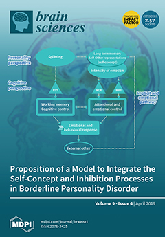
Borderline Personality Disorder (BPD) has been the subject of extensive research, particularly its symptom of impulsivity, which is considered a key component of neurobehavioral models of BPD and often leads to severe negative consequences for the person. Impulsivity and the measurements used to assess it have greatly evolved over time. Recently, the study of inhibition processes with behavioral tasks has highlighted some cognitive and affective deficits in this population. However, the literature presents important inconsistencies which raise questions about the potential role played by personality processes such as the self-concept. We investigated this question via a systematic review and our results lead us to propose a new theoretical model which integrates inhibition processes and the self-concept in order to explain the occurrence of borderline impulsive behavior. View this paper.
  • Issues are regarded as officially published after their release is announced to the table of contents alert mailing list.
  • You may sign up for e-mail alerts to receive table of contents of newly released issues.
  • PDF is the official format for papers published in both, html and pdf forms. To view the papers in pdf format, click on the "PDF Full-text" link, and use the free Adobe Reader to open them.
Order results
Result details
Section
Select all
Export citation of selected articles as:
9 pages, 1601 KiB  
Case Report
Wireless Computer-Supported Cooperative Work: A Pilot Experiment on Art and Brain–Computer Interfaces
by Gabriel G. De la Torre, Sara Gonzalez-Torre, Carlos Muñoz and Manuel A. Garcia
Brain Sci. 2019, 9(4), 94; https://doi.org/10.3390/brainsci9040094 - 25 Apr 2019
Cited by 1 | Viewed by 3719
Abstract
The present case study looked into the feasibility of using brain–computer interface (BCI) technology combined with computer-supported cooperative work (CSCW) in a wireless network. We had two objectives; first, to test the wireless BCI-based configuration and the practical use of this idea we [...] Read more.
The present case study looked into the feasibility of using brain–computer interface (BCI) technology combined with computer-supported cooperative work (CSCW) in a wireless network. We had two objectives; first, to test the wireless BCI-based configuration and the practical use of this idea we assessed workload perception in participants located several kilometers apart taking part in the same drawing task. Second, we studied the cortical activation patterns of participants performing the drawing task with and without the BCI technology. Results showed higher mental workload perception and broader cortical activation (frontal-temporal-occipital) under BCI experimental conditions. This idea shows a possible application of BCI research in the social field, where two or more users could engage in a computer networking task using BCI technology over the internet. New research avenues for CSCW are discussed and possibilities for future research are given. Full article
(This article belongs to the Special Issue Collection on Neural Engineering)
Show Figures

Figure 1

17 pages, 732 KiB  
Article
Descriptive Psychopathology of the Acute Effects of Intravenous Delta-9-Tetrahydrocannabinol Administration in Humans
by Marco Colizzi, Nathalie Weltens, Philip McGuire, Lukas Van Oudenhove and Sagnik Bhattacharyya
Brain Sci. 2019, 9(4), 93; https://doi.org/10.3390/brainsci9040093 - 25 Apr 2019
Cited by 17 | Viewed by 4368
Abstract
Background: Cannabis use can increase the risk of psychosis, and the acute administration of its key psychoactive ingredient, delta-9-tetrahydrocannabinol (∆9-THC), can induce transient psychotomimetic symptoms. Methods: A double-blind, randomized, placebo-controlled crossover design was used to investigate the symptomatic effects of acute intravenous administration [...] Read more.
Background: Cannabis use can increase the risk of psychosis, and the acute administration of its key psychoactive ingredient, delta-9-tetrahydrocannabinol (∆9-THC), can induce transient psychotomimetic symptoms. Methods: A double-blind, randomized, placebo-controlled crossover design was used to investigate the symptomatic effects of acute intravenous administration of ∆9-THC (1.19 mg/2 mL) in 16 healthy participants (seven males) with modest previous cannabis exposure. Results: In the 20 min following acute ∆9-THC administration, symptomatic effects of at least mild severity were present in 94% of the cohort, with moderate to severe symptoms having a much lower prevalence (19%). Nearly one-third (31%) of the volunteers were still experiencing protracted mild symptomatic effects 2.5 h after exposure to ∆9-THC. Compared to the Δ9-THC challenge, most of the study participants did not experience any symptomatic effects following placebo administration (62%). Acute physical reactions were 2.5 times more frequent after Δ9-THC (31%) than placebo (12%). Male and female participants differed in terms of acute Δ9-THC effects, with some negative symptoms occurring more frequently in female (56% to 89%) than male participants (0% to 29%), and acute physical reactions occurring exclusively in the female gender (56%). Conclusions: These results have implications for future research, also in light of cannabis being the most widely used illicit drug. Full article
(This article belongs to the Special Issue Cannabis: Neuropsychiatry and Its Effects on Brain and Behavior)
Show Figures

Figure 1

13 pages, 594 KiB  
Article
Relation of Serum Plasmalogens and APOE Genotype to Cognition and Dementia in Older Persons in a Cross-Sectional Study
by Dayan B. Goodenowe and Vijitha Senanayake
Brain Sci. 2019, 9(4), 92; https://doi.org/10.3390/brainsci9040092 - 24 Apr 2019
Cited by 14 | Viewed by 7175
Abstract
Using a community sample of 1205 elderly persons, we investigated the associations and potential interactions between Apolipoprotein E (APOE) genotype and serum phosphatidylethanolamine (PlsEtn) on cognition and dementia. For each person, APOE genotype, PlsEtn Biosynthesis value (PBV, the combination of three key [...] Read more.
Using a community sample of 1205 elderly persons, we investigated the associations and potential interactions between Apolipoprotein E (APOE) genotype and serum phosphatidylethanolamine (PlsEtn) on cognition and dementia. For each person, APOE genotype, PlsEtn Biosynthesis value (PBV, the combination of three key PlsEtn species), cognition (the combination of five specific cognitive domains), and diagnosis of dementia was determined. APOE genotype and PBV were observed to be non-interacting (p > 0.05) and independently associated with cognition: APOE (relative to ε3ε3:ε2ε3 (Coef = 0.14, p = 4.2 × 10⁻²); ε3ε4/ε4ε4 (Coef = −0.22, p = 6.2 × 10⁻⁵); PBV (Coef = 0.12, p = 1.7 × 10⁻⁷) and dementia: APOE (relative to ε3ε3:ε2ε3 (Odds Ratio OR = 0.44, p = 3.0 × 10⁻²); ε3ε4/ε4ε4 (OR = 2.1, p = 2.2 × 10⁻⁴)); PBV (OR = 0.61, p = 3.3 × 10⁻⁶). Associations are expressed per standard deviation (SD) and adjusted for serum lipids and demographics. Due to the independent and non-interacting nature of the APOE and PBV associations, the prevalence of dementia in APOE ε3ε4/ε4ε4 persons with high PBV values (>1 SD from mean) was observed to be the same as APOE ε3ε3 persons (14.3% versus 14.0%). Similarly, the prevalence of dementia in APOE ε3ε3 persons with high PBV values was only 5.7% versus 6.7% for APOE ε2ε3 persons. The results of these analyses indicate that the net effect of APOE genotype on cognition and the prevalence of dementia is dependent upon the plasmalogen status of the person. Full article
(This article belongs to the Special Issue Dementia and Cognitive Ageing)
Show Figures

Figure 1

24 pages, 2103 KiB  
Article
Music Training Positively Influences the Preattentive Perception of Voice Onset Time in Children with Dyslexia: A Longitudinal Study
by Aline Frey, Clément François, Julie Chobert, Jean-Luc Velay, Michel Habib and Mireille Besson
Brain Sci. 2019, 9(4), 91; https://doi.org/10.3390/brainsci9040091 - 21 Apr 2019
Cited by 24 | Viewed by 7243
Abstract
Previous results showed a positive influence of music training on linguistic abilities at both attentive and preattentive levels. Here, we investigate whether six months of active music training is more efficient than painting training to improve the preattentive processing of phonological parameters based [...] Read more.
Previous results showed a positive influence of music training on linguistic abilities at both attentive and preattentive levels. Here, we investigate whether six months of active music training is more efficient than painting training to improve the preattentive processing of phonological parameters based on durations that are often impaired in children with developmental dyslexia (DD). Results were also compared to a control group of Typically Developing (TD) children matched on reading age. We used a Test–Training–Retest procedure and analysed the Mismatch Negativity (MMN) and the N1 and N250 components of the Event-Related Potentials to syllables that differed in Voice Onset Time (VOT), vowel duration, and vowel frequency. Results were clear-cut in showing a normalization of the preattentive processing of VOT in children with DD after music training but not after painting training. They also revealed increased N250 amplitude to duration deviant stimuli in children with DD after music but not painting training, and no training effect on the preattentive processing of frequency. These findings are discussed in view of recent theories of dyslexia pointing to deficits in processing the temporal structure of speech. They clearly encourage the use of active music training for the rehabilitation of children with language impairments. Full article
(This article belongs to the Special Issue Advances in the Neurocognition of Music and Language)
Show Figures

Figure 1

14 pages, 879 KiB  
Review
Parallel Emergence of a Compartmentalized Striatum with the Phylogenetic Development of the Cerebral Cortex
by Tadashi Hamasaki and Satoshi Goto
Brain Sci. 2019, 9(4), 90; https://doi.org/10.3390/brainsci9040090 - 19 Apr 2019
Cited by 7 | Viewed by 5524
Abstract
The intricate neuronal architecture of the striatum plays a pivotal role in the functioning of the basal ganglia circuits involved in the control of various aspects of motor, cognitive, and emotional functions. Unlike the cerebral cortex, which has a laminar structure, the striatum [...] Read more.
The intricate neuronal architecture of the striatum plays a pivotal role in the functioning of the basal ganglia circuits involved in the control of various aspects of motor, cognitive, and emotional functions. Unlike the cerebral cortex, which has a laminar structure, the striatum is primarily composed of two functional subdivisions (i.e., the striosome and matrix compartments) arranged in a mosaic fashion. This review addresses whether striatal compartmentalization is present in non-mammalian vertebrates, in which simple cognitive and behavioral functions are executed by primitive sensori-motor systems. Studies show that neuronal subpopulations that share neurochemical and connective properties with striosomal and matrix neurons are present in the striata of not only anamniotes (fishes and amphibians), but also amniotes (reptiles and birds). However, these neurons do not form clearly segregated compartments in these vertebrates, suggesting that such compartmentalization is unique to mammals. In the ontogeny of the mammalian forebrain, the later-born matrix neurons disperse the early-born striosome neurons into clusters to form the compartments in tandem with the development of striatal afferents from the cortex. We propose that striatal compartmentalization in mammals emerged in parallel with the evolution of the cortex and possibly enhanced complex processing of sensory information and behavioral flexibility phylogenetically. Full article
(This article belongs to the Collection Collection on Developmental Neuroscience)
Show Figures

Figure 1

12 pages, 1632 KiB  
Article
Reliability of Fronto–Amygdala Coupling during Emotional Face Processing
by Camilla L Nord, Alan Gray, Oliver J Robinson and Jonathan P Roiser
Brain Sci. 2019, 9(4), 89; https://doi.org/10.3390/brainsci9040089 - 19 Apr 2019
Cited by 12 | Viewed by 6206
Abstract
One of the most exciting translational prospects for brain imaging research is the potential use of functional magnetic resonance imaging (fMRI) ‘biomarkers’ to predict an individual’s risk of developing a neuropsychiatric disorder or the likelihood of responding to a particular intervention. This proposal [...] Read more.
One of the most exciting translational prospects for brain imaging research is the potential use of functional magnetic resonance imaging (fMRI) ‘biomarkers’ to predict an individual’s risk of developing a neuropsychiatric disorder or the likelihood of responding to a particular intervention. This proposal depends critically on reliable measurements at the level of the individual. Several previous studies have reported relatively poor reliability of amygdala activation during emotional face processing, a key putative fMRI ‘biomarker’. However, the reliability of amygdala connectivity measures is much less well understood. Here, we assessed the reliability of task-modulated coupling between three seed regions (left and right amygdala and the subgenual anterior cingulate cortex) and the dorsomedial frontal/cingulate cortex (DMFC), measured using a psychophysiological interaction analysis in 29 healthy individuals scanned approximately two weeks apart. We performed two runs on each day of three different emotional face-processing tasks: emotion identification, emotion matching, and gender classification. We tested both between-day reliability and within-day (between-run) reliability. We found good-to-excellent within-subject reliability of amygdala–DMFC coupling, both between days (in two tasks), and within day (in one task). This suggests that disorder-relevant regional coupling may be sufficiently reliable to be used as a predictor of treatment response or clinical risk in future clinical studies. Full article
Show Figures

Figure 1

21 pages, 11907 KiB  
Article
Optimization of the Navigated TMS Mapping Algorithm for Accurate Estimation of Cortical Muscle Representation Characteristics
by Dmitry O. Sinitsyn, Andrey Yu. Chernyavskiy, Alexandra G. Poydasheva, Ilya S. Bakulin, Natalia A. Suponeva and Michael A. Piradov
Brain Sci. 2019, 9(4), 88; https://doi.org/10.3390/brainsci9040088 - 19 Apr 2019
Cited by 7 | Viewed by 3633
Abstract
Navigated transcranial magnetic stimulation (nTMS) mapping of cortical muscle representations allows noninvasive assessment of the state of a healthy or diseased motor system, and monitoring changes over time. These applications are hampered by the heterogeneity of existing mapping algorithms and the lack of [...] Read more.
Navigated transcranial magnetic stimulation (nTMS) mapping of cortical muscle representations allows noninvasive assessment of the state of a healthy or diseased motor system, and monitoring changes over time. These applications are hampered by the heterogeneity of existing mapping algorithms and the lack of detailed information about their accuracy. We aimed to find an optimal motor evoked potential (MEP) sampling scheme in the grid-based mapping algorithm in terms of the accuracy of muscle representation parameters. The abductor pollicis brevis (APB) muscles of eight healthy subjects were mapped three times on consecutive days using a seven-by-seven grid with ten stimuli per cell. The effect of the MEP variability on the parameter accuracy was assessed using bootstrapping. The accuracy of representation parameters increased with the number of stimuli without saturation up to at least ten stimuli per cell. The detailed sampling showed that the between-session representation area changes in the absence of interventions were significantly larger than the within-session fluctuations and thus could not be explained solely by the trial-to-trial variability of MEPs. The results demonstrate that the number of stimuli has no universally optimal value and must be chosen by balancing the accuracy requirements with the mapping time constraints in a given problem. Full article
(This article belongs to the Section Neurotechnology and Neuroimaging)
Show Figures

Figure 1

21 pages, 1051 KiB  
Review
The Temporal Effects of Acute Exercise on Episodic Memory Function: Systematic Review with Meta-Analysis
by Paul D. Loprinzi, Jeremiah Blough, Lindsay Crawford, Seungho Ryu, Liye Zou and Hong Li
Brain Sci. 2019, 9(4), 87; https://doi.org/10.3390/brainsci9040087 - 18 Apr 2019
Cited by 83 | Viewed by 10360
Abstract
Background: Accumulating research demonstrates that the timing of exercise plays an important role in influencing episodic memory. However, we have a limited understanding as to the factors that moderate this temporal effect. Thus, the purpose of this systematic review with meta-analysis was to [...] Read more.
Background: Accumulating research demonstrates that the timing of exercise plays an important role in influencing episodic memory. However, we have a limited understanding as to the factors that moderate this temporal effect. Thus, the purpose of this systematic review with meta-analysis was to evaluate the effects of study characteristics (e.g., exercise modality, intensity and duration of acute exercise) and participant attributes (e.g., age, sex) across each of the temporal periods of acute exercise on episodic memory (i.e., acute exercise occurring before memory encoding, and during memory encoding, early consolidation, and late consolidation). Methods: The following databases were used for our computerized searches: Embase/PubMed, Web of Science, Google Scholar, Sports Discus and PsychInfo. Studies were included if they: (1) Employed an experimental design with a comparison to a control group/visit, (2) included human participants, (3) evaluated exercise as the independent variable, (4) employed an acute bout of exercise (defined as a single bout of exercise), (5) evaluated episodic memory as the outcome variable (defined as the retrospective recall of information either in a spatial or temporal manner), and (6) provided sufficient data (e.g., mean, SD, and sample size) for a pooled effect size estimate. Results: In total, 25 articles met our inclusionary criteria and were meta-analyzed. Acute exercise occurring before memory encoding (d = 0.11, 95% CI: −0.01, 0.23, p = 0.08), during early memory consolidation (d = 0.47, 95% CI: 0.28, 0.67; p < 0.001) and during late memory consolidation (d = 1.05, 95% CI: 0.32, 1.78; p = 0.005) enhanced episodic memory function. Conversely, acute exercise occurring during memory encoding had a negative effect on episodic memory (d = −0.12, 95% CI: −0.22, −0.02; p = 0.02). Various study designs and participant characteristics moderated the temporal effects of acute exercise on episodic memory function. For example, vigorous-intensity acute exercise, and acute exercise among young adults, had greater effects when the acute bout of exercise occurred before memory encoding or during the early memory consolidation period. Conclusions: The timing of acute exercise plays an important role in the exercise-memory interaction. Various exercise- and participant-related characteristics moderate this temporal relationship. Full article
(This article belongs to the Special Issue Collection on Cognitive Neuroscience)
Show Figures

Figure 1

14 pages, 381 KiB  
Article
Psychiatric Disorders and Alcohol Consumption Among Low-Income African Americans:Gender Differences
by Sharon Cobb and Shervin Assari
Brain Sci. 2019, 9(4), 86; https://doi.org/10.3390/brainsci9040086 - 18 Apr 2019
Cited by 5 | Viewed by 4340
Abstract
Background: Although cooccurrence of nonsubstance use disorders (non-SUDs) and substance use is well-established in the literature, most of what we know in this regard is derived from studies that have recruited predominantly White sample populations. As a result, there is a gap in [...] Read more.
Background: Although cooccurrence of nonsubstance use disorders (non-SUDs) and substance use is well-established in the literature, most of what we know in this regard is derived from studies that have recruited predominantly White sample populations. As a result, there is a gap in knowledge on this link among low-income African Americans (AAs). There is also a need to understand how low-income AA men and women differ in these associations. Objective: To study whether there is an association between number of non-SUDs and amount of alcohol consumption by AA adults, and whether this association varies between AA men and women. Methods: This cross-sectional study recruited a nonrandom sample of 150 AA adults with non-SUDs (i.e., major depression, bipolar disorders, obsessive–compulsive disorder, paranoid disorder, panic disorder, posttraumatic stress disorder (PTSD), and schizoaffective disorder). The independent variable was the number of non-SUDs. The dependent variable was the amount of alcohol consumption. Age, socioeconomic status (educational attainment and household income), and self-rated health were covariates. Gender was the moderator. Linear regression models were used to analyze the data. Results: A higher number of non-SUDs was not associated with a higher amount of alcohol use in the pooled sample of AA adults. We, however, found a significant interaction between gender and number of non-SUDs on the amount of alcohol use, suggesting a stronger effect of non-SUDs on alcohol consumption in AA men than in AA women. Gender-stratified linear regression models showed a positive association between number of non-SUDs and amount of alcohol consumption in AA men but not in AA women. Conclusion: Non-SUDs impact alcohol use of AA men but not women. Future research should test whether AA men may have a higher tendency to turn to alcohol to regulate their emotions and cope with psychological pain due to multiple non-SUDs. The results also suggest that integration of services for SUDs and non-SUDs may be more relevant to provision of mental health services for AA men than AA women. Full article
Show Figures

Figure 1

14 pages, 604 KiB  
Perspective
Cognitive Function in Primary Sjögren’s Syndrome: A Systematic Review
by Ciro Manzo, Eva Martinez-Suarez, Melek Kechida, Marco Isetta and Jordi Serra-Mestres
Brain Sci. 2019, 9(4), 85; https://doi.org/10.3390/brainsci9040085 - 15 Apr 2019
Cited by 21 | Viewed by 8107
Abstract
Background: Cognitive disorders are reported to be common in patients with primary Sjogren’s syndrome (pSS). In some cases, they are the first clinical manifestation, preceding the diagnosis of pSS by two years on average. Aim: A systematic review was conducted to explore cognitive [...] Read more.
Background: Cognitive disorders are reported to be common in patients with primary Sjogren’s syndrome (pSS). In some cases, they are the first clinical manifestation, preceding the diagnosis of pSS by two years on average. Aim: A systematic review was conducted to explore cognitive impairment in pSS, with reference to diagnostic methods and their relationship with laboratory data and clinical manifestations. Materials and Methods: According to the PRISMA 2009 checklist, we carried out a comprehensive literature search in the three main bibliographic databases: MEDLINE, EMBASE, and PsycINFO (NICE HDAS interface). The following main search terms were used: primary Sjogren syndrome, neurological manifestations, fatigue, cognitive functions, psychiatric manifestations, mild cognitive impairment, dementia, and neurocognitive disorder. The search was made on 14 September, 2018. References from all selected studies were also examined. Inclusion criteria were: all studies and case-reports published in any language from 2002 that assessed the association of pSS (according to classification criteria proposed by the 2002 American/European collaborative group (AECG)) with all types of cognitive impairment (including dementia). Exclusion criteria were: reviews, abstracts, secondary Sjögren’s syndrome (SS), and all articles in which other classification criteria were used. Results: The initial search yielded 352 articles, of which 253 were excluded based on the title and abstract review. A total of 54 articles underwent a full-length review, and 32 articles were excluded. Data were extracted from 18 studies and three case-reports involving a total of 6196 participants. In most cases, cognitive dysfunction was a brain fog or a mild cognitive impairment (MCI). Occasionally, an autoimmune dementia was present. The relationship between pSS and degenerative dementias, such as Alzheimer’s disease (AD), was a controversial issue, even if some investigators hypothesized that pSS could be a risk factor. Several unmet needs were highlighted. First, some of the included studies had not reported the severity of pSS; hence, few correlations between disease severity and cognitive function were possible. Secondly, the evaluation of the pathogenetic role of comorbid diseases was often absent. The lack of information on the type of dementia represented a third critical point in the majority of the included studies. Conclusions: This systematic review confirmed that adequate studies on cognitive function in pSS are scarce, mostly performed on small-sized samples, and often conflicting. The routine assessment of cognitive function in patients with pSS seems advisable and it will help to elucidate some of the unmet needs highlighted by this review in future appropriately designed studies. Full article
(This article belongs to the Special Issue Collection on Cognitive Neuroscience)
Show Figures

Figure 1

11 pages, 1335 KiB  
Article
Olfactory Event-Related Potentials and Exhaled Organic Volatile Compounds: The Slow Link Between Olfactory Perception and Breath Metabolic Response. A Pilot Study on Phenylethyl Alcohol and Vaseline Oil
by Sara Invitto and Andrea Mazzatenta
Brain Sci. 2019, 9(4), 84; https://doi.org/10.3390/brainsci9040084 - 15 Apr 2019
Cited by 13 | Viewed by 4760
Abstract
Olfactory processing starts with the breath and elicits neuronal, metabolic and cortical responses. This process can be investigated centrally via the Olfactory Event-Related Potentials (OERPs) and peripherally via exhaled Volatile Organic Compounds (VOCs). Despite this, the relationship between OERPs (i.e., N1 and Late [...] Read more.
Olfactory processing starts with the breath and elicits neuronal, metabolic and cortical responses. This process can be investigated centrally via the Olfactory Event-Related Potentials (OERPs) and peripherally via exhaled Volatile Organic Compounds (VOCs). Despite this, the relationship between OERPs (i.e., N1 and Late Positive Component LPC) and exhaled VOCs has not been investigated enough. The aim of this research is to study OERPs and VOCs connection to two different stimuli: phenylethyl alcohol (PEA) and Vaseline Oil (VO). Fifteen healthy subjects performed a perceptual olfactory task with PEA as a smell target stimulus and VO as a neutral stimulus. The results suggest that OERPs and VOCs distributions follow the same amplitude trend and that PEA is highly arousing in both psychophysiological measures. PEA shows ampler and faster N1, a component related to the sensorial aspect of the stimulus. The N1 topographic localization is different between PEA and VO: PEA stimulus evokes greater N1 in the left centroparietal site. LPC, a component elicited by the perceptual characteristic of the stimulus, shows faster latency in the Frontal lobe and decreased amplitude in the Central and Parietal lobe elicited by the PEA smell. Moreover, the delayed time between the onset of N1-LPC and the onset of VOCs seems to be about 3 s. This delay could be identified as the internal metabolic time in which the odorous stimulus, once perceived at the cortical level, is metabolized and subsequently exhaled. Furthermore, the VO stimulus does not allocate the attentive, perceptive and metabolic resource as with PEA. Full article
(This article belongs to the Special Issue Collection on Cognitive Neuroscience)
Show Figures

Figure 1

11 pages, 4166 KiB  
Communication
Hypothesis: Astrocyte Foot Processes Detachment from the Neurovascular Unit in Female Diabetic Mice May Impair Modulation of Information Processing—Six Degrees of Separation
by Melvin R. Hayden
Brain Sci. 2019, 9(4), 83; https://doi.org/10.3390/brainsci9040083 - 14 Apr 2019
Cited by 14 | Viewed by 5097
Abstract
Astrocytes via their foot processes (ACfp) are specialized connecting cells, and they structurally connect the neurovascular unit (NVU) mural cells to neurons. Astrocytes provide homeostatic mechanisms for structural connections and provide communication between the NVU and regional neurons for functional hyperemia in regions [...] Read more.
Astrocytes via their foot processes (ACfp) are specialized connecting cells, and they structurally connect the neurovascular unit (NVU) mural cells to neurons. Astrocytes provide homeostatic mechanisms for structural connections and provide communication between the NVU and regional neurons for functional hyperemia in regions of increased neuronal activity (neurovascular coupling). Previously, our group has demonstrated a detachment, separation, and retraction of ACfp in diabetic db/db females (DBC). It was hypothesized that a loss of adherent ACfp/NVU could result in the known impaired cognition in DBC. Additionally hypothesized was that empagliflozin treatment could protect DBC ACfp/NVU remodeling. This study demonstrates a significant loss of ACfp/NVU numbers in DBC and a protection of this loss by empagliflozin treatment (DBE). The number of intact ACfp/NVU was 6.45 ± 1.1 in control heterozygous (CKC) vs. 1.88 ± 0.72 in DBC (p < 0.05) and 5.86 ± 0.88 in DBE vs. DBC (p < 0.05) by visually hand-counting the capillary NVUs (22 in CKC, 25 in DBC, and 22 in DBE). These findings suggest that empagliflozin provides neuroprotection via the prevention of ACfp separation in DBE as compared to diabetic DBC. Furthermore, a loss of ACfp/NVU numbers in DBC may correspond with a negative modulation of informational processing, and the protection of ACfp/NVU numbers could provide a protective modulation in DBE models. Full article
(This article belongs to the Section Neuroglia)
Show Figures

Graphical abstract

9 pages, 225 KiB  
Opinion
Noninvasive Brain Stimulation and Psychotherapy in Anxiety and Depressive Disorders: A Viewpoint
by Moussa A. Chalah and Samar S. Ayache
Brain Sci. 2019, 9(4), 82; https://doi.org/10.3390/brainsci9040082 - 14 Apr 2019
Cited by 32 | Viewed by 5190
Abstract
Among the most prevalent psychiatric conditions stand anxiety and depression. Psychotherapy and medications are considered effective treatments in these clinical settings. However, pharmacotherapy and psychotherapy (i.e., cognitive behavioral therapy (CBT)) administered in monotherapy or in a combined regimen do not result in satisfactory [...] Read more.
Among the most prevalent psychiatric conditions stand anxiety and depression. Psychotherapy and medications are considered effective treatments in these clinical settings. However, pharmacotherapy and psychotherapy (i.e., cognitive behavioral therapy (CBT)) administered in monotherapy or in a combined regimen do not result in satisfactory outcomes in all patients. Therefore, finding new treatments would be of great help. In the last three decades, noninvasive brain stimulation (NIBS) has emerged as a safe tool to improve several neuropsychiatric symptoms. The following work revisits the available reports that assessed the add-on value of NIBS techniques when combined to psychotherapy (CBT or related interventions) in mood and anxiety disorders. The available protocols targeted the prefrontal cortex, a region that was previously found to have an enhanced activity or functional connectivity after psychotherapeutic interventions. Promising yet scarce evidence exists on this matter. A discrepancy exists among the available reports regarding the type and duration of interventions, the patients’ clinical profiles, and the presence of a sham intervention. NIBS may have acted by enhancing psychotherapy effects on the top-down cognitive control of emotions. Combining both therapies may result in promising effects, but future large-scale trials are needed to judge the utility of this combination in psychiatric populations. Full article
(This article belongs to the Special Issue Collection on Clinical Neuroscience)
14 pages, 1223 KiB  
Article
EEG Window Length Evaluation for the Detection of Alzheimer’s Disease over Different Brain Regions
by Katerina D. Tzimourta, Nikolaos Giannakeas, Alexandros T. Tzallas, Loukas G. Astrakas, Theodora Afrantou, Panagiotis Ioannidis, Nikolaos Grigoriadis, Pantelis Angelidis, Dimitrios G. Tsalikakis and Markos G. Tsipouras
Brain Sci. 2019, 9(4), 81; https://doi.org/10.3390/brainsci9040081 - 14 Apr 2019
Cited by 39 | Viewed by 6041
Abstract
Alzheimer’s Disease (AD) is a neurogenerative disorder and the most common type of dementia with a rapidly increasing world prevalence. In this paper, the ability of several statistical and spectral features to detect AD from electroencephalographic (EEG) recordings is [...] Read more.
Alzheimer’s Disease (AD) is a neurogenerative disorder and the most common type of dementia with a rapidly increasing world prevalence. In this paper, the ability of several statistical and spectral features to detect AD from electroencephalographic (EEG) recordings is evaluated. For this purpose, clinical EEG recordings from 14 patients with AD (8 with mild AD and 6 with moderate AD) and 10 healthy, age-matched individuals are analyzed. The EEG signals are initially segmented in nonoverlapping epochs of different lengths ranging from 5 s to 12 s. Then, a group of statistical and spectral features calculated for each EEG rhythm (δ, θ, α, β, and γ) are extracted, forming the feature vector that trained and tested a Random Forests classifier. Six classification problems are addressed, including the discrimination from whole-brain dynamics and separately from specific brain regions in order to highlight any alterations of the cortical regions. The results indicated a high accuracy ranging from 88.79% to 96.78% for whole-brain classification. Also, the classification accuracy was higher at the posterior and central regions than at the frontal area and the right side of temporal lobe for all classification problems. Full article
(This article belongs to the Special Issue Collection on Clinical Neuroscience)
Show Figures

Figure 1

18 pages, 3799 KiB  
Article
Extracellular S100β Disrupts Bergman Glia Morphology and Synaptic Transmission in Cerebellar Purkinje Cells
by Olga S. Belozor, Dariya A. Yakovleva, Ilya V. Potapenko, Andrey N. Shuvaev, Marina V. Smolnikova, Alex Vasilev, Elena A. Pozhilenkova and Anton N. Shuvaev
Brain Sci. 2019, 9(4), 80; https://doi.org/10.3390/brainsci9040080 - 12 Apr 2019
Cited by 6 | Viewed by 5364
Abstract
Astrogliosis is a pathological process that affects the density, morphology, and function of astrocytes. It is a common feature of brain trauma, autoimmune diseases, and neurodegeneration including spinocerebellar ataxia type 1 (SCA1), a poorly understood neurodegenerative disease. S100β is a Ca2+ binding [...] Read more.
Astrogliosis is a pathological process that affects the density, morphology, and function of astrocytes. It is a common feature of brain trauma, autoimmune diseases, and neurodegeneration including spinocerebellar ataxia type 1 (SCA1), a poorly understood neurodegenerative disease. S100β is a Ca2+ binding protein. In SCA1, excessive excretion of S100β by reactive astrocytes and its uptake by Purkinje cells has been demonstrated previously. Under pathological conditions, excessive extracellular concentration of S100β stimulates the production of proinflammatory cytokines and induces apoptosis. We modeled astrogliosis by S100β injections into cerebellar cortex in mice. Injections of S100β led to significant changes in Bergmann glia (BG) cortical organization and affected their processes. S100β also changed morphology of the Purkinje cells (PCs), causing a significant reduction in the dendritic length. Moreover, the short-term synaptic plasticity and depolarization-induced suppression of synaptic transmission were disrupted after S100β injections. We speculate that these effects are the result of Ca2+-chelating properties of S100β protein. In summary, exogenous S100β induced astrogliosis in cerebellum could lead to neuronal dysfunction, which resembles a natural neurodegenerative process. We suggest that astrocytes play an essential role in SCA1 pathology, and that astrocytic S100β is an important contributor to this process. Full article
(This article belongs to the Section Neuroglia)
Show Figures

Graphical abstract

18 pages, 341 KiB  
Review
Sensorimotor Control in Dystonia
by Phillip Desrochers, Alexander Brunfeldt, Christos Sidiropoulos and Florian Kagerer
Brain Sci. 2019, 9(4), 79; https://doi.org/10.3390/brainsci9040079 - 11 Apr 2019
Cited by 20 | Viewed by 6928
Abstract
This is an overview of the sensorimotor impairments in dystonia, a syndrome characterized by sustained or intermittent aberrant movement patterns leading to abnormal movements and/or postures with or without a tremulous component. Dystonia can affect the entire body or specific body regions and [...] Read more.
This is an overview of the sensorimotor impairments in dystonia, a syndrome characterized by sustained or intermittent aberrant movement patterns leading to abnormal movements and/or postures with or without a tremulous component. Dystonia can affect the entire body or specific body regions and results from a plethora of etiologies, including subtle changes in gray and white matter in several brain regions. Research over the last 25 years addressing topics of sensorimotor control has shown functional sensorimotor impairments related to sensorimotor integration, timing, oculomotor and head control, as well as upper and lower limb control. In the context of efforts to update the classification of dystonia, sensorimotor research is highly relevant for a better understanding of the underlying pathology, and potential mechanisms contributing to global and regional dysfunction within the central nervous system. This overview of relevant research regarding sensorimotor control in humans with idiopathic dystonia attempts to frame the dysfunction with respect to what is known regarding motor control in patients and healthy individuals. We also highlight promising avenues for the future study of neuromotor control that may help to further elucidate dystonia etiology, pathology, and functional characteristics. Full article
(This article belongs to the Special Issue Collection on Clinical Neuroscience)
17 pages, 2446 KiB  
Article
High Cervical Spinal Cord Stimulation: A One Year Follow-Up Study on Motor and Non-Motor Functions in Parkinson’s Disease
by Paolo Mazzone, Fabio Viselli, Stefano Ferraina, Margherita Giamundo, Massimo Marano, Marco Paoloni, Francesco Masedu, Annamaria Capozzo and Eugenio Scarnati
Brain Sci. 2019, 9(4), 78; https://doi.org/10.3390/brainsci9040078 - 03 Apr 2019
Cited by 23 | Viewed by 5254
Abstract
Background: The present study investigated the effectiveness of stimulation applied at cervical levels on pain and Parkinson’s disease (PD) symptoms using either tonic or burst stimulation mode. Methods: Tonic high cervical spinal cord stimulation (T-HCSCS) was applied on six PD patients suffering from [...] Read more.
Background: The present study investigated the effectiveness of stimulation applied at cervical levels on pain and Parkinson’s disease (PD) symptoms using either tonic or burst stimulation mode. Methods: Tonic high cervical spinal cord stimulation (T-HCSCS) was applied on six PD patients suffering from low back pain and failed back surgery syndrome, while burst HCSCS (B-HCSCS) was applied in twelve PD patients to treat primarily motor deficits. Stimulation was applied percutaneously with quadripolar or octapolar electrodes. Clinical evaluation was assessed by the Unified Parkinson’s Disease Rating Scale (UPDRS) and the Hoehn and Yahr (H&Y) scale. Pain was evaluated by a visual analog scale. Evaluations of gait and of performance in a cognitive motor task were performed in some patients subjected to B-HCSCS. One patient who also suffered from severe autonomic cardiovascular dysfunction was investigated to evaluate the effectiveness of B-HCSCS on autonomic functions. Results: B-HCSCS was more effective and had more consistent effects than T-HCSCS in reducing pain. In addition, B-HCSCS improved UPDRS scores, including motor sub-items and tremor and H&Y score. Motor benefits appeared quickly after the beginning of B-HCSCS, in contrast to long latency improvements induced by T-HCSCS. A slight decrease of effectiveness was observed 12 months after implantation. B-HCSCS also improved gait and ability of patients to correctly perform a cognitive–motor task requiring inhibition of a prepared movement. Finally, B-HCSCS ameliorated autonomic control in the investigated patient. Conclusions: The results support a better usefulness of B-HCSCS compared to T-HCSCS in controlling pain and specific aspects of PD motor and non-motor deficits for at least one year. Full article
(This article belongs to the Section Systems Neuroscience)
Show Figures

Figure 1

23 pages, 1066 KiB  
Review
The Modulating Role of Self-Referential Stimuli and Processes in the Effect of Stress and Negative Emotion on Inhibition Processes in Borderline Personality Disorder: Proposition of a Model to Integrate the Self-Concept and Inhibition Processes
by Jean Gagnon, Joséphine Aldebert, Gasser Saleh and Wan Seo Kim
Brain Sci. 2019, 9(4), 77; https://doi.org/10.3390/brainsci9040077 - 30 Mar 2019
Cited by 4 | Viewed by 4868
Abstract
Impulsivity is an important clinical and diagnostic feature of borderline personality disorder (BPD). Even though it has been reported that BPD individuals’ inhibition performance is significantly reduced in the context of negative emotion or stress, this literature shows mixed results, raising questions about [...] Read more.
Impulsivity is an important clinical and diagnostic feature of borderline personality disorder (BPD). Even though it has been reported that BPD individuals’ inhibition performance is significantly reduced in the context of negative emotion or stress, this literature shows mixed results, raising questions about the possible role played by other factors. Winter (2016) proposed that negative emotion stimuli can be more disruptive for BPD individuals’ attention control performance because they induce higher distractibility self-referential processes. This article aimed to systematically review the literature regarding the effect of stress and negative emotions on three main inhibition processes—prepotent response inhibition, resistance to distractor interference, and resistance to proactive interference—in BPD and to verify the putative modulating role of self-referential stimuli and processes on these inhibition processes. All English and French experimental studies published until August 2018 were searched in PsychINFO and PubMED databases. The following keywords were used: “borderline* AND inhibit* OR interference* OR forget* OR task* AND emotion* OR stress* OR affect*”. A total of 1215 articles were included in the study. After full text revision, twenty-six papers were selected for review. The results of this review indicate that when stimuli or procedures involve self-reference stimuli or processes, BPD individuals’ performance seems to be more disrupted in all three inhibition processes. A model based on Winter’s and Kernberg’s models is proposed with the aim of integrating the self-concept with inhibition processes in BPD. Full article
Show Figures

Figure 1

20 pages, 823 KiB  
Review
Positive and Negative Emotion Regulation in Adolescence: Links to Anxiety and Depression
by Katherine S. Young, Christina F. Sandman and Michelle G. Craske
Brain Sci. 2019, 9(4), 76; https://doi.org/10.3390/brainsci9040076 - 29 Mar 2019
Cited by 212 | Viewed by 38755
Abstract
Emotion regulation skills develop substantially across adolescence, a period characterized by emotional challenges and developing regulatory neural circuitry. Adolescence is also a risk period for the new onset of anxiety and depressive disorders, psychopathologies which have long been associated with disruptions in regulation [...] Read more.
Emotion regulation skills develop substantially across adolescence, a period characterized by emotional challenges and developing regulatory neural circuitry. Adolescence is also a risk period for the new onset of anxiety and depressive disorders, psychopathologies which have long been associated with disruptions in regulation of positive and negative emotions. This paper reviews the current understanding of the role of disrupted emotion regulation in adolescent anxiety and depression, describing findings from self-report, behavioral, peripheral psychophysiological, and neural measures. Self-report studies robustly identified associations between emotion dysregulation and adolescent anxiety and depression. Findings from behavioral and psychophysiological studies are mixed, with some suggestion of specific impairments in reappraisal in anxiety. Results from neuroimaging studies broadly implicate altered functioning of amygdala-prefrontal cortical circuitries, although again, findings are mixed regarding specific patterns of altered neural functioning. Future work may benefit from focusing on designs that contrast effects of specific regulatory strategies, and isolate changes in emotional regulation from emotional reactivity. Approaches to improve treatments based on empirical evidence of disrupted emotion regulation in adolescents are also discussed. Future intervention studies might consider training and measurement of specific strategies in adolescents to better understand the role of emotion regulation as a treatment mechanism. Full article
Show Figures

Figure 1

28 pages, 2114 KiB  
Review
A Review of Functional and Structural Neurobiology of the Action Observation Network in Autism Spectrum Disorder and Developmental Coordination Disorder
by Emily Kilroy, Sharon A. Cermak and Lisa Aziz-Zadeh
Brain Sci. 2019, 9(4), 75; https://doi.org/10.3390/brainsci9040075 - 28 Mar 2019
Cited by 21 | Viewed by 9055
Abstract
Recent research has reported motor impairment similarities between children with developmental coordination disorder (DCD) and a subgroup of individuals with autism spectrum disorder (ASD). However, there is a debate as to whether DCD is a co-occurring diagnosis in individuals with ASD and motor [...] Read more.
Recent research has reported motor impairment similarities between children with developmental coordination disorder (DCD) and a subgroup of individuals with autism spectrum disorder (ASD). However, there is a debate as to whether DCD is a co-occurring diagnosis in individuals with ASD and motor impairments (ASDd), or if motor impairments in ASD are distinct from DCD. However, the etiology of motor impairments is not well understood in either disorder. Clarifying comorbidities in ASD is important to determine different etiopathological phenotyping clusters in ASD and to understand the variety of genetic and environmental factors that contribute to the disorder. Furthermore, this distinction has important therapeutic relevance. Here we explore the current neuroimaging findings in ASD and DCD and discusses possible neural mechanisms that underlie similarities and differences between the disorders. Full article
(This article belongs to the Special Issue Brain Mechanisms of Sensory Processing Disorder)
Show Figures

Figure 1

23 pages, 2766 KiB  
Review
Localization of Free and Bound Metal Species through X-Ray Synchrotron Fluorescence Microscopy in the Rodent Brain and Their Relation to Behavior
by Caroline L. C. Neely, Stephen L. P. Lippi, Antonio Lanzirotti and Jane M. Flinn
Brain Sci. 2019, 9(4), 74; https://doi.org/10.3390/brainsci9040074 - 28 Mar 2019
Cited by 13 | Viewed by 5522
Abstract
Biometals in the brain, such as zinc, copper, and iron, are often discussed in cases of neurological disorders; however, these metals also have important regulatory functions and mediate cell signaling and plasticity. With the use of synchrotron X-ray fluorescence, our lab localized total, [...] Read more.
Biometals in the brain, such as zinc, copper, and iron, are often discussed in cases of neurological disorders; however, these metals also have important regulatory functions and mediate cell signaling and plasticity. With the use of synchrotron X-ray fluorescence, our lab localized total, both bound and free, levels of zinc, copper, and iron in a cross section of one hemisphere of a rat brain, which also showed differing metal distributions in different regions within the hippocampus, the site in the brain known to be crucial for certain types of memory. This review discusses the several roles of these metals in brain regions with an emphasis on hippocampal cell signaling, based on spatial mapping obtained from X-ray fluorescence microscopy. We also discuss the localization of these metals and emphasize different cell types and receptors in regions with metal accumulation, as well as the potential relationship between this physiology and behavior. Full article
Show Figures

Figure 1

15 pages, 1591 KiB  
Review
Found in Translation: The Utility of C. elegans Alpha-Synuclein Models of Parkinson’s Disease
by Anthony L. Gaeta, Kim A. Caldwell and Guy A. Caldwell
Brain Sci. 2019, 9(4), 73; https://doi.org/10.3390/brainsci9040073 - 28 Mar 2019
Cited by 42 | Viewed by 9460
Abstract
Parkinson’s Disease (PD) is the second-most common neurodegenerative disease in the world, yet the fundamental and underlying causes of the disease are largely unknown, and treatments remain sparse and impotent. Several biological systems have been employed to model the disease but the nematode [...] Read more.
Parkinson’s Disease (PD) is the second-most common neurodegenerative disease in the world, yet the fundamental and underlying causes of the disease are largely unknown, and treatments remain sparse and impotent. Several biological systems have been employed to model the disease but the nematode roundworm Caenorhabditis elegans (C. elegans) shows unique promise among these to disinter the elusive factors that may prevent, halt, and/or reverse PD phenotypes. Some of the most salient of these C. elegans models of PD are those that position the misfolding-prone protein alpha-synuclein (α-syn), a hallmark pathological component of PD, as the primary target for scientific interrogation. By transgenic expression of human α-syn in different tissues, including dopamine neurons and muscle cells, the primary cellular phenotypes of PD in humans have been recapitulated in these C. elegans models and have already uncovered multifarious genetic factors and chemical compounds that attenuate dopaminergic neurodegeneration. This review describes the paramount discoveries obtained through the application of different α-syn models of PD in C. elegans and highlights their established utility and respective promise to successfully uncover new conserved genetic modifiers, functional mechanisms, therapeutic targets and molecular leads for PD with the potential to translate to humans. Full article
(This article belongs to the Special Issue Frontiers in Parkinson’s Disease (PD))
Show Figures

Graphical abstract

Previous Issue
Next Issue
Back to TopTop