Next Issue
Volume 10, January
Previous Issue
Volume 9, November


Brain Sci., Volume 9, Issue 12 (December 2019) – 53 articles

Cover Story (view full-size image): 
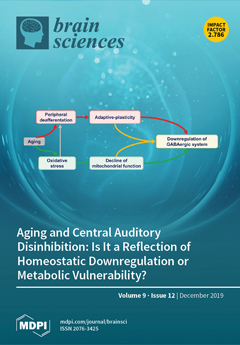
Clinical data suggest that deafferentation-related disinhibition tends to occur primarily in the aged brain. Therefore, aging-related disinhibition may, in part, be related to the high metabolic demands of inhibitory neurons relative to their excitatory counterparts. These findings suggest that both deafferentation-related maladaptive plastic changes and aging-related metabolic factors combine to produce changes in central auditory function. Here, we explore the arguments that downregulation of inhibition may be due to homeostatic responses to diminished afferent input vs. metabolic vulnerability of inhibitory neurons in the aged brain. Understanding the relative importance of these mechanisms will be critical for the development of treatments for the underlying causes of aging-related central disinhibition. View this paper
  • Issues are regarded as officially published after their release is announced to the table of contents alert mailing list.
  • You may sign up for e-mail alerts to receive table of contents of newly released issues.
  • PDF is the official format for papers published in both, html and pdf forms. To view the papers in pdf format, click on the "PDF Full-text" link, and use the free Adobe Reader to open them.
Order results
Result details
Section
Select all
Export citation of selected articles as:
22 pages, 2901 KiB  
Article
Neurologic Injury and Brain Growth in the Setting of Long-Gap Esophageal Atresia Perioperative Critical Care: A Pilot Study
by Samuel S. Rudisill, Jue T. Wang, Camilo Jaimes, Chandler R. L. Mongerson, Anne R. Hansen, Russell W. Jennings and Dusica Bajic
Brain Sci. 2019, 9(12), 383; https://doi.org/10.3390/brainsci9120383 - 17 Dec 2019
Cited by 13 | Viewed by 3118
Abstract
We previously showed that infants born with long-gap esophageal atresia (LGEA) demonstrate clinically significant brain MRI findings following repair with the Foker process. The current pilot study sought to identify any pre-existing (PRE-Foker process) signs of brain injury and to characterize brain and [...] Read more.
We previously showed that infants born with long-gap esophageal atresia (LGEA) demonstrate clinically significant brain MRI findings following repair with the Foker process. The current pilot study sought to identify any pre-existing (PRE-Foker process) signs of brain injury and to characterize brain and corpus callosum (CC) growth. Preterm and full-term infants (n = 3/group) underwent non-sedated brain MRI twice: before (PRE-Foker scan) and after (POST-Foker scan) completion of perioperative care. A neuroradiologist reported on qualitative brain findings. The research team quantified intracranial space, brain, cerebrospinal fluid (CSF), and CC volumes. We report novel qualitative brain findings in preterm and full-term infants born with LGEA before undergoing Foker process. Patients had a unique hospital course, as assessed by secondary clinical end-point measures. Despite increased total body weight and absolute intracranial and brain volumes (cm3) between scans, normalized brain volume was decreased in 5/6 patients, implying delayed brain growth. This was accompanied by both an absolute and relative CSF volume increase. In addition to qualitative findings of CC abnormalities in 3/6 infants, normative CC size (% brain volume) was consistently smaller in all infants, suggesting delayed or abnormal CC maturation. A future larger study group is warranted to determine the impact on the neurodevelopmental outcomes of infants born with LGEA. Full article
(This article belongs to the Section Developmental Neuroscience)
Show Figures

Figure 1

3 pages, 172 KiB  
Commentary
Reports of L-Norvaline Toxicity in Humans May Be Greatly Overstated
by Baruh Polis, Michael A. Gilinsky and Abraham O. Samson
Brain Sci. 2019, 9(12), 382; https://doi.org/10.3390/brainsci9120382 - 17 Dec 2019
Cited by 3 | Viewed by 2927
Abstract
Recently, a study published in “Toxicology In Vitro” (Kate Samardzic and Kenneth J. Rodgers) was entitled: “Cytotoxicity and Mitochondrial Dysfunction Caused by the Dietary Supplement L-Norvaline”. The title may be greatly overstated, and here we provide several arguments showing that norvaline is not [...] Read more.
Recently, a study published in “Toxicology In Vitro” (Kate Samardzic and Kenneth J. Rodgers) was entitled: “Cytotoxicity and Mitochondrial Dysfunction Caused by the Dietary Supplement L-Norvaline”. The title may be greatly overstated, and here we provide several arguments showing that norvaline is not as toxic as reported. Full article
(This article belongs to the Section Neuroglia)
18 pages, 1056 KiB  
Article
A Pathway-Based Genomic Approach to Identify Medications: Application to Alcohol Use Disorder
by Laura B. Ferguson, Shruti Patil, Bailey A. Moskowitz, Igor Ponomarev, Robert A. Harris, Roy D. Mayfield and Robert O. Messing
Brain Sci. 2019, 9(12), 381; https://doi.org/10.3390/brainsci9120381 - 16 Dec 2019
Cited by 5 | Viewed by 3273
Abstract
Chronic, excessive alcohol use alters brain gene expression patterns, which could be important for initiating, maintaining, or progressing the addicted state. It has been proposed that pharmaceuticals with opposing effects on gene expression could treat alcohol use disorder (AUD). Computational strategies comparing gene [...] Read more.
Chronic, excessive alcohol use alters brain gene expression patterns, which could be important for initiating, maintaining, or progressing the addicted state. It has been proposed that pharmaceuticals with opposing effects on gene expression could treat alcohol use disorder (AUD). Computational strategies comparing gene expression signatures of disease to those of pharmaceuticals show promise for nominating novel treatments. We reasoned that it may be sufficient for a treatment to target the biological pathway rather than lists of individual genes perturbed by AUD. We analyzed published and unpublished transcriptomic data using gene set enrichment of Kyoto Encyclopedia of Genes and Genomes (KEGG) pathways to identify biological pathways disrupted in AUD brain and by compounds in the Library of Network-based Cellular Signatures (LINCS L1000) and Connectivity Map (CMap) databases. Several pathways were consistently disrupted in AUD brain, including an up-regulation of genes within the Complement and Coagulation Cascade, Focal Adhesion, Systemic Lupus Erythematosus, and MAPK signaling, and a down-regulation of genes within the Oxidative Phosphorylation pathway, strengthening evidence for their importance in AUD. Over 200 compounds targeted genes within those pathways in an opposing manner, more than twenty of which have already been shown to affect alcohol consumption, providing confidence in our approach. We created a user-friendly web-interface that researchers can use to identify drugs that target pathways of interest or nominate mechanism of action for drugs. This study demonstrates a unique systems pharmacology approach that can nominate pharmaceuticals that target pathways disrupted in disease states such as AUD and identify compounds that could be repurposed for AUD if sufficient evidence is attained in preclinical studies. Full article
Show Figures

Graphical abstract

16 pages, 1437 KiB  
Article
Typical and Aberrant Functional Brain Flexibility: Lifespan Development and Aberrant Organization in Traumatic Brain Injury and Dyslexia
by Stavros I. Dimitriadis, Panagiotis G. Simos, Jack Μ. Fletcher and Andrew C. Papanicolaou
Brain Sci. 2019, 9(12), 380; https://doi.org/10.3390/brainsci9120380 - 16 Dec 2019
Cited by 6 | Viewed by 3288
Abstract
Intrinsic functional connectivity networks derived from different neuroimaging methods and connectivity estimators have revealed robust developmental trends linked to behavioural and cognitive maturation. The present study employed a dynamic functional connectivity approach to determine dominant intrinsic coupling modes in resting-state neuromagnetic data from [...] Read more.
Intrinsic functional connectivity networks derived from different neuroimaging methods and connectivity estimators have revealed robust developmental trends linked to behavioural and cognitive maturation. The present study employed a dynamic functional connectivity approach to determine dominant intrinsic coupling modes in resting-state neuromagnetic data from 178 healthy participants aged 8–60 years. Results revealed significant developmental trends in three types of dominant intra- and inter-hemispheric neuronal population interactions (amplitude envelope, phase coupling, and phase-amplitude synchronization) involving frontal, temporal, and parieto-occipital regions. Multi-class support vector machines achieved 89% correct classification of participants according to their chronological age using dynamic functional connectivity indices. Moreover, systematic temporal variability in functional connectivity profiles, which was used to empirically derive a composite flexibility index, displayed an inverse U-shaped curve among healthy participants. Lower flexibility values were found among age-matched children with reading disability and adults who had suffered mild traumatic brain injury. The importance of these results for normal and abnormal brain development are discussed in light of the recently proposed role of cross-frequency interactions in the fine-grained coordination of neuronal population activity. Full article
(This article belongs to the Special Issue Human Brain Dynamics: Latest Advances and Prospects)
Show Figures

Graphical abstract

16 pages, 982 KiB  
Article
Experience Matters: The Effects of Hypothetical versus Experiential Delays and Magnitudes on Impulsive Choice in Delay Discounting Tasks
by Catherine C. Steele, MacKenzie Gwinner, Travis Smith, Michael E. Young and Kimberly Kirkpatrick
Brain Sci. 2019, 9(12), 379; https://doi.org/10.3390/brainsci9120379 - 16 Dec 2019
Cited by 7 | Viewed by 3159
Abstract
Impulsive choice in humans is typically measured using hypothetical delays and rewards. In two experiments, we determined how experiencing the delay and/or the reward affected impulsive choice behavior. Participants chose between two amounts of real or hypothetical candy (M&Ms) after a real or [...] Read more.
Impulsive choice in humans is typically measured using hypothetical delays and rewards. In two experiments, we determined how experiencing the delay and/or the reward affected impulsive choice behavior. Participants chose between two amounts of real or hypothetical candy (M&Ms) after a real or hypothetical delay (5–30 s), where choosing the shorter delay was the impulsive choice. Experiment 1 compared choice behavior on a real-delay, real-reward (RD/RR) task where participants received M&Ms after experiencing the delays versus a real-delay, hypothetical-reward (RD/HR) task where participants accumulated hypothetical M&Ms after experiencing the delays. Experiment 2 compared the RD/HR task and a hypothetical-delay, hypothetical-reward (HD/HR) task where participants accumulated hypothetical M&Ms after hypothetical delays. The results indicated that choices did not differ between real and hypothetical M&Ms (Experiment 1), and participants were less sensitive to delay and more larger-later (LL)-preferring with hypothetical delays compared to real delays (Experiment 2). Experiencing delays to reward may be important for modeling real-world impulsive choices where delays are typically experienced. These novel experiential impulsive choice tasks may improve translational methods for comparison with animal models and may be improved procedures for predicting real-life choice behavior in humans. Full article
Show Figures

Figure 1

13 pages, 2745 KiB  
Article
Reduced Apoptotic Injury by Phenothiazine in Ischemic Stroke through the NOX-Akt/PKC Pathway
by Yanna Tong, Kenneth B. Elkin, Changya Peng, Jiamei Shen, Fengwu Li, Longfei Guan, Yu Ji, Wenjing Wei, Xiaokun Geng and Yuchuan Ding
Brain Sci. 2019, 9(12), 378; https://doi.org/10.3390/brainsci9120378 - 15 Dec 2019
Cited by 11 | Viewed by 3347
Abstract
Phenothiazine treatment has been shown to reduce post-stroke ischemic injury, though the underlying mechanism remains unclear. This study sought to confirm the neuroprotective effects of phenothiazines and to explore the role of the NOX (nicotinamide adenine dinucleotide phosphate oxidase)/Akt/PKC (protein kinase C) pathway [...] Read more.
Phenothiazine treatment has been shown to reduce post-stroke ischemic injury, though the underlying mechanism remains unclear. This study sought to confirm the neuroprotective effects of phenothiazines and to explore the role of the NOX (nicotinamide adenine dinucleotide phosphate oxidase)/Akt/PKC (protein kinase C) pathway in cerebral apoptosis. Sprague-Dawley rats underwent middle cerebral artery occlusion (MCAO) for 2 h and were randomly divided into 3 different cohorts: (1) saline, (2) 8 mg/kg chlorpromazine and promethazine (C+P), and (3) 8 mg/kg C+P as well as apocynin (NOX inhibitor). Brain infarct volumes were examined, and cell death/NOX activity was determined by assays. Western blotting was used to assess protein expression of kinase C-δ (PKC-δ), phosphorylated Akt (p-Akt), Bax, Bcl-XL, and uncleaved/cleaved caspase-3. Both C+P and C+P/NOX inhibitor administration yielded a significant reduction in infarct volumes and cell death, while the C+P/NOX inhibitor did not confer further reduction. In both treatment groups, anti-apoptotic Bcl-XL protein expression generally increased, while pro-apoptotic Bax and caspase-3 proteins generally decreased. PKC protein expression was decreased in both treatment groups, demonstrating a further decrease by C+P/NOX inhibitor at 6 and 24 h of reperfusion. The present study confirms C+P-mediated neuroprotection and suggests that the NOX/Akt/PKC pathway is a potential target for efficacious therapy following ischemic stroke. Full article
(This article belongs to the Collection Collection on Molecular and Cellular Neuroscience)
Show Figures

Figure 1

9 pages, 1256 KiB  
Article
Characteristics of the Uncinate Fasciculus and Cingulum in Patients with Mild Cognitive Impairment: Diffusion Tensor Tractography Study
by Chan-Hyuk Park, Su-Hong Kim and Han-Young Jung
Brain Sci. 2019, 9(12), 377; https://doi.org/10.3390/brainsci9120377 - 14 Dec 2019
Cited by 11 | Viewed by 3386
Abstract
Many studies have examined the relationship between cognition, and the cingulum and uncinate fasciculus (UF). In this study, diffusion tensor tractography (DTT) was used to investigate the correlation between fractional-anisotropy (FA) values and the number of fibers in the cingulum and UF in [...] Read more.
Many studies have examined the relationship between cognition, and the cingulum and uncinate fasciculus (UF). In this study, diffusion tensor tractography (DTT) was used to investigate the correlation between fractional-anisotropy (FA) values and the number of fibers in the cingulum and UF in patients with and without cognitive impairment. The correlation between cognitive function, and the cingulum and UF was also investigated. Thirty patients (14 males, age = 70.68 ± 7.99 years) were divided into a control group (n = 14) and mild-cognitive-impairment (MCI) group (n = 16). The Seoul Neuropsychological Screening Battery (SNSB) and DTT were performed to assess cognition and bilateral tracts of the cingulum and UF. The relationship between SNSB values and the cingulum and UF was analyzed. The number of fibers in the right cingulum and right UF were significantly different between the two groups. The MCI group showed thinner tracts in both the cingulum and UF compared to the control group. A significant relationship was found between the number of fibers in the right UF and delayed memory recall. In conclusion, memory loss in MCI was associated with a decreased number of fibers in the right UF, while language and visuospatial function were related to the number of fibers in the right cingulum. Full article
(This article belongs to the Special Issue Dementia and Cognitive Ageing)
Show Figures

Figure 1

15 pages, 7550 KiB  
Article
A Hybrid Feature Pool-Based Emotional Stress State Detection Algorithm Using EEG Signals
by Md Junayed Hasan and Jong-Myon Kim
Brain Sci. 2019, 9(12), 376; https://doi.org/10.3390/brainsci9120376 - 13 Dec 2019
Cited by 40 | Viewed by 4453
Abstract
Human stress analysis using electroencephalogram (EEG) signals requires a detailed and domain-specific information pool to develop an effective machine learning model. In this study, a multi-domain hybrid feature pool is designed to identify most of the important information from the signal. The hybrid [...] Read more.
Human stress analysis using electroencephalogram (EEG) signals requires a detailed and domain-specific information pool to develop an effective machine learning model. In this study, a multi-domain hybrid feature pool is designed to identify most of the important information from the signal. The hybrid feature pool contains features from two types of analysis: (a) statistical parametric analysis from the time domain, and (b) wavelet-based bandwidth specific feature analysis from the time-frequency domain. Then, a wrapper-based feature selector, Boruta, is applied for ranking all the relevant features from that feature pool instead of considering only the non-redundant features. Finally, the k-nearest neighbor (k-NN) algorithm is used for final classification. The proposed model yields an overall accuracy of 73.38% for the total considered dataset. To validate the performance of the proposed model and highlight the necessity of designing a hybrid feature pool, the model was compared to non-linear dimensionality reduction techniques, as well as those without feature ranking. Full article
(This article belongs to the Special Issue Brain Plasticity, Cognitive Training and Mental States Assessment)
Show Figures

Figure 1

13 pages, 934 KiB  
Article
Neuroendocrine and Inflammatory Effects of Childhood Trauma Following Psychosocial and Inflammatory Stress in Women with Remitted Major Depressive Disorder
by Laura L.M. Cassiers, Peter Niemegeers, Erik Fransen, Manuel Morrens, Peter De Boer, Luc Van Nueten, Stephan Claes, Bernard G.C. Sabbe and Filip Van Den Eede
Brain Sci. 2019, 9(12), 375; https://doi.org/10.3390/brainsci9120375 - 13 Dec 2019
Cited by 5 | Viewed by 3159
Abstract
The dysregulation of the inflammatory and neuroendocrine systems seen in major depressive disorder (MDD) may persist after remission and this is associated with a higher risk of relapse. This vulnerable subgroup may be characterized by a history of childhood trauma. In a single-blind [...] Read more.
The dysregulation of the inflammatory and neuroendocrine systems seen in major depressive disorder (MDD) may persist after remission and this is associated with a higher risk of relapse. This vulnerable subgroup may be characterized by a history of childhood trauma. In a single-blind randomized placebo-controlled crossover study, 21 women with remitted recurrent MDD and 18 healthy controls were exposed to psychosocial stress (Trier social stress test) or inflammatory stress (typhoid vaccine), or both, to investigate the effects of childhood trauma on the neuroendocrine and inflammatory responses. Childhood trauma was assessed using the Childhood Trauma Questionnaire and participants were dichotomized into a traumatized and non-traumatized group. Serum adrenocorticotropic hormone (ACTH), cortisol, interferon (IFN)-γ, tumor necrosis factor (TNF)-α, and interleukin (IL)-6 were measured at regular intervals after each intervention. The effects of trauma, time, and intervention on these parameters were modeled by fitting linear mixed models. Childhood trauma in itself did not have a main effect on the outcome measurements. However, an interactional effect of trauma with stressor type was found in the remitted MDD group: trauma was associated with higher cortisol levels only after adding immunological to psychosocial stress, and with lower TNF-α levels in response to vaccination. This suggests the existence of a vulnerable trauma-associated MDD endophenotype. Full article
(This article belongs to the Special Issue Neuroimmunology of Major Psychiatric Disorders)
Show Figures

Figure 1

10 pages, 219 KiB  
Article
Spatial Neglect in Stroke: Identification, Disease Process and Association with Outcome During Inpatient Rehabilitation
by Ulrike Hammerbeck, Matthew Gittins, Andy Vail, Lizz Paley, Sarah F Tyson and Audrey Bowen
Brain Sci. 2019, 9(12), 374; https://doi.org/10.3390/brainsci9120374 - 13 Dec 2019
Cited by 31 | Viewed by 6507
Abstract
We established spatial neglect prevalence, disease profile and amount of therapy that inpatient stroke survivors received, and outcomes at discharge using Sentinel Stroke National Audit Programme (SSNAP) data. We used data from 88,664 National Health Service (NHS) admissions in England, Wales and Northern [...] Read more.
We established spatial neglect prevalence, disease profile and amount of therapy that inpatient stroke survivors received, and outcomes at discharge using Sentinel Stroke National Audit Programme (SSNAP) data. We used data from 88,664 National Health Service (NHS) admissions in England, Wales and Northern Ireland (July 2013–July 2015), for stroke survivors still in hospital after 3 days with a completed baseline neglect National Institute for Health Stroke Scale (NIHSS) score. Thirty percent had neglect (NIHSS item 11 ≥ 1) and they were slightly older (78 years) than those without neglect (75 years). Neglect was observed more commonly in women (33 vs. 27%) and in individuals with a premorbid dependency (37 vs. 28%). Survivors of mild stroke were far less likely to present with neglect than those with severe stroke (4% vs. 84%). Those with neglect had a greatly increased length of stay (27 vs. 10 days). They received a comparable amount of average daily occupational and physiotherapy during their longer inpatient stay but on discharge a greater percentage of individuals with neglect were dependent on the modified Rankin scale (76 vs. 57%). Spatial neglect is common and associated with worse clinical outcomes. These results add to our understanding of neglect to inform clinical guidelines, service provision and priorities for future research. Full article
(This article belongs to the Special Issue Unilateral Neglect Assessment and Rehabilitation)
14 pages, 2406 KiB  
Article
Cortical Thickness Links Impulsive Personality Traits and Risky Behavior
by Rickie Miglin, Nadia Bounoua, Shelly Goodling, Ana Sheehan, Jeffrey M. Spielberg and Naomi Sadeh
Brain Sci. 2019, 9(12), 373; https://doi.org/10.3390/brainsci9120373 - 13 Dec 2019
Cited by 19 | Viewed by 3935
Abstract
Impulsive personality traits are often predictive of risky behavior, but not much is known about the neurobiological basis of this relationship. We investigated whether thickness of the cortical mantle varied as a function of impulsive traits and whether such variation also explained recent [...] Read more.
Impulsive personality traits are often predictive of risky behavior, but not much is known about the neurobiological basis of this relationship. We investigated whether thickness of the cortical mantle varied as a function of impulsive traits and whether such variation also explained recent risky behavior. A community sample of 107 adults (ages 18–55; 54.2% men) completed self-report measures of impulsive traits and risky behavior followed by a neuroimaging protocol. Using the three-factor model of impulsive traits derived from the UPPS-P Impulsive Behavior Scale, analysis of the entire cortical mantle identified three thickness clusters that related to impulsive traits. Sensation seeking was negatively related to thickness in the right pericalcarine cortex, whereas impulsive urgency was positively associated with thickness in the left superior parietal and right paracentral lobule. Notably, follow-up analyses showed that thickness in the right pericalcarine cortex also related to recent risky behavior, with the identified cluster mediating the association between sensation seeking and risky behavior. Findings suggest that reduced thickness in the pericalcarine region partially explains the link between sensation seeking and the tendency to engage in risky behavior, providing new insight into the neurobiological basis of these relationships. Full article
Show Figures

Figure 1

14 pages, 632 KiB  
Article
A Comprehensive sLORETA Study on the Contribution of Cortical Somatomotor Regions to Motor Imagery
by Mustafa Yazici, Mustafa Ulutas and Mukadder Okuyan
Brain Sci. 2019, 9(12), 372; https://doi.org/10.3390/brainsci9120372 - 13 Dec 2019
Cited by 6 | Viewed by 3347
Abstract
Brain–computer interface (BCI) is a technology used to convert brain signals to control external devices. Researchers have designed and built many interfaces and applications in the last couple of decades. BCI is used for prevention, detection, diagnosis, rehabilitation, and restoration in healthcare. EEG [...] Read more.
Brain–computer interface (BCI) is a technology used to convert brain signals to control external devices. Researchers have designed and built many interfaces and applications in the last couple of decades. BCI is used for prevention, detection, diagnosis, rehabilitation, and restoration in healthcare. EEG signals are analyzed in this paper to help paralyzed people in rehabilitation. The electroencephalogram (EEG) signals recorded from five healthy subjects are used in this study. The sensor level EEG signals are converted to source signals using the inverse problem solution. Then, the cortical sources are calculated using sLORETA methods at nine regions marked by a neurophysiologist. The features are extracted from cortical sources by using the common spatial pattern (CSP) method and classified by a support vector machine (SVM). Both the sensor and the computed cortical signals corresponding to motor imagery of the hand and foot are used to train the SVM algorithm. Then, the signals outside the training set are used to test the classification performance of the classifier. The 0.1–30 Hz and mu rhythm band-pass filtered activity is also analyzed for the EEG signals. The classification performance and recognition of the imagery improved up to 100% under some conditions for the cortical level. The cortical source signals at the regions contributing to motor commands are investigated and used to improve the classification of motor imagery. Full article
(This article belongs to the Collection Collection on Theoretical and Computational Neuroscience)
Show Figures

Figure 1

18 pages, 1077 KiB  
Review
Co-Morbid Insomnia and Sleep Apnea (COMISA): Prevalence, Consequences, Methodological Considerations, and Recent Randomized Controlled Trials
by Alexander Sweetman, Leon Lack and Célyne Bastien
Brain Sci. 2019, 9(12), 371; https://doi.org/10.3390/brainsci9120371 - 12 Dec 2019
Cited by 104 | Viewed by 8233
Abstract
Co-morbid insomnia and sleep apnea (COMISA) is a highly prevalent and debilitating disorder, which results in additive impairments to patients’ sleep, daytime functioning, and quality of life, and complex diagnostic and treatment decisions for clinicians. Although the presence of COMISA was first recognized [...] Read more.
Co-morbid insomnia and sleep apnea (COMISA) is a highly prevalent and debilitating disorder, which results in additive impairments to patients’ sleep, daytime functioning, and quality of life, and complex diagnostic and treatment decisions for clinicians. Although the presence of COMISA was first recognized by Christian Guilleminault and colleagues in 1973, it received very little research attention for almost three decades, until the publication of two articles in 1999 and 2001 which collectively reported a 30%–50% co-morbid prevalence rate, and re-ignited research interest in the field. Since 1999, there has been an exponential increase in research documenting the high prevalence, common characteristics, treatment complexities, and bi-directional relationships of COMISA. Recent trials indicate that co-morbid insomnia symptoms may be treated with cognitive and behavioral therapy for insomnia, to increase acceptance and use of continuous positive airway pressure therapy. Hence, the treatment of COMISA appears to require nuanced diagnostic considerations, and multi-faceted treatment approaches provided by multi-disciplinary teams of psychologists and physicians. In this narrative review, we present a brief overview of the history of COMISA research, describe the importance of measuring and managing insomnia symptoms in the presence of sleep apnea, discuss important methodological and diagnostic considerations for COMISA, and review several recent randomized controlled trials investigating the combination of CBTi and CPAP therapy. We aim to provide clinicians with pragmatic suggestions and tools to identify, and manage this prevalent COMISA disorder in clinical settings, and discuss future avenues of research to progress the field. Full article
(This article belongs to the Special Issue Insomnia: Beyond Hyperarousal)
Show Figures

Figure 1

16 pages, 1224 KiB  
Article
Helicobacter pylori, Vascular Risk Factors and Cognition in U.S. Older Adults
by Víctor M. Cárdenas, François Boller and Gustavo C. Román
Brain Sci. 2019, 9(12), 370; https://doi.org/10.3390/brainsci9120370 - 12 Dec 2019
Cited by 18 | Viewed by 3946
Abstract
Previous studies suggested that Helicobacter pylori infection could be a risk factor for stroke, dementia, and Alzheimer’s disease (AD). The authors examined data from participants, 60 years old and older in the Third National Health and Nutrition Examination Survey (NHANES-III) to assess the [...] Read more.
Previous studies suggested that Helicobacter pylori infection could be a risk factor for stroke, dementia, and Alzheimer’s disease (AD). The authors examined data from participants, 60 years old and older in the Third National Health and Nutrition Examination Survey (NHANES-III) to assess the relation between Helicobacter pylori infection and results of the Mini-Mental State Examination (n = 1860) using logistic regression analysis controlling for age, gender, race/ethnicity, education, poverty and history of medically diagnosed diabetes. Moreover, we examined performance on the digit-symbol substitution test (DSST) of 1031 participants in the 1999–2000 NHANES according to their H. pylori infection status controlling for potential confounders using multiple linear regression analyses. In 1988–1991, older adults infected with CagA strains of H. pylori had a 50% borderline statistically significant increased level of cognitive impairment, as measured by low Mini-Mental State Examination (MMSE) scores (age–education adjusted prevalence ratio: 1.5; 95% confidence interval: 1.0, 2.0). In 1999–2000, older US adults infected with H. pylori scored 2.6 fewer points in the DSST than those uninfected (mean adjusted difference: −2.6; 95% confidence interval −5.1, −0.1). The authors concluded that H. pylori infection might be a risk factor for cognitive decline in the elderly. They also found that low cobalamin and elevated homocysteine were associated with cognitive impairment. Full article
Show Figures

Figure 1

11 pages, 1971 KiB  
Article
Protective Effects of Scolopendra Water Extract on Trimethyltin-Induced Hippocampal Neurodegeneration and Seizures in Mice
by Yun-Soo Seo, Mary Jasmin Ang, Byeong Cheol Moon, Hyo Seon Kim, Goya Choi, Hye-Sun Lim, Sohi Kang, Mijin Jeon, Sung-Ho Kim, Changjong Moon and Joong Sun Kim
Brain Sci. 2019, 9(12), 369; https://doi.org/10.3390/brainsci9120369 - 12 Dec 2019
Cited by 5 | Viewed by 2696
Abstract
Trimethyltin (TMT) is an organotin compound with potent neurotoxic action characterized by neuronal degeneration in the hippocampus. This study evaluated the protective effects of a Scolopendra water extract (SWE) against TMT intoxication in hippocampal neurons, using both in vitro and in vivo model [...] Read more.
Trimethyltin (TMT) is an organotin compound with potent neurotoxic action characterized by neuronal degeneration in the hippocampus. This study evaluated the protective effects of a Scolopendra water extract (SWE) against TMT intoxication in hippocampal neurons, using both in vitro and in vivo model systems. Specifically, we examined the actions of SWE on TMT- (5 mM) induced cytotoxicity in primary cultures of mouse hippocampal neurons (7 days in vitro) and the effects of SWE on hippocampal degeneration in adult TMT- (2.6 mg/kg, intraperitoneal) treated C57BL/6 mice. We found that SWE pretreatment (0–100 μg/mL) significantly reduced TMT-induced cytotoxicity in cultured hippocampal neurons in a dose-dependent manner, as determined by lactate dehydrogenase and 3-[4,5-dimethylthiazol-2-yl]-2,5-diphenyltetrazolium bromide assays. Additionally, this study showed that perioral administration of SWE (5 mg/kg), from −6 to 0 days before TMT injection, significantly attenuated hippocampal cell degeneration and seizures in adult mice. Furthermore, quantitative analysis of Iba-1 (Allograft inflammatory factor 1)- and GFAP (Glial fibrillary acidic protein)-immunostained cells revealed a significant reduction in the levels of Iba-1- and GFAP-positive cell bodies in the dentate gyrus (DG) of mice treated with SWE prior to TMT injection. These data indicated that SWE pretreatment significantly protected the hippocampus against the massive activation of microglia and astrocytes elicited by TMT. In addition, our data showed that the SWE-induced reduction of immune cell activation was linked to a significant reduction in cell death and a significant improvement in TMT-induced seizure behavior. Thus, we conclude that SWE ameliorated the detrimental effects of TMT toxicity on hippocampal neurons, both in vivo and in vitro. Altogether, our findings hint at a promising pharmacotherapeutic use of SWE in hippocampal degeneration and dysfunction. Full article
Show Figures

Figure 1

11 pages, 240 KiB  
Perspective
Noli Me Tangere: Social Touch, Tactile Defensiveness, and Communication in Neurodevelopmental Disorders
by Daniela Smirni, Pietro Smirni, Marco Carotenuto, Lucia Parisi, Giuseppe Quatrosi and Michele Roccella
Brain Sci. 2019, 9(12), 368; https://doi.org/10.3390/brainsci9120368 - 12 Dec 2019
Cited by 19 | Viewed by 6868
Abstract
Tactile defensiveness is a common feature in neurodevelopmental disorders (NDDs). Since the first studies, tactile defensiveness has been described as the result of an abnormal response to sensory stimulation. Moreover, it has been studied how the tactile system is closely linked to socio-communicative [...] Read more.
Tactile defensiveness is a common feature in neurodevelopmental disorders (NDDs). Since the first studies, tactile defensiveness has been described as the result of an abnormal response to sensory stimulation. Moreover, it has been studied how the tactile system is closely linked to socio-communicative development and how the interoceptive sensory system supports both a discriminating touch and an affective touch. Therefore, several neurophysiological studies have been conducted to investigate the neurobiological basis of the development and functioning of the tactile system for a better understanding of the tactile defensiveness behavior and the social touch of NDDs. Given the lack of recent literature on tactile defensiveness, the current study provides a brief overview of the original contributions on this research topic in children with NDDs focusing attention on how this behavior has been considered over the years in the clinical setting. Full article
(This article belongs to the Special Issue Recent Advances in Neurodevelopmental Disorders)
13 pages, 1238 KiB  
Article
Individual Differences in Ethanol Drinking and Seeking Behaviors in Rats Exposed to Chronic Intermittent Ethanol Vapor Exposure is Associated with Altered CaMKII Autophosphorylation in the Nucleus Accumbens Shell
by Sucharita S. Somkuwar and Chitra D. Mandyam
Brain Sci. 2019, 9(12), 367; https://doi.org/10.3390/brainsci9120367 - 11 Dec 2019
Cited by 3 | Viewed by 2930
Abstract
Chronic intermittent ethanol vapor exposure (CIE) in rodents produces reliable and high blood ethanol concentration and behavioral symptoms associated with moderate to severe alcohol use disorder (AUD)—for example, escalation of operant ethanol self-administration, a feature suggestive of transition from recreational to addictive use, [...] Read more.
Chronic intermittent ethanol vapor exposure (CIE) in rodents produces reliable and high blood ethanol concentration and behavioral symptoms associated with moderate to severe alcohol use disorder (AUD)—for example, escalation of operant ethanol self-administration, a feature suggestive of transition from recreational to addictive use, is a widely replicated behavior in rats that experience CIE. Herein, we present evidence from a subset of rats that do not demonstrate escalation of ethanol self-administration following seven weeks of CIE. These low responders (LR) maintain low ethanol self-administration during CIE, demonstrate lower relapse to drinking during abstinence and reduced reinstatement of ethanol seeking triggered by ethanol cues when compared with high responders (HR). We examined the blood ethanol levels in LR and HR rats during CIE and show higher levels in LR compared with HR. We also examined peak corticosterone levels during CIE and show that LR rats have higher levels compared with HR rats. Lastly, we evaluated the levels of Ca2+/calmodulin-dependent protein kinase II (CaMKII) in the nucleus accumbens shell and reveal that the activity of CaMKII, which is autophosphorylated at site Tyr-286, is significantly reduced in HR rats compared with LR rats. These findings demonstrate that dysregulation of the hypothalamic–pituitary–adrenal axis activity and plasticity-related proteins regulating molecular memory in the nucleus accumbens shell are associated with higher ethanol-drinking and -seeking in HR rats. Future mechanistic studies should evaluate CaMKII autophosphorylation-dependent remodeling of glutamatergic synapses in the ventral striatum as a plausible mechanism for the CIE-induced enhanced ethanol drinking and seeking behaviors. Full article
Show Figures

Figure 1

14 pages, 572 KiB  
Article
Inflammatory Biomarkers are Correlated with Some Forms of Regressive Autism Spectrum Disorder
by Margherita Prosperi, Letizia Guiducci, Diego G. Peroni, Chiara Narducci, Melania Gaggini, Sara Calderoni, Raffaella Tancredi, Maria Aurora Morales, Amalia Gastaldelli, Filippo Muratori and Elisa Santocchi
Brain Sci. 2019, 9(12), 366; https://doi.org/10.3390/brainsci9120366 - 11 Dec 2019
Cited by 21 | Viewed by 4118
Abstract
Background: Several studies have tried to investigate the role of inflammatory biomarkers in Autism Spectrum Disorder (ASD), and their correlations with clinical phenotypes. Despite the growing research in this topic, existing data are mostly contradictory. Methods: Eighty-five ASD preschoolers were assessed [...] Read more.
Background: Several studies have tried to investigate the role of inflammatory biomarkers in Autism Spectrum Disorder (ASD), and their correlations with clinical phenotypes. Despite the growing research in this topic, existing data are mostly contradictory. Methods: Eighty-five ASD preschoolers were assessed for developmental level, adaptive functioning, gastrointestinal (GI), socio-communicative and psychopathological symptoms. Plasma levels of leptin, resistin, plasminogen activator inhibitor-1 (PAI-1), macrophage chemoattractant protein-1 (CCL2), tumor necrosis factor-alfa (TNF-α), and interleukin-6 (IL-6) were correlated with clinical scores and were compared among different ASD subgroups according to the presence or absence of: (i) GI symptoms, (ii) regressive onset of autism. Results: Proinflammatory cytokines (TNF-α, IL-6 and CCL2) were lower than those reported in previous studies in children with systemic inflammatory conditions. GI symptoms were not correlated with levels of inflammatory biomarkers except for resistin that was lower in ASD-GI children (p = 0.032). Resistin and PAI-1 levels were significantly higher in the group with “regression plus a developmental delay” onset (Reg+DD group) compared to groups without regression or with regression without a developmental delay (p < 0.01 for all). Conclusions: Our results did not highlight the presence of any systemic inflammatory state in ASD subjects neither disentangling children with/without GI symptoms. The Reg + DD group significantly differed from others in some plasmatic values, but these differences failed to discriminate the subgroups as possible distinct ASD endo-phenotypes. Full article
(This article belongs to the Special Issue Advances in Autism Research)
Show Figures

Graphical abstract

23 pages, 2590 KiB  
Article
Get Set or Get Distracted? Disentangling Content-Priming and Attention-Catching Effects of Background Lure Stimuli on Identifying Targets in Two Simultaneously Presented Series
by Rolf Verleger, Kamila Śmigasiewicz, Lars Michael, Laura Heikaus and Michael Niedeggen
Brain Sci. 2019, 9(12), 365; https://doi.org/10.3390/brainsci9120365 - 11 Dec 2019
Cited by 1 | Viewed by 2586
Abstract
In order to study the changing relevance of stimulus features in time and space, we used a task with rapid serial presentation of two stimulus streams where two targets (“T1” and “T2”) had to be distinguished from background stimuli and where the difficult [...] Read more.
In order to study the changing relevance of stimulus features in time and space, we used a task with rapid serial presentation of two stimulus streams where two targets (“T1” and “T2”) had to be distinguished from background stimuli and where the difficult T2 distinction was impeded by background stimuli presented before T1 that resemble T2 (“lures”). Such lures might actually have dual characteristics: Their capturing attention might interfere with target identification, whereas their similarity to T2 might result in positive priming. To test this idea here, T2 was a blue digit among black letters, and lures resembled T2 either by alphanumeric category (black digits) or by salience (blue letters). Same-category lures were expected to prime T2 identification whereas salient lures would impede T2 identification. Results confirmed these predictions, yet the precise pattern of results did not fit our conceptual framework. To account for this pattern, we speculate that lures serve to confuse participants about the order of events, and the major factor distinguishing color lures and digit lures is their confusability with T2. Mechanisms of effects were additionally explored by measuring event-related EEG potentials. Consistent with the assumption that they attract more attention, color lures evoked larger N2pc than digit lures and affected the ensuing T1-evoked N2pc. T2-evoked N2pc was indistinguishably reduced by all kinds of preceding lures, though. Lure-evoked mesio-frontal negativity increased from first to third lures both with digit and color lures and, thereby, might have reflected expectancy for T1. Full article
(This article belongs to the Special Issue ERP and EEG Markers of Brain Visual Attentional Processing)
Show Figures

Figure 1

19 pages, 485 KiB  
Article
Estimating the Parameters of Fitzhugh–Nagumo Neurons from Neural Spiking Data
by Resat Ozgur Doruk and Laila Abosharb
Brain Sci. 2019, 9(12), 364; https://doi.org/10.3390/brainsci9120364 - 09 Dec 2019
Cited by 10 | Viewed by 2809
Abstract
A theoretical and computational study on the estimation of the parameters of a single Fitzhugh–Nagumo model is presented. The difference of this work from a conventional system identification is that the measured data only consist of discrete and noisy neural spiking (spike times) [...] Read more.
A theoretical and computational study on the estimation of the parameters of a single Fitzhugh–Nagumo model is presented. The difference of this work from a conventional system identification is that the measured data only consist of discrete and noisy neural spiking (spike times) data, which contain no amplitude information. The goal can be achieved by applying a maximum likelihood estimation approach where the likelihood function is derived from point process statistics. The firing rate of the neuron was assumed as a nonlinear map (logistic sigmoid) relating it to the membrane potential variable. The stimulus data were generated by a phased cosine Fourier series having fixed amplitude and frequency but a randomly shot phase (shot at each repeated trial). Various values of amplitude, stimulus component size, and sample size were applied to examine the effect of stimulus to the identification process. Results are presented in tabular and graphical forms, which also include statistical analysis (mean and standard deviation of the estimates). We also tested our model using realistic data from a previous research (H1 neurons of blowflies) and found that the estimates have a tendency to converge. Full article
(This article belongs to the Collection Collection on Theoretical and Computational Neuroscience)
Show Figures

Figure 1

29 pages, 6194 KiB  
Article
Brain Connectivity Analysis Under Semantic Vigilance and Enhanced Mental States
by Fares Al-Shargie, Usman Tariq, Omnia Hassanin, Hasan Mir, Fabio Babiloni and Hasan Al-Nashash
Brain Sci. 2019, 9(12), 363; https://doi.org/10.3390/brainsci9120363 - 09 Dec 2019
Cited by 30 | Viewed by 4977
Abstract
In this paper, we present a method to quantify the coupling between brain regions under vigilance and enhanced mental states by utilizing partial directed coherence (PDC) and graph theory analysis (GTA). The vigilance state is induced using a modified version of stroop color-word [...] Read more.
In this paper, we present a method to quantify the coupling between brain regions under vigilance and enhanced mental states by utilizing partial directed coherence (PDC) and graph theory analysis (GTA). The vigilance state is induced using a modified version of stroop color-word task (SCWT) while the enhancement state is based on audio stimulation with a pure tone of 250 Hz. The audio stimulation was presented to the right and left ears simultaneously for one-hour while participants perform the SCWT. The quantification of mental states was performed by means of statistical analysis of indexes based on GTA, behavioral responses of time-on-task (TOT), and Brunel Mood Scale (BRMUS). The results show that PDC is very sensitive to vigilance decrement and shows that the brain connectivity network is significantly reduced with increasing TOT, p < 0.05. Meanwhile, during the enhanced state, the connectivity network maintains high connectivity as time passes and shows significant improvements compared to vigilance state. The audio stimulation enhances the connectivity network over the frontal and parietal regions and the right hemisphere. The increase in the connectivity network correlates with individual differences in the magnitude of the vigilance enhancement assessed by response time to stimuli. Our results provide evidence for enhancement of cognitive processing efficiency with audio stimulation. The BRMUS was used to evaluate the emotional states of vigilance task before and after using the audio stimulation. BRMUS factors, such as fatigue, depression, and anger, significantly decrease in the enhancement group compared to vigilance group. On the other hand, happy and calmness factors increased with audio stimulation, p < 0.05. Full article
(This article belongs to the Special Issue Recent Advances in Human Brain Connectivity)
Show Figures

Graphical abstract

17 pages, 2111 KiB  
Article
The Influence of Subclinical Neck Pain on Neurophysiological and Behavioral Measures of Multisensory Integration
by Antonia M. Karellas, Paul Yielder, James J. Burkitt, Heather S. McCracken and Bernadette A. Murphy
Brain Sci. 2019, 9(12), 362; https://doi.org/10.3390/brainsci9120362 - 09 Dec 2019
Cited by 3 | Viewed by 2903
Abstract
Multisensory integration (MSI) is necessary for the efficient execution of many everyday tasks. Alterations in sensorimotor integration (SMI) have been observed in individuals with subclinical neck pain (SCNP). Altered audiovisual MSI has previously been demonstrated in this population using performance measures, such as [...] Read more.
Multisensory integration (MSI) is necessary for the efficient execution of many everyday tasks. Alterations in sensorimotor integration (SMI) have been observed in individuals with subclinical neck pain (SCNP). Altered audiovisual MSI has previously been demonstrated in this population using performance measures, such as reaction time. However, neurophysiological techniques have not been combined with performance measures in the SCNP population to determine differences in neural processing that may contribute to these behavioral characteristics. Electroencephalography (EEG) event-related potentials (ERPs) have been successfully used in recent MSI studies to show differences in neural processing between different clinical populations. This study combined behavioral and ERP measures to characterize MSI differences between healthy and SCNP groups. EEG was recorded as 24 participants performed 8 blocks of a simple reaction time (RT) MSI task, with each block consisting of 34 auditory (A), visual (V), and audiovisual (AV) trials. Participants responded to the stimuli by pressing a response key. Both groups responded fastest to the AV condition. The healthy group demonstrated significantly faster RTs for the AV and V conditions. There were significant group differences in neural activity from 100–140 ms post-stimulus onset, with the control group demonstrating greater MSI. Differences in brain activity and RT between individuals with SCNP and a control group indicate neurophysiological alterations in how individuals with SCNP process audiovisual stimuli. This suggests that SCNP alters MSI. This study presents novel EEG findings that demonstrate MSI differences in a group of individuals with SCNP. Full article
(This article belongs to the Collection Collection on Systems Neuroscience)
Show Figures

Figure 1

13 pages, 1759 KiB  
Article
Role of MyD88 in IL-1β and Ethanol Modulation of GABAergic Transmission in the Central Amygdala
by Michal Bajo, Reesha R. Patel, David M. Hedges, Florence P. Varodayan, Roman Vlkolinsky, Tony D. Davis, Michael D. Burkart, Yuri A. Blednov and Marisa Roberto
Brain Sci. 2019, 9(12), 361; https://doi.org/10.3390/brainsci9120361 - 07 Dec 2019
Cited by 14 | Viewed by 3092
Abstract
Myeloid differentiation primary response protein (MyD88) is a critical neuroimmune adaptor protein in TLR (Toll-like receptor) and IL-1R (Interleukin-1 receptor) signaling complexes. These two pro-inflammatory families play an important role in the neurobiology of alcohol use disorder, specifically MyD88 regulates ethanol drinking, ethanol-induced [...] Read more.
Myeloid differentiation primary response protein (MyD88) is a critical neuroimmune adaptor protein in TLR (Toll-like receptor) and IL-1R (Interleukin-1 receptor) signaling complexes. These two pro-inflammatory families play an important role in the neurobiology of alcohol use disorder, specifically MyD88 regulates ethanol drinking, ethanol-induced sedation, and ethanol-induced deficits in motor coordination. In this study, we examined the role of MyD88 in mediating the effects of IL-1β and ethanol on GABAergic transmission in the central amygdala (CeA) of male mice using whole-cell patch-clamp recordings in combination with pharmacological (AS-1, a mimetic that prevents MyD88 recruitment by IL-1R) and genetic (Myd88 knockout mice) approaches. We demonstrate through both approaches that IL-1β and ethanol’s modulatory effects at CeA GABA synapses are not dependent on MyD88. Myd88 knockout potentiated IL-1β’s actions in reducing postsynaptic GABAA receptor function. Pharmacological inhibition of MyD88 modulates IL-1β’s action at CeA GABA synapses similar to Myd88 knockout mice. Additionally, ethanol-induced CeA GABA release was greater in Myd88 knockout mice compared to wildtype controls. Thus, MyD88 is not essential to IL-1β or ethanol regulation of CeA GABA synapses but plays a role in modulating the magnitude of their effects, which may be a potential mechanism by which it regulates ethanol-related behaviors. Full article
Show Figures

Figure 1

13 pages, 1638 KiB  
Article
Neuronal Transmembrane Chloride Transport Has a Time-Dependent Influence on Survival of Hippocampal Cultures to Oxygen-Glucose Deprivation
by Ana-Maria Zagrean, Ioana-Florentina Grigoras, Mara Ioana Iesanu, Rosana-Bristena Ionescu, Diana Maria Chitimus, Robert Mihai Haret, Bogdan Ianosi, Mihai Ceanga and Leon Zagrean
Brain Sci. 2019, 9(12), 360; https://doi.org/10.3390/brainsci9120360 - 06 Dec 2019
Cited by 7 | Viewed by 3879
Abstract
Neuronal ischemia results in chloride gradient alterations which impact the excitatory–inhibitory balance, volume regulation, and neuronal survival. Thus, the Na+/K+/Cl co-transporter (NKCC1), the K+/ Cl co-transporter (KCC2), and the gamma-aminobutyric acid A (GABAA) [...] Read more.
Neuronal ischemia results in chloride gradient alterations which impact the excitatory–inhibitory balance, volume regulation, and neuronal survival. Thus, the Na+/K+/Cl co-transporter (NKCC1), the K+/ Cl co-transporter (KCC2), and the gamma-aminobutyric acid A (GABAA) receptor may represent therapeutic targets in stroke, but a time-dependent effect on neuronal viability could influence the outcome. We, therefore, successively blocked NKCC1, KCC2, and GABAA (with bumetanide, DIOA, and gabazine, respectively) or activated GABAA (with isoguvacine) either during or after oxygen-glucose deprivation (OGD). Primary hippocampal cultures were exposed to a 2-h OGD or sham normoxia treatment, and viability was determined using the resazurin assay. Neuronal viability was significantly reduced after OGD, and was further decreased by DIOA treatment applied during OGD (p < 0.01) and by gabazine applied after OGD (p < 0.05). Bumetanide treatment during OGD increased viability (p < 0.05), while isoguvacine applied either during or after OGD did not influence viability. Our data suggests that NKCC1 and KCC2 function has an important impact on neuronal viability during the acute ischemic episode, while the GABAA receptor plays a role during the subsequent recovery period. These findings suggest that pharmacological modulation of transmembrane chloride transport could be a promising approach during stroke and highlight the importance of the timing of treatment application in relation to ischemia-reoxygenation. Full article
(This article belongs to the Collection Collection on Molecular and Cellular Neuroscience)
Show Figures

Figure 1

19 pages, 1280 KiB  
Article
Endocrine Disruptors Induced Distinct Expression of Thyroid and Estrogen Receptors in Rat versus Mouse Primary Cerebellar Cell Cultures
by Gergely Jocsak, Eniko Ioja, David Sandor Kiss, Istvan Toth, Zoltan Barany, Tibor Bartha, Laszlo V. Frenyo and Attila Zsarnovszky
Brain Sci. 2019, 9(12), 359; https://doi.org/10.3390/brainsci9120359 - 05 Dec 2019
Cited by 8 | Viewed by 3372
Abstract
The endocrine system of animals consists of fine-tuned self-regulating mechanisms that maintain the hormonal and neuronal milieu during tissue development. This complex system can be influenced by endocrine disruptors (ED)—substances that can alter the hormonal regulation even in small concentrations. By now, thousands [...] Read more.
The endocrine system of animals consists of fine-tuned self-regulating mechanisms that maintain the hormonal and neuronal milieu during tissue development. This complex system can be influenced by endocrine disruptors (ED)—substances that can alter the hormonal regulation even in small concentrations. By now, thousands of substances—either synthesized by the plastic, cosmetic, agricultural, or medical industry or occurring naturally in plants or in polluted groundwater—can act as EDs. Their identification and testing has been a hard-to-solve problem; Recent indications that the ED effects may be species-specific just further complicated the determination of biological ED effects. Here we compare the effects of bisphenol-A, zearalenone, and arsenic (well-known EDs) exerted on mouse and rat neural cell cultures by measuring the differences of the ED-affected neural estrogen- and thyroid receptors. EDs alters the receptor expression in a species-like manner detectable in the magnitude as well as in the nature of biological responses. It is concluded that the interspecies differences (or species specificity) in ED effects should be considered in the future testing of ED effects. Full article
(This article belongs to the Section Molecular and Cellular Neuroscience)
Show Figures

Graphical abstract

11 pages, 293 KiB  
Perspective
Human Periapical Cyst-Derived Stem Cells Can Be A Smart “Lab-on-A-Cell” to Investigate Neurodegenerative Diseases and the Related Alteration of the Exosomes’ Content
by Marco Tatullo, Bruna Codispoti, Gianrico Spagnuolo and Barbara Zavan
Brain Sci. 2019, 9(12), 358; https://doi.org/10.3390/brainsci9120358 - 05 Dec 2019
Cited by 13 | Viewed by 4111
Abstract
Promising researches have demonstrated that the alteration of biological rhythms may be consistently linked to neurodegenerative pathologies. Parkinson’s disease (PD) has a multifactorial pathogenesis, involving both genetic and environmental and/or molecular co-factors. Generally, heterogeneous alterations in circadian rhythm (CR) are a typical finding [...] Read more.
Promising researches have demonstrated that the alteration of biological rhythms may be consistently linked to neurodegenerative pathologies. Parkinson’s disease (PD) has a multifactorial pathogenesis, involving both genetic and environmental and/or molecular co-factors. Generally, heterogeneous alterations in circadian rhythm (CR) are a typical finding in degenerative processes, such as cell aging and death. Although numerous genetic phenotypes have been discovered in the most common forms of PD, it seems that severe deficiencies in synaptic transmission and high vesicular recycling are frequently found in PD patients. Neuron-to-neuron interactions are often ensured by exosomes, a specific type of extracellular vesicle (EV). Neuron-derived exosomes may carry several active compounds, including miRNAs: Several studies have found that circulating miRNAs are closely associated with an atypical oscillation of circadian rhythm genes, and they are also involved in the regulation of clock genes, in animal models. In this context, a careful analysis of neural-differentiated Mesenchymal Stem Cells (MSCs) and the molecular and genetic characterization of their exosome content, both in healthy cells and in PD-induced cells, could be a strategic field of investigation for early diagnosis and better treatment of PD and similar neurodegenerative pathologies. A novel MSC population, called human periapical cyst–mesenchymal stem cells (hPCy–MSCs), has demonstrated that it naively expresswa the main neuronal markers, and may differentiate towards functional neurons. Therefore, hPCy–MSCs can be considered of particular interest for testing of in vitro strategies to treat neurological diseases. On the other hand, the limitations of using stem cells is an issue that leads researchers to perform experimental studies on the exosomes released by MCSs. Human periapical cyst-derived mesenkymal stem cells can be a smart “lab-on-a-cell” to investigate neurodegenerative diseases and the related exosomes’ content alteration. Full article
(This article belongs to the Collection Collection on Molecular and Cellular Neuroscience)
9 pages, 242 KiB  
Article
Cortical Excitability Measures May Predict Clinical Response to Fampridine in Patients with Multiple Sclerosis and Gait Impairment
by Rechdi Ahdab, Madiha M. Shatila, Abed Rahman Shatila, George Khazen, Joumana Freiha, Maher Salem, Karim Makhoul, Rody El Nawar, Shaza El Nemr, Samar S. Ayache and Naji Riachi
Brain Sci. 2019, 9(12), 357; https://doi.org/10.3390/brainsci9120357 - 05 Dec 2019
Cited by 10 | Viewed by 3190
Abstract
Background: Most multiple sclerosis (MS) patients will develop walking limitations during the disease. Sustained-release oral fampridine is the only approved drug that will improve gait in a subset of MS patients. Objectives: (1) Evaluate fampridine cortical excitability effect in MS patients with gait [...] Read more.
Background: Most multiple sclerosis (MS) patients will develop walking limitations during the disease. Sustained-release oral fampridine is the only approved drug that will improve gait in a subset of MS patients. Objectives: (1) Evaluate fampridine cortical excitability effect in MS patients with gait disability. (2) Investigate whether cortical excitability changes can predict the therapeutic response to fampridine. Method: This prospective observational study enrolled 20 adult patients with MS and gait impairment planned to receive fampridine 10 mg twice daily for two consecutive weeks. Exclusion criteria included: Recent relapse (<3 months), modification of disease modifying drugs (<6 months), or Expanded Disability Status Scale (EDSS) score >7. Neurological examination, timed 25-foot walk test (T25wt), EDSS, and cortical excitability studies were performed upon inclusion and 14 days after initiation of fampridine. Results: After treatment, the mean improvement of T25wt (ΔT25wt) was 4.9 s. Significant enhancement of intra-cortical facilitation was observed (139% versus 241%, p = 0.01) following treatment. A positive correlation was found between baseline resting motor threshold (rMT) and both EDSS (r = 0.57; p < 0.01) and ΔT25wt (r = 0.57, p = 0.01). rMT above 52% of the maximal stimulator output was found to be a good predictor of a favorable response to fampridine (accuracy: 75%). Discussion: Fampridine was found to have a significant modulatory effect on the cerebral cortex, demonstrated by an increase in excitatory intracortical processes as unveiled by paired-pulse transcranial magnetic stimulation. rMT could be useful in selecting patients likely to experience a favorable response to fampridine. Full article
(This article belongs to the Special Issue Advances in Multiple Sclerosis Research—Series I)
9 pages, 533 KiB  
Article
Efficacy of Verbally Describing One’s Own Body Movement in Motor Skill Acquisition
by Tsubasa Kawasaki, Masashi Kono and Ryosuke Tozawa
Brain Sci. 2019, 9(12), 356; https://doi.org/10.3390/brainsci9120356 - 04 Dec 2019
Cited by 2 | Viewed by 2810
Abstract
The present study examined whether (a) verbally describing one’s own body movement can be potentially effective for acquiring motor skills, and (b) if the effects are related to motor imagery. The participants in this study were 36 healthy young adults (21.2 ± 0.7 [...] Read more.
The present study examined whether (a) verbally describing one’s own body movement can be potentially effective for acquiring motor skills, and (b) if the effects are related to motor imagery. The participants in this study were 36 healthy young adults (21.2 ± 0.7 years), randomly assigned into two groups (describing and control). They performed a ball rotation activity, with the describing group being asked by the examiner to verbally describe their own ball rotation, while the control group was asked to read a magazine aloud. The participants’ ball rotation performances were measured before the intervention, then again immediately after, five minutes after, and one day after. In addition, participants’ motor imagery ability (mental chronometry) of their upper extremities was measured. The results showed that the number of successful ball rotations (motor smoothness) and the number of ball drops (motor error) significantly improved in the describing group. Moreover, improvement in motor skills had a significant correlation with motor imagery ability. This suggests that verbally describing an intervention is an effective tool for learning motor skills, and that motor imagery is a potential mechanism for such verbal descriptions. Full article
Show Figures

Figure 1

22 pages, 12818 KiB  
Article
Improved EOG Artifact Removal Using Wavelet Enhanced Independent Component Analysis
by Mohamed F. Issa and Zoltan Juhasz
Brain Sci. 2019, 9(12), 355; https://doi.org/10.3390/brainsci9120355 - 04 Dec 2019
Cited by 40 | Viewed by 4641
Abstract
Electroencephalography (EEG) signals are frequently contaminated with unwanted electrooculographic (EOG) artifacts. Blinks and eye movements generate large amplitude peaks that corrupt EEG measurements. Independent component analysis (ICA) has been used extensively in manual and automatic methods to remove artifacts. By decomposing the signals [...] Read more.
Electroencephalography (EEG) signals are frequently contaminated with unwanted electrooculographic (EOG) artifacts. Blinks and eye movements generate large amplitude peaks that corrupt EEG measurements. Independent component analysis (ICA) has been used extensively in manual and automatic methods to remove artifacts. By decomposing the signals into neural and artifactual components and artifact components can be eliminated before signal reconstruction. Unfortunately, removing entire components may result in losing important neural information present in the component and eventually may distort the spectral characteristics of the reconstructed signals. An alternative approach is to correct artifacts within the independent components instead of rejecting the entire component, for which wavelet transform based decomposition methods have been used with good results. An improved, fully automatic wavelet-based component correction method is presented for EOG artifact removal that corrects EOG components selectively, i.e., within EOG activity regions only, leaving other parts of the component untouched. In addition, the method does not rely on reference EOG channels. The results show that the proposed method outperforms other component rejection and wavelet-based EOG removal methods in its accuracy both in the time and the spectral domain. The proposed new method represents an important step towards the development of accurate, reliable and automatic EOG artifact removal methods. Full article
(This article belongs to the Special Issue Handling Uncertainty in EEG Signal Pattern Recognition)
Show Figures

Figure 1

14 pages, 3560 KiB  
Article
Diffusion Tensor Magnetic Resonance Imaging for Differentiating Multiple System Atrophy Cerebellar Type and Spinocerebellar Ataxia Type 3
by Chi-Wen Jao, Bing-Wen Soong, Chao-Wen Huang, Chien-An Duan, Chih-Chun Wu, Yu-Te Wu and Po-Shan Wang
Brain Sci. 2019, 9(12), 354; https://doi.org/10.3390/brainsci9120354 - 03 Dec 2019
Cited by 11 | Viewed by 3478
Abstract
Multiple system atrophy cerebellar type (MSA-C) and spinocerebellar ataxia type 3 (SCA3) demonstrate similar manifestations, including ataxia, pyramidal and extrapyramidal signs, as well as atrophy and signal intensity changes in the cerebellum and brainstem. MSA-C and SCA3 cannot be clinically differentiated through T1-weighted [...] Read more.
Multiple system atrophy cerebellar type (MSA-C) and spinocerebellar ataxia type 3 (SCA3) demonstrate similar manifestations, including ataxia, pyramidal and extrapyramidal signs, as well as atrophy and signal intensity changes in the cerebellum and brainstem. MSA-C and SCA3 cannot be clinically differentiated through T1-weighted magnetic resonance imaging (MRI) alone; therefore, clinical consensus criteria and genetic testing are also required. Here, we used diffusion tensor imaging (DTI) to measure water molecular diffusion of white matter and investigate the difference between MSA-C and SCA3. Four measurements were calculated from DTI images, including fractional anisotropy (FA), axial diffusivity (AD), radial diffusivity (RD), and mean diffusivity (MD). Fifteen patients with MSA-C, 15 patients with SCA3, and 30 healthy individuals participated in this study. Both patient groups demonstrated a significantly decreased FA but a significantly increased AD, RD, and MD in the cerebello-ponto-cerebral tracts. Moreover, patients with SCA3 demonstrated a significant decrease in FA but more significant increases in AD, RD, and MD in the cerebello-cerebral tracts than patients with MSAC. Our results may suggest that FA and MD can be effectively used for differentiating SCA3 and MSA-C, both of which are cerebellar ataxias and have many common atrophied regions in the cerebral and cerebellar cortex. Full article
(This article belongs to the Section Neurotechnology and Neuroimaging)
Show Figures

Figure 1

Previous Issue
Next Issue
Back to TopTop