

Therapeutic Strategies to Ameliorate Neuronal Damage in Epilepsy by Regulating Oxidative Stress, Mitochondrial Dysfunction, and Neuroinflammation
Abstract
:1. Introduction
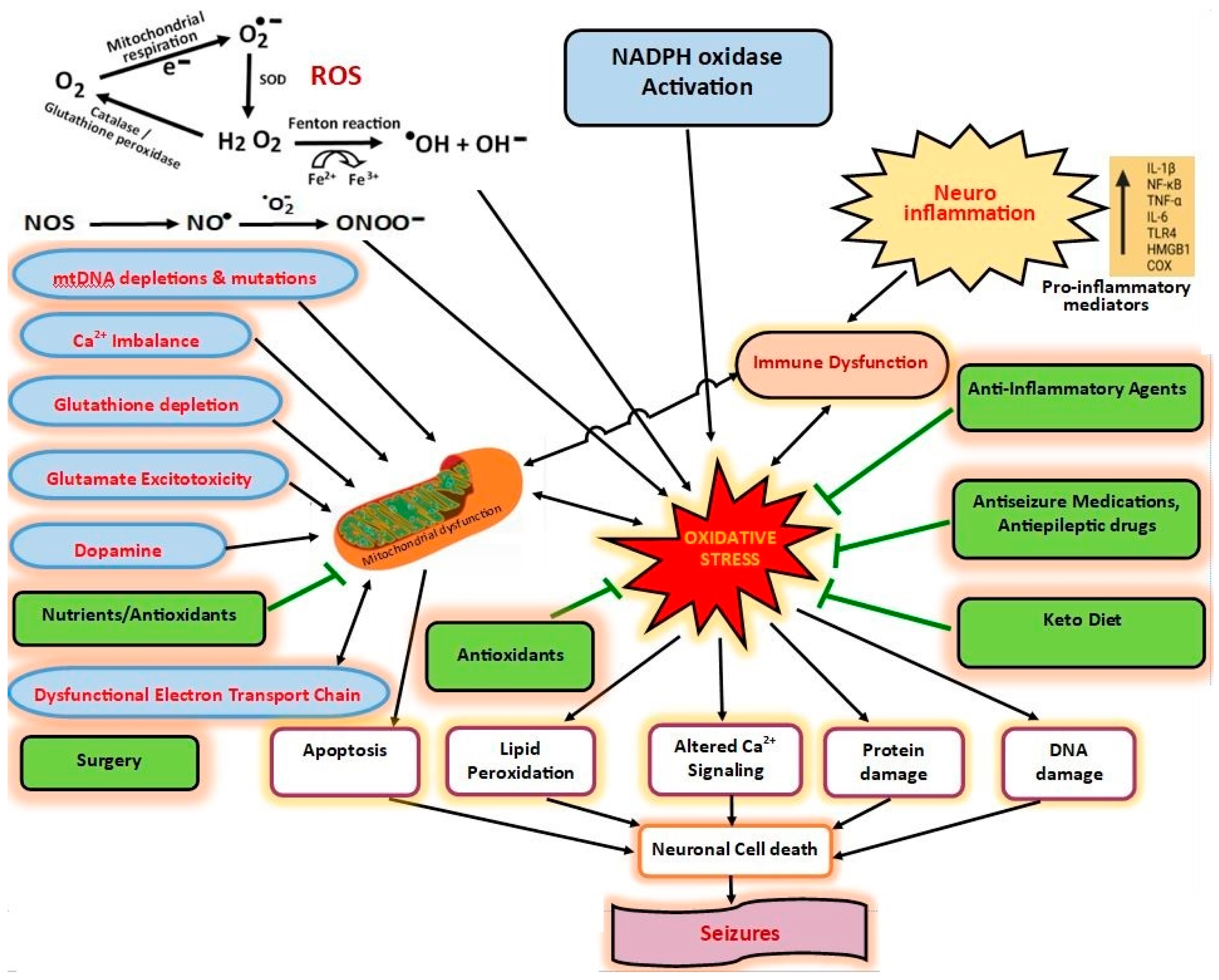
2. Epilepsy and Oxidative Stress
3. Epilepsy and Mitochondrial Dysfunction
4. Lipid Peroxidation
5. Epilepsy and Inflammation
6. Epilepsy and NOX
7. Epilepsy and Excitotoxicity
8. BBB Dysfunction
9. Epilepsy and Antioxidants (Antioxidant Therapies)
9.1. Acetyl-l-carnitine
9.2. Melatonin
9.3. NAC
9.4. Baicalein
9.5. CoQ10
9.6. Astaxanthin
10. Epilepsy and AEDs
10.1. Valproic Acid
10.2. Levetiracetam
11. Epilepsy and ASMs
11.1. CBD
11.2. Brivaracetam
11.3. Ursolic Acid
11.4. Curcumin
12. Epilepsy and Neuromodulation
12.1. VNS
12.2. Epilepsy and Surgery
13. Epilepsy and Diet Therapy
14. Epilepsy and Nutrients
14.1. Fish Oil and Fatty Acids
14.2. Magnesium and Zinc
14.3. Polyphenols and Flavonoids
15. Conclusions
Author Contributions
Funding
Conflicts of Interest
References
- Qian, X.; Wang, Z.R.; Zheng, J.J.; Ding, J.Q.; Zhong, J.G.; Zhang, T.Y.; Li, W.; Zhang, M. Baicalein improves cognitive deficits and hippocampus impairments in temporal lobe epilepsy rats. Brain Res. 2019, 1714, 111–118. [Google Scholar] [CrossRef] [PubMed]
- Tashakori-Miyanroudi, M.; Souresrafil, A.; Hashemi, P.; Ehsanzadeh, S.J.; Farrahizadeh, M.; Behroozi, Z. Prevalence of depression, anxiety, and psychological distress in patients with epilepsy during COVID-19: A systematic review. Epilepsy Behav. 2021, 125, 108410. [Google Scholar] [CrossRef] [PubMed]
- Imdad, K.; Abualait, T.; Kanwal, A.; AlGhannam, Z.T.; Bashir, S.; Farrukh, A.; Khattak, S.H.; Albaradie, R.; Bashir, S. The Metabolic Role of Ketogenic Diets in Treating Epilepsy. Nutrients 2022, 14, 5074. [Google Scholar] [CrossRef] [PubMed]
- Tang, F.; Hartz, A.M.S.; Bauer, B. Drug-resistant epilepsy, multiple hypotheses, few answers. Front. Neurol. 2017, 8, 301. [Google Scholar] [CrossRef] [PubMed]
- Devinsky, O.; Vezzani, A.; O’Brien, T.J.; Jette, N.; Scheffer, I.E.; de Curtis, M.; Perucca, P. Epilepsy. Nat. Rev. Dis. Prim. 2018, 4, 18024. [Google Scholar] [CrossRef]
- Manford, M. Recent advances in epilepsy. J. Neurol. 2017, 264, 1811–1824. [Google Scholar] [CrossRef] [PubMed]
- Guerreiro, C.A. Epilepsy: Is there hope? Indian J. Med. Res. 2016, 144, 657–660. [Google Scholar] [CrossRef]
- Ricci, L.; Croce, P.; Pulitano, P.; Boscarino, M.; Zappasodi, F.; Narducci, F.; Lanzone, J.; Sancetta, B.; Mecarelli, O.; Di Lazzaro, V.; et al. Levetiracetam Modulates EEG Microstates in Temporal Lobe Epilepsy. Brain Topogr. 2022, 35, 680–691. [Google Scholar] [CrossRef]
- Bernasconi, A.; Bernasconi, N. The Role of MRI in the Treatment of Drug-Resistant Focal Epilepsy. Eur. Neurol. 2022, 85, 333–341. [Google Scholar] [CrossRef]
- Sun, H.; Li, X.; Guo, Q.; Liu, S. Research progress on oxidative stress regulating different types of neuronal death caused by epileptic seizures. Neurol. Sci. 2022, 43, 6279–6298. [Google Scholar] [CrossRef]
- Hodges, S.L.; Lugo, J.N. Therapeutic role of targeting mTOR signaling and neuroinflammation in epilepsy. Epilepsy Res. 2020, 161, 106282. [Google Scholar] [CrossRef]
- Patel, D.C.; Tewari, B.P.; Chaunsali, L.; Sontheimer, H. Neuron-glia interactions in the pathophysiology of epilepsy. Nat. Rev. Neurosci. 2019, 20, 282–297. [Google Scholar] [CrossRef]
- Kong, H.; Wang, H.; Zhuo, Z.; Li, Z.; Tian, P.; Wu, J.; Liu, J.; Chen, Z.; Zhang, J.; Luo, Q. Inhibition of miR-181a-5p reduces astrocyte and microglia activation and oxidative stress by activating SIRT1 in immature rats with epilepsy. Lab. Investig. 2020, 100, 1223–1237. [Google Scholar] [CrossRef]
- Mikulecká, A.; Druga, R.; Stuchlík, A.; Mareš, P.; Kubová, H. Comorbidities of Early-Onset Temporal Epilepsy: Cognitive, Social, Emotional, and Morphologic Dimensions. Exp. Neurol. 2019, 320, 113005. [Google Scholar] [CrossRef]
- Allone, C.; Bonanno, L.; Buono, V.L.; Corallo, F.; Palmeri, R.; Micchia, K.; Pollicino, P.; Bramanti, A.; Marino, S. Neuropsychological assessment and clinical evaluation in temporal lobe epilepsy with associated cortical dysplasia. J. Clin. Neurosci. 2020, 72, 146–150. [Google Scholar] [CrossRef]
- Dong, X.; Hao, X.; Xu, P.; Fan, M.; Wang, X.; Huang, X.; Jiang, P.; Zeng, L.; Xie, Y. RNA sequencing analysis of cortex and hippocampus in a kainic acid rat model of temporal lobe epilepsy to identify mechanisms and therapeutic targets related to inflammation, immunity and cognition. Int. Immunopharmacol. 2020, 87, 106825. [Google Scholar] [CrossRef]
- Trinka, E.; Hofler, J.; Leitinger, M.; Brigo, F. Pharmacotherapy for status epilepticus. Drugs 2015, 75, 1499–1521. [Google Scholar] [CrossRef]
- Erdogan, M.A.; Erdogan, A.; Erbas, O. The Anti-Seizure Effect of Liraglutide on PTZ–Induced Convulsions Through its Antioxidant and Anti-Inflammatory Properties. Neurochem. Res. 2023, 48, 188–195. [Google Scholar] [CrossRef]
- Mao, X.Y.; Zhou, H.H.; Jin, W.L. Redox-related neuronal death and crosstalk as drug targets: Focus on epilepsy. Front. Neurosci. 2019, 13, 512. [Google Scholar] [CrossRef]
- Grosso, S.; Longini, M.; Rodriguez, A.; Proietti, F.; Piccini, B.; Balestri, P.; Buonocore, G. Oxidative stress in children affected by epileptic encephalopathies. J. Neurol. Sci. 2011, 300, 103–106. [Google Scholar] [CrossRef]
- Attia, G.M.; Elmansy, R.A.; Elsaed, W.M. Neuroprotective effect of nilotinib on pentylenetetrazol-induced epilepsy in adult rat hippocampus: Involvement of oxidative stress, autophagy, inflammation, and apoptosis. Folia Neuropathol. 2019, 57, 146–160. [Google Scholar] [CrossRef] [PubMed]
- He, L.-Y.; Hu, M.-B.; Li, R.-L.; Zhao, R.; Fan, L.-H.; He, L.; Lu, F.; Huang, Y.-L.; Wu, C.-J. Natural medicines for the treatment of epilepsy: Bioactive components, pharmacology and mechanism. Front. Pharmacol. 2021, 12, 604040. [Google Scholar] [CrossRef] [PubMed]
- Alyami, N.M.; Abdi, S.; Alyami, H.M.; Almeer, R. Proanthocyanidins alleviate pentylenetetrazole-induced epileptic seizures in mice via the antioxidant activity. Neurochem. Res. 2022, 47, 3012–3023. [Google Scholar] [CrossRef] [PubMed]
- Huang, W.Y.; Lin, S.; Chen, H.Y.; Chen, Y.P.; Chen, T.Y.; Hsu, K.S.; Wu, H.M. NADPH oxidases as potential pharmacological targets against increased seizure susceptibility after systemic inflammation. J. Neuroinflam. 2018, 15, 140. [Google Scholar] [CrossRef]
- Roganovic, M.; Pantovic, S.; Dizdarevic, S. Role of the oxidative stress in the pathogenesis of epilepsy. Neurol. Sci. Neurophysiol. 2019, 36, 11632. [Google Scholar] [CrossRef]
- Alqarni, F.; Eweis, H.S.; Ali, A.; Alrafiah, A.; Alsieni, M.; Karim, S.; Alkathyri, M.A. The Effect of Coenzyme Q10 on Liver Injury Induced by Valproic Acid and Its Antiepileptic Activity in Rats. Biomedicines 2022, 10, 168. [Google Scholar] [CrossRef]
- Ambrogini, P.; Torquato, P.; Bartolini, D.; Albertini, M.C.; Lattanzi, D.; Palma, M.; Marinelli, R.; Betti, M.; Minelli, A.; Cuppini, R.; et al. Excitotoxicity, neuroinflammation and oxidant stress as molecular bases of epileptogenesis and epilepsy-derived neurodegeneration: The role of vitamin E. Biochim. Biophys. Acta (BBA)—Mol. Basis Dis. 2019, 1865, 1098–1112. [Google Scholar] [CrossRef]
- Yuen, A.W.; Keezer, M.R.; Sander, J.W. Epilepsy is a neurological and a systemic disorder. Epilepsy Behav. 2018, 78, 57–61. [Google Scholar] [CrossRef]
- Tashakori-Miyanroudi, M.; Ramazi, S.; Hashemi, P.; Nazari-Serenjeh, M.; Baluchnejadmojarad, T.; Roghani, M. Acetyl-L–Carnitine Exerts Neuroprotective and Anticonvulsant Effect in Kainate Murine Model of Temporal Lobe Epilepsy. J. Mol. Neurosci. 2022, 72, 1224–1233. [Google Scholar] [CrossRef]
- Khamse, S.; Haftcheshmeh, S.M.; Sadr, S.S.; Roghani, M.; Kamalinejad, M.; Moghaddam, P.M.; Golchoobian, R.; Ebrahimi, F. The potential neuroprotective roles of olive leaf extract in an epilepsy rat model induced by kainic acid. Res. Pharm. Sci. 2020, 16, 48–57. [Google Scholar] [CrossRef]
- Vezzani, A.; Balosso, S.; Ravizza, T. Neuroinflammatory pathways as treatment targets and biomarkers in epilepsy. Nat. Rev. Neurol. 2019, 15, 459–472. [Google Scholar] [CrossRef]
- Gershen, L.D.; Zanotti-Fregonara, P.; Dustin, I.H.; Liow, J.S.; Hirvonen, J.; Kreisl, W.C.; Jenko, K.J.; Inati, S.K.; Fujita, M.; Morse, C.L.; et al. Neuroinflammation in temporal lobe epilepsy measured using positron emission tomographic imaging of translocator protein. JAMA Neurol. 2015, 72, 882–888. [Google Scholar] [CrossRef]
- Dickstein, L.P.; Liow, J.S.; Austermuehle, A.; Zoghbi, S.; Inati, S.K.; Zaghloul, K.; Zanotti-Fregonara, P.; Theodore, W.H. Neuroinflammation in neocortical epilepsy measured by PET imaging of translocator protein. Epilepsia 2019, 60, 1248–1254. [Google Scholar] [CrossRef]
- de Zorzi, V.N.; Haupenthal, F.; Cardoso, A.S.; Cassol, G.; Facundo, V.A.; Bálico, L.J.; Lima, D.K.S.; Santos, A.R.S.; Furian, A.F.; Oliveira, M.S.; et al. Galangin Prevents Increased Susceptibility to Pentylenetetrazol-Stimulated. Seizures by Prostaglandin E2. Neuroscience 2019, 413, 154–168. [Google Scholar] [CrossRef]
- Vezzani, A.; Lang, B.; Aronica, E. Immunity and inflammation in epilepsy. Cold Spring Harb. Perspect. Med. 2015, 6, a022699. [Google Scholar] [CrossRef]
- Loscher, W.; Klitgaard, H.; Twyman, R.E.; Schmidt, D. New avenues for anti-epileptic drug discovery and development. Nat. Rev. Drug Discov. 2013, 12, 757–776. [Google Scholar] [CrossRef]
- Löscher, W.; Klein, P. The Pharmacology and Clinical Efficacy of Antiseizure Medications: From Bromide Salts to Cenobamate and Beyond. CNS Drugs 2021, 35, 935–963. [Google Scholar] [CrossRef]
- Dhir, A. Natural polyphenols in preclinical models of epilepsy. Phytother. Res. 2020, 34, 1268–1281. [Google Scholar] [CrossRef]
- Terrone, G.; Frigerio, F.; Balosso, S.; Ravizza, T.; Vezzani, A. Inflammation and reactive oxygen species in status epilepticus: Biomarkers and implications for therapy. Epilepsy Behav. 2019, 101, 106275. [Google Scholar] [CrossRef]
- Mehdizadeh, A.; Barzegar, M.; Negargar, S.; Yahyavi, A.; Raeisi, S. The current and emerging therapeutic approaches in drug-resistant epilepsy management. Acta Neurol. Belg. 2019, 119, 155–162. [Google Scholar] [CrossRef]
- Dalic, L.; Cook, M.J. Managing drug-resistant epilepsy: Challenges and solutions. Neuropsychiatr. Dis. Treat. 2016, 12, 2605–2616. [Google Scholar] [CrossRef] [PubMed]
- Jaiswal, G.; Kumar, P. Neuroprotective role of apocynin against pentylenetetrazole kindling epilepsy and associated comorbidities in mice by suppression of ROS/RNS. Behav. Brain Res. 2022, 419, 113699. [Google Scholar] [CrossRef] [PubMed]
- Li, S.; Luo, Z.; Lu, B.; Xia, S.; Li, C.; Guan, X.; Zhang, J.; Huang, K.; Xian, F. Protective effects of lycopene on kainic acid-induced seizures. Epilepsy Res. 2019, 151, 1–6. [Google Scholar] [CrossRef] [PubMed]
- Madireddy, S.; Madireddy, S. Therapeutic Interventions to Mitigate Mitochondrial Dysfunction and Oxidative Stress-Induced Damage in Patients with Bipolar Disorder. Int. J. Mol. Sci. 2022, 23, 1844. [Google Scholar] [CrossRef]
- Boll, K.M.; Noto, C.; Bonifácio, K.L.; Bortolasci, C.C.; Gadelha, A.; Bressan, R.A.; Barbosa, D.S.; Maes, M.; Moreira, E.G. Oxidative and nitrosative stress biomarkers in chronic schizophrenia. Psychiatry Res. 2017, 253, 43–48. [Google Scholar] [CrossRef]
- Fraguas, D.; Diaz-Caneja, C.M.; Ayora, M.; Hernandez-Alvarez, F.; Rodriguez-Quiroga, A.; Recio, S.; Leza, J.C.; Arango, C. Oxidative stress and inflammation in first-episode psychosis: A systematic review and meta-analysis. Schizophr. Bull. 2019, 45, 742–751. [Google Scholar] [CrossRef]
- Jordan, W.; Dobrowolny, H.; Bahn, S.; Bernstein, H.G.; Brigadski, T.; Frodl, T.; Isermann, B.; Lessmann, V.; Pilz, J.; Rodenbeck, A.; et al. Oxidative stress in drug-naïve first episode patients with schizophrenia and major depression: Effects of disease acuity and potential confounders. Eur. Arch. Psychiatry Clin. Neurosci. 2018, 268, 129–143. [Google Scholar] [CrossRef]
- Magaji, M.G.; Iniaghe, L.O.; Abolarin, M.; Abdullahi, O.I.; Magaji, R.A. Neurobehavioural evaluation of resveratrol in murine models of anxiety and schizophrenia. Metab. Brain Dis. 2017, 32, 437–442. [Google Scholar] [CrossRef]
- Steullet, P.; Cabungcal, J.; Coyle, J.; Didriksen, M.; Gill, K.; Grace, A.A.; Hensch, T.K.; LaMantia, A.-S.; Lindemann, L.; Maynard, T.M.; et al. Oxidative stress-driven parvalbumin interneuron impairment as a common mechanism in models of schizophrenia. Mol. Psychiatr. 2017, 22, 936–943. [Google Scholar] [CrossRef]
- Wu, J.Q.; Kosten, T.R.; Zhang, X.Y. Free radicals, antioxidant defense systems, and schizophrenia. Prog. Neuropsychopharmacol. Biol. Psychiatry 2013, 46, 200–206. [Google Scholar] [CrossRef]
- Mueller, T.M.; Meador-Woodruff, J.H. Post-translational protein modifications in schizophrenia. NPJ Schizophr. 2020, 6, 5. [Google Scholar] [CrossRef]
- Conn, K.A.; Burne, T.H.J.; Kesby, J.P. Subcortical dopamine and cognition in schizophrenia: Looking beyond psychosis in preclinical models. Front. Neurosci. 2020, 14, 542. [Google Scholar] [CrossRef]
- González-Blanco, L.; García-Portilla, M.P.; García-Álvarez, L.; de la Fuente-Tomás, L.; Iglesias García, C.; Sáiz, P.A.; Rodríguez-González, S.; Coto-Montes, A.; Bobes, J. Oxidative stress biomarkers and clinical dimensions in first 10 years of schizophrenia. Rev. Psiquiatr. Salud Ment. 2018, 11, 130–140. [Google Scholar] [CrossRef]
- Miljević, Č.D.; Nikolić-Kokić, A.; Blagojević, D.; Milovanović, M.; Munjiza, A.; Jukić, M.M.; Pešić, V.; Lečić-Toševski, D.; Spasić, M.B. Association between neurological soft signs and antioxidant enzyme activity in schizophrenic patients. Psychiatry Res. 2018, 269, 746–752. [Google Scholar] [CrossRef]
- Morera-Fumero, A.L.; Diaz-Mesa, E.; Abreu-Gonzalez, P.; Fernandez-Lopez, L.; Cejas-Mendez, M.D. Low levels of serum total antioxidant capacity and presence at admission andabsence at discharge of a day/night change as a marker of acute paranoid schizophrenia relapse. Psychiatry Res. 2017, 249, 200–205. [Google Scholar] [CrossRef]
- Nucifora, L.G.; Tanaka, T.; Hayes, L.N.; Kim, M.; Lee, B.J.; Matsuda, T.; Nucifora, F.C.; Sedlak, T.; Mojtabai, R.; Eaton, W.; et al. Reduction of plasma glutathione in psychosis associated with schizophrenia and bipolar disorder in translational psychiatry. Transl. Psychiatry 2017, 7, e1215. [Google Scholar] [CrossRef]
- Madireddy, S.; Madireddy, S. Regulation of Reactive Oxygen Species-Mediated Damage in the Pathogenesis of Schizophrenia. Brain Sci. 2020, 10, 742. [Google Scholar] [CrossRef]
- Zhang, Y.; Catts, V.S.; Shannon Weickert, C. Lower antioxidant capacity in the prefrontal cortex of individuals with schizophrenia. Aust. N. Z. J. Psychiatry 2018, 52, 690–698. [Google Scholar] [CrossRef]
- Zhang, X.Y.; Chen, D.-C.; Tan, Y.-L.; Tan, S.-P.; Wang, Z.-R.; Yang, F.-D.; Okusaga, O.O.; Zunta-Soares, G.B.; Soares, J.C. The interplay between BDNF and oxidative stress in chronic schizophrenia. Psychoneuroendocrinology 2015, 51, 201–208. [Google Scholar] [CrossRef]
- Cobb, C.A.; Cole, M.P. Oxidative and nitrative stress in neurodegeneration. Neurobiol. Dis. 2015, 84, 4–21. [Google Scholar] [CrossRef]
- Parsons, A.L.M.; Bucknor, E.M.V.; Castroflorio, E.; Soares, T.R.; Oliver, P.L.; Rial, D. The Interconnected Mechanisms of Oxidative Stress and Neuroinflammation in Epilepsy. Antioxidants 2022, 11, 157. [Google Scholar] [CrossRef] [PubMed]
- Mendez-Armenta, M.; Nava-Ruiz, C.; Juarez-Rebollar, D.; Rodríguez-Martínez, E.; Gómez, P.Y. Oxidative stress associated with neuronal apoptosis in experimental models of epilepsy. Oxidative Med. Cell. Longev. 2014, 2014, 293689. [Google Scholar] [CrossRef] [PubMed]
- Lim, A.; Thomas, R.H. The mitochondrial epilepsies. Eur. J. Paediatr. Neurol. 2020, 24, 47–52. [Google Scholar] [CrossRef] [PubMed]
- Singh, S.; Singh, T.G. Emerging perspectives on mitochondrial dysfunctioning and inflammation in epileptogenesis. Inflamm. Res. 2021, 70, 1027–1042. [Google Scholar] [CrossRef] [PubMed]
- Pisochi, A.M.; Pop, A. The role of antioxidants in the chemistry of oxidative stress: A review. Eur. J. Med. Chem. 2015, 97, 55–74. [Google Scholar] [CrossRef]
- da Fonsêca, D.V.; da Silva Maia Bezerra Filho, C.; Lima, T.C.; de Almeida, R.N.; de Sousa, D.P. Anticonvulsant Essential Oils and Their Relationship with Oxidative Stress in Epilepsy. Biomolecules 2019, 9, 835. [Google Scholar] [CrossRef]
- Williams, S.; Hamil, N.; Abramov, A.Y.; Walker, M.C.; Kovac, S. Status epilepticus results in persistent overproduction of reactive oxygen species: Inhibition of which is neuroprotective. Neuroscience 2015, 303, 160–165. [Google Scholar] [CrossRef]
- Sandouka, S.; Shekh-Ahmad, T. Induction of the Nrf2 Pathway by Sulforaphane Is Neuroprotective in a Rat Temporal Lobe Epilepsy Model. Antioxidants 2021, 10, 1702. [Google Scholar] [CrossRef]
- Liu, D.H.; Agbo, E.; Zhang, S.H.; Zhu, J.L. Anticonvulsant and Neuroprotective Effects of Paeonol in Epileptic Rats. Neurochem. Res. 2019, 44, 2556–2565. [Google Scholar] [CrossRef]
- Kaproń, B.; Czarnomysy, R.; Wysokiński, M.; Andrys, R.; Musilek, K.; Angeli, A.; Supuran, C.T.; Plech, T. 1,2,4-Triazole-based anticonvulsant agents with additional ROS scavenging activity are effective in a model of pharmacoresistant epilepsy. J. Enzym. Inhib. Med. Chem. 2020, 35, 993–1002. [Google Scholar] [CrossRef]
- Geronzi, U.; Lotti, F.; Grosso, S. Oxidative stress in epilepsy. Expert Rev. Neurother. 2018, 18, 427–434. [Google Scholar] [CrossRef]
- de Araújo Filho, G.M.; Martins, D.P.; Lopes, A.M.; de Jesus Brait, B.; Furlan, A.E.R.; Oliveira, C.I.F.; Marques, L.H.N.; Souza, D.R.S.; de Almeida, E.A. Oxidative stress in patients with refractory temporal lobe epilepsy and mesial temporal sclerosis: Possible association with major depressive disorder? Epilepsy Behav. 2018, 80, 191–196. [Google Scholar] [CrossRef]
- Pearson-Smith, J.N.; Patel, M. Metabolic dysfunction and oxidative stress in epilepsy. Int. J. Mol. Sci. 2017, 18, 2365. [Google Scholar] [CrossRef]
- Kalita, J.; Misra, U.K.; Singh, L.S.; Tiwari, A. Oxidative stress in status Epilepticus: A clinical-radiological correlation. Brain Res. 2019, 1704, 85–93. [Google Scholar] [CrossRef]
- Grewal, G.K.; Kukal, S.; Kanojia, N.; Saso, L.; Kukreti, S.; Kukreti, R. Effect of oxidative stress on ABC transporters: Contribution to epilepsy pharmacoresistance. Molecules 2017, 22, 365. [Google Scholar] [CrossRef]
- Terrone, G.; Balosso, S.; Pauletti, A.; Ravizza, T.; Vezzani, A. Inflammation and reactive oxygen species as disease modifiers in epilepsy. Neuropharmacology 2020, 167, 107742. [Google Scholar] [CrossRef]
- Kovac, S.; Domijan, A.M.; Walker, M.C.; Abramov, A.Y. Seizure activity results in calcium– and mitochondria-independent ROS production via NADPH and xanthine oxidase activation. Cell Death Dis. 2014, 5, e1442. [Google Scholar] [CrossRef]
- Zimmer, T.S.; David, B.; Broekaart, D.W.M.; Schidlowski, M.; Ruffolo, G.; Korotkov, A.; van der Wel, N.N.; van Rijen, P.C.; Mühlebner, A.; van Hecke, W.; et al. Seizure-mediated iron accumulation and dysregulated iron metabolism after status epilepticus and in temporal lobe epilepsy. Acta Neuropathol. 2021, 142, 729–759. [Google Scholar] [CrossRef]
- Patel, M. Mitochondrial dysfunction and oxidative stress: Cause and consequence of epileptic seizures. Free Radic. Biol. Med. 2004, 37, 1951–1962. [Google Scholar] [CrossRef]
- Koshal, P.; Kumar, P. Neurochemical modulation involved in the beneficial effect of liraglutide, GLP-1 agonist on PTZ kindling epilepsy-induced comorbidities in mice. Mol. Cell. Biochem. 2016, 415, 77–87. [Google Scholar] [CrossRef]
- Fulton, R.E.; Pearson-Smith, J.N.; Huynh, C.Q.; Fabisiak, T.; Liang, L.P.; Aivazidis, S.; High, B.A.; Buscaglia, G.; Corrigan, T.; Valdez, R.; et al. Neuron-specific mitochondrial oxidative stress results in epilepsy, glucose dysregulation and a striking astrocyte response. Neurobiol. Dis. 2021, 158, 105470. [Google Scholar] [CrossRef] [PubMed]
- Akbar, M.; Essa, M.M.; Daradkeh, G.; Abdelmegeed, M.A.; Choi, Y.; Mahmood, L.; Song, B.J. Mitochondrial dysfunction and cell death in neurodegenerative diseases through nitroxidative stress. Brain Res. 2016, 1637, 34–55. [Google Scholar] [CrossRef] [PubMed]
- Kovac, S.; Dinkova Kostova, A.T.; Herrmann, A.M.; Melzer, N.; Meuth, S.G.; Gorji, A. Metabolic and Homeostatic changes in seizures and acquired epilepsy-mitochondria, calcium dynamics and reactive oxygen species. Int. J. Mol. Sci. 2017, 18, 1935. [Google Scholar] [CrossRef] [PubMed]
- Walters, G.C.; Usachev, Y.M. Mitochondrial calcium cycling in neuronal function and neurodegeneration. Front. Cell Dev. Biol. 2023, 11, 1094356. [Google Scholar] [CrossRef] [PubMed]
- Al-Shorbagy, M.Y.; Wadie, W.; El-Tanbouly, D.M. Trimetazidine Modulates Mitochondrial Redox Status and Disrupted Glutamate Homeostasis in a Rat Model of Epilepsy. Front. Pharmacol. 2021, 12, 735165. [Google Scholar] [CrossRef]
- Martinez, L.A.; Lai, Y.C.; Holder, J.L.; Anderson, A.E. Genetics in Epilepsy. Neurol. Clin. 2021, 39, 743–777. [Google Scholar] [CrossRef]
- Vezzani, A.; Maroso, M.; Balosso, S.; Sanchez, M.A.; Bartfai, T. IL-1 receptor/Toll-like receptor signaling in infection, inflammation, stress and neurodegeneration couples hyperexcitability and seizures. Brain Behav. Immun. 2011, 25, 1281–1289. [Google Scholar] [CrossRef]
- Szegezdi, E.; Logue, S.E.; Gorman, A.M.; Samali, A. Mediators of endoplasmic reticulum stress-induced apoptosis. EMBO Rep. 2006, 7, 880–885. [Google Scholar] [CrossRef]
- Puttachary, S.; Sharma, S.; Stark, S.; Thippeswamy, T. Seizure-induced oxidative stress in temporal lobe epilepsy. Biomed. Res. Int. 2015, 2015, 745613. [Google Scholar] [CrossRef]
- Yang, N.; Guan, Q.W.; Chen, F.H.; Xia, Q.X.; Yin, X.X.; Zhou, H.H.; Mao, X.Y. Antioxidants targeting mitochondrial oxidative stress: Promising neuroprotectants for epilepsy. Oxidative Med. Cell. Longev. 2020, 2020, 6687185. [Google Scholar] [CrossRef]
- Folbergrová, J.; Kunz, W.S. Mitochondrial dysfunction in epilepsy. Mitochondrion 2012, 12, 35–40. [Google Scholar] [CrossRef]
- Kudin, A.P.; Zsurka, G.; Elger, C.E.; Kunz, W.S. Mitochondrial involvement in temporal lobe epilepsy. Exp. Neurol. 2009, 218, 326–332. [Google Scholar] [CrossRef]
- Khurana, D.S.; Valencia, I.; Goldenthal, M.J.; Legido, A. Mitochondrial dysfunction in epilepsy. Semin. Pediatr. Neurol. 2013, 20, 176–187. [Google Scholar] [CrossRef]
- Jarrett, S.G.; Liang, L.-P.; Hellier, J.L.; Staley, K.J.; Patel, M. Mitochondrial DNA damage and impaired base excision repair during epileptogenesis. Neurobiol. Dis. 2008, 30, 130–138. [Google Scholar] [CrossRef]
- Waldbaum, S.; Liang, L.P.; Patel, M. Persistent impairment of mitochondrial and tissue redox status during lithium-pilocarpine-induced epileptogenesis. J. Neurochem. 2010, 115, 1172–1182. [Google Scholar] [CrossRef]
- Harrison, J.F.; Hollensworth, S.B.; Spitz, D.R.; Copeland, W.C.; Wilson, G.L.; LeDoux, S.P. Oxidative stress-induced apoptosis in neurons correlates with mitochondrial DNA base excision repair pathway imbalance. Nucleic Acids Res. 2005, 33, 4660–4671. [Google Scholar] [CrossRef]
- Brookes, P.S.; Yoon, Y.; Robotham, J.L.; Anders, M.W.; Sheu, S.S. Calcium, ATP, and ROS: A mitochondrial love-hate triangle. Am. J. Physiol. Cell. Physiol. 2004, 287, C817–C833. [Google Scholar] [CrossRef]
- Rowley, S.; Patel, M. Mitochondrial involvement and oxidative stress in temporal lobe epilepsy. Free Radic. Biol. Med. 2013, 62, 121–131. [Google Scholar] [CrossRef]
- Shin, E.-J.; Jeong, J.H.; Chung, Y.H.; Kim, W.-K.; Ko, K.-H.; Bach, J.-H.; Hong, J.-S.; Yoneda, Y.; Kim, H.-C. Role of oxidative stress in epileptic seizures. Neurochem. Int. 2011, 59, 122–137. [Google Scholar] [CrossRef]
- Patel, M.; Liang, L.P.; Roberts, L.J. Enhanced hippocampal F2-isoprostane formation following kainateinduced seizures. J. Neurochem. 2001, 79, 1065–1069. [Google Scholar] [CrossRef]
- Patel, M.; Liang, L.P.; Hou, H.; Williams, B.B.; Kmiec, M.; Swartz, H.M.; Fessel, J.P.; Roberts, L.J. Seizure induced formation of isofurans: Novel products of lipid peroxidation whose formation is positively modulated by oxygen tension. J. Neurochem. 2008, 104, 264–270. [Google Scholar] [CrossRef] [PubMed]
- Tejada, S.; Sureda, A.; Roca, C.; Gamundí, A.; Esteban, S. Antioxidant response and oxidative damage in brain cortex after high dose of pilocarpine. Brain Res. Bull. 2007, 71, 372–375. [Google Scholar] [CrossRef] [PubMed]
- Chiang, N.; Serhan, C.N. Structural elucidation and physiologic functions of specialized pro-resolving mediators and their receptors. Mol. Asp. Med. 2017, 58, 114–129. [Google Scholar] [CrossRef] [PubMed]
- Choi, J.; Koh, S. Role of brain inflammation in epileptogenesis. Yonsei Med. J. 2008, 49, 1–18. [Google Scholar] [CrossRef] [PubMed]
- Mukhtar, I. Inflammatory and immune mechanisms underlying epileptogenesis and epilepsy: From pathogenesis to treatment target. Seizure 2020, 82, 65–79. [Google Scholar] [CrossRef]
- Siebenbrodt, K.; Schütz, V.; Costard, L.S.; Neubert, V.; Alvarez-Fischer, D.; Seidel, K.; Schmeck, B.; Meuth, S.G.; Rosenow, F.; Bauer, S. Hippocampal Cytokine Release in Experimental Epileptogenesis-A Longitudinal In Vivo Microdialysis Study. Brain Sci. 2022, 12, 677. [Google Scholar] [CrossRef]
- Wolinski, P.; Ksiazek-Winiarek, D.; Glabinski, A. Cytokines and Neurodegeneration in Epileptogenesis. Brain Sci. 2022, 12, 380. [Google Scholar] [CrossRef]
- Vezzani, A.; Granata, T. Brain Inflammation in Epilepsy: Experimental and Clinical Evidence. Epilepsia 2005, 46, 1724–1743. [Google Scholar] [CrossRef]
- Vezzani, A.; French, J.; Bartfai, T.; Baram, T.Z. The role of inflammation in epilepsy. Nat. Rev. Neurol. 2011, 7, 31–40. [Google Scholar] [CrossRef]
- Kobylarek, D.; Iwanowski, P.; Lewandowska, Z.; Limphaibool, N.; Szafranek, S.; Labrzycka, A.; Kozubski, W. Advances in the potential biomarkers of epilepsy. Front. Neurol. 2019, 10, 685. [Google Scholar] [CrossRef]
- Volmering, E.; Niehusmann, P.; Peeva, V.; Grote, A.; Zsurka, G.; Altmüller, J.; Nürnberg, P.; Becker, A.J.; Schoch, S.; Elger, C.E.; et al. Neuropathological signs of inflammation correlate with mitochondrial DNA deletions in mesial temporal lobe epilepsy. Acta Neuropathol. 2016, 132, 277–288. [Google Scholar] [CrossRef]
- Ravizza, T.; Gagliardi, B.; Noé, F.; Boer, K.; Aronica, E.; Vezzani, A. Innate and adaptive immunity during epileptogenesis and spontaneous seizures: Evidence from experimental models and human temporal lobe epilepsy. Neurobiol. Dis. 2008, 29, 142–160. [Google Scholar] [CrossRef]
- Wilcox, K.S.; Vezzani, A. Does brain inflammation mediate pathological outcomes in epilepsy? Adv. Exp. Med. Biol. 2014, 813, 169–183. [Google Scholar]
- Sanz, P.; Garcia-Gimeno, M.A. Reactive glia inflammatory signaling pathways and epilepsy. Int. J. Mol. Sci. 2020, 21, 4096. [Google Scholar] [CrossRef]
- Singh, S.; Singh, T.G.; Rehni, A.K.; Sharma, V.; Singh, M.; Kaur, R. Reviving mitochondrial bioenergetics: A relevant approach in epilepsy. Mitochondrion 2021, 58, 213–226. [Google Scholar] [CrossRef]
- Aronica, E.; Crino, P.B. Inflammation in epilepsy: Clinical observations. Epilepsia 2011, 52, 26–32. [Google Scholar] [CrossRef]
- Salari, V.; Mengoni, F.; Del Gallo, F.; Bertini, G.; Fabene, P.F. The Anti-Inflammatory Properties of Mesenchymal Stem Cells in Epilepsy: Possible Treatments and Future Perspectives. Int. J. Mol. Sci. 2020, 21, 9683. [Google Scholar] [CrossRef]
- Borges, K. Neuronal and glial pathological changes during epileptogenesis in the mouse pilocarpine model. Exp. Neurol. 2003, 182, 21–34. [Google Scholar] [CrossRef]
- Liu, D.; Li, S.; Gong, L.; Yang, Y.; Han, Y.; Xie, M.; Zhang, C. Suppression of microRNA-141 suppressed p53 to protect against neural apoptosis in epilepsy by SIRT1 expression. J. Cell. Biochem. 2019, 120, 9409–9420. [Google Scholar] [CrossRef]
- Vinet, J.; Vainchtein, I.D.; Spano, C.; Giordano, C.; Bordini, D.; Curia, G.; Dominici, M.; Boddeke, H.W.; Eggen, B.J.; Biagini, G. Microglia are less pro-inflammatory than myeloid infiltrates in the hippocampus of mice exposed to status epilepticus. Glia 2016, 64, 1350–1362. [Google Scholar] [CrossRef]
- Chmielewska, N.; Maciejak, P.; Osuch, B.; Kursa, M.B.; Szyndler, J. Proinflammatory cytokines, but not brain– and extracellular matrix-derived proteins, are increased in the plasma following electrically induced kindling of seizures. Pharmacol. Rep. 2021, 73, 506–515. [Google Scholar] [CrossRef] [PubMed]
- Scott, R.C. What are the effects of prolonged seizures in the brain? Epileptic Disord. 2014, 16, S6–S11. [Google Scholar] [CrossRef] [PubMed]
- Riikonen, R. Infantile spasms: Therapy and outcome. J. Child Neurol. 2004, 19, 401–404. [Google Scholar] [CrossRef] [PubMed]
- Lee, S.H.; Lee, M.; Ko, D.G.; Choi, B.Y.; Suh, S.W. The Role of NADPH Oxidase in Neuronal Death and Neurogenesis after Acute Neurological Disorders. Antioxidants 2021, 10, 739. [Google Scholar] [CrossRef] [PubMed]
- Villalpando-Rodriguez, G.E.; Gibson, S.B. Reactive oxygen species (ROS) regulates different types of cell death by acting as a rheostat. Oxidative Med. Cell. Longev. 2021, 2021, 9912436. [Google Scholar] [CrossRef]
- Kim, J.H.; Jang, B.G.; Choi, B.Y.; Kim, H.S.; Sohn, M.; Chung, T.N.; Choi, H.C.; Song, H.K.; Suh, S.W. Post-treatment of an NADPH oxidase inhibitor prevents seizureinduced neuronal death. Brain Res. 2013, 1499, 163–172. [Google Scholar] [CrossRef]
- Abramov, A.Y.; Scorziello, A.; Duchen, M.R. Three distinct mechanisms generate oxygen free radicals in neurons and contribute to cell death during anoxia and reoxygenation. J. Neurosci. 2007, 27, 1129–1138. [Google Scholar] [CrossRef]
- Patel, M.; Li, Q.; Chang, L.; Crapo, J.; Liang, L. Activation of NADPH oxidase and extracellular superoxide production in seizure-induced hippocampal damage. J. Neurochem. 2005, 92, 123–131. [Google Scholar] [CrossRef]
- Zhu, X.; Shen, K.; Bai, Y.; Zhang, A.; Xia, Z.; Chao, J.; Yao, H. NADPH oxidase activation is required for pentylenetetrazole kindling-induced hippocampal autophagy. Free Radic. Biol. Med. 2016, 94, 230–242. [Google Scholar] [CrossRef]
- Pecorelli, A.; Natrella, F.; Belmonte, G.; Miracco, C.; Cervellati, F.; Ciccoli, L.; Mariottini, A.; Rocchi, R.; Vatti, G.; Bua, A.; et al. NADPH oxidase activation and 4-hydroxy-2-nonenal/aquaporin-4 adducts as possible new players in oxidative neuronal damage presents in drugresistant epilepsy. Biochim. Biophys. Acta 2015, 1852, 507–519. [Google Scholar] [CrossRef]
- Pascual, O.; Ben Achour, S.; Rostaing, P.; Triller, A.; Bessis, A. Microglia activation triggers astrocyte-mediated modulation of excitatory neurotransmission. Proc. Natl. Acad. Sci. USA 2012, 109, 197–205. [Google Scholar] [CrossRef]
- Erickson, M.A.; Banks, W.A. Cytokine and chemokine responses in serum and brain after single and repeated injections of lipopolysaccharide: Multiplex quantification with path analysis. Brain Behav. Immun. 2011, 25, 1637–1648. [Google Scholar] [CrossRef]
- Lee, S.H.; Choi, B.Y.; Kho, A.R.; Jeong, J.H.; Hong, D.K.; Kang, D.H.; Kang, B.S.; Song, H.K.; Choi, H.C.; Suh, S.W. Inhibition of NADPH oxidase activation by Apocynin rescues seizure-induced reduction of adult hippocampal neurogenesis. Int. J. Mol. Sci. 2018, 19, 3087. [Google Scholar] [CrossRef]
- Shekh-Ahmad, T.; Lieb, A.; Kovac, S.; Gola, L.; Christian Wigley, W.; Abramov, A.Y.; Walker, M.C. Combination antioxidant therapy prevents epileptogenesis and modifies chronic epilepsy. Redox Biol. 2019, 26, 101278. [Google Scholar] [CrossRef]
- di Maio, R.; Mastroberardino, P.G.; Hu, X.; Montero, L.; Greenamyre, J.T. Pilocapine alters NMDA receptor expression and function in hippocampal neurons: NADPH oxidase and ERK1/2 mechanisms. Neurobiol. Dis. 2011, 42, 482–495. [Google Scholar] [CrossRef]
- Diniz, T.C.; Silva, J.C.; de Lima-Saraiva, S.R.; Ribeiro, F.P.; Pacheco, A.G.; de Freitas, R.M.; Quintans-Júnior, L.J.; Quintans Jde, S.; Mendes, R.L.; Almeida, J.R. The role of flavonoids on oxidative stress in epilepsy. Oxidative Med. Cell. Longev. 2015, 2015, 171756. [Google Scholar] [CrossRef]
- Deshpande, L.S.; Lou, J.K.; Mian, A.; Blair, R.E.; Sombati, S.; Attkisson, E.; DeLorenzo, R.J. Time course and mechanism of hippocampal neuronal death in an in vitro model of status epilepticus: Role of NMDA receptor activation and NMDA dependent calcium entry. Eur. J. Pharmacol. 2008, 583, 73–83. [Google Scholar] [CrossRef]
- Borowicz-Reutt, K.K.; Czuczwar, S.J. Role of oxidative stress in epileptogenesis and potential implications for therapy. Pharmacol. Rep. 2020, 72, 1218–1226. [Google Scholar] [CrossRef]
- Mao, X.Y.; Zhou, H.H.; Li, X.; Liu, Z.Q.; Huperzine, A. Huperzine A alleviates oxidative glutamate toxicity in hippocampal HT22 cells via activating BDNF/TrkB–dependent PI3K/Akt/mTOR signaling pathway. Cell. Mol. Neurobiol. 2016, 36, 915–925. [Google Scholar] [CrossRef]
- Svenningsen, A.B.; Madsen, K.D.; Liljefors, T.; Stafford, G.I.; Staden, J.V.; Jager, A.K. Biflavones from Rhus species with affinity for the GABAA/benzodiazepine receptor. J. Ethnopharmacol. 2006, 103, 276–280. [Google Scholar] [CrossRef]
- Li, Q.; Li, Q.-Q.; Jia, J.-N.; Cao, S.; Wang, Z.B.; Wang, X.; Luo, C.; Zhou, H.H.; Liu, Z.Q.; Mao, X.Y. Sodium valproate ameliorates neuronal apoptosis in a kainic acid model of epilepsy via enhancing PKC–dependent GABAR γ2 serine 327 phosphorylation. Neurochem. Res. 2018, 43, 2343–2352. [Google Scholar] [CrossRef] [PubMed]
- Beltran Gonzalez, A.N.; Lopez Pazos, M.I.; Calvo, D.J. Reactive oxygen species in the regulation of the GABA mediated inhibitory neurotransmission. Neuroscience 2020, 439, 137–145. [Google Scholar] [CrossRef]
- Penna, A.; Wang, D.-S.; Yu, J.; Lecker, I.; Brown, P.M.; Bowie, D.; Orser, B.A. Hydrogen peroxide increases GABAA receptor-mediated tonic current in hippocampal neurons. J. Neurosci. 2014, 34, 10624–10634. [Google Scholar] [CrossRef] [PubMed]
- Profaci, C.P.; Munji, R.N.; Pulido, R.S.; Daneman, R. The blood-brain barrier in health and disease: Important unanswered questions. J. Exp. Med. 2020, 217, e20190062. [Google Scholar] [CrossRef] [PubMed]
- Löscher, W.; Friedman, A. Structural, Molecular, and Functional Alterations of the Blood-Brain Barrier during Epileptogenesis and Epilepsy: A Cause, Consequence, or Both? Int. J. Mol. Sci. 2020, 21, 591. [Google Scholar] [CrossRef]
- Rempe, R.G.; Hartz, A.M.; Soldner, E.L.; Sokola, B.S.; Alluri, S.R.; Abner, E.L.; Kryscio, R.J.; Pekcec, A.; Schlichtiger, J.; Bauer, B. Matrix Metalloproteinase-Mediated Blood-Brain Barrier Dysfunction in Epilepsy. J. Neurosci. 2018, 38, 4301–4315. [Google Scholar] [CrossRef]
- van Vliet, E.; Aronica, E.; Gorter, J. Blood-brain barrier dysfunction, seizures and epilepsy. Semin. Cell. Dev. Biol. 2014, 38, 26–34. [Google Scholar] [CrossRef]
- Kim, S.; Park, S.; Choi, T.G.; Kim, S.S. Role of Short Chain Fatty Acids in Epilepsy and Potential Benefits of Probiotics and Prebiotics: Targeting “Health” of Epileptic Patients. Nutrients 2022, 14, 2982. [Google Scholar] [CrossRef]
- Rizwana, N.; Agarwal, V.; Nune, M. Antioxidant for Neurological Diseases and Neurotrauma and Bioengineering Approaches. Antioxidants 2021, 11, 72. [Google Scholar] [CrossRef]
- Cao, B.; Qin, J.; Pan, B.; Qazi, I.H.; Ye, J.; Fang, Y.; Zhou, G. Oxidative Stress and Oocyte Cryopreservation: Recent Advances in Mitigation Strategies Involving Antioxidants. Cells 2022, 11, 3573. [Google Scholar] [CrossRef]
- Ponnampalam, E.N.; Kiani, A.; Santhiravel, S.; Holman, B.W.B.; Lauridsen, C.; Dunshea, F.R. The Importance of Dietary Antioxidants on Oxidative Stress, Meat and Milk Production, and Their Preservative Aspects in Farm Animals: Antioxidant Action, Animal Health, and Product Quality-Invited Review. Animals 2022, 12, 3279. [Google Scholar] [CrossRef]
- Kośmider, K.; Kamieniak, M.; Czuczwar, S.J.; Miziak, B. Second Generation of Antiepileptic Drugs and Oxidative Stress. Int. J. Mol. Sci. 2023, 24, 3873. [Google Scholar] [CrossRef]
- Karadag, A.; Ozcelik, B.; Saner, S. Review of Methods to Determine Antioxidant Capacities. Food Anal. Methods 2009, 2, 41–60. [Google Scholar] [CrossRef]
- Russo, A.; Borrelli, F.; Campisi, A.; Acquaviva, R.; Raciti, G.; Vanella, A. Nitric oxide-related toxicity in cultured astrocytes: Effect of Bacopa monniera. Life Sci. 2003, 73, 1517–1526. [Google Scholar] [CrossRef]
- Sudha, K.; Rao, A.V.; Rao, A. Oxidative stress and antioxidants in epilepsy. Clin. Chim. Acta. 2001, 303, 19–24. [Google Scholar] [CrossRef]
- Lin, T.K.; Chen, S.D.; Lin, K.J.; Chuang, Y.C. Seizure-Induced Oxidative Stress in Status Epilepticus: Is Antioxidant Beneficial? Antioxidants 2020, 9, 1029. [Google Scholar] [CrossRef]
- Liu, Y.F.; Gao, F.; Li, X.W.; Jia, R.H.; Meng, X.D.; Zhao, R.; Jing, Y.Y.; Wang, Y.; Jiang, W. The anticonvulsant and neuroprotective effects of Baicalin on pilocarpine-induced epileptic model in rats. Neurochem. Res. 2012, 37, 1670–1680. [Google Scholar] [CrossRef]
- Aseervatham, G.S.B.; Sivasudha, T.; Sasikumar, J.M.; Christabel, P.H.; Jeyadevi, R.; Ananth, D.A. Antioxidant and hepatoprotective potential of Pouteria campechiana on acetaminophen-induced hepatic toxicity in rats. J. Physiol. Biochem. 2014, 70, 1–14. [Google Scholar] [CrossRef]
- Kazak, F.; Yarim, G.F. Neuroprotective effects of acetyl-l-carnitine on lipopolysaccharide-induced neuroinflammation in mice: Involvement of brain-derived neurotrophic factor. Neurosci. Lett. 2017, 658, 32–36. [Google Scholar] [CrossRef]
- Zidan, A.; Hedya, S.E.; Elfeky, D.M.; Abdin, A.A. The possible anti-apoptotic and antioxidant effects of acetyl l-carnitine as an add-on therapy on a relapsing-remitting model of experimental autoimmune encephalomyelitis in rats. Biomed. Pharmacother. 2018, 103, 1302–1311. [Google Scholar] [CrossRef]
- Waldbaum, S.; Patel, M. Mitochondria, oxidative stress, and temporal lobe epilepsy. Epilepsy Res. 2010, 88, 23–45. [Google Scholar] [CrossRef] [PubMed]
- Maghbooli, M.; Alyan NajafAbadi, S.; Malek Mahmoudi, G.; Molseghi, M.H. Effect of add-on melatonin on seizure outcomes and quality of sleep in epilepsy with idiopathic generalized tonic-clonic seizures alone in adult patients: Cross-sectional, randomized, double-blind, placebo-controlled clinical trial. Brain Behav. 2023, 13, e2860. [Google Scholar] [CrossRef] [PubMed]
- Vasileva, Z. Melatonin and Epilepsy. Folia Med. 2021, 63, 827–833. [Google Scholar] [CrossRef] [PubMed]
- Yildirim, M.; Marangoz, C. Anticonvulsant effects of melatonin on penicillin-induced epileptiform activity in rats. Brain Res. 2006, 1099, 183–188. [Google Scholar] [CrossRef]
- Mohanan, P.V.; Yamamoto, H.A. Preventive effect of melatonin against brain mitochondria DNA damage, lipid peroxidation and seizures induced by kainic acid. Toxicol. Lett. 2002, 129, 99–105. [Google Scholar] [CrossRef]
- Yamamoto, H.A.; Mohanan, P.V. Ganglioside GT1B and melatonin inhibit brain mitochondrial DNA damage and seizures induced by kainic acid in mice. Brain Res. 2003, 964, 100–106. [Google Scholar] [CrossRef]
- Dabbeni-Sala, F.; Floreani, M.; Franceschini, D.; Skaper, S.D.; Giusti, P. Kainic acid induces selective mitochondrial oxidative phosphorylation enzyme dysfunction in cerebellar granule neurons: Protective effects of melatonin and GSH ethyl ester. FASEB J. 2001, 15, 1786–1788. [Google Scholar] [CrossRef]
- Xu, K.; Stringer, J.L. Antioxidants and free radical scavengers do not consistently delay seizure onset in animal models of acute seizures. Epilepsy Behav. 2008, 13, 77–82. [Google Scholar] [CrossRef]
- Tenório, M.C.D.S.; Graciliano, N.G.; Moura, F.A.; Oliveira, A.C.M.; Goulart, M.O.F. N-Acetylcysteine (NAC): Impacts on Human Health. Antioxidants 2021, 10, 967. [Google Scholar] [CrossRef]
- Aldini, G.; Altomare, A.; Baron, G.; Vistoli, G.; Carini, M.; Borsani, L.; Sergio, F. N-Acetylcysteine as an antioxidant and disulphide breaking agent: The reasons why. Free. Radic. Res. 2018, 52, 751–762. [Google Scholar] [CrossRef]
- Klein, P.; Friedman, A.; Hameed, M.Q.; Kaminski, R.M.; Bar-Klein, G.; Klitgaard, H.; Koepp, M.; Jozwiak, S.; Prince, D.A.; Rotenberg, A.; et al. Repurposed molecules for antiepileptogenesis: Missing an opportunity to prevent epilepsy? Epilepsia 2020, 61, 359–386. [Google Scholar] [CrossRef]
- Pauletti, A.; Terrone, G.; Shekh-Ahmad, T.; Salamone, A.; Ravizza, T.; Rizzi, M.; Pastore, A.; Pascente, R.; Liang, L.P.; Villa, B.R.; et al. Targeting oxidative stress improves disease outcomes in a rat model of acquired epilepsy. Brain 2019, 142, e39. [Google Scholar] [CrossRef]
- Silva, L.F.A.; Hoffmann, M.S.; Rambo, L.M.; Ribeiro, L.R.; Lima, F.D.; Furian, A.F.; Oliveira, M.S.; Fighera, M.R.; Royes, L.F. The involvement of Na+, K+-ATPase activity and free radical generation in the susceptibility to pentylenetetrazol-induced seizures after experimental traumatic brain injury. J. Neurol. Sci. 2011, 308, 35–40. [Google Scholar] [CrossRef]
- Lehtinen, M.K.; Tegelberg, S.; Schipper, H.; Su, H.; Zukor, H.; Manninen, O.; Kopra, O.; Joensuu, T.; Hakala, P.; Bonni, A.; et al. Cystatin B deficiency sensitizes neurons to oxidative stress in progressive myoclonus epilepsy, EPM1. J. Neurosci. 2009, 29, 5910–5915. [Google Scholar] [CrossRef]
- Fu, P.; Yuan, Q.; Sun, Y.; Wu, X.; Du, Z.; Li, Z.; Yu, J.; Lv, K.; Hu, J. Baicalein Ameliorates Epilepsy Symptoms in a Pilocarpine-Induced Rat Model by Regulation of IGF1R. Neurochem. Res. 2020, 45, 3021–3033. [Google Scholar] [CrossRef]
- Li, Q.; Li, Q.Q.; Jia, J.N.; Sun, Q.Y.; Zhou, H.H.; Jin, W.L.; Mao, X.Y. Baicalein Exerts Neuroprotective Effects in FeCl3-Induced Posttraumatic Epileptic Seizures via Suppressing Ferroptosis. Front. Pharmacol. 2019, 10, 638. [Google Scholar] [CrossRef]
- Simani, L.; Rezaei, O.; Ryan, F.; Sadeghi, M.; Hooshmandi, E.; Ramezani, M.; Pakdaman, H. Coenzyme Q10 Insufficiency Contributes to the Duration and Frequency of Seizures in Epileptic Patients. Basic Clin. Neurosci. 2020, 11, 765–771. [Google Scholar] [CrossRef]
- Liao, W.C.; Huang, C.W.; Hsiao, Y.H.; Sung, P.S.; Fu, T.F.; Chang, A.Y.W.; Chang, H.H. Association between the Serum Coenzyme Q10 Level and Seizure Control in Patients with Drug-Resistant Epilepsy. Healthcare 2021, 9, 1118. [Google Scholar] [CrossRef]
- Sattarinezhad, E.; Shafaroodi, H.; Sheikhnouri, K.; Mousavi, Z.; Moezi, L. The effects of coenzyme Q10 on seizures in mice: The involvement of nitric oxide. Epilepsy Behav. 2014, 37, 36–42. [Google Scholar] [CrossRef]
- Pradhan, N.; Singh, C.; Singh, A. Coenzyme Q10 a mitochondrial restorer for various brain disorders. Naunyn Schmiedebergs Arch. Pharmacol. 2021, 394, 2197–2222. [Google Scholar] [CrossRef]
- Deng, X.; Wang, M.; Hu, S.; Feng, Y.; Shao, Y.; Xie, Y.; Wu, M.; Chen, Y.; Shi, X. The Neuroprotective Effect of Astaxanthin on Pilocarpine-Induced Status Epilepticus in Rats. Front. Cell. Neurosci. 2019, 13, 123. [Google Scholar] [CrossRef] [PubMed]
- Ambati, R.R.; Phang, S.M.; Ravi, S.; Aswathanarayana, R.G. Astaxanthin: Sources, extraction, stability, biological activities and its commercial applications—A review. Mar. Drugs 2014, 12, 128–152. [Google Scholar] [CrossRef] [PubMed]
- Ata Yaseen Abdulqader, Y.; Abdel Kawy, H.S.; Mohammed Alkreathy, H.; Abdullah Rajeh, N. The potential antiepileptic activity of astaxanthin in epileptic rats treated with valproic acid. Saudi Pharm. J. 2021, 29, 418–426. [Google Scholar] [CrossRef] [PubMed]
- Lu, Y.; Xie, T.; He, X.X.; Mao, Z.F.; Jia, L.J.; Wang, W.P.; Zhen, J.L.; Liu, L.M. Astaxanthin rescues neuron loss and attenuates oxidative stress induced by amygdala kindling in adult rat hippocampus. Neurosci. Lett. 2015, 597, 49–53. [Google Scholar] [CrossRef] [PubMed]
- Huang, J.N.; Wen, B.; Li, X.X.; Xu, L.; Gao, J.Z.; Chen, Z.Z. Astaxanthin mitigates oxidative stress caused by microplastics at the expense of reduced skin pigmentation in discus fish. Sci. Total Environ. 2023, 874, 162494. [Google Scholar] [CrossRef]
- Ying, C.J.; Zhang, F.; Zhou, X.Y.; Hu, X.T.; Chen, J.; Wen, X.R.; Sun, Y.; Zheng, K.Y.; Tang, R.X.; Song, Y.J. Anti-inflammatory Effect of astaxanthin on the sickness behavior induced by diabetes mellitus. Cell. Mol. Neurobiol. 2015, 35, 1027–1037. [Google Scholar] [CrossRef]
- Kim, B.; Farruggia, C.; Ku, C.S.; Pham, T.X.; Yang, Y.; Bae, M.; Wegner, C.J.; Farrell, N.J.; Harness, E.; Park, Y.K.; et al. Astaxanthin inhibits inflammation and fibrosis in the liver and adipose tissue of mouse models of diet-induced obesity and nonalcoholic steatohepatitis. J. Nutr. Biochem. 2017, 43, 27–35. [Google Scholar] [CrossRef]
- Lu, Y.; Wang, X.; Feng, J.; Xie, T.; Si, P.; Wang, W. Neuroprotective effect of astaxanthin on newborn rats exposed to prenatal maternal seizures. Brain Res. Bull. 2019, 148, 63–69. [Google Scholar] [CrossRef]
- Ji, X.; Peng, D.; Zhang, Y.; Zhang, J.; Wang, Y.; Gao, Y.; Lu, N.; Tang, P. Astaxanthin improves cognitive performance in mice following mild traumatic brain injury. Brain Res. 2017, 1659, 88–95. [Google Scholar] [CrossRef]
- Reddy, S.D.; Younus, I.; Sridhar, V.; Reddy, D.S. Neuroimaging Biomarkers of Experimental Epileptogenesis and Refractory Epilepsy. Int. J. Mol. Sci. 2019, 20, 220. [Google Scholar] [CrossRef]
- Brodie, M.J.; Besag, F.; Ettinger, A.B.; Mula, M.; Gobbi, G.; Comai, S.; Aldenkamp, A.P.; Steinhoff, B.J. Epilepsy, Antiepileptic Drugs, and Aggression: An Evidence-Based Review. Pharmacol. Rev. 2016, 68, 563–602. [Google Scholar] [CrossRef] [PubMed]
- Thijs, R.D.; Surges, R.; O’Brien, T.J.; Sander, J.W. Epilepsy in adults. Lancet 2019, 393, 689–701. [Google Scholar] [CrossRef] [PubMed]
- Perucca, P.; Scheffer, I.E.; Kiley, M. The management of epilepsy in children and adults. Med. J. Aust. 2018, 208, 226–233. [Google Scholar] [CrossRef] [PubMed]
- Rusinowska, B. Anti-seizures therapeutics in patients with epilepsy: An approach to levetiracetam—Review. J. Educ. Health Sport 2022, 12, 580–590. [Google Scholar] [CrossRef]
- Striano, P.; Belcastro, V. Update on pharmacotherapy of myoclonic seizures. Expert Opin. Pharmacother. 2017, 18, 187–193. [Google Scholar] [CrossRef]
- Arhan, E.; Kurt, A.N.C.; Neselioglu, S.; Yerel, O.; Uçar, H.K.; Aydin, K.; Serdaroglu, A. Effects of antiepileptic drugs on dynamic thiol/disulphide homeostasis in children with idiopathic epilepsy. Seizure 2019, 65, 89–93. [Google Scholar] [CrossRef]
- Fujimoto, A.; Enoki, H.; Hatano, K.; Sato, K.; Okanishi, T. Replacement of Valproic Acid with New Anti-Seizure Medications in Idiopathic Generalized Epilepsy. J. Clin. Med. 2022, 11, 4582. [Google Scholar] [CrossRef]
- Ferrara, P.; Gatto, A.; Blasi, V.; DI Ruscio, F.; Battaglia, D. The impact of valproic acid treatment on weight gain in pediatric patients with epilepsy. Minerva Pediatr. 2022, 74, 408–411. [Google Scholar] [CrossRef]
- Guo, H.L.; Dong, N.; Chen, F.; Zeng, Y.Y.; Hu, Y.H.; Xia, Y.; Tian, M.; Lu, X.P.; Qiu, J.C. Effect of long-term valproic acid therapy on lipid profiles in paediatric patients with epilepsy: A meta-analysis. Epileptic. Disord. 2022, 24, 822–830. [Google Scholar]
- Abdelkader, N.F.; Elyamany, M.; Gad, A.M.; Assaf, N.; Fawzy, H.M.; Elesawy, W.H. Ellagic acid attenuates liver toxicity induced by valproic acid in rats. J. Pharmacol. Sci. 2020, 143, 23–29. [Google Scholar] [CrossRef]
- Abou-Khalil, B.W. Update on Antiepileptic Drugs 2019. Continuum 2019, 25, 508–536. [Google Scholar] [CrossRef]
- Sánchez-Villalobos, J.M.; Aledo-Serrano, Á.; Serna-Berna, A.; Salinas-Ramos, J.; Martínez-Alonso, E.; Pérez-Vicente, J.A.; Alcaraz-Baños, M. Antiseizure medication for brain metastasis-related epilepsy: Findings of optimal choice from a retrospective cohort. Epilepsy Res. 2021, 178, 106812. [Google Scholar] [CrossRef]
- Mahdavi, A.; Naeini, A.A.; Najafi, M.; Maracy, M.; Ghazvini, M.A. Effect of levetiracetam drug on antioxidant and liver enzymes in epileptic patients: Case-control study. Afr. Health Sci. 2020, 20, 984–990. [Google Scholar] [CrossRef]
- van der Meer, P.B.; Maschio, M.; Dirven, L.; Taphoorn, M.J.B.; Koekkoek, J.A.F. Italian League Against Epilepsy Brain Tumor-Related Epilepsy Study Group. First-line levetiracetam versus enzyme-inducing antiseizure medication in glioma patients with epilepsy. Epilepsia 2023, 64, 162–169. [Google Scholar] [CrossRef]
- Yamamoto, Y.; Ohta, A.; Usui, N.; Imai, K.; Kagawa, Y.; Takahashi, Y. Clinical value of therapeutic drug monitoring for levetiracetam in pediatric patients with epilepsy. Brain Dev. 2023, 45, 285–292. [Google Scholar] [CrossRef]
- Lyseng-Williamson, K.A. Levetiracetam: A review of its use in epilepsy. Drugs 2011, 71, 489–514. [Google Scholar]
- Sills, G.J.; Rogawski, M.A. Mechanisms of action of currently used antiseizure drugs. Neuropharmacology 2020, 168, 107966. [Google Scholar] [CrossRef]
- Cao, Y.; He, X.; Zhao, L.; He, Y.; Wang, S.; Zhang, T.; Jiang, J. Efficacy and safety of Levetiracetam as adjunctive treatment in children with focal onset seizures: A systematic review and meta-analysis. Epilepsy Res. 2019, 153, 40–48. [Google Scholar] [CrossRef]
- McHugh, D.C.; Lancaster, S.; Manganas, L.N. A Systematic Review of the Efficacy of Levetiracetam in Neonatal Seizures. Neuropediatrics 2018, 49, 12–17. [Google Scholar] [CrossRef]
- Kapur, J.; Elm, J.; Chamberlain, J.M.; Barsan, W.; Cloyd, J.; Lowenstein, D.; Shinnar, S.; Conwit, R.; Meinzer, C.; Cock, H.; et al. NETT and PECARN Investigators. Randomized Trial of Three Anticonvulsant Medications for Status Epilepticus. N. Engl. J. Med. 2019, 381, 2103–2113. [Google Scholar] [CrossRef]
- Rehman, Z.; Farooq, T.; Javaid, S.; Ashraf, W.; Rasool, M.F.; Samad, N.; Tariq, M.; Anjum, S.M.M.; Sivandzade, F.; Alotaibi, F.; et al. Combination of levetiracetam with sodium selenite prevents pentylenetetrazole-induced kindling and behavioral comorbidities in rats. Saudi Pharm. J. 2022, 30, 494–507. [Google Scholar] [CrossRef] [PubMed]
- Steriade, C.; French, J.; Devinsky, O. Epilepsy: Key experimental therapeutics in early clinical development. Expert Opin. Investig. Drugs 2020, 29, 373–383. [Google Scholar] [CrossRef] [PubMed]
- Kanner, A.M.; Bicchi, M.M. Antiseizure Medications for Adults With Epilepsy: A Review. JAMA 2022, 327, 1269–1281. [Google Scholar] [CrossRef] [PubMed]
- Hofi, E.; Medvedovsky, M.; Nassar, M.; Levy, N.K.; Eyal, S.; Ekstein, D. Antiseizure Medications Withdrawal Seizures in Patients with Juvenile Myoclonic Epilepsy. Isr. Med. Assoc. J. 2022, 24, 253–257. [Google Scholar]
- Wang, M.; Perera, K.; Josephson, C.B.; Lamidi, M.; Lawal, O.A.; Awosoga, O.; Roach, P.; Patten, S.B.; Wiebe, S.; Sajobi, T.T. Association between antiseizure medications and quality of life in epilepsy: A mediation analysis. Epilepsia 2022, 63, 440–450. [Google Scholar] [CrossRef]
- Liu, K.M.; Huang, Y.; Wan, P.P.; Lu, Y.H.; Zhou, N.; Li, J.J.; Yu, C.Y.; Chou, J.J.; Zhang, L.; Zhang, C.; et al. Ursolic Acid Protects Neurons in Temporal Lobe Epilepsy and Cognitive Impairment by Repressing Inflammation and Oxidation. Front. Pharmacol. 2022, 13, 877898. [Google Scholar] [CrossRef]
- Akyüz, E.; Köklü, B.; Ozenen, C.; Arulsamy, A.; Shaikh, M.F. Elucidating the Potential Side Effects of Current Anti-Seizure Drugs for Epilepsy. Curr. Neuropharmacol. 2021, 19, 1865–1883. [Google Scholar] [CrossRef]
- Turner, A.L.; Perry, M.S. Outside the Box: Medications worth Considering when Traditional Antiepileptic Drugs Have Failed. Seizure 2017, 50, 173–185. [Google Scholar] [CrossRef]
- Gómez, C.T.; Lairion, F.; Repetto, M.; Ettcheto, M.; Merelli, A.; Lazarowski, A.; Auzmendi, J. Cannabidiol (CBD) Alters the Functionality of Neutrophils (PMN). Implications in the Refractory Epilepsy Treatment. Pharmaceuticals 2021, 14, 220. [Google Scholar] [CrossRef]
- Reddy, D.S. Therapeutic and clinical foundations of cannabidiol therapy for difficult-to-treat seizures in children and adults with refractory epilepsies. Exp. Neurol. 2023, 359, 114237. [Google Scholar] [CrossRef]
- Braun, E.; Gualano, F.M.; Siddarth, P.; Segal, E. Second-line cannabis therapy in patients with epilepsy. Clin. Neurol. Neurosurg. 2023, 227, 107638. [Google Scholar] [CrossRef]
- Ożarowski, M.; Karpiński, T.M.; Zielińska, A.; Souto, E.B.; Wielgus, K. Cannabidiol in Neurological and Neoplastic Diseases: Latest Developments on the Molecular Mechanism of Action. Int. J. Mol. Sci. 2021, 22, 4294. [Google Scholar] [CrossRef]
- Gaston, T.E.; Szaflarski, J.P. Cannabis for the Treatment of Epilepsy: An Update. Curr. Neurol. Neurosci. Rep. 2018, 18, 73. [Google Scholar] [CrossRef]
- von Wrede, R.; Helmstaedter, C.; Surges, R. Cannabidiol in the Treatment of Epilepsy. Clin. Drug Investig. 2021, 41, 211–220. [Google Scholar] [CrossRef]
- Cheung, K.A.K.; Peiris, H.; Wallace, G.; Holland, O.J.; Mitchell, M.D. The Interplay between the Endocannabinoid System, Epilepsy and Cannabinoids. Int. J. Mol. Sci. 2019, 20, 6079. [Google Scholar] [CrossRef]
- O’Brien, T.J.; Berkovic, S.F.; French, J.A.; Messenheimer, J.A.; Sebree, T.B.; Bonn-Miller, M.O.; Gutterman, D.L. STAR 1/STAR 2 Study Group. Adjunctive Transdermal Cannabidiol for Adults With Focal Epilepsy: A Randomized Clinical Trial. JAMA Netw. Open 2022, 5, e2220189. [Google Scholar] [CrossRef]
- Elkommos, S.; Mula, M. Current and future pharmacotherapy options for drug-resistant epilepsy. Expert Opin. Pharmacother. 2022, 23, 2023–2034. [Google Scholar] [CrossRef]
- Prager, C.; Kühne, F.; Tietze, A.; Kaindl, A.M. Is cannabidiol worth a trial in Rasmussen encephalitis? Eur. J. Paediatr. Neurol. 2022, 37, 53–55. [Google Scholar] [CrossRef]
- Franco, V.; Perucca, E. Pharmacological and Therapeutic Properties of Cannabidiol for Epilepsy. Drugs 2019, 79, 1435–1454. [Google Scholar] [CrossRef]
- Latimer, D.; Le, D.; Falgoust, E.; Ingraffia, P.; Abd-Elsayed, A.; Cornett, E.M.; Singh, R.; Choi, J.; Varrassi, G.; Kaye, A.M.; et al. Brivaracetam to Treat Partial Onset Seizures in Adults. Health Psychol. Res. 2023, 10, 56782. [Google Scholar] [CrossRef]
- Green, S.F.; Hare, N.; Kassam, M.; Rugg-Gunn, F.; Koepp, M.J.; Sander, J.W.; Rajakulendran, S. Retention of brivaracetam in adults with drug-resistant epilepsy at a single tertiary care center. Epilepsy Behav. 2022, 135, 108868. [Google Scholar] [CrossRef] [PubMed]
- Hwang, S.T.; Stevens, S.J.; Fu, A.X.; Proteasa, S.V. Intractable Generalized Epilepsy: Therapeutic Approaches. Curr. Neurol. Neurosci. Rep. 2019, 19, 16. [Google Scholar] [CrossRef] [PubMed]
- Naddell, S.; Manuel, M.; Cavill, R.; White, P.; Sieradzan, K. BRIVEST: A ‘real-world’ observational, single-centre study investigating the efficacy, safety and tolerability of Brivaracetam. Epilepsy Behav. 2023, 138, 108985. [Google Scholar] [CrossRef] [PubMed]
- Brandt, C.; Dimova, S.; Elmoufti, S.; Laloyaux, C.; Nondonfaz, X.; Klein, P. Retention, efficacy, tolerability, and quality of life during long-term adjunctive brivaracetam treatment by number of lifetime antiseizure medications: A post hoc analysis of phase 3 trials in adults with focal seizures. Epilepsy Behav. 2023, 138, 108967. [Google Scholar] [CrossRef]
- Verrotti, A.; Grasso, E.A.; Cacciatore, M.; Matricardi, S.; Striano, P. Potential role of brivaracetam in pediatric epilepsy. Acta Neurol. Scand. 2021, 143, 19–26. [Google Scholar] [CrossRef] [PubMed]
- Mumoli, L.; Palleria, C.; Gasparini, S.; Citraro, R.; Labate, A.; Ferlazzo, E.; Gambardella, A.; De Sarro, G.; Russo, E. Brivaracetam: Review of its pharmacology and potential use as adjunctive therapy in patients with partial onset seizures. Drug Des. Devel. Ther. 2015, 9, 5719–5725. [Google Scholar]
- French, J.A.; Costantini, C.; Brodsky, A.; von Rosenstiel, P.; Group, N.S. Adjunctive brivaracetam for refractory partial-onset seizures: A randomized, controlled trial. Neurology 2010, 75, 519–525. [Google Scholar] [CrossRef]
- Santamarina, E.; Parejo Carbonell, B.; Sala, J.; Gutiérrez-Viedma, Á.; Miró, J.; Asensio, M.; Abraira, L.; Falip, M.; Ojeda, J.; López-González, F.J.; et al. Use of intravenous brivaracetam in status Epilepticus: A multicenter registry. Epilepsia 2019, 60, 1593–1601. [Google Scholar] [CrossRef]
- Yates, S.L.; Fakhoury, T.; Liang, W.; Eckhardt, K.; Borghs, S.; D’Souza, J. An open-label, prospective, exploratory study of patients with epilepsy switching from levetiracetam to brivaracetam. Epilepsy Behav. 2015, 52, 165–168. [Google Scholar] [CrossRef]
- Hirsch, M.; Hintz, M.; Specht, A.; Schulze-Bonhage, A. Tolerability, efficacy and retention rate of Brivaracetam in patients previously treated with Levetiracetam: A monocenter retrospective outcome analysis. Seizure 2018, 61, 98–103. [Google Scholar] [CrossRef]
- Ramos-Hryb, A.B.; Pazini, F.L.; Kaster, M.P.; Rodrigues, A.L.S. Therapeutic Potential of Ursolic Acid to Manage Neurodegenerative and Psychiatric Diseases. CNS Drugs 2017, 31, 1029–1041. [Google Scholar] [CrossRef]
- Wang, X.T.; Gong, Y.; Zhou, B.; Yang, J.J.; Cheng, Y.; Zhao, J.G.; Qi, M.Y. Ursolic acid ameliorates oxidative stress, inflammation and fibrosis in diabetic cardiomyopathy rats. Biomed. Pharmacother. 2018, 97, 1461–1467. [Google Scholar] [CrossRef]
- Salau, V.F.; Erukainure, O.L.; Ayeni, G.; Ibeji, C.U.; Islam, M.S. Modulatory effect of ursolic acid on neurodegenerative activities in oxidative brain injury: An ex vivo study. J. Food Biochem. 2021, 45, e13597. [Google Scholar] [CrossRef]
- Zhang, T.; Su, J.; Wang, K.; Zhu, T.; Li, X. Ursolic acid reduces oxidative stress to alleviate early brain injury following experimental subarachnoid hemorrhage. Neurosci. Lett. 2014, 579, 12–17. [Google Scholar] [CrossRef]
- Teismann, P.; Schulz, J.B. Cellular Pathology of Parkinson’s Disease: Astrocytes, Microglia and Inflammation. Cell. Tissue Res. 2004, 318, 149–161. [Google Scholar] [CrossRef]
- Habtemariam, S.; Iongevity, C. Antioxidant and Anti-inflammatory Mechanisms of Neuroprotection by Ursolic Acid: Addressing Brain Injury, Cerebral Ischemia, Cognition Deficit, Anxiety, and Depression. Oxidative Med. Cell. Longev. 2019, 2019, 8512048. [Google Scholar] [CrossRef]
- Sun, C.; Mtchedlishvili, Z.; Bertram, E.H.; Erisir, A.; Kapur, J. Selective Loss of Dentate Hilar Interneurons Contributes to Reduced Synaptic Inhibition of Granule Cells in an Electrical Stimulation-Based Animal Model of Temporal Lobe Epilepsy. J. Comp. Neurol. 2007, 500, 876–893. [Google Scholar] [CrossRef]
- Ibarra, A.; Feuillere, N.; Roller, M.; Lesburgere, E.; Beracochea, D. Effects of Chronic Administration of Melissa Officinalis, L. Extract on Anxiety-like Reactivity and on Circadian and Exploratory Activities in Mice. Phytomedicine 2010, 17, 397–403. [Google Scholar] [CrossRef]
- Choo, B.K.M.; Shaikh, M.F. Mechanism of Curcuma longa and Its Neuroactive Components for the Management of Epileptic Seizures: A Systematic Review. Curr. Neuropharmacol. 2021, 19, 1496–1518. [Google Scholar] [CrossRef]
- Laurindo, L.F.; de Carvalho, G.M.; de Oliveira Zanuso, B.; Figueira, M.E.; Direito, R.; de Alvares Goulart, R.; Buglio, D.S.; Barbalho, S.M. Curcumin-Based Nanomedicines in the Treatment of Inflammatory and Immunomodulated Diseases: An Evidence-Based Comprehensive Review. Pharmaceutics 2023, 15, 229. [Google Scholar] [CrossRef]
- Erfani, M.; Ashrafzadeh, F.; Rahimi, H.R.; Ebrahimi, S.A.; Kalali, K.; Beiraghi Toosi, M.; Faraji Rad, E. Effect of Curcumin on Pediatric Intractable Epilepsy. Iran. J. Child Neurol. 2022, 16, 35–45. [Google Scholar] [PubMed]
- Nascimento, C.P.; Ferreira, L.O.; da Silva, A.L.M.; da Silva, A.B.N.; Rodrigues, J.C.M.; Teixeira, L.L.; Azevedo, J.E.C.; de Araujo, D.B.; Hamoy, A.O.; Gonçalves, B.H.; et al. A Combination of Curcuma longa and Diazepam Attenuates Seizures and Subsequent Hippocampal Neurodegeneration. Front. Cell. Neurosci. 2022, 16, 884813. [Google Scholar] [CrossRef] [PubMed]
- Drion, C.M.; Kooijman, L.; Chan, D.; Berkhout, J.; van Vliet, E.A.; Wadman, W.J.; Gorter, J.A. No persistent effects of intracerebral curcumin administration on seizure progression and neuropathology in the kindling rat model for temporal lobe epilepsy. Epilepsy Res. 2022, 181, 106873. [Google Scholar] [CrossRef] [PubMed]
- Choudhary, K.M.; Mishra, A.; Poroikov, V.V.; Goel, R.K. Ameliorative effect of Curcumin on seizure severity, depression like behavior, learning and memory deficit in post-pentylenetetrazole-kindled mice. Eur. J. Pharmacol. 2013, 704, 33–40. [Google Scholar] [CrossRef]
- Agarwal, N.B.; Jain, S.; Agarwal, N.K.; Mediratta, P.K.; Sharma, K.K. Modulation of pentylenetetrazole-induced kindling and oxidative stress by curcumin in mice. Phytomedicine 2011, 18, 756–759. [Google Scholar] [CrossRef]
- Jyoti, A.; Sethi, P.; Sharma, D. Curcumin protects against electrobehavioral progression of seizures in the iron-induced experimental model of epileptogenesis. Epilepsy Behav. 2009, 14, 300–308. [Google Scholar] [CrossRef]
- Mahmoudi, T.; Lorigooini, Z.; Rafieian-Kopaei, M.; Arabi, M.; Rabiei, Z.; Bijad, E.; Kazemi, S. Effect of Curcuma zedoaria hydro-alcoholic extract on learning, memory deficits and oxidative damage of brain tissue following seizures induced by pentylenetetrazole in rat. Behav. Brain Funct. 2020, 16, 7. [Google Scholar] [CrossRef]
- Riva, A.; Guglielmo, A.; Balagura, G.; Marchese, F.; Amadori, E.; Iacomino, M.; Minassian, B.A.; Zara, F.; Striano, P. Emerging treatments for progressive myoclonus epilepsies. Expert Rev. Neurother. 2020, 20, 341–350. [Google Scholar] [CrossRef]
- Kwon, C.S.; Ripa, V.; Al-Awar, O.; Panov, F.; Ghatan, S.; Jetté, N. Epilepsy and Neuromodulation-Randomized Controlled Trials. Brain Sci. 2018, 8, 69. [Google Scholar] [CrossRef]
- Haeusermann, T.; Lechner, C.R.; Fong, K.C.; Bernstein Sideman, A.; Jaworska, A.; Chiong, W.; Dohan, D. Closed-Loop Neuromodulation and Self-Perception in Clinical Treatment of Refractory Epilepsy. AJOB Neurosci. 2023, 14, 32–44. [Google Scholar] [CrossRef]
- Piper, R.J.; Richardson, R.M.; Worrell, G.; Carmichael, D.W.; Baldeweg, T.; Litt, B.; Denison, T.; Tisdall, M.M. Towards network-guided neuromodulation for epilepsy. Brain 2022, 145, 3347–3362. [Google Scholar] [CrossRef]
- Middlebrooks, E.H.; He, X.; Grewal, S.S.; Keller, S.S. Neuroimaging and thalamic connectomics in epilepsy neuromodulation. Epilepsy Res. 2022, 182, 106916. [Google Scholar] [CrossRef]
- Simpson, H.D.; Schulze-Bonhage, A.; Cascino, G.D.; Fisher, R.S.; Jobst, B.C.; Sperling, M.R.; Lundstrom, B.N. Practical considerations in epilepsy neurostimulation. Epilepsia 2022, 63, 2445–2460. [Google Scholar] [CrossRef]
- Cramer, S.W.; McGovern, R.A.; Chen, C.C.; Park, M.C. Clinical Benefit of Vagus Nerve Stimulation for Epilepsy: Assessment of Randomized Controlled Trials and Prospective Non-Randomized Studies. J. Cent. Nerv. Syst. Dis. 2023, 15, 11795735231151830. [Google Scholar] [CrossRef]
- Xie, H.; Ma, J.; Ji, T.; Liu, Q.; Cai, L.; Wu, Y. Efficacy of vagus nerve stimulation in 95 children of drug-resistant epilepsy with structural etiology. Epilepsy Behav. 2023, 140, 109107. [Google Scholar] [CrossRef]
- Remore, L.G.; Omidbeigi, M.; Tsolaki, E.; Bari, A.A. Deep brain stimulation of thalamic nuclei for the treatment of drug-resistant epilepsy: Are we confident with the precise surgical target? Seizure 2023, 105, 22–28. [Google Scholar] [CrossRef]
- Fisher, R.S. Deep brain stimulation of thalamus for epilepsy. Neurobiol. Dis. 2023, 179, 106045. [Google Scholar] [CrossRef]
- Tan, H.; Nerison, C.S.; Bakr, S.M.; Stedelin, B.; Basha, A.K.; Ernst, L.D.; Kellogg, M.A.; Raslan, A.M. Therapeutic value and challenges of responsive neurostimulation for intractable bilateral neocortical temporal lobe epilepsy involving the superior temporal gyrus. Clin. Neurophysiol. 2023, 149, 9–11. [Google Scholar] [CrossRef]
- Hilz, M.J.; Bolz, A. Transcutaneous vagus nerve stimulation and the realm of its therapeutic hopes and physiologic enigmas. Auton. Neurosci. 2022, 243, 103039. [Google Scholar] [CrossRef]
- Ashrafzadeh, F.; Akhondian, J.; Hashemi, N.; Esmaeilzadeh, M.; Ghanaee, A.; Yavarzadeh, H.; Imannezhad, S.; Saeedi Zand, N.; Mirzadeh, H.S.; Beiraghi Toosi, M. Therapeutical impacts of transcranial direct current stimulation on drug-resistant epilepsy in pediatric patients: A double-blind parallel-group randomized clinical trial. Epilepsy Res. 2023, 190, 107074. [Google Scholar] [CrossRef]
- Yang, D.; Wang, Q.; Xu, C.; Fang, F.; Fan, J.; Li, L.; Du, Q.; Zhang, R.; Wang, Y.; Lin, Y.; et al. Transcranial direct current stimulation reduces seizure frequency in patients with refractory focal epilepsy: A randomized, double-blind, sham-controlled, and three-arm parallel multicenter study. Brain Stimul. 2020, 13, 109–116. [Google Scholar] [CrossRef] [PubMed]
- San-Juan, D.; Espinoza López, D.A.; Vázquez Gregorio, R.; Trenado, C.; Fernández-González Aragón, M.; Morales-Quezada, L.; Hernandez Ruiz, A.; Hernandez-González, F.; Alcaraz-Guzmán, A.; Anschel, D.J.; et al. Transcranial direct current stimulation in mesial temporal lobe epilepsy and hippocampal sclerosis. Brain Stimul. 2017, 10, 28–35. [Google Scholar] [CrossRef] [PubMed]
- Fisher, R.S.; McGinn, R.J.; Von Stein, E.L.; Wu, T.Q.; Qing, K.Y.; Fogarty, A.; Razavi, B.; Venkatasubramanian, C. Transcranial direct current stimulation for focal status epilepticus or lateralized periodic discharges in four patients in a critical care setting. Epilepsia 2023, 64, 875–887. [Google Scholar] [CrossRef]
- Gil-López, F.; Boget, T.; Manzanares, I.; Donaire, A.; Conde-Blanco, E.; Baillés, E.; Pintor, L.; Setoaín, X.; Bargalló, N.; Navarro, J.; et al. External trigeminal nerve stimulation for drug resistant epilepsy: A randomized controlled trial. Brain Stimul. 2020, 13, 1245–1253. [Google Scholar] [CrossRef] [PubMed]
- DeGiorgio, C.M.; Soss, J.; Cook, I.A.; Markovic, D.; Gornbein, J.; Murray, D.; Oviedo, S.; Gordon, S.; Corralle-Leyva, G.; Kealey, C.P.; et al. Randomized controlled trial of trigeminal nerve stimulation for drug-resistant epilepsy. Neurology 2013, 80, 786–791. [Google Scholar] [CrossRef]
- Kamel, L.Y.; Xiong, W.; Gott, B.M.; Kumar, A.; Conway, C.R. Vagus nerve stimulation: An update on a novel treatment for treatment-resistant depression. J. Neurol. Sci. 2022, 434, 120171. [Google Scholar] [CrossRef]
- Park, M.C.; Goldman, M.A.; Carpenter, L.L.; Price, L.H.; Friehs, G.M. Vagus nerve stimulation for depression: Rationale, anatomical and physiological basis of efficacy and future prospects. Acta Neurochir. Suppl. 2007, 97, 407–416. [Google Scholar]
- Tsai, J.D.; Yang, R.C.; Chang, M.Y.; Fan, H.C.; Hung, K.L.; Tcns, V.N.S. Vagus nerve stimulation for patients with refractory epilepsy: Demographic features and neuropsychological outcomes of the VNS Taiwan child neurology society database. Epilepsy Behav. 2020, 111, 107186. [Google Scholar] [CrossRef]
- Wheless, J.W.; Gienapp, A.J.; Ryvlin, P. Vagus nerve stimulation (VNS) therapy update. Epilepsy Behav. 2018, 88, 2–10. [Google Scholar] [CrossRef]
- Ryvlin, P.; Rheims, S.; Hirsch, L.J.; Sokolov, A.; Jehi, L. Neuromodulation in epilepsy: State-of-the-art approved therapies. Lancet Neurol. 2021, 20, 1038–1047. [Google Scholar] [CrossRef]
- Englot, D.J.; Chang, E.F.; Auguste, K.I. Vagus nerve stimulation for epilepsy: A meta-analysis of efficacy and predictors of response. J. Neurosurg. 2011, 115, 1248–1255. [Google Scholar] [CrossRef]
- Bonaz, B.; Picq, C.; Sinniger, V.; Mayol, J.F.; Clarençon, D. Vagus nerve stimulation: From epilepsy to the cholinergic anti-inflammatory pathway. Neurogastroenterol. Motil. 2013, 25, 208–221. [Google Scholar] [CrossRef]
- Krahl, S.E.; Clark, K. Vagus nerve stimulation for epilepsy: A review of central mechanisms. Surg. Neurol. Int. 2012, 3, S255–S259. [Google Scholar] [CrossRef]
- Rugg-Gunn, F.; Miserocchi, A.; McEvoy, A. Epilepsy surgery. Pract. Neurol. 2020, 20, 4–14. [Google Scholar] [CrossRef]
- Rosenow, F.; Lüders, H. Presurgical evaluation of epilepsy. Brain 2001, 124, 1683–1700. [Google Scholar] [CrossRef]
- Ryvlin, P.; Cross, J.H.; Rheims, S. Epilepsy surgery in children and adults. Lancet Neurol. 2014, 13, 1114–1126. [Google Scholar] [CrossRef]
- Lukmanji, S.; Altura, K.C.; Rydenhag, B.; Malmgren, K.; Wiebe, S.; Jetté, N. Accuracy of an online tool to assess appropriateness for an epilepsy surgery evaluation-A population-based Swedish study. Epilepsy Res. 2018, 145, 140–144. [Google Scholar] [CrossRef]
- Koutsouras, G.W.; Hall, W.A. Surgery for pediatric drug resistant epilepsy: A narrative review of its history, surgical implications, and treatment strategies. Transl. Pediatr. 2023, 12, 245–259. [Google Scholar] [CrossRef]
- Barba, C.; Cossu, M.; Guerrini, R.; Di Gennaro, G.; Villani, F.; De Palma, L.; Grisotto, L.; Consales, A.; Battaglia, D.; Zamponi, N.; et al. Temporal lobe epilepsy surgery in children and adults: A multicenter study. Epilepsia 2021, 62, 128–142. [Google Scholar] [CrossRef]
- Wiebe, S.; Blume, W.T.; Girvin, J.P.; Eliasziw, M. Effectiveness and Efficiency of Surgery for Temporal Lobe Epilepsy Study Group. A randomized, controlled trial of surgery for temporal-lobe epilepsy. N. Engl. J. Med. 2001, 345, 311–318. [Google Scholar] [CrossRef]
- Dahlin, M.; Singleton, S.S.; David, J.A.; Basuchoudhary, A.; Wickström, R.; Mazumder, R.; Prast-Nielsen, S. Higher levels of Bifidobacteria and tumor necrosis factor in children with drug-resistant epilepsy are associated with anti-seizure response to the ketogenic diet. EBioMedicine 2022, 80, 104061. [Google Scholar] [CrossRef] [PubMed]
- Dowis, K.; Banga, S. The Potential Health Benefits of the Ketogenic Diet: A Narrative Review. Nutrients 2021, 13, 1654. [Google Scholar] [CrossRef] [PubMed]
- Zhu, H.; Bi, D.; Zhang, Y.; Kong, C.; Du, J.; Wu, X.; Wei, Q.; Qin, H. Ketogenic diet for human diseases: The underlying mechanisms and potential for clinical implementations. Signal Transduct. Target Ther. 2022, 7, 11. [Google Scholar] [CrossRef] [PubMed]
- Jiang, Z.; Yin, X.; Wang, M.; Chen, T.; Wang, Y.; Gao, Z.; Wang, Z. Effects of Ketogenic Diet on Neuroinflammation in Neurodegenerative Diseases. Aging Dis. 2022, 13, 1146–1165. [Google Scholar] [CrossRef] [PubMed]
- Sharma, S.; Whitney, R.; Kossoff, E.H.; RamachandranNair, R. Does the ketogenic ratio matter when using ketogenic diet therapy in pediatric epilepsy? Epilepsia 2023, 64, 284–291. [Google Scholar] [CrossRef] [PubMed]
- Rho, J.M.; Boison, D. The metabolic basis of epilepsy. Nat. Rev. Neurol. 2022, 18, 333–347. [Google Scholar] [CrossRef]
- El-Rashidy, O.F.; Nassar, M.F.; Shokair, W.A.; El Gendy, Y.G.A. Ketogenic diet for epilepsy control and enhancement in adaptive behavior. Sci. Rep. 2023, 13, 2102. [Google Scholar] [CrossRef]
- Shen, J.; Jiang, T.; Gao, F.; Jiang, K. Efficacy, Retention Rate, and Influencing Factors of Ketogenic Diet Therapy in Children with Refractory Epilepsy: A Retrospective Study. Neuropediatrics 2023, 54, 37–43. [Google Scholar] [CrossRef]
- Neal, E.G.; Chaffe, H.; Schwartz, R.H.; Lawson, M.S.; Edwards, N.; Fitzsimmons, G.; Whitney, A.; Cross, J.H. The ketogenic diet for the treatment of childhood epilepsy: A randomized controlled trial. Lancet Neurol. 2008, 7, 500–506. [Google Scholar] [CrossRef]
- El-Rashidy, O.F.; Nassar, M.F.; Abdel-Hamid, I.A.; Shatla, R.H.; Abdel-Hamid, M.H.; Gabr, S.S.; LaMohamed, S.G.; El-Sayed, W.S.; Shaaban, S.Y. Modified Atkins diet vs classic ketogenic formula in intractable epilepsy. Acta Neurol. Scand. 2013, 128, 402–408. [Google Scholar] [CrossRef]
- El-Rashidy, O.F.; Youssef, M.M.; Elgendy, Y.G.; Mohsen, M.A.; Morsy, S.M.; Dawh, S.A.; Saad, K. Selenium and antioxidant levels in children with intractable epilepsy receiving ketogenic diet. Acta Neurol. Belg. 2020, 120, 375–380. [Google Scholar] [CrossRef]
- Lambrechts, D.A.J.E.; de Kinderen, R.J.A.; Vles, J.S.H.; de Louw, A.J.A.; Aldenkamp, A.P.; Majoie, H.J.M. A randomized controlled trial of the ketogenic diet in refractory childhood epilepsy. Acta Neurol. Scand. 2017, 135, 231–239. [Google Scholar] [CrossRef]
- van Berkel, A.A.; IJff, D.M.; Verkuyl, J.M. Cognitive benefits of the ketogenic diet in patients with epilepsy: A systematic overview. Epilepsy Behav. 2018, 87, 69–77. [Google Scholar] [CrossRef]
- Masino, S.A.; Rho, J.M. Metabolism and epilepsy: Ketogenic diets as a homeostatic link. Brain Res. 2019, 1703, 26–30. [Google Scholar] [CrossRef]
- Bough, K.J.; Wetherington, J.; Hassel, B.; Pare, J.F.; Gawryluk, J.W.; Greene, J.G.; Shaw, R.; Smith, Y.; Geiger, J.D.; Dingledine, R.J. Mitochondrial biogenesis in the anticonvulsant mechanism of the ketogenic diet. Ann. Neurol. 2006, 60, 223–235. [Google Scholar] [CrossRef]
- Rudy, L.; Carmen, R.; Daniel, R.; Artemio, R.; Moisés, R.O. Anticonvulsant mechanisms of the ketogenic diet and caloric restriction. Epilepsy Res. 2020, 168, 106499. [Google Scholar] [CrossRef]
- Zarnowska, I.M. Therapeutic Use of the Ketogenic Diet in Refractory Epilepsy: What We Know and What Still Needs to Be Learned. Nutrients 2020, 12, 2616. [Google Scholar] [CrossRef]
- Kim, D.Y.; Abdelwahab, M.G.; Lee, S.H.; O’Neill, D.; Thompson, R.J.; Duff, H.J.; Sullivan, P.G.; Rho, J.M. Ketones prevent oxidative impairment of hippocampal synaptic integrity through KATP channels. PLoS ONE 2015, 10, e0119316. [Google Scholar] [CrossRef]
- Youngson, N.A.; Morris, M.J.; Ballard, J.W.O. The mechanisms mediating the antiepileptic effects of the ketogenic diet, and potential opportunities for improvement with metabolism-altering drugs. Seizure 2017, 52, 15–19. [Google Scholar] [CrossRef]
- Lu, Y.; Yang, Y.; Zhou, M.; Liu, N.; Xing, H.; Liu, X.; Li, F. Ketogenic diet attenuates oxidative stress and inflammation after spinal cord injury by activating Nrf2 and suppressing the NF–Kb signaling pathways. Neurosci. Lett. 2018, 14, 13–18. [Google Scholar] [CrossRef]
- Li, R.J.; Liu, Y.; Liu, H.Q.; Li, J. Ketogenic diets and protective mechanisms in epilepsy, metabolic disorders, cancer, neuronal loss, and muscle and nerve degeneration. J. Food Biochem. 2020, 44, e13140. [Google Scholar] [CrossRef] [PubMed]
- Thambi, M.; Nathan, J.; Bailur, S.; Unnikrishnan, M.K.; Ballal, M.; Radhakrishnan, K. Is the antiseizure effect of ketogenic diet in children with drug-resistant epilepsy mediated through proinflammatory cytokines? Epilepsy Res. 2012, 176, 106724. [Google Scholar] [CrossRef] [PubMed]
- Rogawski, M.A.; Löscher, W.; Rho, J.M. Mechanisms of action of antiseizure drugs and the ketogenic diet. Cold Spring Harb. Perspect. Med. 2016, 6, a022780. [Google Scholar] [CrossRef] [PubMed]
- Rasmus, P.; Kozłowska, E. Antioxidant and Anti-Inflammatory Effects of Carotenoids in Mood Disorders: An Overview. Antioxidants 2023, 12, 676. [Google Scholar] [CrossRef]
- Madireddy, S.; Madireddy, S. Protection from the pathogenesis of neurodegenerative disorders including Alzheimer’s disease, amyotrophic lateral sclerosis, Huntington’s disease, and Parkinson’s diseases through the mitigation of reactive oxygen species. J. Neurosci. Neurol. Disord. 2019, 3, 148–161. [Google Scholar] [CrossRef]
- Winiarska-Mieczan, A.; Kwiecień, M.; Jachimowicz-Rogowska, K.; Donaldson, J.; Tomaszewska, E.; Baranowska-Wójcik, E. Anti-Inflammatory, Antioxidant, and Neuroprotective Effects of Polyphenols-Polyphenols as an Element of Diet Therapy in Depressive Disorders. Int. J. Mol. Sci. 2023, 24, 2258. [Google Scholar] [CrossRef]
- Wu, Y.; Zhang, J.; Feng, X.; Jiao, W. Omega-3 polyunsaturated fatty acids alleviate early brain injury after traumatic brain injury by inhibiting neuroinflammation and necroptosis. Transl. Neurosci. 2023, 14, 20220277. [Google Scholar] [CrossRef]
- Yang, M.T.; Chou, I.C.; Wang, H.S. Role of vitamins in epilepsy. Epilepsy Behav. 2023, 139, 109062. [Google Scholar] [CrossRef]
- Didier, A.J.; Stiene, J.; Fang, L.; Watkins, D.; Dworkin, L.D.; Creeden, J.F. Antioxidant and Anti-Tumor Effects of Dietary Vitamins, A, C, and E. Antioxidants 2023, 12, 632. [Google Scholar] [CrossRef]
- Madireddy, S.; Madireddy, S. Nutritional interventions for the prevention and treatment of neurological disorders such as anxiety, bipolar disorder, depression, epilepsy, multiple sclerosis, and schizophrenia. J. Neurosci. Neurol. Disord. 2022, 6, 052–071. [Google Scholar]
- Verrotti, A.; Iapadre, G.; Di Francesco, L.; Zagaroli, L.; Farello, G. Diet in the Treatment of Epilepsy: What We Know So Far. Nutrients 2020, 12, 2645. [Google Scholar] [CrossRef] [PubMed]
- Dreischmeier, E.; Zuloaga, A.; Kotloski, R.J.; Karasov, A.O.; Gidal, B.E. Levetiracetam-associated irritability and potential role of vitamin B6 use in veterans with epilepsy. Epilepsy Behav. Rep. 2021, 16, 100452. [Google Scholar] [CrossRef] [PubMed]
- Kim, J.E.; Cho, K.O. Functional Nutrients for Epilepsy. Nutrients 2019, 11, 1309. [Google Scholar] [CrossRef] [PubMed]
- Ahmed, S.; DeBerardinis, R.J.; Ni, M.; Afroze, B. Vitamin B6-dependent epilepsy due to pyridoxal phosphate-binding protein (PLPBP) defect—First case report from Pakistan and review of literature. Ann. Med. Surg. 2020, 60, 721–727. [Google Scholar] [CrossRef] [PubMed]
- Mastrangelo, M.; Cesario, S. Update on the treatment of vitamin B6 dependent epilepsies. Expert Rev. Neurother. 2019, 19, 1135–1147. [Google Scholar] [CrossRef]
- Alhaidari, H.M.; Babtain, F.; Alqadi, K.; Bouges, A.; Baeesa, S.; Al-Said, Y.A. Association between serum vitamin D levels and age in patients with epilepsy: A retrospective study from an epilepsy center in Saudi Arabia. Ann. Saudi. Med. 2022, 42, 262–268. [Google Scholar] [CrossRef]
- Chaudhuri, J.R.; Mridula, K.R.; Rathnakishore, C.; Balaraju, B.; Bandaru, V.S. Association of 25-Hydroxyvitamin D Deficiency in Pediatric Epileptic Patients. Iran. J. Child. Neurol. 2017, 11, 48–56. [Google Scholar]
- Dobson, R.; Cock, H.R.; Brex, P.; Giovannoni, G. Vitamin D supplementation. Pract. Neurol. 2018, 18, 35–42. [Google Scholar] [CrossRef]
- Dong, N.; Guo, H.L.; Hu, Y.H.; Yang, J.; Xu, M.; Ding, L.; Qiu, J.C.; Jiang, Z.Z.; Chen, F.; Lu, X.P.; et al. Association between serum vitamin D status and the anti-seizure treatment in Chinese children with epilepsy. Front. Nutr. 2022, 9, 968868. [Google Scholar] [CrossRef]
- Holló, A.; Clemens, Z.; Kamondi, A.; Lakatos, P.; Szűcs, A. Correction of vitamin D deficiency improves seizure control in epilepsy: A pilot study. Epilepsy Behav. 2012, 24, 131–133. [Google Scholar] [CrossRef]
- Abdel-Wahab, A.F.; Afify, M.A.; Mahfouz, A.M.; Shahzad, N.; Bamagous, G.A.; Al Ghamdi, S.S. Vitamin D enhances antiepileptic and cognitive effects of lamotrigine in pentylenetetrazole-kindled rats. Brain Res. 2017, 1673, 78–85. [Google Scholar] [CrossRef]
- Mehvari, J.; Motlagh, F.G.; Najafi, M.; Ghazvini, M.R.; Naeini, A.A.; Zare, M. Effects of Vitamin E on seizure frequency, electroencephalogram findings, and oxidative stress status of refractory epileptic patients. Adv. Biomed. Res. 2016, 5, 36. [Google Scholar]
- Upaganlawar, A.B.; Wankhede, N.L.; Kale, M.B.; Umare, M.D.; Sehgal, A.; Singh, S.; Bhatia, S.; Al-Harrasi, A.; Najda, A.; Nurzyńska-Wierdak, R.; et al. Interweaving epilepsy and neurodegeneration: Vitamin E as a treatment approach. Biomed. Pharmacother. 2021, 143, 112146. [Google Scholar] [CrossRef]
- Zakharova, I.O.; Sokolova, T.V.; Vlasova, Y.A.; Bayunova, L.V.; Rychkova, M.P.; Avrova, N.F. α–Tocopherol at Nanomolar Concentration Protects Cortical Neurons against Oxidative Stress. Int. J. Mol. Sci. 2017, 18, 216. [Google Scholar] [CrossRef]
- Simeone, K.A.; Matthews, S.A.; Samson, K.K.; Simeone, T.A. Targeting deficiencies in mitochondrial respiratory complex I and functional uncoupling exerts anti-seizure effects in a genetic model of temporal lobe epilepsy and in a model of acute temporal lobe seizures. Exp. Neurol. 2014, 251, 84–90. [Google Scholar] [CrossRef]
- Barros, D.O.; Xavier, S.M.; Barbosa, C.O.; Silva, R.F.; Freitas, R.L.; Maia, F.D.; Oliveira, A.A.; Freitas, R.M.; Takahashi, R.N. Effects of the vitamin E in catalase activities in hippocampus after status epilepticus induced by pilocarpine in Wistar rats. Neurosci. Lett. 2007, 416, 227–230. [Google Scholar] [CrossRef]
- Das, A.; Sarwar, M.S.; Hossain, M.S.; Karmakar, P.; Islam, M.S.; Hussain, M.E.; Banik, S. Elevated Serum Lipid Peroxidation and Reduced Vitamin C and Trace Element Concentrations Are Correlated With Epilepsy. Clin. EEG Neurosci. 2019, 50, 63–72. [Google Scholar] [CrossRef]
- Ayyildiz, M.; Coskun, S.; Yildirim, M.; Agar, E. The effects of ascorbic acid on penicillin-induced epileptiform activity in rats. Epilepsia 2007, 48, 1388–1395. [Google Scholar] [CrossRef]
- González-Ramírez, M.; Razo-Juárez, L.I.; Sauer-Ramírez, J.L.; González-Trujano, M.E.; Salgado-Ceballos, H.; Orozco-Suarez, S. Anticonvulsive effect of vitamin C on pentylenetetrazol-induced seizures in immature rats. Pharmacol. Biochem. Behav. 2010, 97, 267–272. [Google Scholar] [CrossRef] [PubMed]
- Santos, I.M.; Tomé Ada, R.; Saldanha, G.B.; Ferreira, P.M.; Militão, G.C.; Freitas, R.M. Oxidative stress in the hippocampus during experimental seizures can be ameliorated with the antioxidant ascorbic acid. Oxidative Med. Cell. Longev. 2009, 2, 214–221. [Google Scholar] [CrossRef] [PubMed]
- Nejm, M.B.; Haidar, A.A.; Marques, M.J.; Hirata, A.E.; Nogueira, F.N.; Cavalheiro, E.A.; Scorza, F.A.; Cysneiros, R.M. Fish oil provides protection against the oxidative stress in pilocarpine model of epilepsy. Metab. Brain Dis. 2015, 30, 903–909. [Google Scholar] [CrossRef] [PubMed]
- Tejada, S.; Martorell, M.; Capó, X.; Tur, J.A.; Pons, A.; Sureda, A. Omega-3 Fatty Acids in the Management of Epilepsy. Curr. Top. Med. Chem. 2016, 16, 1897–1905. [Google Scholar] [CrossRef]
- Taha, A.Y.; Trepanier, M.O.; Ciobanu, F.A.; Taha, N.M.; Ahmed, M.; Zeng, Q.; Cheuk, W.I.; Ip, B.; Filo, E.; Scott, B.W.; et al. A minimum of 3 months of dietary fish oil supplementation is required to raise amygdaloid after discharge seizure thresholds in rats––implications for treating complex partial seizures. Epilepsy Behav. 2013, 27, 49–58. [Google Scholar] [CrossRef]
- Scorza, F.A.; Cysneiros, R.M.; Arida, R.M.; Terra, V.C.; Machado, H.R.; Rabello, G.M.; Albuquerque, M.; Cavalheiro, E.A. Fish consumption, contaminants and sudden unexpected death in epilepsy: Many more benefits than risks. Braz. J. Biol. 2010, 70, 665–670. [Google Scholar] [CrossRef]
- DeGiorgio, C.M.; Miller, P.R.; Harpr, R.; Gornbein, J.; Schrader, L.; Soss, J.; Meymandi, S. Fish oil (n-3 fatty acids) in drug resistant epilepsy: A randomised placebo-controlled crossover study. J. Neurol. Neurosurg. Psychiatry 2015, 86, 65–70. [Google Scholar] [CrossRef]
- Omrani, S.; Taheri, M.; Omrani, M.D.; Arsang-Jang, S.; Ghafouri-Fard, S. The effect of omega-3 fatty acids on clinical and paraclinical features of intractable epileptic patients: A triple blind randomized clinical trial. Clin. Transl. Med. 2019, 8, 3. [Google Scholar] [CrossRef]
- Reda, D.M.; Abd-El-Fatah, N.K.; Omar, T.S.; Darwish, O.A. Fish Oil Intake and Seizure Control in Children with Medically Resistant Epilepsy. N. Am. J. Med. Sci. 2015, 7, 317–321. [Google Scholar] [CrossRef]
- González-Bosch, C.; Boorman, E.; Zunszain, P.A.; Mann, G.E. Short-chain fatty acids as modulators of redox signaling in health and disease. Redox. Biol. 2021, 47, 102165. [Google Scholar] [CrossRef]
- Abdullahi, I.; Watila, M.M.; Shahi, N.; Nyandaiti, Y.W.; Bwala, S.A. Serum magnesium in adult patients with idiopathic and symptomatic epilepsy in Maiduguri, Northeast Nigeria. Niger. J. Clin. Pract. 2019, 22, 186–193. [Google Scholar]
- Osborn, K.E.; Shytle, R.D.; Frontera, A.T.; Soble, J.R.; Schoenberg, M.R. Addressing potential role of magnesium dyshomeostasis to improve treatment efficacy for epilepsy: A reexamination of the literature. J. Clin. Pharmacol. 2016, 56, 260–265. [Google Scholar] [CrossRef]
- Yuen, A.W.; Sander, J.W. Can magnesium supplementation reduce seizures in people with epilepsy? A hypothesis. Epilepsy Res. 2012, 100, 152–156. [Google Scholar] [CrossRef] [PubMed]
- Yary, T.; Kauhanen, J. Dietary intake of magnesium and the risk of epilepsy in middle-aged and older Finnish men: A 22-year followup study in a general population. Nutrition 2019, 58, 36–39. [Google Scholar] [CrossRef] [PubMed]
- Kirazlar, M.; Erdogan, M.A.; Erbas, O. Anti-seizure effect of zinc on PTZ–induced epilepsy in rat model. Bratisl. Lek. Listy. 2022, 123, 648–652. [Google Scholar] [CrossRef]
- Szala-Rycaj, J.; Zagaja, M.; Szewczyk, A.; Andres-Mach, M. Selected flavonoids and their role in the treatment of epilepsy—A review of the latest reports from experimental studies. Acta Neurobiol. Exp. 2021, 81, 151–160. [Google Scholar] [CrossRef]
- Madireddy, S.; Madireddy, S. Most Effective Combination of Nutraceuticals for Improved Memory and Cognitive Performance in the House Cricket, Acheta domesticus. Nutrients 2021, 13, 362. [Google Scholar] [CrossRef]
- Mishra, V.; Shuai, B.; Kodali, M.; Shetty, G.A.; Hattiangady, B.; Rao, X.; Shetty, A.K. Resveratrol treatment after status epilepticus restrains neurodegeneration and abnormal neurogenesis with suppression of oxidative stress and inflammation. Sci. Rep. 2015, 5, 17807. [Google Scholar] [CrossRef]
- Wu, Z.; Xu, Q.; Zhang, L.; Kong, D.; Ma, R.; Wang, L. Protective effect of resveratrol against kainate-induced temporal lobe epilepsy in rats. Neurochemical. Res. 2009, 34, 1393–1400. [Google Scholar] [CrossRef]
- Iftikhar, A.; Nausheen, R.; Muzaffar, H.; Naeem, M.A.; Farooq, M.; Khurshid, M.; Almatroudi, A.; Alrumaihi, F.; Allemailem, K.S.; Anwar, H. Potential Therapeutic Benefits of Honey in Neurological Disorders: The Role of Polyphenols. Molecules 2022, 27, 3297. [Google Scholar] [CrossRef]

Disclaimer/Publisher’s Note: The statements, opinions and data contained in all publications are solely those of the individual author(s) and contributor(s) and not of MDPI and/or the editor(s). MDPI and/or the editor(s) disclaim responsibility for any injury to people or property resulting from any ideas, methods, instructions or products referred to in the content. |
© 2023 by the authors. Licensee MDPI, Basel, Switzerland. This article is an open access article distributed under the terms and conditions of the Creative Commons Attribution (CC BY) license (https://creativecommons.org/licenses/by/4.0/).
Share and Cite
Madireddy, S.; Madireddy, S. Therapeutic Strategies to Ameliorate Neuronal Damage in Epilepsy by Regulating Oxidative Stress, Mitochondrial Dysfunction, and Neuroinflammation. Brain Sci. 2023, 13, 784. https://doi.org/10.3390/brainsci13050784
Madireddy S, Madireddy S. Therapeutic Strategies to Ameliorate Neuronal Damage in Epilepsy by Regulating Oxidative Stress, Mitochondrial Dysfunction, and Neuroinflammation. Brain Sciences. 2023; 13(5):784. https://doi.org/10.3390/brainsci13050784
Chicago/Turabian StyleMadireddy, Sahithi, and Samskruthi Madireddy. 2023. "Therapeutic Strategies to Ameliorate Neuronal Damage in Epilepsy by Regulating Oxidative Stress, Mitochondrial Dysfunction, and Neuroinflammation" Brain Sciences 13, no. 5: 784. https://doi.org/10.3390/brainsci13050784