
Effects on Corticospinal Tract Homology of Faremus Personalized Neuromodulation Relieving Fatigue in Multiple Sclerosis: A Proof-of-Concept Study

 , , , , ,
Abstract
:1. Introduction
1.1. MS Fatigue
1.2. Why Applying Excitatory Neuromodulation over S1 in Treating MS Fatigue
1.3. Balance between Hemilateral Homologs Is Critical for Functional Ability
1.4. Corticospinal Tracts and MEP Morphology
1.5. Study Aim
2. Methods
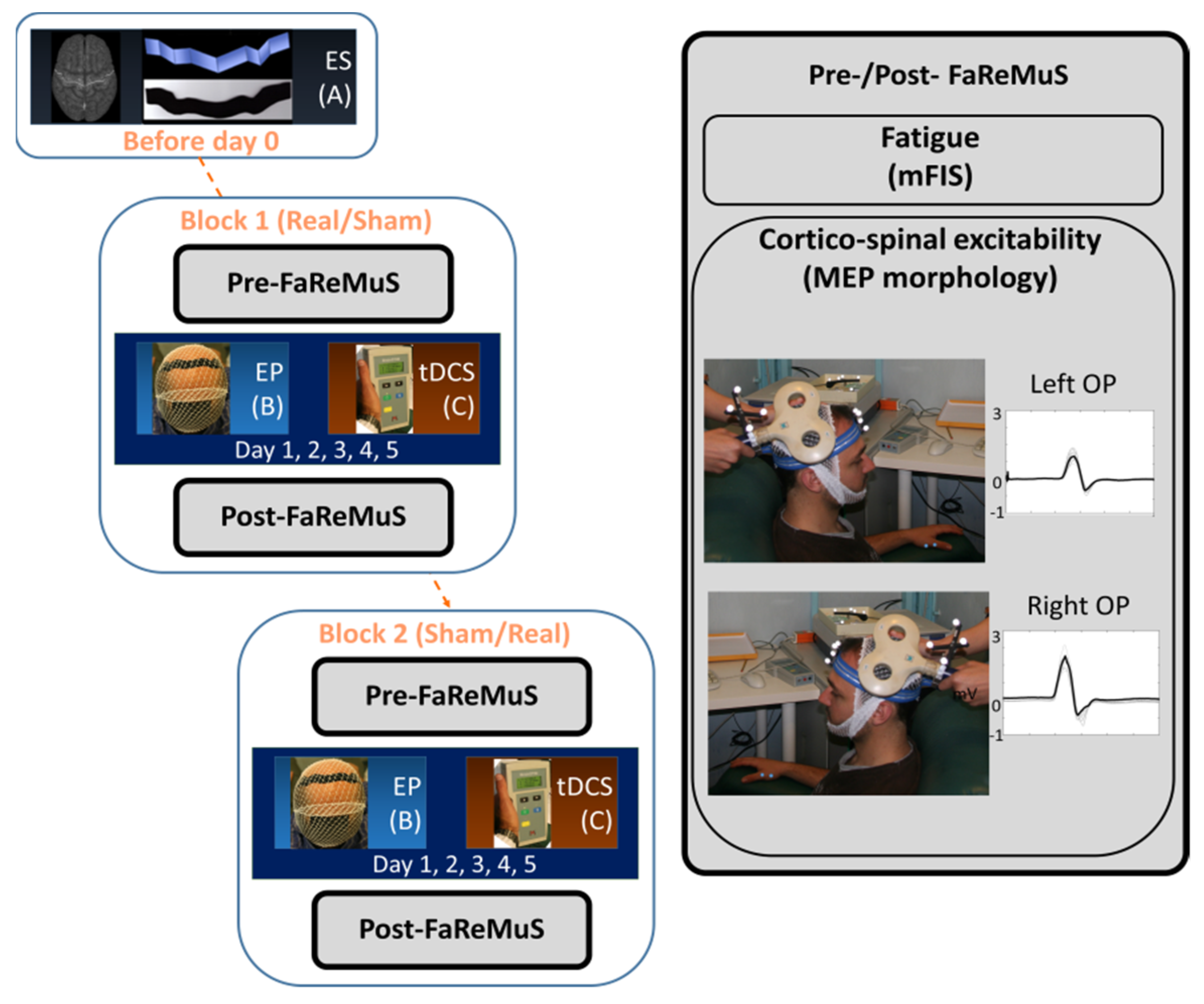
2.1. Study Design
- Absence of clinical or radiological evidence of disease activity (NEDA) for at least 3 months preceding the study;
- Low degree of disability as estimated by Expanded Disability Status Scale (EDSS, Kurtzke 1983) < 2.5;
- Fatigue as estimated by mFIS > 30.
- Exclusion Criteria were as follows:
- Current or prior (within less than 12 weeks before enrolment) exposure to psychotropic drug(s) (antidepressant, anxiolytic, antipsychotics, anticonvulsants, and myorelaxant drugs);
- Coexistence of other condition(s) potentially associated with fatigue (i.e., anemia and pregnancy);
- Current or prior (within less than 4 weeks before enrolment) exposure to anti-fatigue products;
- History of epilepsy.
Faremus Treatment (5-Day Anodal tDCS with Personalized S1 Electrode)
2.2. MEP Collection and Analysis
Stimulation and Recording Setup
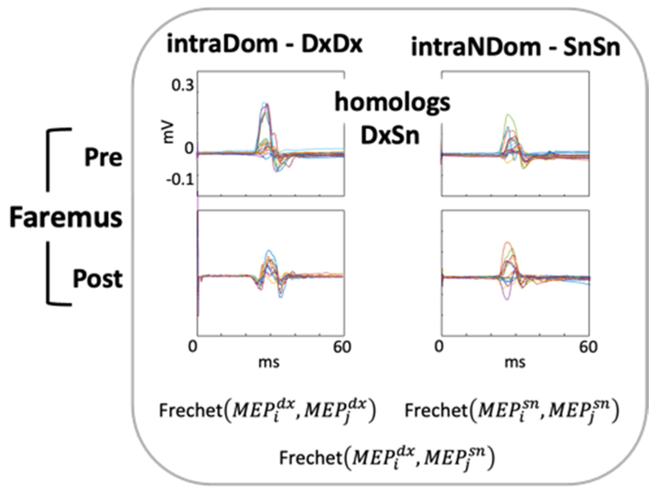
2.3. MEP Morphology Similarity
2.4. Statistical Analysis
2.5. Data Availability
3. Results
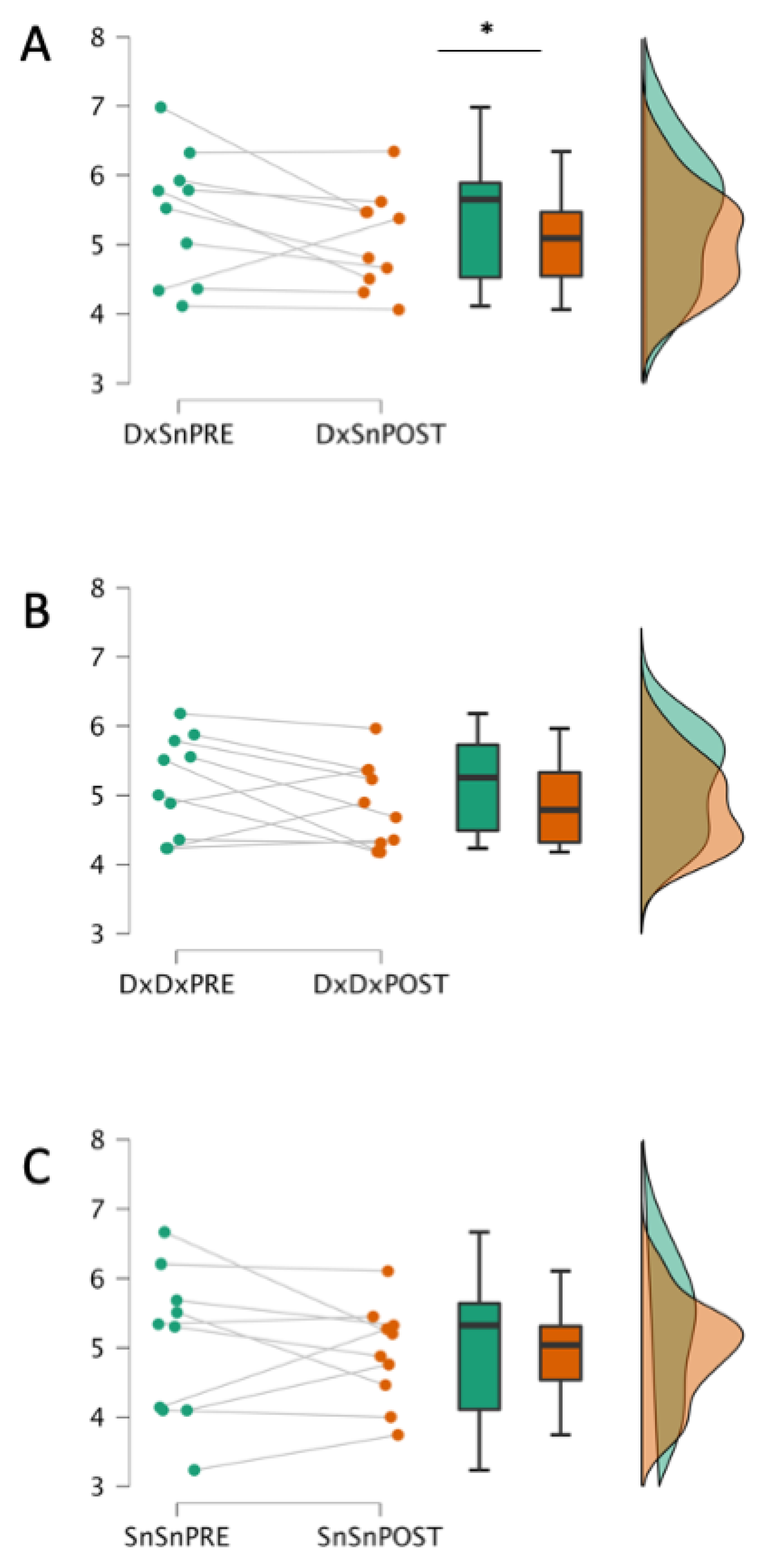
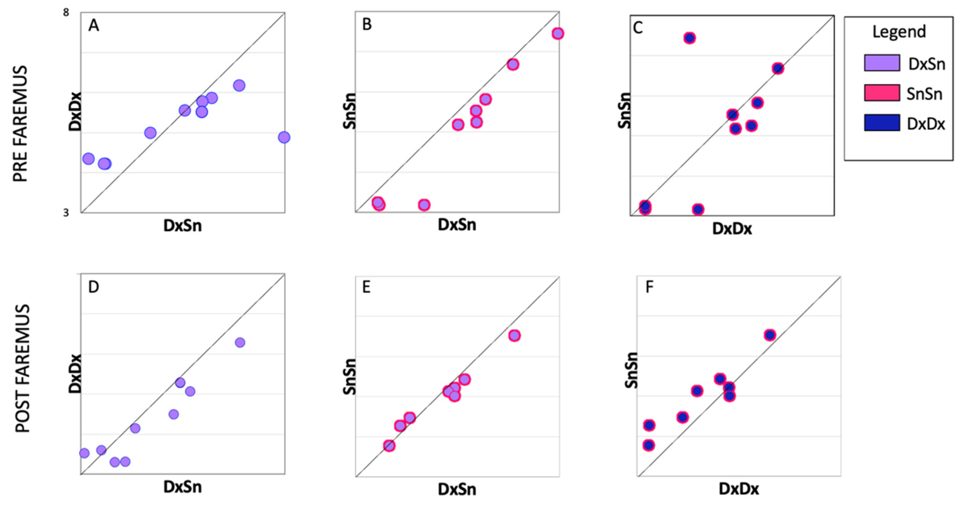
3.1. Faremus Effects on MEP Morphology Similarity as An Index of the Two CSTs’ Homology
3.2. Relationships between MEP Shape Similarities Changes and Fatigue Amelioration
3.3. MEP Amplitude and Latency
4. Discussion
4.1. CST Asymmetries and Fatigue
4.2. CST Homology Modified by Faremus S1 Neuromodulation
4.3. Central More Than Peripheral Origin of MS Fatigue
4.4. MEP Shape vs. Amplitude
4.5. Limitations of the Present Work
5. Conclusions
Author Contributions
Funding
Institutional Review Board Statement
Informed Consent Statement
Data Availability Statement
Acknowledgments
Conflicts of Interest
References
- Filippi, M.; Bar-Or, A.; Piehl, F.; Preziosa, P.; Solari, A.; Vukusic, S.; Rocca, M.A. Multiple Sclerosis. Nat. Rev. Dis. Primers 2018, 4, 43. [Google Scholar] [CrossRef]
- Kesselring, J.; Beer, S. Symptomatic Therapy and Neurorehabilitation in Multiple Sclerosis. Lancet Neurol. 2005, 4, 643–652. [Google Scholar] [CrossRef]
- Rudroff, T.; Kindred, J.H.; Ketelhut, N.B. Fatigue in Multiple Sclerosis: Misconceptions and Future Research Directions. Front. Neurol. 2016, 7, 122. [Google Scholar] [CrossRef] [PubMed] [Green Version]
- Enoka, R.M.; Almuklass, A.M.; Alenazy, M.; Alvarez, E.; Duchateau, J. Distinguishing between Fatigue and Fatigability in Multiple Sclerosis. Neurorehabil. Neural. Repair 2021, 35, 960–973. [Google Scholar] [CrossRef] [PubMed]
- Nourbakhsh, B.; Revirajan, N.; Morris, B.; Cordano, C.; Creasman, J.; Manguinao, M.; Krysko, K.; Rutatangwa, A.; Auvray, C.; Aljarallah, S.; et al. Safety and Efficacy of Amantadine, Modafinil, and Methylphenidate for Fatigue in Multiple Sclerosis: A Randomised, Placebo-Controlled, Crossover, Double-Blind Trial. Lancet Neurol. 2021, 20, 38–48. [Google Scholar] [CrossRef] [PubMed]
- Bertoli, M.; Tecchio, F. Fatigue in Multiple Sclerosis: Does the Functional or Structural Damage Prevail? SAGE Publications Ltd.: New York, NY, USA, 2020; Volume 26, pp. 1809–1815. [Google Scholar]
- Yusuf, A.; Koski, L. A Qualitative Review of the Neurophysiological Underpinnings of Fatigue in Multiple Sclerosis. J. Neurol. Sci. 2013, 330, 4–9. [Google Scholar] [CrossRef]
- Tecchio, F.; Zito, G.; Zappasodi, F.; Dell’Acqua, M.L.; Landi, D.; Nardo, D.; Lupoi, D.; Rossini, P.M.; Filippi, M.M. Intra-Cortical Connectivity in Multiple Sclerosis: A Neurophysiological Approach. Brain 2008, 131, 1783–1792. [Google Scholar] [CrossRef] [PubMed] [Green Version]
- Pellicano, C.; Gallo, A.; Li, X.; Ikonomidou, V.N.; Evangelou, I.E.; Ohayon, J.M.; Stern, S.K.; Ehrmantraut, M.; Cantor, F.; McFarland, H.F.; et al. Relationship of Cortical Atrophy to Fatigue in Patients with Multiple Sclerosis. Arch. Neurol. 2010, 67, 447–453. [Google Scholar] [CrossRef] [Green Version]
- Dell’Acqua, M.L.; Landi, D.; Zito, G.; Zappasodi, F.; Lupoi, D.; Rossini, P.M.; Filippi, M.M.; Tecchio, F. Thalamocortical Sensorimotor Circuit in Multiple Sclerosis: An Integrated Structural and Electrophysiological Assessment. Hum. Brain Mapp. 2010, 31, 1588–1600. [Google Scholar] [CrossRef]
- Porcaro, C.; Cottone, C.; Cancelli, A.; Rossini, P.M.; Zito, G.; Tecchio, F. Cortical Neurodynamics Changes Mediate the Efficacy of a Personalized Neuromodulation against Multiple Sclerosis Fatigue. Sci. Rep. 2019, 9, 18213. [Google Scholar] [CrossRef] [Green Version]
- Leocani, L.; Colombo, B.; Magnani, G.; Martinelli-Boneschi, F.; Cursi, M.; Rossi, P.; Martinelli, V.; Comi, G. Fatigue in Multiple Sclerosis Is Associated with Abnormal Cortical Activation to Voluntary Movement--EEG Evidence. Neuroimage 2001, 13, 1186–1192. [Google Scholar] [CrossRef] [PubMed]
- Liepert, J.; Mingers, D.; Heesen, C.; Bäumer, T.; Weiller, C. Motor Cortex Excitability and Fatigue in Multiple Sclerosis: A Transcranial Magnetic Stimulation Study. Mult. Scler. 2005, 11, 316–321. [Google Scholar] [CrossRef] [PubMed]
- Nielsen, J.F.; Norgaard, P. Increased Post-Exercise Facilitation of Motor Evoked Potentials in Multiple Sclerosis. Clin. Neurophysiol. 2002, 113, 1295–1300. [Google Scholar] [CrossRef]
- Thickbroom, G.W.; Sacco, P.; Kermode, A.G.; Archer, S.A.; Byrnes, M.L.; Guilfoyle, A.; Mastaglia, F.L. Central Motor Drive and Perception of Effort during Fatigue in Multiple Sclerosis. J. Neurol. 2006, 253, 1048–1053. [Google Scholar] [CrossRef] [PubMed]
- Famm, K.; Litt, B.; Tracey, K.J.; Boyden, E.S.; Slaoui, M. Drug Discovery: A Jump-Start for Electroceuticals. Nature 2013, 496, 159–161. [Google Scholar] [CrossRef] [PubMed] [Green Version]
- García-Alías, G.; del Valle, J.; Delgado-Martínez, I.; Navarro, X. Electroceutical Therapies for Injuries of the Nervous System. In Handbook of Innovations in Central Nervous System Regenerative Medicine; Elsevier: Amsterdam, The Netherlands, 2020; pp. 511–537. [Google Scholar]
- Paulus, W. Transcranial Electrical Stimulation (TES—TDCS; TRNS, TACS) Methods. Neuropsychol. Rehabil. 2011, 21, 602–617. [Google Scholar] [CrossRef]
- Nitsche, M.A.; Cohen, L.G.; Wassermann, E.M.; Priori, A.; Lang, N.; Antal, A.; Paulus, W.; Hummel, F.; Boggio, P.S.; Fregni, F.; et al. Transcranial Direct Current Stimulation: State of the Art 2008. Brain Stimul. 2008, 1, 206–223. [Google Scholar] [CrossRef]
- Brunoni, A.R.; Nitsche, M.A.; Bolognini, N.; Bikson, M.; Wagner, T.; Merabet, L.; Edwards, D.J.; Valero-Cabre, A.; Rotenberg, A.; Pascual-Leone, A.; et al. Clinical Research with Transcranial Direct Current Stimulation (TDCS): Challenges and Future Directions. Brain Stimul. 2012, 5, 175–195. [Google Scholar] [CrossRef] [Green Version]
- Romero Lauro, L.J.; Rosanova, M.; Mattavelli, G.; Convento, S.; Pisoni, A.; Opitz, A.; Bolognini, N.; Vallar, G. TDCS Increases Cortical Excitability: Direct Evidence from TMS-EEG. Cortex 2014, 58, 99–111. [Google Scholar] [CrossRef]
- Liu, M.; Fan, S.; Xu, Y.; Cui, L. Non-Invasive Brain Stimulation for Fatigue in Multiple Sclerosis Patients: A Systematic Review and Meta-Analysis. Mult. Scler. Relat. Disord. 2019, 36, 101375. [Google Scholar] [CrossRef]
- Gianni, E.; Bertoli, M.; Simonelli, I.; Paulon, L.; Tecchio, F.; Pasqualetti, P. TDCS Randomized Controlled Trials in No-Structural Diseases: A Quantitative Review. Sci. Rep. 2021, 11, 16311. [Google Scholar] [CrossRef]
- Capone, F.; Motolese, F.; Falato, E.; Rossi, M.; Di Lazzaro, V. The Potential Role of Neurophysiology in the Management of Multiple Sclerosis-Related Fatigue. Front. Neurol. 2020, 11, 251. [Google Scholar] [CrossRef] [Green Version]
- Cogiamanian, F.; Marceglia, S.; Ardolino, G.; Barbieri, S.; Priori, A. Improved Isometric Force Endurance after Transcranial Direct Current Stimulation over the Human Motor Cortical Areas. Eur. J. Neurosci. 2007, 26, 242–249. [Google Scholar] [CrossRef]
- Tecchio, F.; Cancelli, A.; Cottone, C.; Zito, G.; Pasqualetti, P.; Ghazaryan, A.; Rossini, P.M.; Filippi, M.M. Multiple Sclerosis Fatigue Relief by Bilateral Somatosensory Cortex Neuromodulation. J. Neurol. 2014, 261, 1552–1558. [Google Scholar] [CrossRef]
- Cancelli, A.; Cottone, C.; Giordani, A.; Migliore, S.; Lupoi, D.; Porcaro, C.; Mirabella, M.; Rossini, P.M.; Filippi, M.M.; Tecchio, F. Personalized, Bilateral Whole-Body Somatosensory Cortex Stimulation to Relieve Fatigue in Multiple Sclerosis. Mult. Scler. J. 2018, 24, 1366–1374. [Google Scholar] [CrossRef] [PubMed]
- Tecchio, F.; Cancelli, A.; Pizzichino, A.; L’Abbate, T.; Gianni, E.; Bertoli, M.; Paulon, L.; Zannino, S.; Giordani, A.; Lupoi, D.; et al. Home Treatment against Fatigue in Multiple Sclerosis by a Personalized, Bilateral Whole-Body Somatosensory Cortex Stimulation. Mult. Scler. Relat. Disord. 2022, 63, 103813. [Google Scholar] [CrossRef] [PubMed]
- Tecchio, F.; Cancelli, A.; Cottone, C.; Tomasevic, L.; Devigus, B.; Zito, G.; Ercolani, M.; Carducci, F. Regional Personalized Electrodes to Select Transcranial Current Stimulation Target. Front. Hum. Neurosci. 2013, 7, 131. [Google Scholar] [CrossRef] [Green Version]
- Radhu, N.; Blumberger, D.M.; Daskalakis, Z.J. Cortical Inhibition and Excitation in Neuropsychiatric Disorders Using Transcranial Magnetic Stimulation. Transcranial Direct Current Stimulation in Neuropsychiatric Disorders: Clinical Principles and Management; Springer: Cham, Switzerland, 2016; pp. 85–102. [Google Scholar] [CrossRef]
- Cancelli, A.; Cottone, C.; Di Giorgio, M.; Carducci, F.; Tecchio, F. Personalizing the Electrode to Neuromodulate an Extended Cortical Region. Brain Stimul. 2015, 8, 555–560. [Google Scholar] [CrossRef]
- Ashrafi, A.; Mohseni-Bandpei, M.A.; Seydi, M. The Effect of TDCS on the Fatigue in Patients with Multiple Sclerosis: A Systematic Review of Randomized Controlled Clinical Trials. J Clin Neurosci. 2020, 78, 277–283. [Google Scholar] [CrossRef] [PubMed]
- Mortezanejad, M.; Ehsani, F.; Masoudian, N.; Zoghi, M.; Jaberzadeh, S. Comparing the Effects of Multi-Session Anodal Trans-Cranial Direct Current Stimulation of Primary Motor and Dorsolateral Prefrontal Cortices on Fatigue and Quality of Life in Patients with Multiple Sclerosis: A Double-Blind, Randomized, Sham-Controlled Trial. Clin. Rehabil. 2020, 34, 1103–1111. [Google Scholar] [CrossRef]
- Carson, R.G. Inter-Hemispheric Inhibition Sculpts the Output of Neural Circuits by Co-Opting the Two Cerebral Hemispheres. J. Physiol. 2020, 598, 4781–4802. [Google Scholar] [CrossRef]
- Merchant, H.; Naselaris, T.; Georgopoulos, A.P. Dynamic Sculpting of Directional Tuning in the Primate Motor Cortex during Three-Dimensional Reaching. J. Neurosci. 2008, 28, 9164–9172. [Google Scholar] [CrossRef] [Green Version]
- Georgopoulos, A.P.N.; Stefanis, C. The Motor Cortical Circuit. In Handbook of Brain Microcircuits; Oxford University Press: Oxford, UK, 2013; pp. 39–45. [Google Scholar]
- Buyukturkoglu, K.; Porcaro, C.; Cottone, C.; Cancelli, A.; Inglese, M.; Tecchio, F. Simple Index of Functional Connectivity at Rest in Multiple Sclerosis Fatigue. Clin. Neurophysiol. 2017, 128, 807–813. [Google Scholar] [CrossRef] [PubMed]
- Cogliati Dezza, I.; Zito, G.; Tomasevic, L.; Filippi, M.M.; Ghazaryan, A.; Porcaro, C.; Squitti, R.; Ventriglia, M.; Lupoi, D.; Tecchio, F. Functional and Structural Balances of Homologous Sensorimotor Regions in Multiple Sclerosis Fatigue. J. Neurol. 2015, 262, 614–622. [Google Scholar] [CrossRef] [PubMed]
- Manson, S.C.; Palace, J.; Frank, J.A.; Matthews, P.M. Loss of Interhemispheric Inhibition in Patients with Multiple Sclerosis Is Related to Corpus Callosum Atrophy. Exp. Brain Res. 2006, 174, 728–733. [Google Scholar] [CrossRef] [PubMed]
- Zito, G.; Luders, E.; Tomasevic, L.; Lupoi, D.; Toga, A.W.; Thompson, P.M.; Rossini, P.M.; Filippi, M.M.; Tecchio, F. Inter-Hemispheric Functional Connectivity Changes with Corpus Callosum Morphology in Multiple Sclerosis. Neuroscience 2014, 266, 47–55. [Google Scholar] [CrossRef] [Green Version]
- Dum, R.P.; Strick, P.L. The Origin of Corticospinal Projections from the Premotor Areas in the Frontal Lobe. J. Neurosci. 1991, 11, 667–689. [Google Scholar] [CrossRef] [Green Version]
- Seo, J.P.; Jang, S.H. Different Characteristics of the Corticospinal Tract According to the Cerebral Origin: DTI Study. American J. Neuroradiol. 2013, 34, 1359–1363. [Google Scholar] [CrossRef] [Green Version]
- Lemon, R.N. Descending Pathways in Motor Control. Annu Rev Neurosci 2008, 31, 195–218. [Google Scholar] [CrossRef] [Green Version]
- Chaves, A.R.; Wallack, E.M.; Kelly, L.P.; Pretty, R.W.; Wiseman, H.D.; Chen, A.; Moore, C.S.; Stefanelli, M.; Ploughman, M. Asymmetry of Brain Excitability: A New Biomarker That Predicts Objective and Subjective Symptoms in Multiple Sclerosis. Behav. Brain Res. 2019, 359, 281–291. [Google Scholar] [CrossRef]
- Bauer, C.; Dyrby, T.B.; Sellebjerg, F.; Madsen, K.S.; Svolgaard, O.; Blinkenberg, M.; Siebner, H.R.; Andersen, K.W. Motor Fatigue Is Associated with Asymmetric Connectivity Properties of the Corticospinal Tract in Multiple Sclerosis. Neuroimage Clin. 2020, 28, 102393. [Google Scholar] [CrossRef]
- Barker, A.T.; Jalinous, R.; Freeston, I.L. Non-Invasive Magnetic Stimulation of Human Motor Cortex. Lancet 1985, 1, 1106–1107. [Google Scholar] [CrossRef] [PubMed]
- Tecchio, F.; Cecconi, F.; Colamartino, E.; Padalino, M.; Valci, L.; Reinert, M. The Morphology of Somatosensory Evoked Potentials During Middle Cerebral Artery Aneurysm Clipping (MoSAC): A Pilot Study. Clin. EEG Neurosci. 2020, 51, 130–136. [Google Scholar] [CrossRef] [PubMed]
- Tecchio, F.; Pasqualetti, P.; Pizzella, V.; Romani, G.; Rossini, P.M. Morphology of Somatosensory Evoked Fields: Inter-Hemispheric Similarity as a Parameter for Physiological and Pathological Neural Connectivity. Neurosci. Lett. 2000, 287, 203–206. [Google Scholar] [CrossRef]
- Tecchio, F.; Zappasodi, F.; Pasqualetti, P.; Rossini, P.M. Neural Connectivity in Hand Sensorimotor Brain Areas: An Evaluation by Evoked Field Morphology. Hum. Brain Mapp. 2005, 24, 99–108. [Google Scholar] [CrossRef]
- Yperman, J.; Becker, T.; Valkenborg, D.; Hellings, N.; Cambron, M.; Dive, D.; Laureys, G.; Popescu, V.; Van Wijmeersch, B.; Peeters, L.M. Deciphering the Morphology of Motor Evoked Potentials. Front. Neuroinform. 2020, 14, 28. [Google Scholar] [CrossRef]
- Fréchet, M.M. Sur Quelques Points Du Calcul Fonctionnel. Rend. Circ. Mat. Palermo. 1906, 22, 1–72. [Google Scholar] [CrossRef] [Green Version]
- Wylie, T.; Zhu, B. Following a Curve with the Discrete Fréchet Distance. Theor. Comput. Sci 2014, 556, 34–44. [Google Scholar] [CrossRef]
- Pagliara, M.R.; Cecconi, F.; Pasqualetti, P.; Bertoli, M.; Armonaite, K.; Gianni, E.; Grifoni, J.; L’Abbate, T.; Marinozzi, F.; Conti, L.; et al. On the Homology of the Dominant and Non-Dominant Corticospinal Tracts: A Novel Neurophysiological Assessment. Brain Sci. 2023, 13, 278. [Google Scholar] [CrossRef] [PubMed]
- Téllez, N.; Río, J.; Tintoré, M.; Nos, C.; Galán, I.; Montalban, X. Does the Modified Fatigue Impact Scale Offer a More Comprehensive Assessment of Fatigue in MS? Mult. Scler. 2005, 11, 198–202. [Google Scholar] [CrossRef]
- McDonald, W.I.; Compston, A.; Edan, G.; Goodkin, D.; Hartung, H.P.; Lublin, F.D.; McFarland, H.F.; Paty, D.W.; Polman, C.H.; Reingold, S.C.; et al. Recommended Diagnostic Criteria for Multiple Sclerosis: Guidelines from the International Panel on the Diagnosis of Multiple Sclerosis. Ann. Neurol. 2001, 50, 121–127. [Google Scholar] [CrossRef] [PubMed]
- Rossini, P.M.; Barker, A.T.; Berardelli, A.; Caramia, M.D.; Caruso, G.; Cracco, R.Q.; Dimitrijević, M.R.; Hallett, M.; Katayama, Y.; Lücking, C.H.; et al. Non-Invasive Electrical and Magnetic Stimulation of the Brain, Spinal Cord and Roots: Basic Principles and Procedures for Routine Clinical Application. Report of an IFCN Committee. Electroencephalogr. Clin. Neurophysiol. 1994, 91, 79–92. [Google Scholar] [CrossRef] [PubMed]
- Cottone, C.; Cancelli, A.; Pasqualetti, P.; Porcaro, C.; Salustri, C.; Tecchio, F. A New, High-Efficacy, Noninvasive Transcranial Electric Stimulation Tuned to Local Neurodynamics. J. Neurosci. 2018, 38, 586–594. [Google Scholar] [CrossRef] [PubMed] [Green Version]
- Suckley, J.J.; Waters, T.J.; Tran, M.; Stapley, P.J.; Shemmell, J.; Walsh, J.A.; McAndrew, D.J. Randomising Stimulus Intensity Improves the Variability and Reliability of the Assessment of Corticospinal Excitability. J. Neurosci. Methods 2020, 342, 108813. [Google Scholar] [CrossRef]
- Eiter, T.; Mannila, H. Computing Discrete Frechet Distance; Technical report CD-TR 94/64; Technical University of Vienna: Vienna, Austria, 1994. [Google Scholar]
- Pellegrino, G.; Tomasevic, L.; Tombini, M.; Assenza, G.; Bravi, M.; Sterzi, S.; Giacobbe, V.; Zollo, L.; Guglielmelli, E.; Cavallo, G.; et al. Inter-Hemispheric Coupling Changes Associate with Motor Improvements after Robotic Stroke Rehabilitation. Restor. Neurol. Neurosci. 2012, 30, 497–510. [Google Scholar] [CrossRef]
- Rossi, S.; Pasqualetti, P.; Tecchio, F.; Sabato, A.; Rossini, P.M. Modulation of Corticospinal Output to Human Hand Muscles Following Deprivation of Sensory Feedback. Neuroimage 1998, 8, 163–175. [Google Scholar] [CrossRef]
- Fink, A.J.P.; Croce, K.R.; Huang, Z.J.; Abbott, L.F.; Jessell, T.M.; Azim, E. Presynaptic Inhibition of Spinal Sensory Feedback Ensures Smooth Movement HHS Public Access. Nature 2014, 509, 43–48. [Google Scholar] [CrossRef] [Green Version]
- Tomasevic, L.; Zito, G.; Pasqualetti, P.; Filippi, M.M.; Landi, D.; Ghazaryan, A.; Lupoi, D.; Porcaro, C.; Bagnato, F.; Rossini, P.M.; et al. Cortico-Muscular Coherence as an Index of Fatigue in Multiple Sclerosis. Mult. Scler. J. 2013, 19, 334–343. [Google Scholar] [CrossRef]
- Mordillo-Mateos, L.; Soto-Leon, V.; Torres-Pareja, M.; Peinado-Palomino, D.; Mendoza-Laiz, N.; Alonso-Bonilla, C.; Dileone, M.; Rotondi, M.; Aguilar, J.; Oliviero, A. Fatigue in Multiple Sclerosis: General and Perceived Fatigue Does Not Depend on Corticospinal Tract Dysfunction. Front. Neurol. 2019, 10, 339. [Google Scholar] [CrossRef]


| Mean Median | SD [Min-Max] | ||
|---|---|---|---|
| Patients’ Personal and Clinical Profile | Sex 9 Women, 1 Man | ||
| Age (years) | 35.3 | 9.3 | |
| Disease duration (years) | 3.2 | 2.0 | |
| Annual relapse rate | 0 | [0,1] | |
| Expanded Disability Status Scale | 0 | [0–2.5] | |
| Beck Depression Inventory | 8.5 | 0.7 | |
| Modified Fatigue Impact Scale | Pre Faremus | 47 * | 15 |
| Post Faremus | 38 * | 22 | |
| % | 27 | 33 | |
| MEP Latency | MEP Amplitude | |||
|---|---|---|---|---|
| MEP dx | MEP sn | MEP dx | MEP sn | |
| Pre Faremus | 27.1 | 27.3 | 324 | 445 |
| (7.2) | (3.2) | (793) | (651) | |
| Post Faremus | 27.6 | 27.5 | 291 | 329 |
| (6.9) | (4.2) | (925) | (378) | |
Disclaimer/Publisher’s Note: The statements, opinions and data contained in all publications are solely those of the individual author(s) and contributor(s) and not of MDPI and/or the editor(s). MDPI and/or the editor(s) disclaim responsibility for any injury to people or property resulting from any ideas, methods, instructions or products referred to in the content. |
© 2023 by the authors. Licensee MDPI, Basel, Switzerland. This article is an open access article distributed under the terms and conditions of the Creative Commons Attribution (CC BY) license (https://creativecommons.org/licenses/by/4.0/).
Share and Cite
Bertoli, M.; Tataranni, A.; Porziani, S.; Pasqualetti, P.; Gianni, E.; Grifoni, J.; L’Abbate, T.; Armonaite, K.; Conti, L.; Cancelli, A.; et al. Effects on Corticospinal Tract Homology of Faremus Personalized Neuromodulation Relieving Fatigue in Multiple Sclerosis: A Proof-of-Concept Study. Brain Sci. 2023, 13, 574. https://doi.org/10.3390/brainsci13040574
Bertoli M, Tataranni A, Porziani S, Pasqualetti P, Gianni E, Grifoni J, L’Abbate T, Armonaite K, Conti L, Cancelli A, et al. Effects on Corticospinal Tract Homology of Faremus Personalized Neuromodulation Relieving Fatigue in Multiple Sclerosis: A Proof-of-Concept Study. Brain Sciences. 2023; 13(4):574. https://doi.org/10.3390/brainsci13040574
Chicago/Turabian StyleBertoli, Massimo, Angela Tataranni, Susanna Porziani, Patrizio Pasqualetti, Eugenia Gianni, Joy Grifoni, Teresa L’Abbate, Karolina Armonaite, Livio Conti, Andrea Cancelli, and et al. 2023. "Effects on Corticospinal Tract Homology of Faremus Personalized Neuromodulation Relieving Fatigue in Multiple Sclerosis: A Proof-of-Concept Study" Brain Sciences 13, no. 4: 574. https://doi.org/10.3390/brainsci13040574