Article

Standardization of Strategies to Perform a Parafascicular Tubular Approach for the Resection of Brain Tumors in Eloquent Areas

by
Nadin J. Abdala-Vargas
1,
Giuseppe E. Umana
2,*,
Javier G. Patiño-Gomez
1,
Edgar Ordoñez-Rubiano
1,
Hernando A. Cifuentes-Lobelo
1,
Paolo Palmisciano
3,
Gianluca Ferini
4,
Anna Viola
4,
Valentina Zagardo
4,
Daniel Casanova-Martínez
5,
Ottavio S. Tomasi
6,
Alvaro Campero
7 and
Matias Baldoncini
8
1
Neurosurgery Department, Fundación Universitaria de Ciencias de la Salud (FUCS), Hospital Infantil Universitario de San José, Cra. 19 #8A-32, Bogotá 111221, Colombia
2
Department of Neurosurgery, Trauma and Gamma-Knife Center Cannizzaro Hospital, 95126 Catania, Italy
3
Department of Neurosurgery, University of Cincinnati College of Medicine, Cincinnati, OH 45220, USA
4
Department of Radiation Oncology, REM Radioterapia s.r.l., 95029 Vaigrande, Italy
5
San Felipe Campus, Medical Faculty, University of Valparaíso, Valparaíso 2170000, Chile
6
Department of Neurosurgery, Christian-Doppler-Klinik, Paracelsus Private Medical University, 5020 Salzburg, Austria
7
Department of Neurological Surgery, Padilla Hospital, Tucumán T4000, Argentina
8
Department of Neurological Surgery, San Fernando Hospital, Buenos Aires B1646, Argentina
*
Author to whom correspondence should be addressed.
Brain Sci. 2023, 13(3), 498; https://doi.org/10.3390/brainsci13030498
Submission received: 15 February 2023 / Revised: 10 March 2023 / Accepted: 13 March 2023 / Published: 15 March 2023
(This article belongs to the Special Issue Novel Strategies for Surgery of Brain Tumors in Eloquent Areas)

Abstract

:
Objective: The aim of this work is to define a methodological strategy for the minimally invasive tubular retractor (MITR) parafascicular transulcal approach (PTA) for the management of brain tumors sited in eloquent areas. Methods: An observational prospective study was designed to evaluate the benefits of PTA associated with MITRs, tractography and intraoperative cortical stimulation. They study was conducted from June 2018 to June 2021. Information regarding white matter tracts was processed, preventing a potential damage during the approach and/or resection. All patients older than 18 years who had a single brain tumor lesion were included in the study. Patients with a preoperative Karnofsky Performance Scale (KPS) score greater than 70% and a Glasgow Coma Scale (GCS) score > 14 points were included. Results: 72 patients were included in the study, the mean age was 49.6, the most affected gender was male, 12.5% presented aphasia, 11.1% presented paraphasia, 41.6% had motor deficit, 9.7% had an affection in the optic pathway, the most frequently affected region was the frontal lobe (26.3%), the most frequent lesions were high-grade gliomas (34.7%) and the measurement of the incisions was on average 5.58 cm. Of the patients, 94.4% underwent a total macroscopic resection and 90.2% did not present new postoperative neurological deficits. In all cases, a PTA was used. Conclusion: Tubular minimally invasive approaches (MIAs) allow one to perform maximal safe resection of brain tumors in eloquent areas, through small surgical corridors. Future comparative studies between traditional and minimally invasive techniques are required to further investigate the potential of these surgical nuances.

1. Introduction

From the first non-accidental craniotomies, in the late Paleolithic and early Neonolithic periods, to the present day, craniotomy has undergone substantial modifications. Initially, large craniotomies with great brain exposure were necessary for adequate anatomical orientation, better intra-surgical luminosity and larger surgical route considering the size of the surgical instruments and the number of people required for an intervention [1].
Similarly to the advent of microsurgery, the role of minimally invasive techniques in the field of brain neurosurgery has grown in recent decades [2]. The introduction of the neuroendoscope in the management of intracranial pathologies has been known since 1910 [3], but it was popularized only starting from 1988, when Kelly et al. described the surgical technique for the resection of brain lesions with tubular retractor and stereotaxis [4]. Some years before, Jane JA proposed the modification of the orbitozygomatic approach in the supraorbital approach, by means of a minimal craniotomy area that allowed less brain manipulation and theoretically a comparable surgical corridor, a technique which was further popularized by Reisch and Perneczky [5,6].
New surgical technologies, which include high-power lights, improved anesthetic control, high-definition microscopes, advanced imaging and stereotaxic navigation, have led to the planning of smaller, tumor-centered craniotomies that can represent lower complication rates and shorter hospital stays [7,8]. The mini-craniotomy approaches or the keyhole approaches are usually defined as a minimally invasive alternatives to a conventional craniotomy, and they offer the same advantageous approach with less brain manipulation [9]. Since its design, this approach has been partially accepted and has been implemented in the management of selected neurosurgical lesions.
Brain retraction is necessary to access skull base or intra-axial lesions and this can cause brain injury and related neurological or vascular damage [10]. At the end of the 19th century, it was clear that the excessive use of retraction, the retraction time and the uneven distribution of pressure over brain tissue play a key role for a safer brain manipulation/retraction. Thus, retraction systems have been implemented and developed like tubular retractors, and new surgical nuances have been introduced and continue to evolve nowadays. Minimally invasive tubular retractors (MITRs) are able to create a safe approach, theoretically minimizing brain damage, approach time and improving the accuracy with respect to accessing brain lesions. The aim of this study is to propose a methodological strategy for the planning of a parafascicular transulcal approach (PTA) with the help of MITRs in the management of brain tumors in eloquent areas.

2. Materials and Methods

2.1. Study Design

An observational prospective study was conducted from June 2018 to June 2021. The study was carried out after the approval of the Ethics and Research Committee of the Hospital Infantil Universitario de San José, Bogotá, Colombia. Written consent was obtained from each patient for the use and manipulation of the diagnostic and intraoperative images. The confidentiality of personal data was protected by assigning a number to each patient. All patients older than 18 years who had a single brain tumor lesion in eloquent areas were included in the study. Patients with a preoperative Karnofsky Performance Scale (KPS) score greater than 70% and a Glasgow Coma Scale (GSC) score > 14 points were included. Muscle strength was assessed in each patient using the Medical Research Council (MRC) muscle strength scale, before and after the procedure. In patients with lesions in the occipital lobe, visual acuity was evaluated by performing computerized visual campimetry. Patients were evaluated to discern whether any type of aphasia was present or not. The location of the tumors was recorded in the cerebral regions: frontal, parietal, occipital, temporal, insular, frontoparietal, parietooccipital, frontotemporal, parietotemporal and temporo-occipital. Brain tumors were classified into low-grade gliomas (LGGs), high-grade gliomas (HGGs), metastases and others.

2.2. Diagnostic Imaging

Gadolinium MRI data were acquired with a General Electric Signa Excite HDXT scanner (1.5T GE Healthcare, Milwaukee, WI, USA). For each patient, a presurgical brain magnetic resonance imaging (MRI) image was acquired with diffusion tensor imaging (DTI) of 140 T1 slices (1 mm thick) without GAP. After obtaining the images, they were merged and processed with the help of the neuronavigation system (Kick, Curve TM and SmartBrush, BrainLab, Munich, Germany). Information regarding white matter tracts was processed, preventing potential damage during the approach and/or resection. According to the location of the tumor, the following tracts were considered for reconstruction: Aslant’s tract, superior longitudinal fasciculus (SLF), arcuate fasciculus, inferior longitudinal fasciculus (ILF), uncinate fasciculus (UF), geniculo-calcarine fasciculus (GCT), corticospinal tract (CST) and thalamocortical tract. A Gadolinium MRI scan and a CT with contrast were performed 24–48 h postoperatively in each case.

2.3. Incision, Craniotomy and Extent of Resection

The size of the incision was determined intraoperatively with the help of a ruler. The size of the craniotomy was measured on its diameter using the bone algorithm of the postoperative CT scan. The degree of macroscopic resection was evaluated, comparing the pre/postoperative tumor volume and post-contrast enhancement characteristics. Tumor volume was calculated with the neuronavigation system software (Elements, Brainlab, Munich, Germany). The extent of resection (EOR) was classified as follows: total: 100%; near-total: >90%; and partial: <70%.

2.4. Intraoperative Technical Features

Intraoperative cortical and subcortical mapping was performed in all cases. A bipolar electrode (NIM—Eclipse NS—Medtronic 6743 Southpoint Drive N Jacksonville, USA) was used, with 1 mm electrodes and 0.5 mm distance between each one. The amplitude of the current was progressively increased stepwise by 1 mA until 2 mA was reached. We stimulated with biphasic square wave pulses of 1 ms at 60 Hz, with a maximum train duration of 4 s. Functional mapping was performed according to the preoperative planning related to the neuroimaging, using intraopeatively with the neuronavigation, and compared to the cortical stimulation responses. The marked areas were deemed inoperable. ViewSite-type MITRs (Brain Access System, Vycor Medical Inc. 951 Broken Sound Park Away, Suite 320, Boca Raton, USA) were used. In each case, the width, length and height of the MITRs used were recorded in the database. The chosen MITRs’ height depended on the distance between the outer table of the cranium and the superficial edge of the tumor.

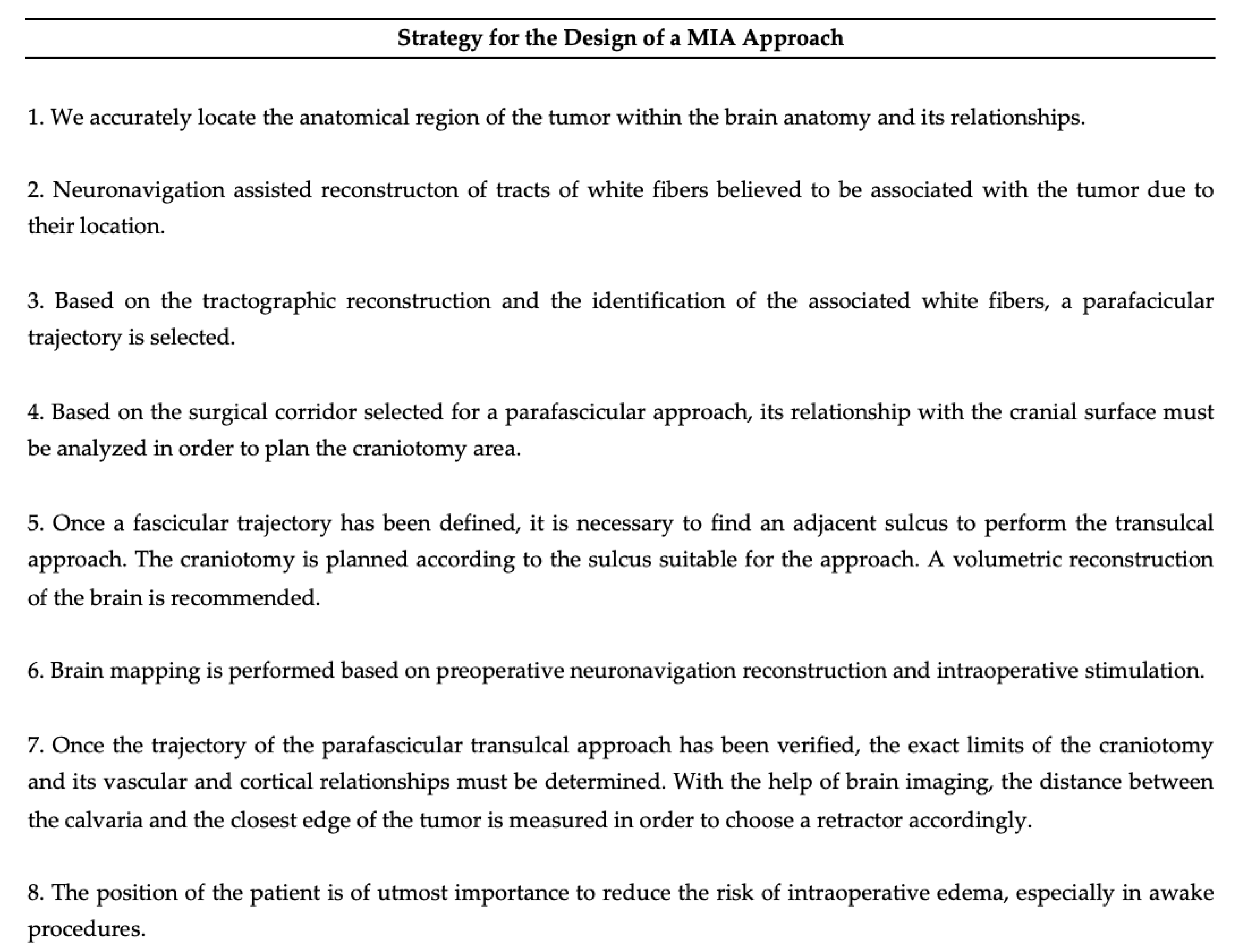
2.5. Eight Step Strategy for PTA Design

A strategy was standardized in eight steps for the approach using a minimally invasive technique: (1) determining the anatomical and functional location of the tumor, (2) tractography reconstruction, (3) determining the white fibers with potential risk of damage, (4) associating cortico-subcortical landmarks, (5) finding a suitable sulcus adjacent or within the surgical corridor, (6) designing the cortical and subcortical areas to be mapped, (7) designing the craniotomy and selecting the MITR and (8) checking the patient’s position (Figure 1).

3. Results

The results obtained in this study are presented in Table 1. A total of 72 operated-on patients were included in the study. The mean age of presentation of the disease in the group of patients was 49.6 years and the most affected gender was male (63.8%). At admission, neurological examination revealed paraphasia in 12.5% of the patients and only 11.1% presented some type of aphasia, described as follows: 2.7% presented anomic aphasia, 2.7% conduction aphasia, 1.3% global aphasia, 2.7% drill bit aphasia and 1.3% transcortical aphasia. Preoperatively, 41.6% had some motor deficit. Computerized campimetry was performed in 9.7% of the patients. Regarding the location of the tumor lesion, the anatomical region was mainly the frontal lobe (26.3%), followed by fronto-parietal (25%) and parietal (11.1%). The most frequent tumor type was HGG (34.72%), followed by LGG (17%), metastases (25%) and other lesions (11.1%). When evaluating the size of the incision, it was observed that the average of these was 5.58 cm, with 5 cm being the most frequent incision. In the craniectomies, it was estimated that the average diameter of these was 4.3 cm, with 4 cm being the most frequent diameter. Of the patients, 94.4% had a total gross resection. Postoperatively, 90.2% of the patients did not present new-onset deficits. Postoperative clinical outcome was evaluated in four aspects, postoperative KPS, long pathway damage, speech integrity and visual pathway integrity. Regarding postoperative KPS, 8.3% of patients had a KPS lower than admission postoperatively. Of the patients, 5.5% presented with impaired muscle strength in the limbs contralateral to the approach. Moreover, 4.1% presented some type of aphasia in the immediate postoperative period and none of the patients had visual impairment. Finally, we found that whenever the area of the tumor in an axial section was up to 5 times greater than the area of exposure of the MITRs, the tumor resection was greater than 90%. The most frequently used MITRs had the following dimensions: 17 mm wide, 11 mm high and 5 cm long.

4. Illustrative Cases

4.1. Case 1

A 29-year-old woman was referred to the emergency department with frequent headaches.
The neurological examination upon admission to the hospital was unremarkable. She had a computerized campimetry without alterations. The head MRI with Gadolinium showed an interventricular tumor (Figure 2A,B). A safe GTR was performed (Figure 3). Histopathology was consistent with a WHO grade I meningioma.

4.2. Case 2

A 46-year-old woman presented with seizures to the emergency department. The patient complained of dysarthria. An MRI was performed showing an intra-axial tumor at the junction of the inferior frontal gyrus and the pre-central sulcus (Figure 4A,B). The patient underwent resection and presented no postoperative neurological decline (Figure 5). Histopathological results were consistent with an HGG.

4.3. Surgical Nuances

Although there is no standardized technique for use of MITRs in brain surgery, the authors of this work believe that brain dissection can be performed with the help of the blunt tip of the MITRs. For an accurate and effective approach, MITRs must be guided with neuronavigation. Neurologists and electrophysiologists are essential for reliable intraoperative monitoring. After cortical stimulation, we recommend washing with cold solution to avoid triggering a seizure. Whenever possible, it is advisable to preserve the vascular, arterial and venous cortical and subcortical structures. In all cases, the MITRs should be fixed to a rigid support to avoid unintended movement that could lead to a change of the resection trajectory. Anesthetic monitoring should be carried out by anesthesiologists with extensive experience in neurological surgery and awake surgery.

5. Discussion

MIA in oncological neurosurgery remains controversial. Until two decades ago, a dogma of neuro-oncology was performing wide surgical approaches, which allowed the adequate management of intraoperative brain swelling, an effective control of bleeding, and a wide margin of maneuverability in case of complications, while achieving maximal EOR [2].
On the other hand, the PTA allows intracerebral access, while preserving the connectivity of the white matter tracts [11,12,13]. In this study, we described a surgical planning strategy for the design of a tubular PTA, guided by neuronavigation, for accurate cortical and subcortical mapping. The search for an ideal incision is mandatory, the traditional and best known techniques are based on the anatomy of the scalp, its vascularization. However, recently linear incisions have been adopted by some surgeons, considering their versatility, applicability and aesthetics [14]. In our work, in all cases, the incisions were linear, and even though no results were documented regarding the incidence of infections or aesthetic effects caused to the patients, we believe these allowed us to perform the craniotomy and the surgical resection of the lesion safely and widely, causing minor aesthetic damage to the skin.
The use of MIA in neurosurgery implies the planning of smaller craniotomies, permitting the brain to be approached in the same way as the prior existing ones [9]. Based on the multiple facts mentioned above, conservative neurosurgeons have given higher popularity to mini-craniotomies. However, even in other cranial pathologies, today it is becoming necessary to evaluate the benefits of brain approaches through these reduced cranial windows [15,16]. In our practice, the craniotomy was planned based on the relationship between the size of the tumor and the cranial area planned with neuronavigation [17]. This technique allowed us a safe approach and a complete resection or an NTR resection in most of the cases. Despite the fact that brain shift [18] and edema were not objectively evaluated, we presume that the small craniotomy openings allowed a lower dissociation curve with respect to the accuracy of navigation during the procedures and, in addition, can become a protective factor that prevents extra calvaria herniation.
PTA is widely described, even though its understanding requires a deep knowledge of anatomy and microsurgical skills, which allows the surgeon to travel through narrow corridors and deep within the brain tissue, causing limited damage to the fibers (Figure 6) [19]. Through the volumetric analysis of the cerebral cortex, its gyri and sulci, a cerebral sulcus was chosen in each patient far from the white fibers associated with the approach. We believe that depending on the location of the tumor, there are associated tracts that are mandatory to find in the reconstruction of the tractography in order to preserve their function (Figure 7).
The tubular MIA technique has been described in detail, as well as its benefits and utilities. This technique is associated with the use of neuronavigation, tractography and intraoperative monitoring, which makes it an ideal approach to prevent damage as much as possible, achieving an optimal resection rate versus neurological integrity [20,21,22,23]. A critical aspect that deserves attention is the preoperative assessment of the white matter tracts that will guide the intraoperative neuronavigation phase. In general, neuronavigation plays the role of allowing the surgeon to reach the lesion, but aiming to reduce the risk of neurological deficits by merging morphological data of the brain’s anatomy with the tractography will provide the safest trajectory to reach the tumor while sparing the fibers. In this context, the use of a tubular retractor perfectly suited the scope thanks to the shape of this tool. It follows that the tractography study is mandatory and paramount. In addition, the preoperative planning phase should be detailed and if possible much improved using the most modern technical tools including navigated trans-cranial magnetic stimulation (nTMS) [24,25,26,27]. nTMS have been proved to improve the reliability of the brain mapping and offers the opportunity to simulate the resection and related risk of deficit by causing a transitory lesion that can be localized, archived and merged with the dataset of the patient to be used during surgery. In this setting, nTMS adds awake information to sleeping surgical procedures.
Despite that the results presented in our study support the use of MIA, it is necessary to continue developing strategies to evaluate other aspects, allowing one to solve the questions of the critics of this tool. We are convinced that MIAs offer to modern neurosurgery the possibility of mutating some practices, in specific cases, which ends up influencing the clinical and surgical results of patients, as has already happened in other surgical specialties.

Limitations

Our study deals with an emerging trend in minimally invasive surgery for brain tumors. However, it does show several weaknesses. First, it is not a comparative study but rather a prospective observational study with its inherent biases. However, we report 9.8% of new-onset deficits that we consider acceptable based on the eloquence of the localization of the tumor. Secondly, the heterogeneity of the population. This may play a role in the clinical outcome of the procedure, but the goal of our study is to describe the steps we found helpful in the planning phase for MITRs. Third, we did not use nTMS in the planning stage. nTMS could provide useful information that could further reduce the risk of neuro-deficit and its use should be investigated as a potential new point of the strategy presented here in future studies.

6. Conclusions

We suggest considering adding MIA in surgical routine planning to perform safe tubular PTA for eloquent area tumors, which, added to the implementation of technologies such as intraoperative cortical and subcortical mapping, as well as tractography reconstruction, may improve maximal safe resection, preserving neurological function. Despite our results, there are currently no studies evaluating the benefits of this procedure compared to conventional techniques, and further studies are needed to evaluate the best indications for each one.

Author Contributions

Conceptualization, N.J.A.-V.; methodology, P.P.; software, J.G.P.-G.; validation, M.B., A.C. and O.S.T.; formal analysis, E.O.-R.; investigation, D.C.-M.; resources, H.A.C.-L.; data curation, N.J.A.-V.; writing—original draft preparation N.J.A.-V.; writing—review and editing, G.E.U.; visualization, G.E.U.; supervision, G.E.U.; project administration, G.F., A.V. and V.Z.; funding acquisition, M.B. All authors have read and agreed to the published version of the manuscript.

Funding

None of the authors received any remuneration or grant to carry out the work.

Institutional Review Board Statement

Number N 100 A. approval of the Ethics and Research Committee of the Hospital Infantil Universitario de San José, Bogotá, Colombia.

Informed Consent Statement

All subjects gave their informed consent for inclusion before they participated in the study. The study was conducted in accordance with the Declaration of Helsinki, and the protocol was approved by the Ethics Committee of the Hospital Infantil Universitario de San José, Bogotá, Colombia.

Data Availability Statement

Not applicable.

Conflicts of Interest

The authors declare no conflict of interest.

Abbreviations

MIAminimally invasive approach (MIA)
MITRsminimally invasive tubular retractors (MITRs)
PTAparafascicular transulcal approach (PTA)
KPSKarnofsky Performance Scale (KPS)
GCSGlasgow Coma Scale (GSC)
MRCMedical Research Council (MRC)
LGGlow-grade glioma (LGG)
HGGhigh-grade glioma (HGG)
MRImagnetic resonance imaging (MRI)
DTIdiffusion tensor imaging (DTI)
SLFsuperior longitudinal fasciculus (SLF)
ILFinferior longitudinal fasciculus (ILF)
GCTgeniculo-calcarine fasciculus (GCT)
UFuncinate fasciculus (UF)
CSTcorticospinal tract (CST)
AFarcuate fasciculus (AF)
AsFanterior superior frontal (AsF)
nTMSnavigated trans-cranial magnetic stimulation (nTMS)

References

  1. Rao, D.; Le, R.T.; Fiester, P.; Patel, J.; Rahmathulla, G. An Illustrative Review of Common Modern Craniotomies. J. Clin. Imaging Sci. 2020, 10, 81. [Google Scholar] [CrossRef]
  2. Teo, C. The Concept of Minimally Invasive Neurosurgery. Neurosurg. Clin. N. Am. 2010, 21, 583–584. [Google Scholar] [CrossRef] [PubMed]
  3. Walker, M.L. History of Ventriculostomy. Neurosurg. Clin. N. Am. 2001, 12, 101–110. [Google Scholar] [CrossRef] [PubMed]
  4. Kelly, P.J.; Goerss, S.J.; Kall, B.A. The stereotaxic retractor in computer-assisted stereotaxic microsurgery. J. Neurosurg. 1988, 69, 301–306. [Google Scholar] [CrossRef] [Green Version]
  5. Jane, J.A.; Park, T.S.; Pobereskin, L.H.; Winn, R.H.; Butler, A.B. The Supraorbital Approach: Technical Note. Neurosurgery 1982, 11, 537–542. [Google Scholar] [CrossRef] [PubMed]
  6. Reisch, R.; Perneczky, A. Ten-year Experience with the Supraorbital Subfrontal Approach through an Eyebrow Skin Incision. Neurosurgery 2005, 57, 242–255. [Google Scholar] [CrossRef] [Green Version]
  7. Perneczky, A.; Reisch, R. Keyhole approaches in neurosurgery, Volume 1, Concept and surgical technique. J. Korean Neurosurg. Soc. 2009, 45, 132. [Google Scholar]
  8. Burkhardt, J.K.; Neidert, M.C.; Woernle, C.M.; Bozinov, O.; Bernays, R.L. Intraoperative low-field MR-guided frameless stereotactic biopsy for intracerebral lesions. Acta Neurochir. 2013, 155, 721–726. [Google Scholar] [CrossRef]
  9. Garrett, M.; Consiglieri, G.; Nakaji, P. Transcranial Minimally Invasive Neurosurgery for Tumors. Neurosurg. Clin. N. Am. 2010, 21, 595–605. [Google Scholar] [CrossRef]
  10. Larson, A.S.; Zuccarello, M.; Grande, A.W. Minimally-invasive tubular retraction ports for intracranial neurosurgery: History and future perspectives. J. Clin. Neurosci. 2021, 89, 97–102. [Google Scholar] [CrossRef]
  11. Sun, G.C.; Chen, X.L.; Zhao, Y.; Wang, F.; Hou, B.-k.; Wang, Y.-b.; Song, Z.-J.; Wang, D.; Xu, B.-n. Intraoperative high-field magnetic resonance imaging combined with fiber tract neu-ronavigation-guided resection of cerebral lesions involving optic radiation. Neurosurgery 2011, 69, 1070–1084. [Google Scholar] [CrossRef] [PubMed]
  12. Muthusami, P.; James, J.; Thomas, B.; Kapilamoorthy, T.R.; Kesavadas, C. Diffusion tensor imaging and tractography of the human language pathways: Moving into the clinical realm. J. Magn. Reson. Imaging 2014, 40, 1041–1053. [Google Scholar] [CrossRef]
  13. Day, J.D. Transsulcal Parafascicular Surgery Using Brain Path® for Subcortical Lesions. Neurosurgery 2017, 64, 151–156. [Google Scholar] [CrossRef] [PubMed] [Green Version]
  14. Adawi, M.; Mosaad, N.; Teama, M. Utility of Linear Scalp Incision in Various Cranial Surgeries. Med. J. Cairo Univ. 2020, 88, 1903–1908. [Google Scholar]
  15. Al-Otaibi, F.; Albloushi, M.; Baeesa, S. Minicraniotomy for Standard Temporal Lobectomy: A Minimally Invasive Surgical Approach. Int. Sch. Res. Not. 2014, 2014, 532523. [Google Scholar] [CrossRef]
  16. Klimo, P.; Matthews, A.; Lew, S.M.; Zwienenberg-Lee, M.; Kaufman, B.A. Minicraniotomy versus bur holes for evacuation of chronic subdural collections in infants—A preliminary single-institution experience. J. Neurosurg. Pediatr. 2011, 8, 423–429. [Google Scholar] [CrossRef] [PubMed]
  17. Stadie, A.T.; Kockro, R.A.; Serra, L.; Fischer, G.; Schwandt, E.; Grunert, P.; Reisch, R. Neurosurgical craniotomy localization using a virtual reality planning system versus intraoperative image-guided navigation. Int. J. Comput. Assist. Radiol. Surg. 2011, 6, 565–572. [Google Scholar] [CrossRef]
  18. Luo, M.; Frisken, S.F.; Narasimhan, S.; Clements, L.W.; Thompson, R.C.; Golby, A.J.; Miga, M.I. A comprehensive model-assisted brain shift correction approach in image-guided neurosurgery: A case study in brain swelling and subsequent sag after craniotomy. In Proceedings of the Medical Imaging 2019: Image-Guided Procedures, Robotic Interventions, and Modeling, San Diego, CA, USA, 17–19 February 2019; Volume 10951, pp. 15–24. [Google Scholar] [CrossRef]
  19. Monroy-Sosa, A.; Chakravarthi, S.S.; Epping, A.; Fukui, M.B.; Jennings, J.; Rovin, R.; Kassam, A.B. Trans-sulcal Parafascicular Surgical Corridor for Resection of Brain Tumors: The Last Frontier. In Principles of Neurooncology; Springer: Cham, Switzerland, 2021. [Google Scholar]
  20. Kinoshita, M.; Miyashita, K.; Tsutsui, T.; Furuta, T.; Nakada, M. Critical Neural Networks in Awake Surgery for Gliomas. Neurol. Med. Chir. 2016, 56, 674–686. [Google Scholar] [CrossRef] [Green Version]
  21. Skrap, M.; Marin, D.; Ius, T.; Fabbro, F.; Tomasino, B. Brain mapping: A novel intraoperative neuropsychological approach. J. Neurosurg. 2016, 125, 877–887. [Google Scholar] [CrossRef] [Green Version]
  22. Greenfield, J.P.; Cobb, W.S.; Tsouris, A.J.; Schwartz, T.H. Stereotactic minimally invasive tubular retractor system for deep brain lesions. Neurosurgery 2008, 63 (Suppl. 2), 334–339. [Google Scholar] [CrossRef]
  23. Kurozumi, K.; Otani, Y.; Ishida, J.; Hiramatsu, M.; Kameda, M.; Ichikawa, T.; Date, I. Combination of the tubular retractor and brain spatulas provides an adequate operative field in surgery for deep-seated lesions: Case series and technical note. Surg. Neurol. Int. 2018, 9, 220. [Google Scholar] [CrossRef]
  24. Umana, G.E.; Scalia, G.; Graziano, F.; Maugeri, R.; Alberio, N.; Barone, F.; Crea, A.; Fagone, S.; Giammalva, G.R.; Brunasso, L.; et al. Navigated Transcranial Magnetic Stimulation Motor Mapping Usefulness in the Surgical Management of Patients Affected by Brain Tumors in Eloquent Areas: A Systematic Review and Meta-Analysis. Front. Neurol. 2021, 12, 644198. [Google Scholar] [CrossRef]
  25. Conti, A.; Raffa, G.; Granata, F.; Rizzo, V.; Germanò, A.; Tomasello, F. Navigated Transcranial Magnetic Stimulation for “Somatotopic” Tractography of the Corticospinal Tract. Neurosurgery 2014, 10 (Suppl. 4), 542–554. [Google Scholar] [CrossRef]
  26. Raffa, G.; Conti, A.; Scibilia, A.; Sindorio, C.; Quattropani, M.C.; Visocchi, M.; Germanò, A.; Tomasello, F. Functional Reconstruction of Motor and Language Pathways Based on Navigated Transcranial Magnetic Stimulation and DTI Fiber Tracking for the Preoperative Planning of Low Grade Glioma Surgery: A New Tool for Preservation and Restoration of Eloquent Networks. Acta Neurochir. 2017, 124, 251–261. [Google Scholar] [CrossRef]
  27. Raffa, G.; Picht, T.; Scibilia, A.; Rösler, J.; Rein, J.; Conti, A.; Ricciardo, G.; Cardali, S.M.; Vajkoczy, P.; Germanò, A. Surgical treatment of meningiomas located in the rolandic area: The role of navigated transcranial magnetic stimulation for preoperative planning, surgical strategy, and prediction of arachnoidal cleavage and motor outcome. J. Neurosurg. 2019, 133, 107–118. [Google Scholar] [CrossRef]
Figure 1. The Eight Step Strategy for PTA Design.
Brainsci 13 00498 g001
Figure 2. (A,B) show MRI rendering of a large intraventricular tumor (red arrow). Arcuate fasciculus (AF), geniculocalcarine tract (GTC) reconstruction helps in the planning stange for the resection of the tumor.
Brainsci 13 00498 g002
Figure 3. (A) Axial and coronal view of a preoperative T2 weighted MRI; (B) the postoperative axial and coronal view of a preoperative T2 weighted MRI that documents the GTR; (C) an intraoperative use of the tubular retractor.
Brainsci 13 00498 g003
Figure 4. An intra-axial tumor at the junction of the inferior frontal gyrus and the pre-central sulcus. Fig (A) shows coronal view of merged T1 weighted MRI scan with corticospinal tract tractography. Fig (B) shows the axial view of the latter.
Brainsci 13 00498 g004
Figure 5. (A) Axial and coronal view of a preoperative T2 weighted MRI; (B) the postoperative axial and coronal view of a preoperative T2 weighted MRI that documents the GTR; (C) an intraoperative view of the cortical surface with the tracts highlighted; (D) the postoperative CT scan with bone algorithm that shows the small craniotomy.
Brainsci 13 00498 g005
Figure 6. Cadaveric white matter dissection showing the fiber tracts commonly taken into account during the planning stage and intraoperatively for a safe tumor resection.
Brainsci 13 00498 g006
Figure 7. Fiber tracts usually reconstruct in our practice during preoperative planning. AsF: Aslant fasciculus; LSF: Superior Longitudinal fasciculus; PT: Pyramidal tract; AF: Arcuate fasciculus; UF: Uncinate fasciculus; ILF: Inferior longitudinal fasciculus; TCT: Thalamic cortical tract; GCT: Geniculo-calcarine tract; MA: Meyer’s loops.
Brainsci 13 00498 g007
Table 1. Tumor characteristics and related clinical outcome.
Neurological Examination%
Altered Force41.6
Language Disturbances
Paraphasia12.5
Aphasia 11.1
Anomic2.7
Conduction2.7
Global1.3
Broca’s2.7
Transcortical1.3
Tumor Localization
Frontal26.3
Parietal11.1
Frontoparietal25
Other37.6
Tumor Type
High Grade Gliomas34.7
Low Grade Gliomas 29.2
Metastasis25
Other11.1
Resection
Total94.4
Sub-Total5.5
Partial0
Disclaimer/Publisher’s Note: The statements, opinions and data contained in all publications are solely those of the individual author(s) and contributor(s) and not of MDPI and/or the editor(s). MDPI and/or the editor(s) disclaim responsibility for any injury to people or property resulting from any ideas, methods, instructions or products referred to in the content.

