
Hemispheric Asymmetry of the Hand Motor Representations in Patients with Highly Malignant Brain Tumors: Implications for Surgery and Clinical Practice
 , , ,
, , ,
Abstract
:1. Introduction
2. Materials and Methods
2.1. Participants
2.2. fMRI Data Acquisition
2.3. fMRI Data Processing
2.4. ROI Analysis
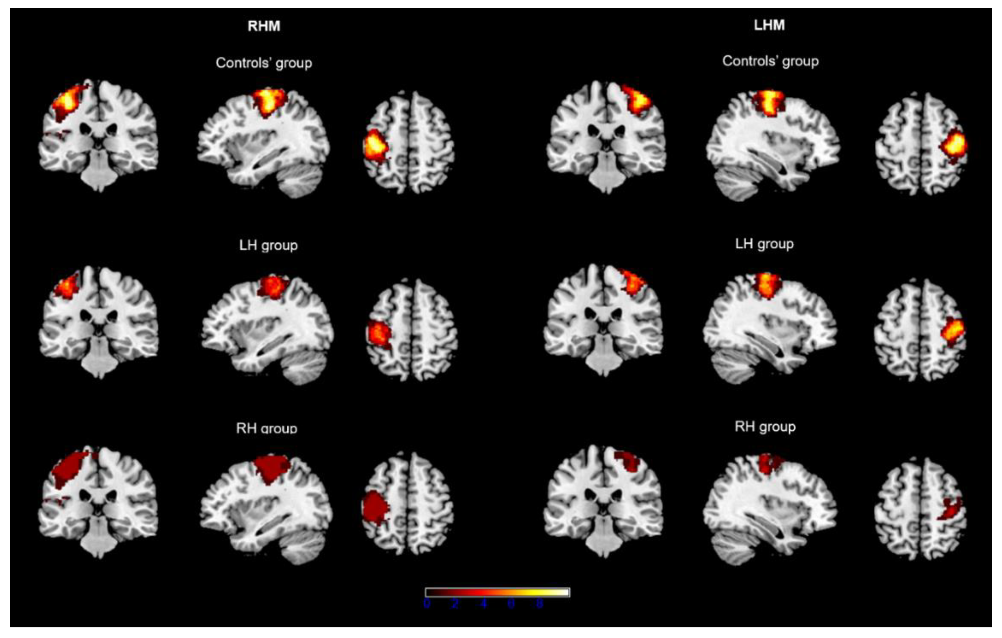
3. Results
3.1. Descriptive Results
3.2. Hemispheric Effects: Within-Group Comparison between RHM and LHM Conditions
3.3. Between-Group Comparisons
3.3.1. Separate Comparisons for RHM and LHM Conditions between Groups
3.3.2. Differences in Hemispheric Asymmetry between Groups
4. Discussion
5. Conclusions
Author Contributions
Funding
Institutional Review Board Statement
Informed Consent Statement
Data Availability Statement
Acknowledgments
Conflicts of Interest
References
- Noll, K.R.; Sullaway, C.; Ziu, M.; Weinberg, J.S.; Wefel, J.S. Relationships between tumor grade and neurocognitive functioning in patients with glioma of the left temporal lobe prior to surgical resection. Neuro Oncol. 2015, 174, 580–587. [Google Scholar] [CrossRef] [PubMed]
- Gibb, W.R.; Kong, N.W.; Tate, M.C. Direct evidence of plasticity within human primary motor and somatosensory cortices of patients with glioblastoma. Neural Plast. 2020, 2020, 8893708. [Google Scholar] [CrossRef] [PubMed]
- Kawashima, A.; Krieg, S.M.; Faust, K.; Schneider, H.; Vajkoczy, P.; Picht, T. Plastic reshaping of cortical language areas evaluated by navigated transcranial magnetic stimulation in a surgical case of glioblastoma multiforme. Clin. Neurol. Neurosurg. 2013, 115, 2226–2229. [Google Scholar] [CrossRef] [PubMed]
- Herbet, G.; Maheu, M.; Costi, E.; Lafargue, G.; Duffau, H. Mapping the neuroplastic potential in brain-damaged patients. Brain 2016, 139, 829–844. [Google Scholar] [CrossRef]
- Ius, T.; Angelini, E.; de Schotten, M.T.; Mandonnet, E.; Duffau, H. Evidence for potentials and limitations of brain plasticity using an atlas of functional resectability of WHO grade II gliomas: Towards a “minimal common brain”. Neuroimage 2011, 56, 992–1000. [Google Scholar] [CrossRef] [PubMed]
- Cargnelutti, E.; Ius, T.; Skrap, M.; Tomasino, B. What do we know about pre-and postoperative plasticity in patients with glioma? A review of neuroimaging and intraoperative mapping studies. Neuroimage Clin. 2020, 28, 102435. [Google Scholar] [CrossRef]
- Cirillo, S.; Caulo, M.; Pieri, V.; Falini, A.; Castellano, A. Role of functional imaging techniques to assess motor and language cortical plasticity in glioma patients: A systematic review. Neural Plast. 2019, 2019, 4056436. [Google Scholar] [CrossRef]
- Majos, A.; Bryszewski, B.; Kośla, K.N.; Pfaifer, L.; Jaskólski, D.; Stefańczyk, L. Process of the functional reorganization of the cortical centers for movement in GBM patients: fMRI study. Clin. Neuroradiol. 2017, 27, 71–79. [Google Scholar] [CrossRef]
- Shinoura, N.; Suzuki, Y.; Yamada, R.; Kodama, T.; Takahashi, M.; Yagi, K. Restored activation of primary motor area from motor reorganization and improved motor function after brain tumor resection. Am. J. Neuroradiol. 2006, 27, 1275–1282. [Google Scholar]
- Conway, N.; Wildschuetz, N.; Moser, T.; Bulubas, L.; Sollmann, N.; Tanigawa, N.; Meyer, B.; Krieg, S.M. Cortical plasticity of motor-eloquent areas measured by navigated transcranial magnetic stimulation in patients with glioma. J. Neurosurg. 2017, 1275, 981–991. [Google Scholar] [CrossRef]
- Zacà, D.; Jovicich, J.; Nadar, S.R.; Voyvodic, J.T.; Pillai, J.J. Cerebrovascular reactivity mapping in patients with low grade gliomas undergoing presurgical sensorimotor mapping with BOLD fMRI. J. Magn. Reason. Imaging 2014, 40, 383–390. [Google Scholar] [CrossRef] [Green Version]
- Zimmermann, M.; Rössler, K.; Kaltenhäuser, M.; Grummich, P.; Brandner, N.; Buchfelder, M.; Dörfler, A.; Kölble, K.; Stadlbauer, A. Comparative fMRI and MEG localization of cortical sensorimotor function: Bimodal mapping supports motor area reorganization in glioma patients. PLoS ONE 2019, 14, e0213371. [Google Scholar] [CrossRef]
- Voets, N.L.; Plaha, P.; Jones, O.P.; Pretorius, P.; Bartsch, A. Presurgical localization of the primary sensorimotor cortex in gliomas. Clin. Neuroradiol. 2021, 31, 245–256. [Google Scholar] [CrossRef]
- Tozakidou, M.; Wenz, H.; Reinhardt, J.; Nennig, E.; Riffel, K.; Blatow, M.; Stippich, C. Primary motor cortex activation and lateralization in patients with tumors of the central region. Neuromage Clin. 2013, 2, 221–228. [Google Scholar] [CrossRef]
- Sun, H.; Vachha, B.; Laino, M.E.; Jenabi, M.; Flynn, J.R.; Zhang, Z.; Holodny, A.I.; Peck, K.K. Decreased hand motor resting-state functional connectivity in patients with glioma: Analysis of factors including neurovascular uncoupling. Radiology 2020, 294, 610–621. [Google Scholar] [CrossRef]
- Tzourio-Mazoyer, N.; Petit, L.; Zago, L.; Crivello, F.; Vinuesa, N.; Joliot, M.; Jobard, G.; Mellet, E.; Mazoyer, B. Between-hand difference in ipsilateral deactivation is associated with hand lateralization: fMRI mapping of 284 volunteers balanced for handedness. Front. Hum. Neurosci. 2015, 9, 5. [Google Scholar] [CrossRef]
- Turesky, T.K.; Olulade, O.A.; Luetje, M.M.; Eden, G.F. An fMRI study of finger tapping in children and adults. Hum. Brain Mapp. 2018, 39, 3203–3215. [Google Scholar] [CrossRef]
- Zhang, J.; Chen, H.; Fang, F.; Liao, W. Quantitative analysis of asymmetrical cortical activity based on power spectrum changes. Brain Topogr. 2010, 23, 257–268. [Google Scholar] [CrossRef]
- Zeng, L.; Chen, H.; Ouyang, L.; Yao, D.; Gao, J.H. Quantitative analysis of asymmetrical cortical activity in motor areas during sequential finger movement. J. Magn. Reason. Imaging 2007, 25, 1370–1375. [Google Scholar] [CrossRef]
- Grabowska, A.; Gut, M.; Binder, M.; Forsberg, L.; Rymarczyk, K.; Urbanik, A. Switching handedness: fMRI study of hand motor control in right-handers, left-handers and converted left-handers. Acta Neurobiol. Exp. 2012, 72, 439–451. [Google Scholar]
- Dassonville, P.; Zhu, X.H.; Ugurbil, K.; Kim, S.G.; Ashe, J. Functional activation in motor cortex reflects the direction and the degree of handedness. Proc. Natl. Acad. Sci. USA 1997, 94, 14015–14018. [Google Scholar] [CrossRef]
- Verstynen, T.; Diedrichsen, J.; Albert, N.; Aparicio, P.; Ivry, R.B. Ipsilateral motor cortex activity during unimanual hand movements relates to task complexity. J. Neurophysiol. 2005, 93, 1209–1222. [Google Scholar] [CrossRef]
- Solodkin, A.; Hlustik, P.; Noll, D.C.; Small, S.L. Lateralization of motor circuits and handedness during finger movements. Eur. J. Neurol. 2001, 8, 425–434. [Google Scholar] [CrossRef]
- Rinehart, J.K.; Singleton, R.D.; Adair, J.C.; Sadek, J.R.; Haaland, K.Y. Arm use after left or right hemiparesis is influenced by hand preference. Stroke 2009, 40, 545–550. [Google Scholar] [CrossRef]
- Oldfield, R.C. The assessment and analysis of handedness: The Edinburgh Inventory. Neuropsychologia 1971, 9, 97–113. [Google Scholar] [CrossRef]
- Gogos, A.J.; Young, J.S.; Morshed, R.A.; Avalos, L.N.; Noss, R.S.; Villanueva-Meyer, J.E.; Hervey-Jumper, S.L.; Berger, M.S. Triple motor mapping: Transcranial, bipolar, and monopolar mapping for supratentorial glioma resection adjacent to motor pathways. J. Neurosurg. 2020, 134, 1728–1737. [Google Scholar] [CrossRef]
- Lettieri, C.; Pauletto, G.; Valiante, G.; Ius, T.; Verriello, L.; Valente, M.; Skrap, M.; Gigli, G.L.; Budai, R. Fast or Slow? A Comparison between two transcranial electrical stimulation techniques for eliciting motor-evoked potentials during supratentorial surgery. J. Clin. Neurophysiol. 2021. [Google Scholar] [CrossRef]
- Engel, J. Update on surgical treatment of the epilepsies: Summary of the second international palm desert conference on the surgical treatment of the epilepsies (1992). Neurology 1993, 43, 1612. [Google Scholar] [CrossRef]
- Friston, K.J.; Frith, C.D.; Turner, R.; Frackowiak, R.S.J. Characterising evoked hemodynamics with fMRI. Neuroimage 1995, 2, 157–165. [Google Scholar] [CrossRef]
- Friston, K.J.; Holmes, A.P.; Worsley, K.J.; Poline, J.-B.; Frith, C.D.; Frackowiak, R.S.J. Statistical parametric maps in functional imaging: A general linear approach. Hum. Brain Mapp. 1995, 2, 189–210. [Google Scholar] [CrossRef]
- Eickhoff, S.; Stephan, K.E.; Mohlberg, H.; Grefkes, C.; Fink, G.R.; Amunts, K.; Zilles, K. A new SPM toolbox for combining probabilistic cytoarchitectonic maps and functional imaging data. Neuroimage 2005, 25, 1325–1335. [Google Scholar] [CrossRef]
- Brett, M.; Anton, J.L.; Valabregue, R.; Poline, J.B. Region of interest analysis using an SPM toolbox. In NeuroImage, Proceedings of the 8th International Conference on Functional Mapping of the Human Brain, Sendai, Japan, 2–6 June 2002; Elsevier: Amsterdam, The Netherlands, 2002; Volume 16, p. 16. [Google Scholar]
- Pasquini, L.; Jenabi, M.; Yildirim, O.; Silveira, P.; Peck, K.K.; Holodny, A.I. Brain Functional Connectivity in Low-and High-Grade Gliomas: Differences in Network Dynamics Associated with Tumor Grade and Location. Cancers 2022, 14, 3327. [Google Scholar] [CrossRef]
- McManus, C. Half a century of handedness research: Myths, truths; fictions, facts; backwards, but mostly forwards. Brain Neurosci. Adv. 2019, 3, 2398212818820513. [Google Scholar] [CrossRef]
- Cho, N.S.; Peck, K.K.; Zhang, Z.; Holodny, A.I. Paradoxical activation in the cerebellum during language fMRI in patients with brain tumors: Possible explanations based on neurovascular uncoupling and functional reorganization. Cerebellum 2018, 173, 286–293. [Google Scholar] [CrossRef]
- Ulmer, J.L.; Krouwer, H.G.; Mueller, W.M.; Ugurel, M.S.; Kocak, M.; Mark, L.P. Pseudo-reorganization of language cortical function at fMR imaging: A consequence of tumorinduced neurovascular uncoupling. Am. J. Neuroradiol. 2003, 24, 213–217. [Google Scholar]
- Shinoura, N.; Yamada, R.; Suzuki, Y.; Kodama, T.; Sekiguchi, K.; Takahashi, M.; Yagi, K. Functional magnetic resonance imaging is more reliable than somatosensory evoked potential or mapping for the detection of the primary motor cortex in proximity to a tumor. Stereotact. Funct. Neurosurg. 2007, 85, 99–105. [Google Scholar] [CrossRef]
- Wengenroth, M.; Blatow, M.; Guenther, J.; Akbar, M.; Tronnier, V.M.; Stippich, C. Diagnostic benefits of presurgical fMRI in patients with brain tumours in the primary sensorimotor cortex. Eur. Radiol. 2011, 2, 1517–1525. [Google Scholar] [CrossRef]
- Amunts, K.; Jäncke, L.; Mohlberg, H.; Steinmetz, H.; Zilles, K. Interhemispheric asymmetry of the human motor cortex related to handedness and gender. Neuropsychologia 2000, 38, 304–312. [Google Scholar] [CrossRef]
- Lissek, S.; Hausmann, M.; Knossalla, F.; Peters, S.; Nicolas, V.; Güntürkün, O.; Tegenthoff, M. Sex differences in cortical and subcortical recruitment during simple and complex motor control: An fMRI study. Neuroimage 2007, 37, 912–926. [Google Scholar] [CrossRef]


| Gender | Age | Edu | Diagnosis | Vol (cm3) | ONA Pre | ONA Post | MEP Pre | MEP Intra | Epil Pre | Epil Post (Engel Class) | |
|---|---|---|---|---|---|---|---|---|---|---|---|
| LH | |||||||||||
| p1 | M | 57 | Meta | 0.52 | R hand myasthenia (4/5) | R superior limb myasthenia | Patho | Normal | Yes | No (Ia) | |
| p2 | F | 59 | Meta | 59.60 | NAD | NAD | Normal | Normal | Yes | No (Ia) | |
| p3 | F | 64 | GBM | 23.15 | N/A | N/A | N/A | N/A | N/A | N/A | |
| p4 | M | 65 | GBM | 22.86 | NAD | R hemiparesis (2/5) | Patho | Normal | Yes | No (Ia) | |
| p5 | M | 70 | GBM | 12.56 | NAD | NAD | Normal | Normal | Yes | Yes (III) | |
| p6 | F | 38 | GBM | 61.79 | NAD | R hemiparesis (3/5) | Patho | Normal | Yes | No (Ia) | |
| p7 | M | 37 | GBM | 96.20 | N/A | N/A | N/A | N/A | N/A | N/A | |
| p8 | F | 52 | GBM | 41.64 | R hemisome myasthenia (3/5) | R hemisome myasthenia (2/5) | Patho | Patho | Yes | Yes (III) | |
| p9 | F | 74 | Meta | 42.50 | NAD | R hemisome myasthenia (2/5) | Normal | Normal | No | No (Ia) | |
| p10 | F | 53 | GBM | 11.03 | N/A | N/A | N/A | N/A | N/A | N/A | |
| RH | |||||||||||
| p1 | M | 44 | Meta | 29.25 | N/A | N/A | N/A | N/A | N/A | N/A | |
| p2 | M | 37 | GBM | 23.82 | NAD | NAD | Normal | Normal | Yes | No (Ia) | |
| p3 | F | 49 | GBM | 30.98 | L hemiparesis (3/5) | L hemiparesis (3/5) | Patho | Patho | Yes | Yes (IV) | |
| p4 | F | 40 | GBM | 48.16 | NAD | Recovering L hemiparesis (4/5) | Normal | Normal | Yes | Yes (IV) | |
| p5 | F | 54 | GBM | 52.21 | N/A | N/A | N/A | N/A | N/A | N/A | |
| p6 | M | 65 | GBM | 39.15 | L superior limb myasthenia (3/5) | L hemiparesis (2/5) | Patho | Patho | Yes | Yes (IV) | |
| p7 | F | 48 | GBM | 8.31 | L superior limb myasthenia (4/5) | L hemiparesis (3/5 superior limb, 1/5 inferior limb) | Patho | Normal | Yes | Yes (III) | |
| p8 | M | 53 | GBM | 22.83 | L superior limb myasthenia (3/5) | L hemiparesis (2/5) | Patho | Patho | Yes | Yes (Ib) | |
| Preoperative MEP | Intraoperative MEP | Preoperative ONA | Postoperative ONA | Engel Class Other than Ia | |
|---|---|---|---|---|---|
| LH group (n = 10) | 4 | 1 | 2 | 4 | 2 |
| RH group (n = 8) | 4 | 3 | 4 | 5 | 5 |
| RHM | LHM | ||||||||
|---|---|---|---|---|---|---|---|---|---|
| ROI Values | ROI Volumes | SM Activation (%) | ROI Values | ROI Volumes | SM Activation (%) | Δ_ROI Values | Δ_ROI Volumes | Δ_SM Activation (%) | |
| Control group | 136.02 (4.85) | 11,656.60 (4874.10) | 16.41 (6.13) | 145.63 (9.11) | 9414.70 (2933.90) | 14.63 (6.13) | 9.53 (3.44) | −2241.90 (4,441.75) | −1.77 (5.85) |
| LH group | 132.13 (13.17) | 4496.89 (1918.73) | 7.22 (2.86) | 136.55 (8.63) | 6725.56 (3293.93) | 10.36 (5.06) | 4.42 (3.63) | 2228.67 (4040.98) | 3.14 (6.13) |
| RH group | 140.23 (13.90) | 5008.50 (3596.33) | 7.64 (5.04) | 134.79 (13.26) | 1895.13 (1705.91) | 3.01 (2.83) | −5.44 (3.85) | −3113.38 (4340.30) | −4.63 (6.39) |
Publisher’s Note: MDPI stays neutral with regard to jurisdictional claims in published maps and institutional affiliations. |
© 2022 by the authors. Licensee MDPI, Basel, Switzerland. This article is an open access article distributed under the terms and conditions of the Creative Commons Attribution (CC BY) license (https://creativecommons.org/licenses/by/4.0/).
Share and Cite
Cargnelutti, E.; Pauletto, G.; Ius, T.; Verriello, L.; Maieron, M.; Skrap, M.; Tomasino, B. Hemispheric Asymmetry of the Hand Motor Representations in Patients with Highly Malignant Brain Tumors: Implications for Surgery and Clinical Practice. Brain Sci. 2022, 12, 1274. https://doi.org/10.3390/brainsci12101274
Cargnelutti E, Pauletto G, Ius T, Verriello L, Maieron M, Skrap M, Tomasino B. Hemispheric Asymmetry of the Hand Motor Representations in Patients with Highly Malignant Brain Tumors: Implications for Surgery and Clinical Practice. Brain Sciences. 2022; 12(10):1274. https://doi.org/10.3390/brainsci12101274
Chicago/Turabian StyleCargnelutti, Elisa, Giada Pauletto, Tamara Ius, Lorenzo Verriello, Marta Maieron, Miran Skrap, and Barbara Tomasino. 2022. "Hemispheric Asymmetry of the Hand Motor Representations in Patients with Highly Malignant Brain Tumors: Implications for Surgery and Clinical Practice" Brain Sciences 12, no. 10: 1274. https://doi.org/10.3390/brainsci12101274