Next Issue
Volume 12, February
Previous Issue
Volume 11, December


Brain Sci., Volume 12, Issue 1 (January 2022) – 122 articles

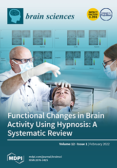
Cover Story (view full-size image): Hypnosis has proven a powerful method in indications such as pain control and anxiety reduction. As recently discussed, it has been yielding increased attention from medical/dental perspectives. This systematic review (PROSPERO-registration-ID-CRD42021259187) aimed to critically evaluate and discuss functional changes in brain activity using hypnosis by means of different imaging techniques. View this paper
  • Issues are regarded as officially published after their release is announced to the table of contents alert mailing list.
  • You may sign up for e-mail alerts to receive table of contents of newly released issues.
  • PDF is the official format for papers published in both, html and pdf forms. To view the papers in pdf format, click on the "PDF Full-text" link, and use the free Adobe Reader to open them.
Order results
Result details
Section
Select all
Export citation of selected articles as:
17 pages, 409 KiB  
Review
Update on Domestic Violence and Traumatic Brain Injury: A Narrative Review
by Kellianne Costello and Brian D. Greenwald
Brain Sci. 2022, 12(1), 122; https://doi.org/10.3390/brainsci12010122 - 17 Jan 2022
Cited by 19 | Viewed by 7628
Abstract
Research on traumatic brain injury (TBI) as a result of domestic violence has greatly increased in the past decade, with publications addressing the prevalence, diagnosis, evaluation, and treatment. Although TBI due to domestic violence has recently been found to occur quite frequently, it [...] Read more.
Research on traumatic brain injury (TBI) as a result of domestic violence has greatly increased in the past decade, with publications addressing the prevalence, diagnosis, evaluation, and treatment. Although TBI due to domestic violence has recently been found to occur quite frequently, it was not widely understood until the 1990s. Individuals who suffer from domestic violence TBI often experience sequelae such as decreased cognitive functioning, memory loss, and PTSD. The goal of this article is to increase awareness about TBI secondary to domestic violence, with the intent that it will highlight areas for future research on the diagnosis, evaluation, and treatment of TBI in this population. The articles in this study were first found using the search terms traumatic brain injury and domestic violence. Although, in recent years, there has been a significant increase in research on TBI due to domestic violence, the overall conclusion of this review article is that there is still a need for future research in many areas including the effects on minority populations, the effects of COVID-19, and improvements of screening tools. Full article
(This article belongs to the Section Neurorehabilitation)
Show Figures

Figure 1

18 pages, 2329 KiB  
Article
Changes in Pronoun Use a Decade before Clinical Diagnosis of Alzheimer’s Dementia—Linguistic Contexts Suggest Problems in Perspective-Taking
by Dagmar Bittner, Claudia Frankenberg and Johannes Schröder
Brain Sci. 2022, 12(1), 121; https://doi.org/10.3390/brainsci12010121 - 17 Jan 2022
Cited by 5 | Viewed by 2143
Abstract
The use of pronouns has been shown to change pathologically in the early phases of Alzheimer’s Dementia (AD). So far, the findings have been of a quantitative nature. Little is known, however, about the developmental path of the change, its onset, the domains [...] Read more.
The use of pronouns has been shown to change pathologically in the early phases of Alzheimer’s Dementia (AD). So far, the findings have been of a quantitative nature. Little is known, however, about the developmental path of the change, its onset, the domains in which it initially occurs, and if and how it spreads to other linguistic domains. The present study investigates pronoun use in six speakers of German a decade before they were clinically diagnosed with AD (LAD) and six biographically matched healthy controls (CTR). The data originate from monologic spoken language elicited by semi-spontaneous biographical interviews. Investigation of nine pronoun types revealed group differences in the use of three pronoun types: D-pronouns—a specific pronoun type of German for reference to persons and objects; the impersonal pronoun man ‘one’, and the propositional pronoun das ‘this/that’. Investigation of the linguistic contexts in which these three pronoun types were used revealed a correlation with declines in elaborative and evaluative information; that is, information the hearer would benefit from in creating an informed model of the discourse. We, therefore, hypothesize that the early changes in language use due to AD point to problems in perspective-taking, specifically in taking the hearer’s perspective. Full article
(This article belongs to the Special Issue Early Recognition of Alzheimer´s Disease)
Show Figures

Figure 1

11 pages, 2521 KiB  
Article
Serum Levels of Lipids and Selected Aminothiols in Epileptic Children—A Pilot Case-Control Study
by Beata Sarecka-Hujar, Izabela Szołtysek-Bołdys and Ilona Kopyta
Brain Sci. 2022, 12(1), 120; https://doi.org/10.3390/brainsci12010120 - 17 Jan 2022
Cited by 3 | Viewed by 1503
Abstract
Background: Standard treatment of epileptic seizures involves the use of antiepileptic drugs (AEDs). Both AEDs themselves and treatment duration may influence the levels of biochemical parameters, e.g., lipids or homocysteine (HCys), that may increase the risk of cardiovascular diseases. The aim of the [...] Read more.
Background: Standard treatment of epileptic seizures involves the use of antiepileptic drugs (AEDs). Both AEDs themselves and treatment duration may influence the levels of biochemical parameters, e.g., lipids or homocysteine (HCys), that may increase the risk of cardiovascular diseases. The aim of the present study was to compare the levels of lipid parameters, as well as the concentrations of selected aminothiols (i.e., HCys, cysteine, and glutathione) between epileptic children treated with multiple AEDs and children without epilepsy. Methods: In the study, 21 children with epilepsy treated with two or more AEDs for at least 6 months (8 girls and 13 boys, mean age 7.03 ± 4.51) and 23 children without epilepsy (7 girls and 16 boys, mean age 7.54 ± 3.90) were prospectively analyzed. Lipid parameters, i.e., total cholesterol (TC), triglycerides (TG), low density lipoprotein (LDL) and high density lipoprotein (HDL), and levels of selected aminothiols were determined in the blood serum. Results: No differences in the mean levels of lipid parameters and in the mean values of lipid ratios (TC/HDL, TG/HDL, LDL/HDL) were observed between the total groups as well as in the sex subgroups. HCys and cysteine levels did not differ between the patients and controls. We observed significantly lower levels of glutathione in children with epilepsy than in children without epilepsy (1.49 ± 0.35 µmol/L vs. 2.39 ± 1.17 µmol/L, respectively) (p < 0.001). Glutathione level was also lower in boys with epilepsy than in boys without epilepsy (p = 0.007). Similarly, epileptic girls had statistically decreased levels of glutathione when compared to girls without epilepsy (p = 0.006). Conclusions: A lower level of glutathione is observed in pediatric patients with epilepsy treated with two or more AEDs for at least 6 months. This indicates the oxidative stress of the patients treated with AEDs, which in turn may affect their well-being, and in the case of chronic occurrence resulting from long-term treatment, also on the function of the liver and the condition of the cardiovascular system. Full article
Show Figures

Figure 1

10 pages, 288 KiB  
Article
Finding the Way to Improve Motor Recovery of Patients with Spinal Cord Lesions: A Case-Control Pilot Study on a Novel Neuromodulation Approach
by Antonino Naro, Luana Billeri, Tina Balletta, Paola Lauria, Maria Pia Onesta and Rocco Salvatore Calabrò
Brain Sci. 2022, 12(1), 119; https://doi.org/10.3390/brainsci12010119 - 17 Jan 2022
Cited by 2 | Viewed by 1825
Abstract
Robot-assisted rehabilitation (RAR) and non-invasive brain stimulation (NIBS) are interventions that, both individually and combined, can significantly enhance motor performance after spinal cord injury (SCI). We sought to determine whether repetitive transcranial magnetic stimulation (rTMS) combined with active transvertebral direct current stimulation (tvDCS) [...] Read more.
Robot-assisted rehabilitation (RAR) and non-invasive brain stimulation (NIBS) are interventions that, both individually and combined, can significantly enhance motor performance after spinal cord injury (SCI). We sought to determine whether repetitive transcranial magnetic stimulation (rTMS) combined with active transvertebral direct current stimulation (tvDCS) (namely, NIBS) in association with RAR (RAR + NIBS) improves lower extremity motor function more than RAR alone in subjects with motor incomplete SCI (iSCI). Fifteen adults with iSCI received one daily session of RAR+NIBS in the early afternoon, six sessions weekly, for eight consecutive weeks. Outcome measures included the 6 min walk test (6MWT), the 10 m walk test (10MWT), the timed up and go (TUG) to test mobility and balance, the Walking Index for Spinal Cord Injury (WISCI II), the Functional Independence Measure-Locomotion (FIM-L), the manual muscle testing for lower extremity motor score (LEMS), the modified Ashworth scale for lower limbs (MAS), and the visual analog scale (VAS) for pain. The data of these subjects were compared with those of 20 individuals matched for clinical and demographic features who previously received the same amount or RAR without NIBS (RAR − NIBS). All patients completed the trial, and none reported any side effects either during or following the training. The 10MWT improved in both groups, but the increase was significantly greater following RAR + NIBS than RAR − NIBS. The same occurred for the FIM-L, LEMS, and WISCI II. No significant differences were appreciable concerning the 6MWT and TUG. Conversely, RAR − NIBS outperformed RAR + NIBS regarding the MAS and VAS. Pairing tvDCS with rTMS during RAR can improve lower extremity motor function more than RAR alone can do. Future research with a larger sample size is recommended to determine longer-term effects on motor function and activities of daily living. Full article
(This article belongs to the Special Issue At the Frontiers of Neurorehabilitation)
8 pages, 821 KiB  
Perspective
Restless Legs Syndrome: Known Knowns and Known Unknowns
by Elena Antelmi, Lorenzo Rocchi, Anna Latorre, Daniele Belvisi, Francesca Magrinelli, Kailash P. Bhatia and Michele Tinazzi
Brain Sci. 2022, 12(1), 118; https://doi.org/10.3390/brainsci12010118 - 16 Jan 2022
Cited by 14 | Viewed by 6042
Abstract
Although restless legs syndrome (RLS) is a common neurological disorder, it remains poorly understood from both clinical and pathophysiological perspectives. RLS is classified among sleep-related movement disorders, namely, conditions characterized by simple, often stereotyped movements occurring during sleep. However, several clinical, neurophysiological and [...] Read more.
Although restless legs syndrome (RLS) is a common neurological disorder, it remains poorly understood from both clinical and pathophysiological perspectives. RLS is classified among sleep-related movement disorders, namely, conditions characterized by simple, often stereotyped movements occurring during sleep. However, several clinical, neurophysiological and neuroimaging observations question this view. The aim of the present review is to summarize and query some of the current concepts (known knowns) and to identify open questions (known unknowns) on RLS pathophysiology. Based on several lines of evidence, we propose that RLS should be viewed as a disorder of sensorimotor interaction with a typical circadian pattern of occurrence, possibly arising from neurochemical dysfunction and abnormal excitability in different brain structures. Full article
Show Figures

Figure 1

8 pages, 227 KiB  
Communication
Decreased Balance Function in School-Aged Children with Behavioral Problems
by Naomichi Matsunaga, Tadashi Ito, Yuji Ito, Jun Mizusawa, Yingzhi Gu, Shota Sanada, Yuya Shirai, Daiki Takahashi, Nobuhiko Ochi, Koji Noritake and Hideshi Sugiura
Brain Sci. 2022, 12(1), 117; https://doi.org/10.3390/brainsci12010117 - 16 Jan 2022
Cited by 2 | Viewed by 1680
Abstract
Children with behavioral problems have a high risk of impaired motor performance. However, the characteristics of balance functions and their associations with behavioral traits are unclear in this population. This study aimed to evaluate balance functions and their relationships with the degree of [...] Read more.
Children with behavioral problems have a high risk of impaired motor performance. However, the characteristics of balance functions and their associations with behavioral traits are unclear in this population. This study aimed to evaluate balance functions and their relationships with the degree of behavioral problems in school-aged children. A total of 209 children, aged 6–10 years, were divided into two groups, those with and those without behavioral problems, using the Strengths and Difficulties Questionnaire (SDQ). Physical assessments included the one-leg standing test (OLST), the two-step test, and the five-times-sit-to-stand test. We compared the data between groups and assessed for correlations in terms of total difficulties and the SDQ subscale scores. Children with behavioral problems showed significantly reduced the OLST results (p < 0.001) and the two-step test results (p = 0.008). The five-times-sit-to-stand test results did not show significant differences between groups. The OLST results were significantly correlated with emotional symptoms (r = −0.22, p < 0.001), hyperactivity/inattention (r = −0.29, p < 0.001), peer relationship problems (r = −0.22, p < 0.001), and total difficulties (r = −0.32, p < 0.001). Meanwhile, the two-step test results showed no significant correlation with the SDQ scores. Children with behavioral problems have poor balance function, thereby increasing the risk for instability. This suggests that the balance function of children with behavioral problems needs to be considered. Full article
(This article belongs to the Special Issue At the Frontiers of Neurorehabilitation)
12 pages, 483 KiB  
Article
The Psychological Impact of the COVID-19 Lockdown: A Comparison between Caregivers of Autistic and Non-Autistic Individuals in Italy
by Laura Fusar-Poli, Miriam Martinez, Teresa Surace, Valeria Meo, Federica Patania, Chiara Avanzato, Maria Salvina Signorelli and Eugenio Aguglia
Brain Sci. 2022, 12(1), 116; https://doi.org/10.3390/brainsci12010116 - 15 Jan 2022
Cited by 5 | Viewed by 1724
Abstract
The COVID-19 outbreak has disrupted the daily routine of the population worldwide, including autistic people and their caregivers, with severe consequences on mental health. On one hand, the reduced social contacts and the interruption of outpatient and daycare services during the lockdown have [...] Read more.
The COVID-19 outbreak has disrupted the daily routine of the population worldwide, including autistic people and their caregivers, with severe consequences on mental health. On one hand, the reduced social contacts and the interruption of outpatient and daycare services during the lockdown have represented a real challenge for autistic people and their caregivers. On the other hand, confinement has allowed individuals to spend more time pursuing their interests and stay home with their family members without feeling the pressure of social expectations. The present study aimed to compare the levels of personal wellbeing, family distress, insomnia, and resilience between caregivers of autistic people and caregivers of people with other neurodevelopmental, psychiatric, or relational disabilities. A web survey was completed by 383 participants, of which 141 were primary caregivers of autistic people. We did not find any significant difference between caregivers of autistic and non-autistic people in any of the considered psychological variables. Lower age of the autistic family member and lower resilience levels were significantly associated with higher individual distress in the group of caregivers of autistic people. Our findings do not corroborate the hypothesis that caregivers of autistic individuals have had more severe consequences than other caregivers during the lockdown. However, they confirm the importance of promoting resilient coping strategies in autistic people and their caregivers. Full article
Show Figures

Figure 1

9 pages, 1165 KiB  
Case Report
Increased Pupil Size during Future Thinking in a Subject with Retrograde Amnesia
by Claire Boutoleau-Bretonnière, Estelle Lamy and Mohamad El Haj
Brain Sci. 2022, 12(1), 115; https://doi.org/10.3390/brainsci12010115 - 15 Jan 2022
Cited by 5 | Viewed by 1661
Abstract
Recent research has assessed pupil size during past thinking in patients with retrograde amnesia. Building on this research, we assessed pupil size during future thinking in a retrograde amnesia patient. To this end, we measured pupil size during past and future thinking in [...] Read more.
Recent research has assessed pupil size during past thinking in patients with retrograde amnesia. Building on this research, we assessed pupil size during future thinking in a retrograde amnesia patient. To this end, we measured pupil size during past and future thinking in L, a 19-year-old, right-handed man free of neurological/psychiatric disorders except for retrograde amnesia that occurred after an episode of fugue. During a past thinking condition, we invited L to retrieve retrograde events (i.e., events that occurred before amnesia) and anterograde events (i.e., events that occurred after amnesia). During a future thinking condition, we invited him to imagine events that might occur the following week, the following month, and in the new year. Past and future thinking occurred while L’s pupil size was monitored with eye-tracking glasses. L demonstrated higher specificity during future than during past thinking. Critically, the results demonstrated a larger pupil size during future than during past thinking. The larger pupil size during future thinking observed in L can be attributed to the high cognitive load involved in future thinking. Our study not only demonstrates preserved future thinking in a patient with dissociative retrograde amnesia, but also shows that pupillometry can be used for the physiological assessment of future thinking in retrograde amnesia patients. Full article
(This article belongs to the Section Behavioral Neuroscience)
Show Figures

Figure 1

22 pages, 2309 KiB  
Article
Validating EEG, MEG and Combined MEG and EEG Beamforming for an Estimation of the Epileptogenic Zone in Focal Cortical Dysplasia
by Frank Neugebauer, Marios Antonakakis, Kanjana Unnwongse, Yaroslav Parpaley, Jörg Wellmer, Stefan Rampp and Carsten H. Wolters
Brain Sci. 2022, 12(1), 114; https://doi.org/10.3390/brainsci12010114 - 14 Jan 2022
Cited by 8 | Viewed by 3133
Abstract
MEG and EEG source analysis is frequently used for the presurgical evaluation of pharmacoresistant epilepsy patients. The source localization of the epileptogenic zone depends, among other aspects, on the selected inverse and forward approaches and their respective parameter choices. In this validation study, [...] Read more.
MEG and EEG source analysis is frequently used for the presurgical evaluation of pharmacoresistant epilepsy patients. The source localization of the epileptogenic zone depends, among other aspects, on the selected inverse and forward approaches and their respective parameter choices. In this validation study, we compare the standard dipole scanning method with two beamformer approaches for the inverse problem, and we investigate the influence of the covariance estimation method and the strength of regularization on the localization performance for EEG, MEG, and combined EEG and MEG. For forward modelling, we investigate the difference between calibrated six-compartment and standard three-compartment head modelling. In a retrospective study, two patients with focal epilepsy due to focal cortical dysplasia type IIb and seizure freedom following lesionectomy or radiofrequency-guided thermocoagulation (RFTC) used the distance of the localization of interictal epileptic spikes to the resection cavity resp. RFTC lesion as reference for good localization. We found that beamformer localization can be sensitive to the choice of the regularization parameter, which has to be individually optimized. Estimation of the covariance matrix with averaged spike data yielded more robust results across the modalities. MEG was the dominant modality and provided a good localization in one case, while it was EEG for the other. When combining the modalities, the good results of the dominant modality were mostly not spoiled by the weaker modality. For appropriate regularization parameter choices, the beamformer localized better than the standard dipole scan. Compared to the importance of an appropriate regularization, the sensitivity of the localization to the head modelling was smaller, due to similar skull conductivity modelling and the fixed source space without orientation constraint. Full article
Show Figures

Figure 1

11 pages, 1523 KiB  
Article
Therapeutic Effects of a Newly Developed 3D Magnetic Finger Rehabilitation Device in Subacute Stroke Patients: A Pilot Study
by Sung-Hoon Kim, Dong-Min Ji, Chan-Yong Kim, Sung-Bok Choi, Min-Cheol Joo and Min-Su Kim
Brain Sci. 2022, 12(1), 113; https://doi.org/10.3390/brainsci12010113 - 14 Jan 2022
Cited by 3 | Viewed by 1617
Abstract
We developed a magnetic-force-based three-dimensional (3D) rehabilitation device that can perform motor rehabilitation treatment for paralyzed fingers, regardless of upper extremity movement and position, and investigated the therapeutic effects of the device. An end-effector type rehabilitation device that can generate magnetic fields in [...] Read more.
We developed a magnetic-force-based three-dimensional (3D) rehabilitation device that can perform motor rehabilitation treatment for paralyzed fingers, regardless of upper extremity movement and position, and investigated the therapeutic effects of the device. An end-effector type rehabilitation device that can generate magnetic fields in three directions was developed using electromagnets and permanent magnetics. A double-blinded randomized controlled pilot study was conducted with a total of 12 patients. The intervention group had rehabilitation treatment using the developed magnetic finger rehabilitation device for 30 min a day for four weeks. The control group underwent exercise rehabilitation treatment. The control group received conventional occupational therapy on the upper limbs, including hands, from an occupational therapist, for the same amount of time. Adverse effects were monitored, and the patient’s sensory or proprioceptive deficits were examined before the intervention. No participants reported safety concerns while the intervention was conducted. The Wolf Motor Function Test (WMFT) scores were significantly improved in the intervention group (from 13.4 ± 3.6 to 20.9 ± 4.0 points) compared to the control group (from 13.1 ± 4.0 to 15.2 ± 3.8 points) (p = 0.016). The patients in the intervention group (from 88 ± 12 to 67 ± 13 s) showed greater improvement of WMFT times compared to the control group (from 89 ± 10 to 73 ± 11 s) (p = 0.042). The Manual Function Test and the upper limb score of the Fugl-Meyer Assessment were significantly improved in the intervention group compared with the control group (p = 0.038 and p = 0.042). The patients in the intervention group also showed significantly greater enhancement of the Korean version of the modified Barthel Index than the control group (p = 0.042). Rehabilitation treatment using the 3D magnetic-force-driven finger rehabilitation device helped improve finger motor function and activities of daily living in subacute stroke patients. Full article
(This article belongs to the Section Neurorehabilitation)
Show Figures

Figure 1

16 pages, 349 KiB  
Review
Revisiting Hemispheric Asymmetry in Mood Regulation: Implications for rTMS for Major Depressive Disorder
by Benjamin C. Gibson, Andrei Vakhtin, Vincent P. Clark, Christopher C. Abbott and Davin K. Quinn
Brain Sci. 2022, 12(1), 112; https://doi.org/10.3390/brainsci12010112 - 14 Jan 2022
Cited by 11 | Viewed by 3746
Abstract
Hemispheric differences in emotional processing have been observed for over half a century, leading to multiple theories classifying differing roles for the right and left hemisphere in emotional processing. Conventional acceptance of these theories has had lasting clinical implications for the treatment of [...] Read more.
Hemispheric differences in emotional processing have been observed for over half a century, leading to multiple theories classifying differing roles for the right and left hemisphere in emotional processing. Conventional acceptance of these theories has had lasting clinical implications for the treatment of mood disorders. The theory that the left hemisphere is broadly associated with positively valenced emotions, while the right hemisphere is broadly associated with negatively valenced emotions, drove the initial application of repetitive transcranial magnetic stimulation (rTMS) for the treatment of major depressive disorder (MDD). Subsequent rTMS research has led to improved response rates while adhering to the same initial paradigm of administering excitatory rTMS to the left prefrontal cortex (PFC) and inhibitory rTMS to the right PFC. However, accumulating evidence points to greater similarities in emotional regulation between the hemispheres than previously theorized, with potential implications for how rTMS for MDD may be delivered and optimized in the near future. This review will catalog the range of measurement modalities that have been used to explore and describe hemispheric differences, and highlight evidence that updates and advances knowledge of TMS targeting and parameter selection. Future directions for research are proposed that may advance precision medicine and improve efficacy of TMS for MDD. Full article
(This article belongs to the Section Psychiatric Diseases)
10 pages, 455 KiB  
Article
Caregiver Burden and Quality of Life in Late Stage Parkinson’s Disease
by Kristina Rosqvist, Anette Schrag, Per Odin and the CLaSP Consortium
Brain Sci. 2022, 12(1), 111; https://doi.org/10.3390/brainsci12010111 - 14 Jan 2022
Cited by 14 | Viewed by 2623
Abstract
Parkinson’s disease (PD) is a chronic, progressive, neurodegenerative disease involving both motor and non-motor symptoms (NMS). In the late stage of the disease, Hoehn and Yahr (HY) stages IV-V, the symptomatology is often severe and patients become increasingly dependent on help in their [...] Read more.
Parkinson’s disease (PD) is a chronic, progressive, neurodegenerative disease involving both motor and non-motor symptoms (NMS). In the late stage of the disease, Hoehn and Yahr (HY) stages IV-V, the symptomatology is often severe and patients become increasingly dependent on help in their daily life, resulting in an increased burden for the informal caregivers. To assess the implications of the caregiver burden, caregiver quality of life (QoL) was assessed in 74 informal caregivers to patients in late stage PD, by the Alzheimer’s Patient Partners Life Impact Questionnaire (APPLIQue), which has been found useful also in PD. The majority of caregivers were the spouse/partner. Individual items provided information on which aspects of caregiver burden were the most common, i.e., items: “feel guilty if not there” (71% affirmed), “situation wears me down” (65% affirmed) and “always on my mind” (61% affirmed). In simple linear regression analyses, female patient gender (p = 0.007), better cognition (p = 0.004), lower NMS burden (p = 0.012) and not being the partner (p = 0.022) were associated with better caregiver QoL. Multivariable linear regression analyses identified better cognition (p = 0.004) and female patient gender (p = 0.035) as independently associated with better informal caregiver QoL. Identifying and treating NMS as well as recognizing and alleviating caregiver burden seem essential to enhance QoL for both patients and caregivers in late stage PD. Full article
(This article belongs to the Special Issue Caregiver Burden in Movement Disorders and Neurodegenerative Diseases)
Show Figures

Figure 1

8 pages, 976 KiB  
Case Report
Recovery Nystagmus in Vestibular Neuritis with Minimal Canal Paresis. Clinical Observation and Interpretation
by Eleni Zoe Gkoritsa
Brain Sci. 2022, 12(1), 110; https://doi.org/10.3390/brainsci12010110 - 14 Jan 2022
Cited by 2 | Viewed by 2376
Abstract
Recovery nystagmus in vestibular neuritis patients is a reversal of spontaneous nystagmus direction, beating towards the affected ear, observed along the time course of central compensation. It is rarely registered due either to its rarity as a phenomenon per se, or to the [...] Read more.
Recovery nystagmus in vestibular neuritis patients is a reversal of spontaneous nystagmus direction, beating towards the affected ear, observed along the time course of central compensation. It is rarely registered due either to its rarity as a phenomenon per se, or to the fact that it is missed between follow-up appointments. The aim of the manuscript is to describe in detail a case of recovery nystagmus found in an atypical case of vestibular neuritis and discuss pathophysiology and clinical considerations regarding this rare finding. A 26-year-old man was referred to our Otorhinolaryngology practice reporting “dizziness” sensation and nausea in the last 48 h. Clinical examination revealed left beating spontaneous nystagmus (average slow phase velocity aSPV 8.1°/s) with absence of fixation. The head impulse test (H.I.T.) was negative. Cervical vestibular evoked myogenic potentials (cVEMP) and Playtone audiometry (PTA) were normal. Romberg and Unterberger tests were not severely affected. A strong directional preponderance to the left was found in caloric vestibular test with minimal canal paresis (CP 13%) on the right. The first follow-up consultation took place on the 9th day after the onset of symptoms. Right beating weak (aSPV 2.4°/s) spontaneous nystagmus was observed with absence of fixation, whereas a strong right directional preponderance (DP) was found in caloric vestibular test. A brain MRI scan was ordered to exclude central causes of vertigo, which was normal. The patient was seen again completely free of symptoms 45 days later. He reported feeling dizzy during dynamic movements of the head and trunk for another 15 days after his second consultation. The unexpected observation of nystagmus direction reversal seven days after the first consultation is a typical sign of recovery nystagmus. Recovery nystagmus (RN) is centrally mediated and when found, it should always be carefully assessed in combination with the particularities of vestibular neuritis. Full article
(This article belongs to the Special Issue Vestibular Neurology)
Show Figures

Graphical abstract

13 pages, 21217 KiB  
Article
CT in the Differentiation of Gliomas from Brain Metastases: The Radiomics Analysis of the Peritumoral Zone
by Lucian Mărginean, Paul Andrei Ștefan, Andrei Lebovici, Iulian Opincariu, Csaba Csutak, Roxana Adelina Lupean, Paul Alexandru Coroian and Bogdan Andrei Suciu
Brain Sci. 2022, 12(1), 109; https://doi.org/10.3390/brainsci12010109 - 14 Jan 2022
Cited by 15 | Viewed by 5404
Abstract
Due to their similar imaging features, high-grade gliomas (HGGs) and solitary brain metastases (BMs) can be easily misclassified. The peritumoral zone (PZ) of HGGs develops neoplastic cell infiltration, while in BMs the PZ contains pure vasogenic edema. As the two PZs cannot be [...] Read more.
Due to their similar imaging features, high-grade gliomas (HGGs) and solitary brain metastases (BMs) can be easily misclassified. The peritumoral zone (PZ) of HGGs develops neoplastic cell infiltration, while in BMs the PZ contains pure vasogenic edema. As the two PZs cannot be differentiated macroscopically, this study investigated whether computed tomography (CT)-based texture analysis (TA) of the PZ can reflect the histological difference between the two entities. Thirty-six patients with solitary brain tumors (HGGs, n = 17; BMs, n = 19) that underwent CT examinations were retrospectively included in this pilot study. TA of the PZ was analyzed using dedicated software (MaZda version 5). Univariate, multivariate, and receiver operating characteristics analyses were used to identify the best-suited parameters for distinguishing between the two groups. Seven texture parameters were able to differentiate between HGGs and BMs with variable sensitivity (56.67–96.67%) and specificity (69.23–100%) rates. Their combined ability successfully identified HGGs with 77.9–99.2% sensitivity and 75.3–100% specificity. In conclusion, the CT-based TA can be a useful tool for differentiating between primary and secondary malignancies. The TA features indicate a more heterogenous content of the HGGs’ PZ, possibly due to the local infiltration of neoplastic cells. Full article
(This article belongs to the Special Issue Imaging and Pathological Markers for Metastatic Brain Tumors)
Show Figures

Figure 1

16 pages, 460 KiB  
Systematic Review
Functional Changes in Brain Activity Using Hypnosis: A Systematic Review
by Thomas Gerhard Wolf, Karin Anna Faerber, Christian Rummel, Ulrike Halsband and Guglielmo Campus
Brain Sci. 2022, 12(1), 108; https://doi.org/10.3390/brainsci12010108 - 13 Jan 2022
Cited by 15 | Viewed by 7371
Abstract
Hypnosis has proven a powerful method in indications such as pain control and anxiety reduction. As recently discussed, it has been yielding increased attention from medical/dental perspectives. This systematic review (PROSPERO-registration-ID-CRD42021259187) aimed to critically evaluate and discuss functional changes in brain activity using [...] Read more.
Hypnosis has proven a powerful method in indications such as pain control and anxiety reduction. As recently discussed, it has been yielding increased attention from medical/dental perspectives. This systematic review (PROSPERO-registration-ID-CRD42021259187) aimed to critically evaluate and discuss functional changes in brain activity using hypnosis by means of different imaging techniques. Randomized controlled trials, cohort, comparative, cross-sectional, evaluation and validation studies from three databases—Cochrane, Embase and Medline via PubMed from January 1979 to August 2021—were reviewed using an ad hoc prepared search string and following the Preferred Reporting Items for Systematic Review and Meta-Analyses (PRISMA) guidelines. A total of 10,404 articles were identified, 1194 duplicates were removed and 9190 papers were discarded after consulting article titles/abstracts. Ultimately, 20 papers were assessed for eligibility, and 20 papers were included after a hand search (ntotal = 40). Despite a broad heterogenicity of included studies, evidence of functional changes in brain activity using hypnosis was identified. Electromyography (EMG) startle amplitudes result in greater activity in the frontal brain area; amplitudes using Somatosensory Event-Related Potentials (SERPs) showed similar results. Electroencephalography (EEG) oscillations of θ activity are positively associated with response to hypnosis. EEG results showed greater amplitudes for highly hypnotizable subjects over the left hemisphere. Less activity during hypnosis was observed in the insula and anterior cingulate cortex (ACC). Full article
(This article belongs to the Section Systems Neuroscience)
Show Figures

Figure 1

21 pages, 11868 KiB  
Article
Presbycusis and the Aging of Eye Movement: Common Attention Mechanisms
by Martin Chavant and Zoï Kapoula
Brain Sci. 2022, 12(1), 107; https://doi.org/10.3390/brainsci12010107 - 13 Jan 2022
Cited by 2 | Viewed by 2204
Abstract
Presbycusis, physiological age-related hearing loss, is a major health problem because it is the most common cause of hearing impairment, and its impact will grow in the coming years with the aging population. Besides auditory consequences, the literature recently found an association between [...] Read more.
Presbycusis, physiological age-related hearing loss, is a major health problem because it is the most common cause of hearing impairment, and its impact will grow in the coming years with the aging population. Besides auditory consequences, the literature recently found an association between hearing loss and cognitive decline over the last two decades, emphasizing the importance of the early detection of presbycusis. However, the current hearing tests are not sufficient to detect presbycusis in some cases. Furthermore, the underlying mechanisms of this association are still under discussion, calling for a new field of research on that topic. In that context, this study investigates for the first time the interaction between presbycusis, eye movement latency and Stroop scores for a normal aging population. Hearing abilities, eye movement latency and the Stroop Victoria test were measured for 69 elderly (mean 66.7 ± 8.4) and 30 young (mean 25.3 ± 2.7) participants. The results indicated a significant relationship between saccade latency and speech audiometry in the silence score, independently from age. These promising results suggest common attentional mechanisms between speech processing and saccade latency. The results are discussed regarding the relationship between hearing and cognition, and regarding the perspective of expanding new tools for presbycusis diagnosis. Full article
(This article belongs to the Special Issue Eye Movements to Evaluate and Treat Attention Deficits)
Show Figures

Figure 1

11 pages, 1995 KiB  
Article
Post-Stroke Outcomes of Patients with Chronic Obstructive Pulmonary Disease
by Aleksandra Szylińska, Katarzyna Kotfis, Marta Bott-Olejnik, Paweł Wańkowicz and Iwona Rotter
Brain Sci. 2022, 12(1), 106; https://doi.org/10.3390/brainsci12010106 - 13 Jan 2022
Cited by 7 | Viewed by 2756
Abstract
Introduction: Research has shown that patients with ischemic stroke and coexisting obstructive respiratory disorders have worse clinical status on admission and increased long-term mortality. Patients with chronic obstructive pulmonary disease (COPD) are at increased risk of stroke, and the risk is even greater [...] Read more.
Introduction: Research has shown that patients with ischemic stroke and coexisting obstructive respiratory disorders have worse clinical status on admission and increased long-term mortality. Patients with chronic obstructive pulmonary disease (COPD) are at increased risk of stroke, and the risk is even greater after exacerbation of COPD. Moreover, COPD and stroke share major risk factors, which are advancing age and smoking. The aim of this study was to analyze the incidence of complications and mortality in acute ischemic stroke (AIS) patients with and without COPD. Material and methods: We analyzed prospectively collected data of 1022 patients with acute is-chemic stroke hospitalized in a district general hospital. The patients were divided into two groups—with coexisting COPD and without COPD. Results: Logistic regression analysis, which allowed for potential confounders, showed an association between coexisting COPD and the fol-lowing complications in acute ischemic stroke patients: heart failure (OR = 1.879, p = 0.048), atrial fibrillation (OR = 4.746, p = 0.012), delirium (OR = 2.803, p < 0.001), pneumonia (OR = 2.424, p = 0.005), bronchospasm (OR = 3.400, p = 0.023), and out-hospital mortality (OR = 2.656, p = 0.001). Conclusion: Patients presenting with acute ischemic stroke and coexisting COPD significantly more often had cardiac and pulmonary complications, as well as delirium following stroke. In a long-term follow-up, the probability of one-year survival was significantly lower in AIS patients with co-existing COPD. Full article
(This article belongs to the Section Neurorehabilitation)
Show Figures

Figure 1

14 pages, 5241 KiB  
Article
Sensitivity of a 29-Channel MEG Source Montage
by Jukka Nenonen, Liisa Helle, Amit Jaiswal, Elizabeth Bock, Nicole Ille and Harald Bornfleth
Brain Sci. 2022, 12(1), 105; https://doi.org/10.3390/brainsci12010105 - 13 Jan 2022
Cited by 1 | Viewed by 1948
Abstract
In this paper, we study the performance of a source montage corresponding to 29 brain regions reconstructed from whole-head magnetoencephalographic (MEG) recordings, with the aim of facilitating the review of MEG data containing epileptiform discharges. Test data were obtained by superposing simulated signals [...] Read more.
In this paper, we study the performance of a source montage corresponding to 29 brain regions reconstructed from whole-head magnetoencephalographic (MEG) recordings, with the aim of facilitating the review of MEG data containing epileptiform discharges. Test data were obtained by superposing simulated signals from 100-nAm dipolar sources to a resting state MEG recording from a healthy subject. Simulated sources were placed systematically to different cortical locations for defining the optimal regularization for the source montage reconstruction and for assessing the detectability of the source activity from the 29-channel MEG source montage. The signal-to-noise ratio (SNR), computed for each source from the sensor-level and source-montage signals, was used as the evaluation parameter. Without regularization, the SNR from the simulated sources was larger in the sensor-level signals than in the source montage reconstructions. Setting the regularization to 2% increased the source montage SNR to the same level as the sensor-level SNR, improving the detectability of the simulated events from the source montage reconstruction. Sources producing a SNR of at least 15 dB were visually detectable from the source-montage signals. Such sources are located closer than about 75 mm from the MEG sensors, in practice covering all areas in the grey matter. The 29-channel source montage creates more focal signals compared to the sensor space and can significantly shorten the detection time of epileptiform MEG discharges for focus localization. Full article
Show Figures

Figure 1

9 pages, 2149 KiB  
Communication
Prediction and Visualization of Non-Enhancing Tumor in Glioblastoma via T1w/T2w-Ratio Map
by Shota Yamamoto, Takahiro Sanada, Mio Sakai, Atsuko Arisawa, Naoki Kagawa, Eku Shimosegawa, Katsuyuki Nakanishi, Yonehiro Kanemura, Manabu Kinoshita and Haruhiko Kishima
Brain Sci. 2022, 12(1), 99; https://doi.org/10.3390/brainsci12010099 - 12 Jan 2022
Cited by 4 | Viewed by 1822
Abstract
One of the challenges in glioblastoma (GBM) imaging is to visualize non-enhancing tumor (NET) lesions. The ratio of T1- and T2-weighted images (rT1/T2) is reported as a helpful imaging surrogate of microstructures of the brain. This research study investigated the possibility of using [...] Read more.
One of the challenges in glioblastoma (GBM) imaging is to visualize non-enhancing tumor (NET) lesions. The ratio of T1- and T2-weighted images (rT1/T2) is reported as a helpful imaging surrogate of microstructures of the brain. This research study investigated the possibility of using rT1/T2 as a surrogate for the T1- and T2-relaxation time of GBM to visualize NET effectively. The data of thirty-four histologically confirmed GBM patients whose T1-, T2- and contrast-enhanced T1-weighted MRI and 11C-methionine positron emission tomography (Met-PET) were available were collected for analysis. Two of them also underwent MR relaxometry with rT1/T2 reconstructed for all cases. Met-PET was used as ground truth with T2-FLAIR hyperintense lesion, with >1.5 in tumor-to-normal tissue ratio being NET. rT1/T2 values were compared with MR relaxometry and Met-PET. rT1/T2 values significantly correlated with both T1- and T2-relaxation times in a logarithmic manner (p < 0.05 for both cases). The distributions of rT1/T2 from Met-PET high and low T2-FLAIR hyperintense lesions were different and a novel metric named Likeliness of Methionine PET high (LMPH) deriving from rT1/T2 was statistically significant for detecting Met-PET high T2-FLAIR hyperintense lesions (mean AUC = 0.556 ± 0.117; p = 0.01). In conclusion, this research study supported the hypothesis that rT1/T2 could be a promising imaging marker for NET identification. Full article
(This article belongs to the Special Issue Advance in Glioma Invasion)
Show Figures

Figure 1

18 pages, 1931 KiB  
Article
OXTR Gene DNA Methylation Levels Are Associated with Discounting Behavior with Untrustworthy Proposers
by Stefano Anzani, Loreta Cannito, Fabio Bellia, Alberto Di Domenico, Bernardo Dell’Osso, Riccardo Palumbo and Claudio D’Addario
Brain Sci. 2022, 12(1), 98; https://doi.org/10.3390/brainsci12010098 - 12 Jan 2022
Cited by 4 | Viewed by 1855
Abstract
Individual differences in temporal and probabilistic discounting are associated with a wide range of life outcomes in literature. Traditional approaches have focused on impulsiveness and cognitive control skills, on goal-oriented personality traits as well as on the psychological perception of time. More recently, [...] Read more.
Individual differences in temporal and probabilistic discounting are associated with a wide range of life outcomes in literature. Traditional approaches have focused on impulsiveness and cognitive control skills, on goal-oriented personality traits as well as on the psychological perception of time. More recently, literature started to consider the role of social and contextual factors in discounting behavior. Between others, higher generalized trust in human beings and specific trust in people who will deliver the future/probabilistic rewards have been related to a stronger willingness to wait and to assume risk. Moreover, the tendency to trust others has been associated with the oxytocin receptor gene regulation that can be modified by life experiences. In this perspective, we hypothesized that differences in the tendency to wait and to take risks for a more desirable reward according to the proposer’s trustworthiness could be related to a different level of DNA methylation at the oxytocin receptor gene. Findings confirmed that participants are less willing to wait and to risk when the proposer is considered highly untrustworthy and revealed how higher oxytocin receptor gene DNA methylation is associated with a stronger effect due to the presence of an untrustworthy proposer. Limits and future directions are outlined. Full article
(This article belongs to the Section Behavioral Neuroscience)
Show Figures

Figure 1

17 pages, 1601 KiB  
Article
The Semantics of Natural Objects and Tools in the Brain: A Combined Behavioral and MEG Study
by Elisa Visani, Davide Rossi Sebastiano, Dunja Duran, Gioacchino Garofalo, Fabio Magliocco, Francesco Silipo and Giovanni Buccino
Brain Sci. 2022, 12(1), 97; https://doi.org/10.3390/brainsci12010097 - 12 Jan 2022
Cited by 8 | Viewed by 2018
Abstract
Current literature supports the notion that the recognition of objects, when visually presented, is sub-served by neural structures different from those responsible for the semantic processing of their nouns. However, embodiment foresees that processing observed objects and their verbal labels should share similar [...] Read more.
Current literature supports the notion that the recognition of objects, when visually presented, is sub-served by neural structures different from those responsible for the semantic processing of their nouns. However, embodiment foresees that processing observed objects and their verbal labels should share similar neural mechanisms. In a combined behavioral and MEG study, we compared the modulation of motor responses and cortical rhythms during the processing of graspable natural objects and tools, either verbally or pictorially presented. Our findings demonstrate that conveying meaning to an observed object or processing its noun similarly modulates both motor responses and cortical rhythms; being natural graspable objects and tools differently represented in the brain, they affect in a different manner both behavioral and MEG findings, independent of presentation modality. These results provide experimental evidence that neural substrates responsible for conveying meaning to objects overlap with those where the object is represented, thus supporting an embodied view of semantic processing. Full article
(This article belongs to the Special Issue The Role of the Sensorimotor System in Cognitive Functions)
Show Figures

Figure 1

14 pages, 3296 KiB  
Article
The Effects of Intraoperative Hypothermia on Postoperative Cognitive Function in the Rat Hippocampus and Its Possible Mechanisms
by Guangyan Xu, Tianjia Li and Yuguang Huang
Brain Sci. 2022, 12(1), 96; https://doi.org/10.3390/brainsci12010096 - 12 Jan 2022
Cited by 7 | Viewed by 2304
Abstract
Intraoperative hypothermia is a common complication during operations and is associated with several adverse events. Postoperative cognitive dysfunction (POCD) and its adverse consequences have drawn increasing attention in recent years. There are currently no relevant studies investigating the correlation between intraoperative hypothermia and [...] Read more.
Intraoperative hypothermia is a common complication during operations and is associated with several adverse events. Postoperative cognitive dysfunction (POCD) and its adverse consequences have drawn increasing attention in recent years. There are currently no relevant studies investigating the correlation between intraoperative hypothermia and POCD. The aim of this study was to assess the effects of intraoperative hypothermia on postoperative cognitive function in rats undergoing exploratory laparotomies and to investigate the possible related mechanisms. We used the Y-maze and Morris Water Maze (MWM) tests to assess the rats’ postoperative spatial working memory, spatial learning, and memory. The morphological changes in hippocampal neurons were examined by haematoxylin-eosin (HE) staining and hippocampal synaptic plasticity-related protein expression. Activity-regulated cytoskeletal-associated protein (Arc), cyclic adenosine monophosphate-response element-binding protein (CREB), S133-phosphorylated CREB (p-CREB [S133]), α-amino-3-hydroxy-5-methyl-4-isoxazole propionic acid receptor 1 (AMPAR1), and S831-phosphorylated AMPAR1 (p-AMPAR1 [S831]) were evaluated by Western blotting. Our results suggest a correlation between intraoperative hypothermia and POCD in rats and that intraoperative hypothermia may lead to POCD regarding impairments in spatial working memory, spatial learning, and memory. POCD induced by intraoperative hypothermia might be due to hippocampal neurons damage and decreased expression of synaptic plasticity-related proteins Arc, p-CREB (S133), and p-AMPAR1 (S831). Full article
(This article belongs to the Section Behavioral Neuroscience)
Show Figures

Figure 1

12 pages, 1221 KiB  
Study Protocol
Non-Invasive Transcutaneous Vagus Nerve Stimulation for the Treatment of Fibromyalgia Symptoms: A Study Protocol
by Andrés Molero-Chamizo, Michael A. Nitsche, Armin Bolz, Rafael Tomás Andújar Barroso, José R. Alameda Bailén, Jesús Carlos García Palomeque and Guadalupe Nathzidy Rivera-Urbina
Brain Sci. 2022, 12(1), 95; https://doi.org/10.3390/brainsci12010095 - 12 Jan 2022
Cited by 9 | Viewed by 7810
Abstract
Stimulation of the vagus nerve, a parasympathetic nerve that controls the neuro-digestive, vascular, and immune systems, induces pain relief, particularly in clinical conditions such as headache and rheumatoid arthritis. Transmission through vagal afferents towards the nucleus of the solitary tract (NST), the central [...] Read more.
Stimulation of the vagus nerve, a parasympathetic nerve that controls the neuro-digestive, vascular, and immune systems, induces pain relief, particularly in clinical conditions such as headache and rheumatoid arthritis. Transmission through vagal afferents towards the nucleus of the solitary tract (NST), the central relay nucleus of the vagus nerve, has been proposed as the main physiological mechanism that reduces pain intensity after vagal stimulation. Chronic pain symptoms of fibromyalgia patients might benefit from stimulation of the vagus nerve via normalization of altered autonomic and immune systems causing their respective symptoms. However, multi-session non-invasive vagal stimulation effects on fibromyalgia have not been evaluated in randomized clinical trials. We propose a parallel group, sham-controlled, randomized study to modulate the sympathetic–vagal balance and pain intensity in fibromyalgia patients by application of non-invasive transcutaneous vagus nerve stimulation (tVNS) over the vagal auricular and cervical branches. We will recruit 136 fibromyalgia patients with chronic moderate to high pain intensity. The primary outcome measure will be pain intensity, and secondary measures will be fatigue, health-related quality of life, sleep disorders, and depression. Heart rate variability and pro-inflammatory cytokine levels will be obtained as secondary physiological measures. We hypothesize that multiple tVNS sessions (five per week, for 4 weeks) will reduce pain intensity and improve quality of life as a result of normalization of the vagal control of nociception and immune–autonomic functions. Since both vagal branches project to the NST, we do not predict significantly different results between the two stimulation protocols. Full article
Show Figures

Figure 1

16 pages, 4000 KiB  
Article
Identification of Autism in Children Using Static Facial Features and Deep Neural Networks
by K. K. Mujeeb Rahman and M. Monica Subashini
Brain Sci. 2022, 12(1), 94; https://doi.org/10.3390/brainsci12010094 - 12 Jan 2022
Cited by 38 | Viewed by 12391
Abstract
Autism spectrum disorder (ASD) is a complicated neurological developmental disorder that manifests itself in a variety of ways. The child diagnosed with ASD and their parents’ daily lives can be dramatically improved with early diagnosis and appropriate medical intervention. The applicability of static [...] Read more.
Autism spectrum disorder (ASD) is a complicated neurological developmental disorder that manifests itself in a variety of ways. The child diagnosed with ASD and their parents’ daily lives can be dramatically improved with early diagnosis and appropriate medical intervention. The applicability of static features extracted from autistic children’s face photographs as a biomarker to distinguish them from typically developing children is investigated in this study paper. We used five pre-trained CNN models: MobileNet, Xception, EfficientNetB0, EfficientNetB1, and EfficientNetB2 as feature extractors and a DNN model as a binary classifier to identify autism in children accurately. We used a publicly available dataset to train the suggested models, which consisted of face pictures of children diagnosed with autism and controls classed as autistic and non-autistic. The Xception model outperformed the others, with an AUC of 96.63%, a sensitivity of 88.46%, and an NPV of 88%. EfficientNetB0 produced a consistent prediction score of 59% for autistic and non-autistic groups with a 95% confidence level. Full article
Show Figures

Figure 1

18 pages, 3944 KiB  
Article
Understanding Estimations of Magnitudes: An fMRI Investigation
by Sarit Ashkenazi, Yarden Gliksman and Avishai Henik
Brain Sci. 2022, 12(1), 104; https://doi.org/10.3390/brainsci12010104 - 12 Jan 2022
Cited by 3 | Viewed by 1729
Abstract
The current study examined whether discrete numerical estimation is based on the same cognitive process as estimation of continuous magnitudes such as weight and time. While the verbal estimation of numerical quantities has a contingent unit of measurement (e.g., how many cookies fit [...] Read more.
The current study examined whether discrete numerical estimation is based on the same cognitive process as estimation of continuous magnitudes such as weight and time. While the verbal estimation of numerical quantities has a contingent unit of measurement (e.g., how many cookies fit in a cookie jar? _X_ cookies), estimation of time and weight does not (e.g., how much time does it take to fill a bath with water? _X_ minutes/hours/seconds). Therefore, estimation of the latter categories has another level of difficulty, requiring extensive involvement of cognitive control. During a functional magnetic resonance imaging (fMRI) scan, 18 students performed estimations with three estimation categories: number, time, and weight. Estimations elicited activity in multiple brain regions, mainly: (1) visual regions including bilateral lingual gyrus), (2) parietal regions including the left angular gyrus and right supramarginal gyrus, and (3) the frontal regions (cingulate gyrus and the inferior frontal cortex). Continuous magnitude estimations (mostly time) produced different frontal activity than discrete numerical estimations did, demonstrating different profiles of brain activations between discrete numerical estimations and estimations of continuous magnitudes. The activity level in the right middle and inferior frontal gyrus correlated with the tendency to give extreme responses, signifying the importance of the right prefrontal lobe in estimations. Full article
Show Figures

Figure 1

18 pages, 344 KiB  
Article
Mindfulness Practice with a Brain-Sensing Device Improved Cognitive Functioning of Elementary School Children: An Exploratory Pilot Study
by Boglarka Vekety, Alexander Logemann and Zsofia K. Takacs
Brain Sci. 2022, 12(1), 103; https://doi.org/10.3390/brainsci12010103 - 12 Jan 2022
Cited by 8 | Viewed by 3480
Abstract
This is the first pilot study with children that has assessed the effects of a brain–computer interface-assisted mindfulness program on neural mechanisms and associated cognitive performance. The participants were 31 children aged 9–10 years who were randomly assigned to either an eight-session mindfulness [...] Read more.
This is the first pilot study with children that has assessed the effects of a brain–computer interface-assisted mindfulness program on neural mechanisms and associated cognitive performance. The participants were 31 children aged 9–10 years who were randomly assigned to either an eight-session mindfulness training with EEG-feedback or a passive control group. Mindfulness-related brain activity was measured during the training, while cognitive tests and resting-state brain activity were measured pre- and post-test. The within-group measurement of calm/focused brain states and mind-wandering revealed a significant linear change. Significant positive changes were detected in children’s inhibition, information processing, and resting-state brain activity (alpha, theta) compared to the control group. Elevated baseline alpha activity was associated with less reactivity in reaction time on a cognitive test. Our exploratory findings show some preliminary support for a potential executive function-enhancing effect of mindfulness supplemented with EEG-feedback, which may have some important implications for children’s self-regulated learning and academic achievement. Full article
(This article belongs to the Special Issue The Brain Goes to School)
14 pages, 479 KiB  
Systematic Review
Correlation between Mild Traumatic Brain Injury-Induced Inflammatory Cytokines and Emotional Symptom Traits: A Systematic Review
by Shazia Malik, Omar Alnaji, Mahnoor Malik, Teresa Gambale and Michel Piers Rathbone
Brain Sci. 2022, 12(1), 102; https://doi.org/10.3390/brainsci12010102 - 12 Jan 2022
Cited by 9 | Viewed by 2849
Abstract
Both mild traumatic brain injuries (mTBI) and systemic injuries trigger a transient neuroinflammatory response that result in similar clinical outcome. The ensuing physical, cognitive, and emotional symptoms fail to subside in approximately 15–20% of the concussed population. Emotional impairments, particularly depression, anxiety, and [...] Read more.
Both mild traumatic brain injuries (mTBI) and systemic injuries trigger a transient neuroinflammatory response that result in similar clinical outcome. The ensuing physical, cognitive, and emotional symptoms fail to subside in approximately 15–20% of the concussed population. Emotional impairments, particularly depression, anxiety, and post-traumatic stress disorder (PTSD), are commonly associated with poor recovery following mTBI. These emotional impairments also have a significant neuroinflammatory component. We hypothesized that the inflammatory cytokines seen in mTBI patients with emotional symptoms would coincide with those commonly seen in patients with emotional symptoms without mTBI. A systematic review was conducted to identify the most common neuroinflammatory cytokines in the mTBI population with psychological symptoms (depression, anxiety, PTSD). The electronic databases EMBASE, MEDLINE, Cochrane Central Register of Controlled Trials (CENTRAL), PUBMED, and PSYCINFO were searched from data inception to 31 August 2021. A systematic screening approach was employed from screening to data analysis. A total of 994 articles were screened, 108 were selected for full article review, and 8 were selected for data analysis. The included studies consisted of 875 patients of which 81.3% were male. The mean sample size of patients with at least one mTBI was 73.8 ± 70.3 (range, 9–213), with a mean age of 33.9 ± 4.8 years. The most common cytokines associated with poor psychological outcomes involving PTSD and/or depression in the chronic mTBI population were IL-6, TNFα, IL-10, and CRP. Full article
(This article belongs to the Special Issue Frontiers in Biomarkers of Brain Injury)
Show Figures

Figure 1

16 pages, 487 KiB  
Systematic Review
Is It Time to Test the Antiseizure Potential of Palmitoylethanolamide in Human Studies? A Systematic Review of Preclinical Evidence
by Riccardo Bortoletto, Matteo Balestrieri, Sagnik Bhattacharyya and Marco Colizzi
Brain Sci. 2022, 12(1), 101; https://doi.org/10.3390/brainsci12010101 - 12 Jan 2022
Cited by 6 | Viewed by 2837
Abstract
Antiseizure medications are the cornerstone pharmacotherapy for epilepsy. They are not devoid of side effects. In search for better-tolerated antiseizure agents, cannabinoid compounds and other N-acylethanolamines not directly binding cannabinoid receptors have drawn significant attention. Among these, palmitoylethanolamide (PEA) has shown neuroprotective, anti-inflammatory, [...] Read more.
Antiseizure medications are the cornerstone pharmacotherapy for epilepsy. They are not devoid of side effects. In search for better-tolerated antiseizure agents, cannabinoid compounds and other N-acylethanolamines not directly binding cannabinoid receptors have drawn significant attention. Among these, palmitoylethanolamide (PEA) has shown neuroprotective, anti-inflammatory, and analgesic properties. All studies examining PEA’s role in epilepsy and acute seizures were systematically reviewed. Preclinical studies indicated a systematically reduced PEA tone accompanied by alterations of endocannabinoid levels. PEA supplementation reduced seizure frequency and severity in animal models of epilepsy and acute seizures, in some cases, similarly to available antiseizure medications but with a better safety profile. The peripheral-brain immune system seemed to be more effectively modulated by subchronic pretreatment with PEA, with positive consequences in terms of better responding to subsequent epileptogenic insults. PEA treatment restored the endocannabinoid level changes that occur in a seizure episode, with potential preventive implications in terms of neural damage. Neurobiological mechanisms for PEA antiseizure effect seemed to include the activation of the endocannabinoid system and the modulation of neuroinflammation and excitotoxicity. Although no human study was identified, there is ground for testing the antiseizure potential of PEA and its safety profile in human studies of epilepsy. Full article
Show Figures

Figure 1

14 pages, 2052 KiB  
Article
OKN-007 Alters Protein Expression Profiles in High-Grade Gliomas: Mass Spectral Analysis of Blood Sera
by Rheal A. Towner, James Hocker, Nataliya Smith, Debra Saunders, James Battiste and Jay Hanas
Brain Sci. 2022, 12(1), 100; https://doi.org/10.3390/brainsci12010100 - 12 Jan 2022
Cited by 2 | Viewed by 2022
Abstract
Current therapies for high-grade gliomas, particularly glioblastomas (GBM), do not extend patient survival beyond 16–22 months. OKN-007 (OKlahoma Nitrone 007), which is currently in phase II (multi-institutional) clinical trials for GBM patients, and has demonstrated efficacy in several rodent and human xenograft glioma [...] Read more.
Current therapies for high-grade gliomas, particularly glioblastomas (GBM), do not extend patient survival beyond 16–22 months. OKN-007 (OKlahoma Nitrone 007), which is currently in phase II (multi-institutional) clinical trials for GBM patients, and has demonstrated efficacy in several rodent and human xenograft glioma models, shows some promise as an anti-glioma therapeutic, as it affects most aspects of tumorigenesis (tumor cell proliferation, angiogenesis, migration, and apoptosis). Combined with the chemotherapeutic agent temozolomide (TMZ), OKN-007 is even more effective by affecting chemo-resistant tumor cells. In this study, mass spectrometry (MS) methodology ESI-MS, mass peak analysis (Leave One Out Cross Validation (LOOCV) and tandem MS peptide sequence analyses), and bioinformatics analyses (Ingenuity® Pathway Analysis (IPA®)), were used to identify up- or down-regulated proteins in the blood sera of F98 glioma-bearing rats, that were either untreated or treated with OKN-007. Proteins of interest identified by tandem MS-MS that were decreased in sera from tumor-bearing rats that were either OKN-007-treated or untreated included ABCA2, ATP5B, CNTN2, ITGA3, KMT2D, MYCBP2, NOTCH3, and VCAN. Conversely, proteins of interest in tumor-bearing rats that were elevated following OKN-007 treatment included ABCA6, ADAMTS18, VWA8, MACF1, and LAMA5. These findings, in general, support our previous gene analysis, indicating that OKN-007 may be effective against the ECM. These findings also surmise that OKN-007 may be more effective against oligodendrogliomas, other brain tumors such as medulloblastoma, and possibly other types of cancers. Full article
(This article belongs to the Section Neuro-oncology)
Show Figures

Graphical abstract

17 pages, 2752 KiB  
Article
Early Schizophrenia and Bipolar Disorder Patients Display Reduced Neural Prepulse Inhibition
by Rodrigo San-Martin, Maria Inês Zimiani, Milton Augusto Vendramini de Ávila, Rosana Shuhama, Cristina Marta Del-Ben, Paulo Rossi Menezes, Francisco José Fraga and Cristiane Salum
Brain Sci. 2022, 12(1), 93; https://doi.org/10.3390/brainsci12010093 - 11 Jan 2022
Cited by 4 | Viewed by 2292
Abstract
Background: Altered sensorimotor gating has been demonstrated by Prepulse Inhibition (PPI) tests in patients with psychosis. Recent advances in signal processing methods allow assessment of neural PPI through electroencephalogram (EEG) recording during acoustic startle response measures (classic muscular PPI). Simultaneous measurements of muscular [...] Read more.
Background: Altered sensorimotor gating has been demonstrated by Prepulse Inhibition (PPI) tests in patients with psychosis. Recent advances in signal processing methods allow assessment of neural PPI through electroencephalogram (EEG) recording during acoustic startle response measures (classic muscular PPI). Simultaneous measurements of muscular (eye-blink) and neural gating phenomena during PPI test may help to better understand sensorial processing dysfunctions in psychosis. In this study, we aimed to assess simultaneously muscular and neural PPI in early bipolar disorder and schizophrenia patients. Method: Participants were recruited from a population-based case-control study of first episode psychosis. PPI was measured using electromyography (EMG) and EEG in pulse alone and prepulse + pulse with intervals of 30, 60, and 120 ms in early bipolar disorder (n = 18) and schizophrenia (n = 11) patients. As control group, 15 socio-economically matched healthy subjects were recruited. All subjects were evaluated with Rating Scale, Hamilton Rating Scale for Depression, and Young Mania Rating Scale questionnaires at recruitment and just before PPI test. Wilcoxon ranked sum tests were used to compare PPI test results between groups. Results: In comparison to healthy participants, neural PPI was significantly reduced in PPI 30 and PPI60 among bipolar and schizophrenia patients, while muscular PPI was reduced in PPI60 and PPI120 intervals only among patients with schizophrenia. Conclusion: The combination of muscular and neural PPI evaluations suggested distinct impairment patterns among schizophrenia and bipolar disorder patients. Simultaneous recording may contribute with novel information in sensory gating investigations. Full article
(This article belongs to the Special Issue Recent Advances in Neuroimaging and Neurophysiology in Psychiatry)
Show Figures

Figure 1

Previous Issue
Next Issue
Back to TopTop