Next Issue
Volume 11, April
Previous Issue
Volume 11, February


Brain Sci., Volume 11, Issue 3 (March 2021) – 128 articles

Cover Story (view full-size image): 
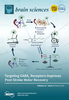
Stroke induces a loss in neurovascular coupling and an increase in reactive astrogliosis while producing abnormal conditions of excitability and plasticity in brain regions surrounding the infarct, which underlies the lasting disabilities observed in stroke patients. Drugs that block tonic GABA signalling promote neuronal excitability, enhance LTP formation, and lead to long-lasting enhancements in learning. Herein, GABAc receptors, which are found on astrocytes, were assessed as novel pharmacological targets for stroke recovery. Dampening GABAc receptor-mediated GABA signalling in astrocytes results in dampened reactive astrogliosis, improved astrocytic GABA uptake, and a lasting improvement in functional recovery. Image created using BioRender.com. View this paper.
  • Issues are regarded as officially published after their release is announced to the table of contents alert mailing list.
  • You may sign up for e-mail alerts to receive table of contents of newly released issues.
  • PDF is the official format for papers published in both, html and pdf forms. To view the papers in pdf format, click on the "PDF Full-text" link, and use the free Adobe Reader to open them.
Order results
Result details
Section
Select all
Export citation of selected articles as:
11 pages, 563 KiB  
Article
Paediatricians’ Views on Pain in Children with Profound Intellectual and Multiple Disabilities
by Lucie Petigas and Christopher J. Newman
Brain Sci. 2021, 11(3), 408; https://doi.org/10.3390/brainsci11030408 - 23 Mar 2021
Cited by 13 | Viewed by 2630
Abstract
Pain is a frequent issue in children with profound intellectual and multiple disabilities (PIMD). Its identification and treatment can prove highly challenging for primary care physicians, mainly because of the children’s limited communication abilities. We used an online survey to explore paediatricians’ views [...] Read more.
Pain is a frequent issue in children with profound intellectual and multiple disabilities (PIMD). Its identification and treatment can prove highly challenging for primary care physicians, mainly because of the children’s limited communication abilities. We used an online survey to explore paediatricians’ views regarding the experience and management of pain in children with PIMD and invited 480 professionals working in the canton of Vaud, Switzerland, to take part. We received 121 responses (participation rate 25.5%). A large majority of respondents provided care to children with PIMD. All paediatricians considered that these children feel pain at least as much as typically developing children. However, paediatricians had mixed views on their tolerance to pain. More than 90% held the view that their pain is under-assessed and undertreated. The principal barriers they reported to appropriate management were communication limitations with the child, difficulties in pain assessment, lack of knowledge about children with disabilities and lack of experience. Paediatricians have complex opinions regarding how children with PIMD experience pain and how to manage this problem. Professional education and training on the specificities of children with PIMD, including how to address their pain, seem necessary to foster paediatricians’ ability and confidence in approaching this complex issue. Full article
(This article belongs to the Special Issue Pain Assessment in Impaired Cognition)
Show Figures

Figure 1

36 pages, 1490 KiB  
Review
Efficacy of the Treatment of Developmental Language Disorder: A Systematic Review
by Sara Rinaldi, Maria Cristina Caselli, Valentina Cofelice, Simonetta D’Amico, Anna Giulia De Cagno, Giuseppina Della Corte, Maria Valeria Di Martino, Brigida Di Costanzo, Maria Chiara Levorato, Roberta Penge, Tiziana Rossetto, Alessandra Sansavini, Simona Vecchi and Pierluigi Zoccolotti
Brain Sci. 2021, 11(3), 407; https://doi.org/10.3390/brainsci11030407 - 23 Mar 2021
Cited by 37 | Viewed by 20992
Abstract
Background. Language disorder is the most frequent developmental disorder in childhood and it has a significant negative impact on children’s development. The goal of the present review was to systematically analyze the effectiveness of interventions in children with developmental language disorder (DLD) from [...] Read more.
Background. Language disorder is the most frequent developmental disorder in childhood and it has a significant negative impact on children’s development. The goal of the present review was to systematically analyze the effectiveness of interventions in children with developmental language disorder (DLD) from an evidence-based perspective. Methods. We considered systematic reviews, meta-analyses of randomized controlled trials (RCTs), control group cohort studies on any type of intervention aimed at improving children’s skills in the phono-articulatory, phonological, semantic-lexical, and morpho-syntactic fields in preschool and primary school children (up to eight years of age) that were diagnosed with DLD. We identified 27 full-length studies, 26 RCT and one review. Results. Early intensive intervention in three- and four-year-old children has a positive effect on phonological expressive and receptive skills and acquisitions are maintained in the medium term. Less evidence is available on the treatment of expressive vocabulary (and no evidence on receptive vocabulary). Intervention on morphological and syntactic skills has effective results on expressive (but not receptive) skills; however, a number of inconsistent results have also been reported. Only one study reports a positive effect of treatment on inferential narrative skills. Limited evidence is also available on the treatment of meta-phonological skills. More studies investigated the effectiveness of interventions on general language skills, which now appears as a promising area of investigation, even though results are not all consistent. Conclusions. The effectiveness of interventions over expressive and receptive phonological skills, morpho-syntactic skills, as well as inferential skills in narrative context underscores the importance that these trainings be implemented in children with DLD. Full article
Show Figures

Figure 1

10 pages, 1347 KiB  
Article
Cortical Representation and Excitability Increases for a Thenar Muscle Mediate Improvement in Short-Term Cellular Phone Text Messaging Ability
by Anthony W. Meek, Joselyn Perez, Brach Poston and Zachary A. Riley
Brain Sci. 2021, 11(3), 406; https://doi.org/10.3390/brainsci11030406 - 23 Mar 2021
Viewed by 1830
Abstract
Cortical representations expand during skilled motor learning. We studied a unique model of motor learning with cellular phone texting, where the thumbs are used exclusively to interact with the device and the prominence of use can be seen where 3200 text messages are [...] Read more.
Cortical representations expand during skilled motor learning. We studied a unique model of motor learning with cellular phone texting, where the thumbs are used exclusively to interact with the device and the prominence of use can be seen where 3200 text messages are exchanged a month in the 18–24 age demographic. The purpose of the present study was to examine the motor cortex representation and input–output (IO) recruitment curves of the abductor pollicis brevis (APB) muscle of the thumb and the ADM muscle with transcranial magnetic stimulation (TMS), relative to individuals’ texting abilities and short-term texting practice. Eighteen individuals performed a functional texting task (FTT) where we scored their texting speed and accuracy. TMS was then used to examine the cortical volumes and areas of activity in the two muscles and IO curves were constructed to measure excitability. Subjects also performed a 10-min practice texting task, after which we repeated the cortical measures. There were no associations between the cortical measures and the FTT scores before practice. However, after practice the APB cortical map expanded and excitability increased, whereas the ADM map constricted. The increase in the active cortical areas in APB correlated with the improvement in the FTT score. Based on the homogenous group of subjects that were already good at texting, we conclude that the cortical representations and excitability for the thumb muscle were already enlarged and more receptive to changes with short-term practice, as noted by the increase in FTT performance after 10-min of practice. Full article
(This article belongs to the Section Systems Neuroscience)
Show Figures

Figure 1

18 pages, 6966 KiB  
Review
Contribution of TMS and TMS-EEG to the Understanding of Mechanisms Underlying Physiological Brain Aging
by Andrea Guerra, Lorenzo Rocchi, Alberto Grego, Francesca Berardi, Concetta Luisi and Florinda Ferreri
Brain Sci. 2021, 11(3), 405; https://doi.org/10.3390/brainsci11030405 - 22 Mar 2021
Cited by 15 | Viewed by 3347
Abstract
In the human brain, aging is characterized by progressive neuronal loss, leading to disruption of synapses and to a degree of failure in neurotransmission. However, there is increasing evidence to support the notion that the aged brain has a remarkable ability to reorganize [...] Read more.
In the human brain, aging is characterized by progressive neuronal loss, leading to disruption of synapses and to a degree of failure in neurotransmission. However, there is increasing evidence to support the notion that the aged brain has a remarkable ability to reorganize itself, with the aim of preserving its physiological activity. It is important to develop objective markers able to characterize the biological processes underlying brain aging in the intact human, and to distinguish them from brain degeneration associated with many neurological diseases. Transcranial magnetic stimulation (TMS), coupled with electromyography or electroencephalography (EEG), is particularly suited to this aim, due to the functional nature of the information provided, and thanks to the ease with which it can be integrated with behavioral manipulation. In this review, we aimed to provide up to date information about the role of TMS and TMS-EEG in the investigation of brain aging. In particular, we focused on data about cortical excitability, connectivity and plasticity, obtained by using readouts such as motor evoked potentials and transcranial evoked potentials. Overall, findings in the literature support an important potential contribution of TMS to the understanding of the mechanisms underlying normal brain aging. Further studies are needed to expand the current body of information and to assess the applicability of TMS findings in the clinical setting. Full article
Show Figures

Figure 1

15 pages, 1111 KiB  
Review
Plasticity in the Hippocampus, Neurogenesis and Drugs of Abuse
by Yosef Avchalumov and Chitra D. Mandyam
Brain Sci. 2021, 11(3), 404; https://doi.org/10.3390/brainsci11030404 - 22 Mar 2021
Cited by 21 | Viewed by 5863
Abstract
Synaptic plasticity in the hippocampus assists with consolidation and storage of long-lasting memories. Decades of research has provided substantial information on the cellular and molecular mechanisms underlying synaptic plasticity in the hippocampus, and this review discusses these mechanisms in brief. Addiction is a [...] Read more.
Synaptic plasticity in the hippocampus assists with consolidation and storage of long-lasting memories. Decades of research has provided substantial information on the cellular and molecular mechanisms underlying synaptic plasticity in the hippocampus, and this review discusses these mechanisms in brief. Addiction is a chronic relapsing disorder with loss of control over drug taking and drug seeking that is caused by long-lasting memories of drug experience. Relapse to drug use is caused by exposure to context and cues associated with the drug experience, and is a major clinical problem that contributes to the persistence of addiction. This review also briefly discusses some evidence that drugs of abuse alter plasticity in the hippocampus, and that development of novel treatment strategies that reverse or prevent drug-induced synaptic alterations in the hippocampus may reduce relapse behaviors associated with addiction. Full article
(This article belongs to the Special Issue Neurogenesis and Gliogenesis in Health and Disease)
Show Figures

Figure 1

23 pages, 8962 KiB  
Review
Hereditary Spastic Paraplegia: From Genes, Cells and Networks to Novel Pathways for Drug Discovery
by Alan Mackay-Sim
Brain Sci. 2021, 11(3), 403; https://doi.org/10.3390/brainsci11030403 - 22 Mar 2021
Cited by 25 | Viewed by 4226
Abstract
Hereditary spastic paraplegia (HSP) is a diverse group of Mendelian genetic disorders affecting the upper motor neurons, specifically degeneration of their distal axons in the corticospinal tract. Currently, there are 80 genes or genomic loci (genomic regions for which the causative gene has [...] Read more.
Hereditary spastic paraplegia (HSP) is a diverse group of Mendelian genetic disorders affecting the upper motor neurons, specifically degeneration of their distal axons in the corticospinal tract. Currently, there are 80 genes or genomic loci (genomic regions for which the causative gene has not been identified) associated with HSP diagnosis. HSP is therefore genetically very heterogeneous. Finding treatments for the HSPs is a daunting task: a rare disease made rarer by so many causative genes and many potential mutations in those genes in individual patients. Personalized medicine through genetic correction may be possible, but impractical as a generalized treatment strategy. The ideal treatments would be small molecules that are effective for people with different causative mutations. This requires identification of disease-associated cell dysfunctions shared across genotypes despite the large number of HSP genes that suggest a wide diversity of molecular and cellular mechanisms. This review highlights the shared dysfunctional phenotypes in patient-derived cells from patients with different causative mutations and uses bioinformatic analyses of the HSP genes to identify novel cell functions as potential targets for future drug treatments for multiple genotypes. Full article
(This article belongs to the Special Issue Factors Responsible for CSMN Vulnerability)
Show Figures

Figure 1

14 pages, 310 KiB  
Perspective
Promoting Generalized Learning in Balance Recovery Interventions
by Sara A. Harper, Anne Z. Beethe, Christopher J. Dakin and David A. E. Bolton
Brain Sci. 2021, 11(3), 402; https://doi.org/10.3390/brainsci11030402 - 22 Mar 2021
Cited by 13 | Viewed by 2587
Abstract
Recent studies have shown balance recovery can be enhanced via task-specific training, referred to as perturbation-based balance training (PBT). These interventions rely on principles of motor learning where repeated exposure to task-relevant postural perturbations results in more effective compensatory balance responses. Evidence indicates [...] Read more.
Recent studies have shown balance recovery can be enhanced via task-specific training, referred to as perturbation-based balance training (PBT). These interventions rely on principles of motor learning where repeated exposure to task-relevant postural perturbations results in more effective compensatory balance responses. Evidence indicates that compensatory responses trained using PBT can be retained for many months and can lead to a reduction in falls in community-dwelling older adults. A notable shortcoming with PBT is that it does not transfer well to similar but contextually different scenarios (e.g., falling sideways versus a forward trip). Given that it is not feasible to train all conditions in which someone could fall, this limited transfer presents a conundrum; namely, how do we best use PBT to appropriately equip people to deal with the enormous variety of fall-inducing scenarios encountered in daily life? In this perspective article, we draw from fields of research that explore how general learning can be promoted. From this, we propose a series of methods, gleaned from parallel streams of research, to inform and hopefully optimize this emerging field where people receive training to specifically improve their balance reactions. Full article
(This article belongs to the Special Issue Balance Rehabilitation in Neurological Disorders)
13 pages, 323 KiB  
Article
Parental Education and Left Lateral Orbitofrontal Cortical Activity during N-Back Task: An fMRI Study of American Adolescents
by Shervin Assari, Shanika Boyce, Mohammed Saqib, Mohsen Bazargan and Cleopatra H. Caldwell
Brain Sci. 2021, 11(3), 401; https://doi.org/10.3390/brainsci11030401 - 22 Mar 2021
Cited by 3 | Viewed by 2905
Abstract
Introduction. The Orbitofrontal Cortex (OFC) is a cortical structure that has implications in cognition, memory, reward anticipation, outcome evaluation, decision making, and learning. As such, OFC activity correlates with these cognitive brain abilities. Despite research suggesting race and socioeconomic status (SES) indicators such [...] Read more.
Introduction. The Orbitofrontal Cortex (OFC) is a cortical structure that has implications in cognition, memory, reward anticipation, outcome evaluation, decision making, and learning. As such, OFC activity correlates with these cognitive brain abilities. Despite research suggesting race and socioeconomic status (SES) indicators such as parental education may be associated with OFC activity, limited knowledge exists on multiplicative effects of race and parental education on OFC activity and associated cognitive ability. Purpose. Using functional brain imaging data from the Adolescent Brain Cognitive Development (ABCD) study, we tested the multiplicative effects of race and parental education on left lateral OFC activity during an N-Back task. In our study, we used a sociological rather than biological theory that conceptualizes race and SES as proxies of access to the opportunity structure and exposure to social adversities rather than innate and non-modifiable brain differences. We explored racial variation in the effect of parental educational attainment, a primary indicator of SES, on left lateral OFC activity during an N-Back task between Black and White 9–10 years old adolescents. Methods. The ABCD study is a national, landmark, multi-center brain imaging investigation of American adolescents. The total sample was 4290 9–10 years old Black or White adolescents. The independent variables were SES indicators, namely family income, parental education, and neighborhood income. The primary outcome was the average beta weight for N-Back (2 back versus 0 back contrast) in ASEG ROI left OFC activity, measured by functional Magnetic Resonance Imaging (fMRI) during an N-Back task. Ethnicity, age, sex, subjective SES, and family structure were the study covariates. For data analysis, we used linear regression models. Results. In White but not Black adolescents, parental education was associated with higher left lateral OFC activity during the N-Back task. In the pooled sample, we found a significant interaction between race and parental education on the outcome, suggesting that high parental education is associated with a larger increase in left OFC activity of White than Black adolescents. Conclusions. For American adolescents, race and SES jointly influence left lateral OFC activity correlated with cognition, memory, decision making, and learning. Given the central role of left lateral OFC activity in learning and memory, our finding calls for additional research on contextual factors that reduce the gain of SES for Black adolescents. Cognitive inequalities are not merely due to the additive effects of race and SES but also its multiplicative effects. Full article
16 pages, 2416 KiB  
Article
Effects of Dizocilpine, Midazolam and Their Co-Application on the Trimethyltin (TMT)-Induced Rat Model of Cognitive Deficit
by Marketa Chvojkova, Hana Kubova and Karel Vales
Brain Sci. 2021, 11(3), 400; https://doi.org/10.3390/brainsci11030400 - 22 Mar 2021
Cited by 2 | Viewed by 2162
Abstract
Research of treatment options addressing the cognitive deficit associated with neurodegenerative disorders is of particular importance. Application of trimethyltin (TMT) to rats represents a promising model replicating multiple relevant features of such disorders. N-methyl-D-aspartate (NMDA) receptor antagonists and gamma-aminobutyric acid type A (GABA [...] Read more.
Research of treatment options addressing the cognitive deficit associated with neurodegenerative disorders is of particular importance. Application of trimethyltin (TMT) to rats represents a promising model replicating multiple relevant features of such disorders. N-methyl-D-aspartate (NMDA) receptor antagonists and gamma-aminobutyric acid type A (GABAA) receptor potentiators have been reported to alleviate the TMT-induced cognitive deficit. These compounds may provide synergistic interactions in other models. The aim of this study was to investigate, whether co-application of NMDA receptor antagonist dizocilpine (MK-801) and GABAA receptor potentiator midazolam would be associated with an improved effect on the TMT-induced model of cognitive deficit. Wistar rats injected with TMT were repeatedly (12 days) treated with MK-801, midazolam, or both. Subsequently, cognitive performance was assessed. Finally, after a 17-day drug-free period, hippocampal neurodegeneration (neuronal density in CA2/3 subfield in the dorsal hippocampus, dentate gyrus morphometry) were analyzed. All three protective treatments induced similar degree of therapeutic effect in Morris water maze. The results of histological analyses were suggestive of minor protective effect of the combined treatment (MK-801 and midazolam), while these compounds alone were largely ineffective at this time point. Therefore, in terms of mitigation of cognitive deficit, the combined treatment was not associated with improved effect. Full article
Show Figures

Figure 1

12 pages, 940 KiB  
Review
An ALE Meta-Analysis on Investment Decision-Making
by Elena Ortiz-Teran, Ibai Diez and Joaquin Lopez-Pascual
Brain Sci. 2021, 11(3), 399; https://doi.org/10.3390/brainsci11030399 - 21 Mar 2021
Cited by 3 | Viewed by 3197
Abstract
It is claimed that investment decision-making should rely on rational analyses based on facts and not emotions. However, trying to make money out of market forecasts can trigger all types of emotional responses. As the question on how investors decide remains controversial, we [...] Read more.
It is claimed that investment decision-making should rely on rational analyses based on facts and not emotions. However, trying to make money out of market forecasts can trigger all types of emotional responses. As the question on how investors decide remains controversial, we carried out an activation likelihood estimation (ALE) meta-analysis using functional magnetic resonance imaging (fMRI) studies that have reported whole-brain analyses on subjects performing an investment task. We identified the ventral striatum, anterior insula, amygdala and anterior cingulate cortex as being involved in this decision-making process. These regions are limbic-related structures which respond to reward, risk and emotional conflict. Our findings support the notion that investment choices are emotional decisions that take into account market information, individual preferences and beliefs. Full article
(This article belongs to the Special Issue Advances in Neuroeconomics)
Show Figures

Figure 1

19 pages, 392 KiB  
Review
Inherited Neuromuscular Disorders: Which Role for Serum Biomarkers?
by Antonino Lupica, Vincenzo Di Stefano, Andrea Gagliardo, Salvatore Iacono, Antonia Pignolo, Salvatore Ferlisi, Angelo Torrente, Sonia Pagano, Massimo Gangitano and Filippo Brighina
Brain Sci. 2021, 11(3), 398; https://doi.org/10.3390/brainsci11030398 - 21 Mar 2021
Cited by 8 | Viewed by 2693
Abstract
Inherited neuromuscular disorders (INMD) are a heterogeneous group of rare diseases that involve muscles, motor neurons, peripheral nerves or the neuromuscular junction. Several different lab abnormalities have been linked to INMD: sometimes they are typical of the disorder, but they usually appear to [...] Read more.
Inherited neuromuscular disorders (INMD) are a heterogeneous group of rare diseases that involve muscles, motor neurons, peripheral nerves or the neuromuscular junction. Several different lab abnormalities have been linked to INMD: sometimes they are typical of the disorder, but they usually appear to be less specific. Sometimes serum biomarkers can point out abnormalities in presymtomatic or otherwise asymptomatic patients (e.g., carriers). More often a biomarker of INMD is evaluated by multiple clinicians other than expert in NMD before the diagnosis, because of the multisystemic involvement in INMD. The authors performed a literature search on biomarkers in inherited neuromuscular disorders to provide a practical approach to the diagnosis and the correct management of INMD. A considerable number of biomarkers have been reported that support the diagnosis of INMD, but the role of an expert clinician is crucial. Hence, the complete knowledge of such abnormalities can accelerate the diagnostic workup supporting the referral to specialists in neuromuscular disorders. Full article
(This article belongs to the Special Issue Biochemical Biomarkers and Neurodegenerative Diseases)
10 pages, 1279 KiB  
Article
Using Functional Connectivity to Examine the Correlation between Mirror Neuron Network and Autistic Traits in a Typically Developing Sample: A fNIRS Study
by Thien Nguyen, Helga O. Miguel, Emma E. Condy, Soongho Park and Amir Gandjbakhche
Brain Sci. 2021, 11(3), 397; https://doi.org/10.3390/brainsci11030397 - 20 Mar 2021
Cited by 4 | Viewed by 2688
Abstract
Mirror neuron network (MNN) is associated with one’s ability to recognize and interpret others’ actions and emotions and has a crucial role in cognition, perception, and social interaction. MNN connectivity and its relation to social attributes, such as autistic traits have not been [...] Read more.
Mirror neuron network (MNN) is associated with one’s ability to recognize and interpret others’ actions and emotions and has a crucial role in cognition, perception, and social interaction. MNN connectivity and its relation to social attributes, such as autistic traits have not been thoroughly examined. This study aimed to investigate functional connectivity in the MNN and assess relationship between MNN connectivity and subclinical autistic traits in neurotypical adults. Hemodynamic responses, including oxy- and deoxy-hemoglobin were measured in the central and parietal cortex of 30 healthy participants using a 24-channel functional Near-Infrared spectroscopy (fNIRS) system during a live action-observation and action-execution task. Functional connectivity was derived from oxy-hemoglobin data. Connections with significantly greater connectivity in both tasks were assigned to MNN connectivity. Correlation between connectivity and autistic traits were performed using Pearson correlation. Connections within the right precentral, right supramarginal, left inferior parietal, left postcentral, and between left supramarginal-left angular regions were identified as MNN connections. In addition, individuals with higher subclinical autistic traits present higher connectivity in both action-execution and action-observation conditions. Positive correlation between MNN connectivity and subclinical autistic traits can be used in future studies to investigate MNN in a developing population with autism spectrum disorder. Full article
Show Figures

Figure 1

11 pages, 1047 KiB  
Article
Association between Previous Statin Use and Alzheimer’s Disease: A Nested Case-Control Study Using a National Health Screening Cohort
by Ji Hee Kim, Heui Seung Lee, Jee Hye Wee, Yoo Hwan Kim, Chan Yang Min, Dae Myoung Yoo and Hyo Geun Choi
Brain Sci. 2021, 11(3), 396; https://doi.org/10.3390/brainsci11030396 - 20 Mar 2021
Cited by 7 | Viewed by 2318
Abstract
A number of studies report the incidence of Alzheimer’s disease (AD) in patients taking statins, but the results are inconsistent. (1) Background: The present study investigated the cross-sectional association between previous statin use and the risk of AD development in Korean residents. (2) [...] Read more.
A number of studies report the incidence of Alzheimer’s disease (AD) in patients taking statins, but the results are inconsistent. (1) Background: The present study investigated the cross-sectional association between previous statin use and the risk of AD development in Korean residents. (2) Methods: We used the Korean National Health Insurance Service-National Sample Cohort; 17,172 AD patients were matched by age, gender, income, and region of residence with 68,688 control participants at a ratio of 1:4. We used a multiple conditional logistic regression model to analyse the association between the number of days of statin use and AD occurrence. Further analyses were performed to identify whether this association is maintained for different ages, genders, socioeconomic status groups, and covariates. (3) Results: The odds ratio, which was adjusted for potential confounders, for the days of statin use per year in the AD group compared to the control group was 0.95 (95% confidence interval = 0.92–0.98; p = 0.003). The number of days of statin use in the AD group was significantly smaller in the subgroups of non-smokers and individuals with normal weight, alcohol consumption less than once a week, total cholesterol level below 200 mg/dL, systolic blood pressure below 140, diastolic blood pressure below 90, and fasting blood glucose below 100 mg/dL. (4) Conclusions: Our results suggest that statin use prevents the occurrence of AD. The effects of statin use in preventing AD may be greater in individuals at relatively low risk. Full article
(This article belongs to the Special Issue Collection on Cognitive Neuroscience)
Show Figures

Figure 1

13 pages, 1502 KiB  
Article
Influence of Brain-Derived Neurotrophic Factor Genotype on Short-Latency Afferent Inhibition and Motor Cortex Metabolites
by Ryoki Sasaki, Naofumi Otsuru, Shota Miyaguchi, Sho Kojima, Hiraku Watanabe, Ken Ohno, Noriko Sakurai, Naoki Kodama, Daisuke Sato and Hideaki Onishi
Brain Sci. 2021, 11(3), 395; https://doi.org/10.3390/brainsci11030395 - 20 Mar 2021
Cited by 10 | Viewed by 2318
Abstract
The Met allele of the brain-derived neurotrophic factor (BDNF) gene confers reduced cortical BDNF expression and associated neurobehavioral changes. BDNF signaling influences the survival, development, and synaptic function of cortical networks. Here, we compared gamma-aminobutyric acid (GABA)ergic network activity in the human primary [...] Read more.
The Met allele of the brain-derived neurotrophic factor (BDNF) gene confers reduced cortical BDNF expression and associated neurobehavioral changes. BDNF signaling influences the survival, development, and synaptic function of cortical networks. Here, we compared gamma-aminobutyric acid (GABA)ergic network activity in the human primary motor cortex (M1) between the Met (Val/Met and Met/Met) and non-Met (Val/Val) genotype groups. Short- and long-interval intracortical inhibition, short-latency afferent inhibition (SAI), and long-latency afferent inhibition were measured using transcranial magnetic stimulation (TMS) as indices of GABAergic activity. Furthermore, the considerable inter-individual variability in inhibitory network activity typically measured by TMS may be affected not only by GABA but also by other pathways, including glutamatergic and cholinergic activities; therefore, we used 3-T magnetic resonance spectroscopy (MRS) to measure the dynamics of glutamate plus glutamine (Glx) and choline concentrations in the left M1, left somatosensory cortex, and right cerebellum. All inhibitory TMS conditions produced significantly smaller motor-evoked potentials than single-pulses. SAI was significantly stronger in the Met group than in the Val/Val group. Only the M1 Glx concentration was significantly lower in the Met group, while the BDNF genotype did not affect choline concentration in any region. Further, a positive correlation was observed between SAI and Glx concentrations only in M1. Our findings provide evidence that the BDNF genotype regulates both the inhibitory and excitatory circuits in human M1. In addition, lower Glx concentration in the M1 of Met carriers may alter specific inhibitory network on M1, thereby influencing the cortical signal processing required for neurobehavioral functions. Full article
Show Figures

Figure 1

28 pages, 1993 KiB  
Review
Processing of Degraded Speech in Brain Disorders
by Jessica Jiang, Elia Benhamou, Sheena Waters, Jeremy C. S. Johnson, Anna Volkmer, Rimona S. Weil, Charles R. Marshall, Jason D. Warren and Chris J. D. Hardy
Brain Sci. 2021, 11(3), 394; https://doi.org/10.3390/brainsci11030394 - 20 Mar 2021
Cited by 6 | Viewed by 4311
Abstract
The speech we hear every day is typically “degraded” by competing sounds and the idiosyncratic vocal characteristics of individual speakers. While the comprehension of “degraded” speech is normally automatic, it depends on dynamic and adaptive processing across distributed neural networks. This presents the [...] Read more.
The speech we hear every day is typically “degraded” by competing sounds and the idiosyncratic vocal characteristics of individual speakers. While the comprehension of “degraded” speech is normally automatic, it depends on dynamic and adaptive processing across distributed neural networks. This presents the brain with an immense computational challenge, making degraded speech processing vulnerable to a range of brain disorders. Therefore, it is likely to be a sensitive marker of neural circuit dysfunction and an index of retained neural plasticity. Considering experimental methods for studying degraded speech and factors that affect its processing in healthy individuals, we review the evidence for altered degraded speech processing in major neurodegenerative diseases, traumatic brain injury and stroke. We develop a predictive coding framework for understanding deficits of degraded speech processing in these disorders, focussing on the “language-led dementias”—the primary progressive aphasias. We conclude by considering prospects for using degraded speech as a probe of language network pathophysiology, a diagnostic tool and a target for therapeutic intervention. Full article
Show Figures

Figure 1

22 pages, 854 KiB  
Systematic Review
A Scientometric Approach to Review the Role of the Medial Preoptic Area (MPOA) in Parental Behavior
by Alessandro Carollo, Jan Paolo Macapinlac Balagtas, Michelle Jin-Yee Neoh and Gianluca Esposito
Brain Sci. 2021, 11(3), 393; https://doi.org/10.3390/brainsci11030393 - 20 Mar 2021
Cited by 24 | Viewed by 5050
Abstract
Research investigating the neural substrates underpinning parental behaviour has recently gained momentum. Particularly, the hypothalamic medial preoptic area (MPOA) has been identified as a crucial region for parenting. The current study conducted a scientometric analysis of publications from 1 January 1972 to 19 [...] Read more.
Research investigating the neural substrates underpinning parental behaviour has recently gained momentum. Particularly, the hypothalamic medial preoptic area (MPOA) has been identified as a crucial region for parenting. The current study conducted a scientometric analysis of publications from 1 January 1972 to 19 January 2021 using CiteSpace software to determine trends in the scientific literature exploring the relationship between MPOA and parental behaviour. In total, 677 scientific papers were analysed, producing a network of 1509 nodes and 5498 links. Four major clusters were identified: “C-Fos Expression”, “Lactating Rat”, “Medial Preoptic Area Interaction” and “Parental Behavior”. Their content suggests an initial trend in which the properties of the MPOA in response to parental behavior were studied, followed by a growing attention towards the presence of a brain network, including the reward circuits, regulating such behavior. Furthermore, while attention was initially directed uniquely to maternal behavior, it has recently been extended to the understanding of paternal behaviors as well. Finally, although the majority of the studies were conducted on rodents, recent publications broaden the implications of previous documents to human parental behavior, giving insight into the mechanisms underlying postpartum depression. Potential directions in future works were also discussed. Full article
(This article belongs to the Special Issue Hypothalamus, Neuropeptides and Socioemotional Behavior)
Show Figures

Figure 1

12 pages, 1306 KiB  
Review
Prevalence of Neural Autoantibodies in Epilepsy of Unknown Etiology: Systematic Review and Meta-Analysis
by Pablo Cabezudo-García, Natalia Mena-Vázquez, Nicolás L. Ciano-Petersen, Guillermina García-Martín, Guillermo Estivill-Torrús and Pedro J. Serrano-Castro
Brain Sci. 2021, 11(3), 392; https://doi.org/10.3390/brainsci11030392 - 19 Mar 2021
Cited by 6 | Viewed by 2889
Abstract
Background: The prevalence of neural autoantibodies in epilepsy of unknown etiology varies among studies. We aimed to conduct a systematic review and meta-analysis to determine the pooled global prevalence and the prevalence for each antibody. Methods: A systematic search was conducted for studies [...] Read more.
Background: The prevalence of neural autoantibodies in epilepsy of unknown etiology varies among studies. We aimed to conduct a systematic review and meta-analysis to determine the pooled global prevalence and the prevalence for each antibody. Methods: A systematic search was conducted for studies that included prospectively patients ≥16 years old with epilepsy of unknown etiology and systematically determined neural autoantibodies. A meta-analysis was undertaken to estimate pooled prevalence in total patients with a positive result for at least one neural autoantibody in serum and/or cerebrospinal fluid (CSF) and for each autoantibody. Results: Ten of the eleven studies that met the inclusion criteria and a total of 1302 patients with epilepsy of unknown etiology were included in themeta-analysis. The global pooled prevalence (IC95%) was 7.6% (4.6–11.2) in a total of 82 patients with a positive result for any neural autoantibody. None of the controls available in the studies had a positive result. Individual pooled prevalence for each autoantibody was: glycine receptor (GlyR) (3.2%), glutamic acid decarboxylase (GAD) (1.9%), N-methyl-d-aspartate receptor (NMDAR) (1.8%), leucine-rich glioma inactivated-1 protein (LGI1) (1.1%), contactin-2-associated protein (CASPR2) (0.6%) and onconeuronal (0.2%). Conclusions: The pooled prevalence of neural autoantibodies in patients with epilepsy of unknown etiology is small but not irrelevant. None of the controls had a positive result. There was high heterogeneity among studies. In the future, a homogeneous protocol for testing neural autoantibodies is recommended. Full article
(This article belongs to the Special Issue The Immune System, Neuroinflammation and Epilepsy)
Show Figures

Graphical abstract

20 pages, 358 KiB  
Article
Single-Nucleotide Polymorphisms in Oxidative Stress-Related Genes and the Risk of a Stroke in a Polish Population—A Preliminary Study
by Ewelina Synowiec, Paulina Wigner, Natalia Cichon, Cezary Watala, Piotr Czarny, Joanna Saluk-Bijak, Elzbieta Miller, Tomasz Sliwinski, Ewa Zielinska-Nowak and Michal Bijak
Brain Sci. 2021, 11(3), 391; https://doi.org/10.3390/brainsci11030391 - 19 Mar 2021
Cited by 6 | Viewed by 1993
Abstract
The present preliminary case-control study was undertaken to detect the potential association of six single nucleotide polymorphisms (SNPs) in oxidative stress-related genes: SOD2 (c.47T > C; rs4880), CAT (c.-89A > T; rs7943316), GPX4 (c.660T > A; rs713041), NOS1 (g.117803515C > T; rs1879417) and [...] Read more.
The present preliminary case-control study was undertaken to detect the potential association of six single nucleotide polymorphisms (SNPs) in oxidative stress-related genes: SOD2 (c.47T > C; rs4880), CAT (c.-89A > T; rs7943316), GPX4 (c.660T > A; rs713041), NOS1 (g.117803515C > T; rs1879417) and NOS2 (c.1823C > T; rs2297518 and c.-227G > C; rs10459953) and the occurrence of a stroke. The SNPs were determined using the TaqMan® Allelic Discrimination Assay in 107 patients with strokes and 107 age- and sex-matched individuals who had not experienced cerebrovascular accidents. The T alleles of the rs4880 were positively correlated with a stroke (bootstrap OR 1.31; 1.07–1.59 95% CI). In the case of the rs713041, an association with the T allele was found (bootstrap OR 1.36; 1.12–1.67). In addition, the occurrence of a stroke was associated with the presence of the C allele of the rs1879417 (bootstrap OR 1.32; 1.09–1.61). We also found that the C/C genotype and C allele of the rs2297518 increased the risk of a stroke (bootstrap ORs 7.00; 4.34–11.29 and 4.96; 3.88–6.34, respectively). Moreover, the C allele of the rs10459953 was associated with an increased occurrence of this disease (bootstrap OR 1.31; 1.08–1.60). These results indicated that genetics variants in the SOD2, GPX4, NOS1 and NOS2 might be associated with susceptibility to strokes in the Polish population. Full article
7 pages, 244 KiB  
Brief Report
Test Anxiety in Autistic University Students—Preliminary Results from a German-Speaking Sample
by Alafia Zavery, Myriam Zäch and Alex Bertrams
Brain Sci. 2021, 11(3), 390; https://doi.org/10.3390/brainsci11030390 - 19 Mar 2021
Cited by 1 | Viewed by 2256
Abstract
Autism is a neurodevelopmental condition associated with increased levels of anxiety. However, whether autism is related to heightened test anxiety as one situation-specific type of anxiety has not yet been examined. This question may be relevant for the achievement and well-being of autistic [...] Read more.
Autism is a neurodevelopmental condition associated with increased levels of anxiety. However, whether autism is related to heightened test anxiety as one situation-specific type of anxiety has not yet been examined. This question may be relevant for the achievement and well-being of autistic people in educational settings (e.g., at universities). In this study, we took a first step to investigate whether autistic university students have increased test anxiety. A sample of 16 German-speaking university students completed an established diagnostic instrument to measure test anxiety and its components of worry, emotionality, cognitive interference, and lack of confidence. The scores of the autistic students were compared with the test anxiety means and percentile ranks of the standardization sample for the applied test anxiety measure (n = 1350). For an additional comparison, the test anxiety means and percentile ranks of non-autistic university students (n = 101) were assessed during the last third of the semester; that is, close to the examinations. Overall, the results suggest that autistic university students have remarkably increased test anxiety. Although the present findings must be considered preliminary, they suggest that text anxiety in educational settings may be a neglected significant problem for autistic people that requires further attention in research and practice. Full article
10 pages, 789 KiB  
Article
Neurocognitive Impairment in Severe Mental Illness. Comparative study with Spanish Speaking Patients
by Gabriel G. De la Torre, Sandra Doval, David López-Sanz, Manuel García-Sedeño, Miguel A. Ramallo, Macarena Bernal and Sara González-Torre
Brain Sci. 2021, 11(3), 389; https://doi.org/10.3390/brainsci11030389 - 19 Mar 2021
Cited by 3 | Viewed by 2315
Abstract
Background. Serious mental illness (SMI) represents a category of psychiatric disorders characterized by specific difficulties of personal and social functioning, derived from suffering severe and persistent mental health problems. Aims. We wanted to look into differences in cognitive performance among different SMI patients. [...] Read more.
Background. Serious mental illness (SMI) represents a category of psychiatric disorders characterized by specific difficulties of personal and social functioning, derived from suffering severe and persistent mental health problems. Aims. We wanted to look into differences in cognitive performance among different SMI patients. Methods. Repeatable Battery for the Assessment of Neuropsychological Status (RBANS) screening was applied in one sample of SMI patients (n = 149) and another of healthy comparison participants (n = 35). Within the SMI sample, three different subsamples were formed: one with 97 patients with schizophrenia, a second with 29 patients with mood disorders, and a third with 23 patients with personality disorder. We performed a comparative study within and between groups. Results. Analysis of covariance was performed. Significant differences were found for cognitive functioning including attention and memory. Conclusions. RBANS can be recommended for the detection of neurocognitive deficits in psychiatric disorders, especially in Schizophrenia. Full article
Show Figures

Figure 1

12 pages, 1888 KiB  
Article
Cortical Plasticity and Interneuron Recruitment in Adolescents Born to Women with Gestational Diabetes Mellitus
by Jago M. Van Dam, Mitchell R. Goldsworthy, William M. Hague, Suzette Coat and Julia B. Pitcher
Brain Sci. 2021, 11(3), 388; https://doi.org/10.3390/brainsci11030388 - 19 Mar 2021
Cited by 3 | Viewed by 2037
Abstract
Exposure to gestational diabetes mellitus (GDM) in utero is associated with a range of adverse cognitive and neurological outcomes. Previously, we reported altered neuroplastic responses to continuous theta burst stimulation (cTBS) in GDM-exposed adolescents. Recent research suggests that the relative excitability of complex [...] Read more.
Exposure to gestational diabetes mellitus (GDM) in utero is associated with a range of adverse cognitive and neurological outcomes. Previously, we reported altered neuroplastic responses to continuous theta burst stimulation (cTBS) in GDM-exposed adolescents. Recent research suggests that the relative excitability of complex oligosynaptic circuits (late I-wave circuits) can predict these responses. We aimed to determine if altered I-wave recruitment was associated with neuroplastic responses in adolescents born to women with GDM. A total of 20 GDM-exposed adolescents and 10 controls (aged 13.1 ± 1.0 years) participated. cTBS was used to induce neuroplasticity. I-wave recruitment was assessed by comparing motor-evoked potential latencies using different TMS coil directions. Recruitment of late I-waves was associated with stronger LTD-like neuroplastic responses to cTBS (p = < 0.001, R2 = 0.36). There were no differences between groups in mean neuroplasticity (p = 0.37), I-wave recruitment (p = 0.87), or the association between these variables (p = 0.41). The relationship between I-wave recruitment and the response to cTBS previously observed in adults is also present in adolescents and does not appear to be altered significantly by in utero GDM exposure. Exposure to GDM does not appear to significantly impair LTD-like synaptic plasticity or interneuron recruitment. Full article
Show Figures

Figure 1

14 pages, 1018 KiB  
Article
Distinctive Frontal and Occipitotemporal Surface Features in Neglectful Parenting
by Inmaculada León, María José Rodrigo, Ileana Quiñones, Juan Andrés Hernández-Cabrera and Lorna García-Pentón
Brain Sci. 2021, 11(3), 387; https://doi.org/10.3390/brainsci11030387 - 18 Mar 2021
Cited by 6 | Viewed by 2040
Abstract
Although the brain signatures of adaptive human parenting are well documented, the cortical features associated with maladaptive caregiving are underexplored. We investigated whether cortical thickness and surface area vary in a small group of mothers who had neglected their children (24 in the [...] Read more.
Although the brain signatures of adaptive human parenting are well documented, the cortical features associated with maladaptive caregiving are underexplored. We investigated whether cortical thickness and surface area vary in a small group of mothers who had neglected their children (24 in the neglect group, NG) compared to a control group of mothers with non-neglectful caregiving (21 in the control group, CG). We also tested whether the cortical differences were related to dyadic mother-child emotional availability (EA) in a play task with their children and whether alexithymia involving low emotional awareness that characterizes the NG could play a role in the cortical-EA associations. Whole-brain analysis of the cortical mantle identified reduced cortical thickness in the right rostral middle frontal gyrus and an increased surface area in the right lingual and lateral occipital cortices for the NG with respect to the CG. Follow-up path analysis showed direct effects of the right rostral middle frontal gyrus (RMFG) on the emotional availability (EA) and on the difficulty to identify feelings (alexithymia factor), with a marginal indirect RMFG-EA effect through this factor. These preliminary findings extend existing work by implicating differences in cortical features associated with neglectful parenting and relevant to mother-child interactive bonding. Full article
Show Figures

Figure 1

30 pages, 2538 KiB  
Review
Against the Resilience of High-Grade Gliomas: The Immunotherapeutic Approach (Part I)
by Alice Giotta Lucifero and Sabino Luzzi
Brain Sci. 2021, 11(3), 386; https://doi.org/10.3390/brainsci11030386 - 18 Mar 2021
Cited by 16 | Viewed by 3690
Abstract
The resilience of high-grade gliomas (HGGs) against conventional chemotherapies is due to their heterogeneous genetic landscape, adaptive phenotypic changes, and immune escape mechanisms. Innovative immunotherapies have been developed to counteract the immunosuppressive capability of gliomas. Nevertheless, further research is needed to assess the [...] Read more.
The resilience of high-grade gliomas (HGGs) against conventional chemotherapies is due to their heterogeneous genetic landscape, adaptive phenotypic changes, and immune escape mechanisms. Innovative immunotherapies have been developed to counteract the immunosuppressive capability of gliomas. Nevertheless, further research is needed to assess the efficacy of the immuno-based approach. The aim of this study is to review the newest immunotherapeutic approaches for glioma, focusing on the drug types, mechanisms of action, clinical pieces of evidence, and future challenges. A PRISMA (Preferred Reporting Items for Systematic Review and Meta-Analysis)-based literature search was performed on PubMed/Medline and ClinicalTrials.gov databases using the keywords “active/adoptive immunotherapy,” “monoclonal antibodies,” “vaccine,” and “engineered T cell.”, combined with “malignant brain tumor”, “high-grade glioma.” Only articles written in English published in the last 10 years were selected, filtered based on best relevance. Active immunotherapies include systemic temozolomide, monoclonal antibodies, and vaccines. In several preclinical and clinical trials, adoptive immunotherapies, including T, natural killer, and natural killer T engineered cells, have been shown to be potential treatment options for relapsing gliomas. Systemic temozolomide is considered the backbone for newly diagnosed HGGs. Bevacizumab and rindopepimut are promising second-line treatments. Adoptive immunotherapies have been proven for relapsing tumors, but further evidence is needed. Full article
Show Figures

Figure 1

10 pages, 457 KiB  
Article
Blood Pressure and Risk of Cognitive Impairment: The Role of Vascular Disease in Neurodegeneration
by Mar Carmona-Abellan, Malwina Trzeciak, Miriam Recio Fernández, Beatriz Echeveste, Laura Imaz, Maria-Rosario Luquin and Mario Riverol
Brain Sci. 2021, 11(3), 385; https://doi.org/10.3390/brainsci11030385 - 18 Mar 2021
Cited by 4 | Viewed by 2146
Abstract
(1) Background: Both cerebral vascular disorders and cognitive decline increase in incidence with age. The role of cerebral vascular disease and hemodynamic changes in the development of cognitive deficits is controversial. The objective of this study was to assess the cardiovascular response during [...] Read more.
(1) Background: Both cerebral vascular disorders and cognitive decline increase in incidence with age. The role of cerebral vascular disease and hemodynamic changes in the development of cognitive deficits is controversial. The objective of this study was to assess the cardiovascular response during cardiac stress testing in neurologically asymptomatic individuals who developed cognitive impairment several years after previous cardiac stress testing. (2) Methods: This was a retrospective cohort study of patients who underwent cardiac stress testing between January 2001 and December 2010. Patients were followed up until May 2015, and we selected those who developed cognitive dysfunction including dementia, mild cognitive impairment, and subjective cognitive decline, after the stress test. Heart rate and blood pressure both at rest and at peak exercise, and the mean R-R interval at rest were recorded. For each patient who developed cognitive impairment, we selected one matched control who did not show cognitive decline by the end of the follow-up period. (3) Results: From the cohort of 7224 patients, 371 developed cognitive impairment; of these, 186 (124 men) met the inclusion criteria, and 186 of the other patients were selected as matched controls. During follow-up, cognitive impairment appeared 6.2 ± 4.7 years after the cardiac stress test. These patients who had subsequently developed cognitive impairment had significantly lower at-rest systolic, diastolic, and mean blood pressure than controls (p < 0.05). Further, compared with controls, their maximum heart rate was significantly higher at peak exercise. (4) Conclusion: The results from this study suggest that differences in cardiovascular response to stress might be present in individuals who develop cognitive decline. These findings challenge the possibility of assessing blood pressure and heart rate variability at rest and during cardiac stress as potential risk factors associated with cognitive impairment. Full article
Show Figures

Figure 1

35 pages, 4839 KiB  
Article
Probing the Brain–Body Connection Using Transcranial Magnetic Stimulation (TMS): Validating a Promising Tool to Provide Biomarkers of Neuroplasticity and Central Nervous System Function
by Arthur R. Chaves, Nicholas J. Snow, Lynsey R. Alcock and Michelle Ploughman
Brain Sci. 2021, 11(3), 384; https://doi.org/10.3390/brainsci11030384 - 17 Mar 2021
Cited by 16 | Viewed by 7438
Abstract
Transcranial magnetic stimulation (TMS) is a non-invasive method used to investigate neurophysiological integrity of the human neuromotor system. We describe in detail, the methodology of a single pulse TMS protocol that was performed in a large cohort of people (n = 110) [...] Read more.
Transcranial magnetic stimulation (TMS) is a non-invasive method used to investigate neurophysiological integrity of the human neuromotor system. We describe in detail, the methodology of a single pulse TMS protocol that was performed in a large cohort of people (n = 110) with multiple sclerosis (MS). The aim was to establish and validate a core-set of TMS variables that predicted typical MS clinical outcomes: walking speed, hand dexterity, fatigue, and cognitive processing speed. We provide a brief and simple methodological pipeline to examine excitatory and inhibitory corticospinal mechanisms in MS that map to clinical status. Delayed and longer ipsilateral silent period (a measure of transcallosal inhibition; the influence of one brain hemisphere’s activity over the other), longer cortical silent period (suggestive of greater corticospinal inhibition via GABA) and higher resting motor threshold (lower corticospinal excitability) most strongly related to clinical outcomes, especially when measured in the hemisphere corresponding to the weaker hand. Greater interhemispheric asymmetry (imbalance between hemispheres) correlated with poorer performance in the greatest number of clinical outcomes. We also show, not surprisingly, that TMS variables related more strongly to motor outcomes than non-motor outcomes. As it was validated in a large sample of patients with varying severities of central nervous system dysfunction, the protocol described herein can be used by investigators and clinicians alike to investigate the role of TMS as a biomarker in MS and other central nervous system disorders. Full article
(This article belongs to the Special Issue Innovations in Neurorehabilitation and Neuroplasticity)
Show Figures

Figure 1

5 pages, 436 KiB  
Communication
Birds of a Feather Flock Together: Disadvantageous Decision Making in Augmented Restless Legs Syndrome Patients with and without Impulse Control Disorders
by Beatrice Heim, Philipp Ellmerer, Ambra Stefani, Anna Heidbreder, Elisabeth Brandauer, Birgit Högl, Klaus Seppi and Atbin Djamshidian
Brain Sci. 2021, 11(3), 383; https://doi.org/10.3390/brainsci11030383 - 17 Mar 2021
Cited by 4 | Viewed by 1781
Abstract
Background: Augmentation (AUG) in patients with restless legs syndrome (RLS) can be associated with impulse control disorder (ICD) symptoms, such as compulsive sexual behavior, gambling disorder or compulsive shopping. In this study, we wanted to assess whether RLS patients with AUG differ in [...] Read more.
Background: Augmentation (AUG) in patients with restless legs syndrome (RLS) can be associated with impulse control disorder (ICD) symptoms, such as compulsive sexual behavior, gambling disorder or compulsive shopping. In this study, we wanted to assess whether RLS patients with AUG differ in decision making from those patients who have augmentation and in addition ICD symptoms (AUG + ICD) in a post hoc analysis of a patient cohort assessed in a previous study. Methods: In total, 40 RLS patients with augmentation (19 AUG + ICD, 21 AUG without ICDs) were included. RLS diagnosis, severity, and diagnosis of augmentation were made by sleep disorder specialists. ICD symptoms were assessed using semi-structured interviews. All patients performed the beads task, which is an information sampling task where participants must decide from which of the two cups colored beads were drawn. Results were compared to 21 healthy controls (HC). Results: There was no difference in information sampling or irrational decision making between AUG and AUG + ICD patients (p = 0.67 and p = 1.00, respectively). Both patient groups drew less beads and made more irrational decisions than HC (all p-values < 0.03, respectively). Conclusions: Our results suggest that augmentation itself is associated with poorer decision making even in the absence of ICD symptoms. Further studies are necessary to explore whether rapid and hasty decision making are a harbinger of augmentation in RLS. Full article
Show Figures

Figure 1

6 pages, 1073 KiB  
Case Report
Intracranial Venous Sinus Stenting in Idiopathic Intracranial Hypertension: A Case Report and Review of the Literature
by Dinesh Ramanathan, Zachary D. Travis, Emmanuel Omosor, Taylor Wilson, Nikhil Sahasrabudhe and Anish Sen
Brain Sci. 2021, 11(3), 382; https://doi.org/10.3390/brainsci11030382 - 17 Mar 2021
Cited by 1 | Viewed by 1903
Abstract
We describe a case of severe headaches, double vision, and progressive vision loss secondary to a ruptured intracranial cyst (IAC) in a 31-year-old woman with no relevant past medical history. The case is peculiar because drainage of the subdural hygroma led to a [...] Read more.
We describe a case of severe headaches, double vision, and progressive vision loss secondary to a ruptured intracranial cyst (IAC) in a 31-year-old woman with no relevant past medical history. The case is peculiar because drainage of the subdural hygroma led to a minimal improvement in vision with persistent elevated intracranial pressure (ICP). Further exploration revealed transverse sinus stenosis necessitating stenting. Evaluation post-stenting showed marked reduction of ICP and improvement in symptoms. This report underscores the importance of comprehensive work-up and suspicion of multiple underlying etiologies that may be crucial to complete resolution of presenting symptoms in some cases. We provide an overview of the clinical indications and evidence for venous sinus stenting in treating idiopathic intracranial hypertension (IIH). Full article
Show Figures

Figure A1

19 pages, 2881 KiB  
Article
Diffusion Kurtosis Imaging Fiber Tractography of Major White Matter Tracts in Neurosurgery
by Miriam H. A. Bopp, Julia Emde, Barbara Carl, Christopher Nimsky and Benjamin Saß
Brain Sci. 2021, 11(3), 381; https://doi.org/10.3390/brainsci11030381 - 17 Mar 2021
Cited by 3 | Viewed by 2560
Abstract
Diffusion tensor imaging (DTI)-based fiber tractography is routinely used in clinical applications to visualize major white matter tracts, such as the corticospinal tract (CST), optic radiation (OR), and arcuate fascicle (AF). Nevertheless, DTI is limited due to its capability of resolving intra-voxel multi-fiber [...] Read more.
Diffusion tensor imaging (DTI)-based fiber tractography is routinely used in clinical applications to visualize major white matter tracts, such as the corticospinal tract (CST), optic radiation (OR), and arcuate fascicle (AF). Nevertheless, DTI is limited due to its capability of resolving intra-voxel multi-fiber populations. Sophisticated models often require long acquisition times not applicable in clinical practice. Diffusion kurtosis imaging (DKI), as an extension of DTI, combines sophisticated modeling of the diffusion process with short acquisition times but has rarely been investigated in fiber tractography. In this study, DTI- and DKI-based fiber tractography of the CST, OR, and AF was investigated in healthy volunteers and glioma patients. For the CST, significantly larger tract volumes were seen in DKI-based fiber tractography. Similar results were obtained for the OR, except for the right OR in patients. In the case of the AF, results of both models were comparable with DTI-based fiber tractography showing even significantly larger tract volumes in patients. In the case of the CST and OR, DKI-based fiber tractography contributes to advanced visualization under clinical time constraints, whereas for the AF, other models should be considered. Full article
Show Figures

Figure 1

10 pages, 7533 KiB  
Article
Variation of Autonomic Nervous System Function by Age and Gender in Thai Ischemic Stroke Patients
by Warawoot Chuangchai, Wiraporn Pothisiri and Phumdecha Chanbenjapipu
Brain Sci. 2021, 11(3), 380; https://doi.org/10.3390/brainsci11030380 - 17 Mar 2021
Cited by 5 | Viewed by 2279
Abstract
Background: Ischemic stroke is one of the major causes of disability and mortality. Its effects on the autonomic nervous system (ANS) through nonlinear heart rate variability (HRV) and pulse transit time (PTT) have not been well explored among Thai patients. Objective: This study [...] Read more.
Background: Ischemic stroke is one of the major causes of disability and mortality. Its effects on the autonomic nervous system (ANS) through nonlinear heart rate variability (HRV) and pulse transit time (PTT) have not been well explored among Thai patients. Objective: This study aims to demonstrate the association between ANS and ischemic stroke through nonlinear HRV and PTT. Methods: In total, 111 patients were enrolled in the study and their short-term HRV and PTT data were collected. Results: Parasympathetic tone was higher in elderly patients (≥60 years). The elderly patients had a higher SD1 but lower SD2 and SD2/SD1 than the younger patients, and a similar pattern was found in the female patients compared to the male patients. These findings were supported by the results of the Poincaré plots. Older and female patients had circular plots and approximately round plots, respectively. Moreover, the parasympathetic nervous system (PNS) response was moderate and positively associated with SD1 (r = 0.47, p < 0.001) and PTT (r = 0.29, p = 0.002), and negatively associated with SD2 and SD2/SD1 (r = −0.47, p < 0.001), after controlling for age and sex. Conclusions: The PNS response was predominant in older and female patients whereas the sympathetic response was lower than in the younger and male patients, which reflected certain characteristics of ANS response to ischemic stroke. Moreover, nonlinear parameters of SD1, SD2, SD2/SD1, and Poincaré plots including PTT are useful and recommended in investigating ANS, particularly in PNS, among ischemic stroke patients. Full article
Show Figures

Figure 1

7 pages, 4438 KiB  
Opinion
Oxygen Therapy in Headache Disorders: A Systematic Review
by Tiziana Ciarambino, Gennaro Sansone, Giovanni Menna, Ombretta Para, Giuseppe Signoriello, Laura Leoncini and Mauro Giordano
Brain Sci. 2021, 11(3), 379; https://doi.org/10.3390/brainsci11030379 - 17 Mar 2021
Cited by 1 | Viewed by 2000
Abstract
Background: The global active prevalence of migraines is approximately 14.7%. Oxygen therapy may reduce the use of non-steroidal anti-inflammatory drugs (NSAIDs) which often have various negative side effects. The purpose of this systematic review is to analyze the literature on the efficacy of [...] Read more.
Background: The global active prevalence of migraines is approximately 14.7%. Oxygen therapy may reduce the use of non-steroidal anti-inflammatory drugs (NSAIDs) which often have various negative side effects. The purpose of this systematic review is to analyze the literature on the efficacy of high flow oxygen for the management of headache disorders, compared to placebo treatment. Methods: Studies were identified by PubMed, Web of Science and Scopus database from 1980 to the 30 October 2020. The search included the following terms: “oxygen therapy” and “headache” and “migraine”. Studies were included if high flow oxygen was used in the treatment of headache disorders. All selected studies were qualitatively analyzed. Results: Our literature search identified 71 studies, of which 65 were discarded and 6 were included in the meta-analysis. The random effect model did not show a pooled significant resolution of headache disorders (OR 2.08 (95% CI 0.92–4.70), p < 0.0001) in the oxygen therapy group compared to the placebo group. Conclusion: In our systematic review of six studies, there were no significant differences between high flow oxygen and placebo treatment groups. Full article
Show Figures

Figure 1

Previous Issue
Next Issue
Back to TopTop