Next Issue
Volume 11, February
Previous Issue
Volume 10, December


Brain Sci., Volume 11, Issue 1 (January 2021) – 127 articles

Cover Story (view full-size image): 
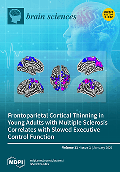
Multiple sclerosis (MS) is a neurodegenerative disease that causes lesion formation and tissue loss or atrophy in the brain. Cognitive and motor challenges are commonly observed in people with MS. Reduced speed of information processing is typically the first sign of cognitive involvement, which in turn broadly affects other cognitive processes including attention, executive function, and memory. The Attention Network Test—Interactions can measure the efficiency of 3 distinct attentional processes—alerting, orienting, and executive control. In this study, we measured attentional efficiencies and gray matter morphometry in a sample of young adults (18–35 years) with relatively mild MS severity. We show that thickness and volume changes in specific gray matter regions correlate with attentional efficiency even in the absence of widespread atrophy. View this paper
  • Issues are regarded as officially published after their release is announced to the table of contents alert mailing list.
  • You may sign up for e-mail alerts to receive table of contents of newly released issues.
  • PDF is the official format for papers published in both, html and pdf forms. To view the papers in pdf format, click on the "PDF Full-text" link, and use the free Adobe Reader to open them.
Order results
Result details
Section
Select all
Export citation of selected articles as:
11 pages, 1785 KiB  
Article
Genetic and Pharmacological Manipulations of Glyoxalase 1 Mediate Ethanol Withdrawal Seizure Susceptibility in Mice
by Amanda M. Barkley-Levenson, Amy Lee and Abraham A. Palmer
Brain Sci. 2021, 11(1), 127; https://doi.org/10.3390/brainsci11010127 - 19 Jan 2021
Cited by 3 | Viewed by 2186
Abstract
Central nervous system (CNS) hyperexcitability is a clinically significant feature of acute ethanol withdrawal. There is evidence for a genetic contribution to withdrawal severity, but specific genetic risk factors have not been identified. The gene glyoxalase 1 (Glo1) has been previously [...] Read more.
Central nervous system (CNS) hyperexcitability is a clinically significant feature of acute ethanol withdrawal. There is evidence for a genetic contribution to withdrawal severity, but specific genetic risk factors have not been identified. The gene glyoxalase 1 (Glo1) has been previously implicated in ethanol consumption in mice, and GLO1 inhibition can attenuate drinking in mice and rats. Here, we investigated whether genetic and pharmacological manipulations of GLO1 activity can also mediate ethanol withdrawal seizure severity in mice. Mice from two transgenic lines overexpressing Glo1 on different genetic backgrounds (C57BL/6J (B6) and FVB/NJ (FVB)) were tested for handling-induced convulsions (HICs) as a measure of acute ethanol withdrawal. Following an injection of 4 g/kg alcohol, both B6 and FVB mice overexpressing Glo1 showed increases in HICs compared to wild-type littermates, though only the FVB line showed a statistically significant difference. We also administered daily ethanol injections (2 g/kg + 9 mg/kg 4-methylpyrazole) to wild-type B6 mice for 10 days and tested them for HICs on the 10th day following treatment with either a vehicle or a GLO1 inhibitor (S-bromobenzylglutathione cyclopentyl diester (pBBG)). Treatment with pBBG reduced HICs, although this effect was only statistically significant following two 10-day cycles of ethanol exposure and withdrawal. These results provide converging genetic and pharmacological evidence that GLO1 can mediate ethanol withdrawal seizure susceptibility. Full article
(This article belongs to the Special Issue The Genetics of Alcohol Use Disorder)
Show Figures

Figure 1

16 pages, 870 KiB  
Article
When, How, and to What Extent Are Individuals with Unresponsive Wakefulness Syndrome Able to Progress? Neurobehavioral Progress
by Enrique Noé, Joan Ferri, José Olaya, María Dolores Navarro, Myrtha O’Valle, Carolina Colomer, Belén Moliner, Camilla Ippoliti, Anny Maza and Roberto Llorens
Brain Sci. 2021, 11(1), 126; https://doi.org/10.3390/brainsci11010126 - 19 Jan 2021
Cited by 6 | Viewed by 2476
Abstract
Accurate estimation of the neurobehavioral progress of patients with unresponsive wakefulness syndrome (UWS) is essential to anticipate their most likely clinical course and guide clinical decision making. Although different studies have described this progress and possible predictors of neurobehavioral improvement in these patients, [...] Read more.
Accurate estimation of the neurobehavioral progress of patients with unresponsive wakefulness syndrome (UWS) is essential to anticipate their most likely clinical course and guide clinical decision making. Although different studies have described this progress and possible predictors of neurobehavioral improvement in these patients, they have methodological limitations that could restrict the validity and generalization of the results. This study investigates the neurobehavioral progress of 100 patients with UWS consecutively admitted to a neurorehabilitation center using systematic weekly assessments based on standardized measures, and the prognostic factors of changes in their neurobehavioral condition. Our results showed that, during the analyzed period, 34% of the patients were able to progress from UWS to minimally conscious state (MCS), 12% of the total sample (near one third from those who progressed to MCS) were able to emerge from MCS, and 10% of the patients died. Transition to MCS was mostly denoted by visual signs, which appeared either alone or in combination with motor signs, and was predicted by etiology and the score on the Coma Recovery Scale-Revised at admission with an accuracy of 75%. Emergence from MCS was denoted in the same proportion by functional communication and object use. Predictive models of emergence from MCS and mortality were not valid and the identified predictors could not be accounted for. Full article
(This article belongs to the Special Issue Recent Advances in the Study of Altered State of Consciousness)
Show Figures

Figure 1

14 pages, 2957 KiB  
Article
Physiological and Medico-Social Research Trends of the Wave P300 and More Late Components of Visual Event-Related Potentials
by Sergey Lytaev and Irina Vatamaniuk
Brain Sci. 2021, 11(1), 125; https://doi.org/10.3390/brainsci11010125 - 18 Jan 2021
Cited by 11 | Viewed by 2333
Abstract
To extend the application of the late waves of the event-related potentials (ERPs) to multiple modalities, devices and software the underlying physiological mechanisms and responses of the brain for a particular sensory system and mental function must be carefully examined. The objective of [...] Read more.
To extend the application of the late waves of the event-related potentials (ERPs) to multiple modalities, devices and software the underlying physiological mechanisms and responses of the brain for a particular sensory system and mental function must be carefully examined. The objective of this study was aimed to study the sensory processes of the “human-computer interaction” model when classifying visual images with an incomplete set of signs based on the analysis of early, middle, late and slow ERPs components. 26 healthy subjects (men) aged 20–26 years were investigated. ERPs in 19 monopolar sites according to the 10/20 system were recorded. Discriminant and factor analyzes (BMDP Statistical Software) were applied. The component N450 is the most specialized indicator of the perception of unrecognizable (oddball) visual images. The amplitude of the ultra-late components N750 and N900 is also higher under conditions of presentation of the oddball image, regardless of the location of the registration points. In brain pathology along with the pronounced asymmetry of the wave distribution, reduction of the N150 wave and lengthening of its peak latency, a line of regularities were noted. These include–a pronounced reduction in peak latency P250 and N350, an increased amplitude of N350 in the frontal and central points of registration, a decrease in the amplitude of N450 in the left frontal cortex and its increase in the occipital registration points, activation of the occipital cortex at a time interval of 400–500 ms, as well as fusion later waves. We called such phenomena of the development of cognitive ERP in brain pathology “the incongruence of ERP components”. The results of the research are discussed in the light of the paradigm of the P300 wave application in brain-computer interface systems, as well as with the peculiarities in brain pathology. Full article
(This article belongs to the Special Issue Quantitative EEG and Cognitive Neuroscience)
Show Figures

Graphical abstract

11 pages, 643 KiB  
Article
Clinical Efficacy and Safety of Silicone Elastomer Sheet during Decompressive Craniectomy: Anti-Adhesive Role in Cranioplasty
by Young Ha Kim, Chi Hyung Lee, Chang Hyeun Kim, Dong Wuk Son, Sang Weon Lee, Geun Sung Song and Soon Ki Sung
Brain Sci. 2021, 11(1), 124; https://doi.org/10.3390/brainsci11010124 - 18 Jan 2021
Cited by 4 | Viewed by 2247
Abstract
(1) Background: Cranioplasty is a surgery to repair a skull bone defect after decompressive craniectomy (DC). If the process of dissection of the epidural adhesion tissue is not performed properly, it can cause many complications. We reviewed the effect of a silicone elastomer [...] Read more.
(1) Background: Cranioplasty is a surgery to repair a skull bone defect after decompressive craniectomy (DC). If the process of dissection of the epidural adhesion tissue is not performed properly, it can cause many complications. We reviewed the effect of a silicone elastomer sheet designed to prevent adhesion. (2) Methods: We retrospectively reviewed 81 consecutive patients who underwent DC and subsequent cranioplasty at our institution between January 2015 and December 2019. We then divided the patients into two groups, one not using the silicone elastomer sheet (n = 50) and the other using the silicone elastomer sheet (n = 31), and compared the surgical outcomes. (3) Results: We found that the use of the sheet shortened the operation time by 24% and reduced the estimated blood loss (EBL) by 43% compared to the control group. Moreover, the complication rate of epidural fluid collection (EFC) in the group using the sheet was 16.7%, which was lower than that in the control group (41.7%, p < 0.023). Multivariate logistic regression analysis showed the sheet (OR 0.294, 95% CI 0.093–0.934, p = 0.039) to be significantly related to EFC. (4) Conclusions: The technique using the silicone elastomer sheet allows surgeons to easily dissect the surgical plane during cranioplasty, which shortens the operation time, reduces EBL, and minimizes complications of EFC. Full article
(This article belongs to the Special Issue Neurosurgery and Neuroanatomy)
Show Figures

Figure 1

10 pages, 1231 KiB  
Article
Brain Metastases in Elderly Patients—The Role of Surgery in the Context of Systemic Treatment
by Martin Proescholdt, Stephanie T. Jünger, Petra Schödel, Karl-Michael Schebesch, Christian Doenitz, Tobias Pukrop, Julius Höhne, Nils-Ole Schmidt, Martin Kocher, Holger Schulz, Maximilian Ruge, Kevin König, Roland Goldbrunner and Stefan Grau
Brain Sci. 2021, 11(1), 123; https://doi.org/10.3390/brainsci11010123 - 18 Jan 2021
Cited by 9 | Viewed by 1824
Abstract
In patients with brain metastases (BM), advanced age is considered a negative prognostic factor. To address the potential reasons for that, we assessed 807 patients who had undergone BM resection; 315 patients aged at least 65 years (group A) were compared with 492 [...] Read more.
In patients with brain metastases (BM), advanced age is considered a negative prognostic factor. To address the potential reasons for that, we assessed 807 patients who had undergone BM resection; 315 patients aged at least 65 years (group A) were compared with 492 younger patients (group B). We analyzed the impact of the pre- and postoperative Karnofsky performance status (KPS), postoperative treatment structure and post-treatment survival. BM resection significantly improved KPS scores in both groups (p = 0.0001). Median survival after BM resection differed significantly between the groups (A: 5.81 vs. B: 8.12 months; p = 0.0015). In both groups, patients who received postoperative systemic treatment showed significantly longer overall survival (p = 0.00001). However, elderly patients less frequently received systemic treatment (p = 0.0001) and the subgroup of elderly patients receiving such therapies had a significantly higher postsurgical KPS score (p = 0.0007). In all patients receiving systemic treatment, age was no longer a negative prognostic factor. Resection of BM improves the functional status of elderly patients, thus enhancing the likeliness to receive systemic treatment, which, in turn, leads to longer overall survival. In the context of such a treatment structure, age alone is no longer a prognostic factor for survival. Full article
Show Figures

Figure 1

21 pages, 2714 KiB  
Article
Potential Effects of Poloxamer 188 on Rat Isolated Brain Mitochondria after Oxidative Stress In Vivo and In Vitro
by Johannes A. Pille and Matthias L. Riess
Brain Sci. 2021, 11(1), 122; https://doi.org/10.3390/brainsci11010122 - 18 Jan 2021
Cited by 7 | Viewed by 2344
Abstract
Outcome after cerebral ischemia is often dismal. Reperfusion adds significantly to the ischemic injury itself. Therefore, new strategies targeting ischemia/reperfusion (I/R) injury are critically needed. Poloxamer (P)188, an amphiphilic triblock copolymer, is a highly promising pharmacological therapeutic as its capability to insert into [...] Read more.
Outcome after cerebral ischemia is often dismal. Reperfusion adds significantly to the ischemic injury itself. Therefore, new strategies targeting ischemia/reperfusion (I/R) injury are critically needed. Poloxamer (P)188, an amphiphilic triblock copolymer, is a highly promising pharmacological therapeutic as its capability to insert into injured cell membranes has been reported to protect against I/R injury in various models. Although mitochondrial function particularly profits from P188 treatment after I/R, it remains unclear if this beneficial effect occurs directly or indirectly. Here, rat isolated brain mitochondria underwent oxidative stress in vivo by asphyxial cardiac arrest or in vitro by the addition of hydrogen peroxide (H2O2) after isolation. Mitochondrial function was assessed by adenosine triphosphate synthesis, oxygen consumption, and calcium retention capacity. Both asphyxia and H2O2 exposure significantly impaired mitochondrial function. P188 did not preserve mitochondrial function after either injury mechanism. Further research is indicated. Full article
(This article belongs to the Special Issue Mitochondria as Therapeutic Target for Acute Brain Pathologies)
Show Figures

Figure 1

14 pages, 2005 KiB  
Article
Modulation of Motor Cortex Plasticity by Repetitive Paired-Pulse TMS at Late I-Wave Intervals Is Influenced by Intracortical Excitability
by George M. Opie, Ryoki Sasaki, Brodie J. Hand and John G. Semmler
Brain Sci. 2021, 11(1), 121; https://doi.org/10.3390/brainsci11010121 - 18 Jan 2021
Cited by 8 | Viewed by 2599
Abstract
The late indirect (I)-waves recruited by transcranial magnetic stimulation (TMS) over primary motor cortex (M1) can be modulated using I-wave periodicity repetitive TMS (iTMS). The purpose of this study was to determine if the response to iTMS is influenced by different interstimulus intervals [...] Read more.
The late indirect (I)-waves recruited by transcranial magnetic stimulation (TMS) over primary motor cortex (M1) can be modulated using I-wave periodicity repetitive TMS (iTMS). The purpose of this study was to determine if the response to iTMS is influenced by different interstimulus intervals (ISIs) targeting late I-waves, and whether these responses were associated with individual variations in intracortical excitability. Seventeen young (27.2 ± 6.4 years, 12 females) healthy adults received iTMS at late I-wave intervals (4.0, 4.5, and 5.0 ms) in three separate sessions. Changes due to each intervention were examined with motor evoked potential (MEP) amplitudes and short-interval intracortical facilitation (SICF) using both posterior-anterior (PA) and anterior-posterior (AP) TMS current directions. Changes in MEP amplitude and SICF were influenced by iTMS ISI, with the greatest facilitation for ISIs at 4 and 5 ms with PA TMS, and 4 ms with AP TMS. Maximum SICF at baseline (irrespective of ISI) was associated with increased iTMS response, but only for PA stimulation. These results suggest that modifying iTMS parameters targeting late I-waves can influence M1 plasticity. They also suggest that maximum SICF may be a means by which responders to iTMS targeting the late I-waves could be identified. Full article
Show Figures

Figure 1

13 pages, 3257 KiB  
Article
The Development of Brain Network in Males with Autism Spectrum Disorders from Childhood to Adolescence: Evidence from fNIRS Study
by Wei Cao, Huilin Zhu, Yan Li, Yu Wang, Wuxia Bai, Uchong Lao, Yingying Zhang, Yan Ji, Sailing He and Xiaobing Zou
Brain Sci. 2021, 11(1), 120; https://doi.org/10.3390/brainsci11010120 - 18 Jan 2021
Cited by 9 | Viewed by 3975
Abstract
In the current study, functional near-infrared spectroscopy (fNIRS) was used to collect resting-state signals from 77 males with autism spectrum disorders (ASD, age: 6~16.25) and 40 typically developing (TD) males (age: 6~16.58) in the theory-of-mind (ToM) network. The graph theory analysis was used [...] Read more.
In the current study, functional near-infrared spectroscopy (fNIRS) was used to collect resting-state signals from 77 males with autism spectrum disorders (ASD, age: 6~16.25) and 40 typically developing (TD) males (age: 6~16.58) in the theory-of-mind (ToM) network. The graph theory analysis was used to obtain the brain network properties in ToM network, and the multiple regression analysis demonstrated that males with ASD showed a comparable global network topology, and a similar age-related decrease in the medial prefrontal cortex area (mPFC) compared to TD individuals. Nevertheless, participants with ASD showed U-shaped trajectories of nodal metrics of right temporo-parietal junction (TPJ), and an age-related decrease in the left middle frontal gyrus (MFG), while trajectories of TD participants were opposite. The nodal metrics of the right TPJ was negatively associated with the social deficits of ASD, while the nodal metrics of the left MFG was negatively associated with the communication deficits of ASD. Current findings suggested a distinct developmental trajectory of the ToM network in males with ASD from childhood to adolescence. Full article
Show Figures

Figure 1

12 pages, 788 KiB  
Article
Cerebrospinal Fluid α-Synuclein Species in Cognitive and Movements Disorders
by Vasilios C. Constantinides, Nour K. Majbour, George P. Paraskevas, Ilham Abdi, Bared Safieh-Garabedian, Leonidas Stefanis, Omar M. El-Agnaf and Elisabeth Kapaki
Brain Sci. 2021, 11(1), 119; https://doi.org/10.3390/brainsci11010119 - 17 Jan 2021
Cited by 15 | Viewed by 2938
Abstract
Total CSF α-synuclein (t-α-syn), phosphorylated α-syn (pS129-α-syn) and α-syn oligomers (o-α-syn) have been studied as candidate biomarkers for synucleinopathies, with suboptimal specificity and sensitivity in the differentiation from healthy controls. Studies of α-syn species in patients with other underlying pathologies are lacking. The [...] Read more.
Total CSF α-synuclein (t-α-syn), phosphorylated α-syn (pS129-α-syn) and α-syn oligomers (o-α-syn) have been studied as candidate biomarkers for synucleinopathies, with suboptimal specificity and sensitivity in the differentiation from healthy controls. Studies of α-syn species in patients with other underlying pathologies are lacking. The aim of this study was to investigate possible alterations in CSF α-syn species in a cohort of patients with diverse underlying pathologies. A total of 135 patients were included, comprising Parkinson’s disease (PD; n = 13), multiple system atrophy (MSA; n = 9), progressive supranuclear palsy (PSP; n = 13), corticobasal degeneration (CBD; n = 9), Alzheimer’s disease (AD; n = 51), frontotemporal degeneration (FTD; n = 26) and vascular dementia patients (VD; n = 14). PD patients exhibited higher pS129-α-syn/α-syn ratios compared to FTD (p = 0.045), after exclusion of samples with CSF blood contamination. When comparing movement disorders (i.e., MSA vs. PD vs. PSP vs. CBD), MSA patients had lower α-syn levels compared to CBD (p = 0.024). Patients with a synucleinopathy (PD and MSA) exhibited lower t-α-syn levels (p = 0.002; cut-off value: ≤865 pg/mL; sensitivity: 95%, specificity: 69%) and higher pS129-/t-α-syn ratios (p = 0.020; cut-off value: ≥0.122; sensitivity: 71%, specificity: 77%) compared to patients with tauopathies (PSP and CBD). There are no significant α-syn species alterations in non-synucleinopathies. Full article
(This article belongs to the Special Issue Cerebrospinal Fluid Biomarkers in Dementia Disorders)
Show Figures

Figure 1

15 pages, 1464 KiB  
Article
Effects of a Motor Imagery Task on Functional Brain Network Community Structure in Older Adults: Data from the Brain Networks and Mobility Function (B-NET) Study
by Blake R. Neyland, Christina E. Hugenschmidt, Robert G. Lyday, Jonathan H. Burdette, Laura D. Baker, W. Jack Rejeski, Michael E. Miller, Stephen B. Kritchevsky and Paul J. Laurienti
Brain Sci. 2021, 11(1), 118; https://doi.org/10.3390/brainsci11010118 - 17 Jan 2021
Cited by 3 | Viewed by 2591
Abstract
Elucidating the neural correlates of mobility is critical given the increasing population of older adults and age-associated mobility disability. In the current study, we applied graph theory to cross-sectional data to characterize functional brain networks generated from functional magnetic resonance imaging data both [...] Read more.
Elucidating the neural correlates of mobility is critical given the increasing population of older adults and age-associated mobility disability. In the current study, we applied graph theory to cross-sectional data to characterize functional brain networks generated from functional magnetic resonance imaging data both at rest and during a motor imagery (MI) task. Our MI task is derived from the Mobility Assessment Tool–short form (MAT-sf), which predicts performance on a 400 m walk, and the Short Physical Performance Battery (SPPB). Participants (n = 157) were from the Brain Networks and Mobility (B-NET) Study (mean age = 76.1 ± 4.3; % female = 55.4; % African American = 8.3; mean years of education = 15.7 ± 2.5). We used community structure analyses to partition functional brain networks into communities, or subnetworks, of highly interconnected regions. Global brain network community structure decreased during the MI task when compared to the resting state. We also examined the community structure of the default mode network (DMN), sensorimotor network (SMN), and the dorsal attention network (DAN) across the study population. The DMN and SMN exhibited a task-driven decline in consistency across the group when comparing the MI task to the resting state. The DAN, however, displayed an increase in consistency during the MI task. To our knowledge, this is the first study to use graph theory and network community structure to characterize the effects of a MI task, such as the MAT-sf, on overall brain network organization in older adults. Full article
Show Figures

Figure 1

16 pages, 1981 KiB  
Article
Traumatic Brain Injury-Related Attention Deficits in Children: A Controlled Treatment Trial with Lisdexamfetamine Dimesylate (Vyvanse)
by Michael G. Tramontana, Jonathan W. Prokop, Edwin Williamson, Tara Duffie and Hayden LaFever
Brain Sci. 2021, 11(1), 117; https://doi.org/10.3390/brainsci11010117 - 16 Jan 2021
Cited by 4 | Viewed by 3135
Abstract
Attention deficits are among the most common and persistent impairments resulting from traumatic brain injury (TBI). This study was the first to examine the effects of lisdexamfetamine dimesylate (LDX, Vyvanse) in treating TBI-related attention deficits in children. It was an extension of a [...] Read more.
Attention deficits are among the most common and persistent impairments resulting from traumatic brain injury (TBI). This study was the first to examine the effects of lisdexamfetamine dimesylate (LDX, Vyvanse) in treating TBI-related attention deficits in children. It was an extension of a previous controlled trial with adults. This was a 12-week, randomized, double-blind, placebo-controlled, dose-titration, crossover trial. In addition to weekly safety monitoring, there were assessments on a broad range of neuropsychological and behavioral measures at baseline, 6-weeks, and 12-weeks. A total of 20 carefully selected children were enrolled, ranging from 10 to 16 years of age. The sample consisted of cases with mainly mild TBI (based on the known details regarding their injuries), but they had persisting attention deficits and other post-concussion symptoms lasting from 2 to 29 months by the time of enrollment. A total of 16 children completed the trial. One of the children withdrew due to a mild anxiety reaction while on LDX. There were no other adverse effects. Positive treatment results were found on both formal testing of sustained attention and in terms of parent ratings of attention, emotional status, behavioral controls, and various aspects of executive functioning. The findings also served to highlight broader insights into the nature of attention deficits and their treatment in children with TBI. Full article
Show Figures

Figure 1

14 pages, 939 KiB  
Review
Epidemiology of Anthropometric Factors in Glioblastoma Multiforme—Literature Review
by Donata Simińska, Jan Korbecki, Klaudyna Kojder, Patrycja Kapczuk, Marta Fabiańska, Izabela Gutowska, Anna Machoy-Mokrzyńska, Dariusz Chlubek and Irena Baranowska-Bosiacka
Brain Sci. 2021, 11(1), 116; https://doi.org/10.3390/brainsci11010116 - 16 Jan 2021
Cited by 12 | Viewed by 5775
Abstract
Although glioblastoma multiforme (GBM) is a widely researched cancer of the central nervous system, we still do not know its full pathophysiological mechanism and we still lack effective treatment methods as the current combination of surgery, radiotherapy, and chemotherapy does not bring about [...] Read more.
Although glioblastoma multiforme (GBM) is a widely researched cancer of the central nervous system, we still do not know its full pathophysiological mechanism and we still lack effective treatment methods as the current combination of surgery, radiotherapy, and chemotherapy does not bring about satisfactory results. The median survival time for GBM patients is only about 15 months. In this paper, we present the epidemiology of central nervous system (CNS) tumors and review the epidemiological data on GBM regarding gender, age, weight, height, and tumor location. The data indicate the possible influence of some anthropometric factors on the occurrence of GBM, especially in those who are male, elderly, overweight, and/or are taller. However, this review of single and small-size epidemiological studies should not be treated as definitive due to differences in the survey methods used. Detailed epidemiological registers could help identify the main at-risk groups which could then be used as homogenous study groups in research worldwide. Such research, with less distortion from various factors, could help identify the pathomechanisms that lead to the development of GBM. Full article
Show Figures

Graphical abstract

12 pages, 1559 KiB  
Article
A Phase 1/2 Study of Flavocoxid, an Oral NF-κB Inhibitor, in Duchenne Muscular Dystrophy
by Gian Luca Vita, Maria Sframeli, Norma Licata, Alessandra Bitto, Sara Romeo, Francesca Frisone, Annamaria Ciranni, Giovanni Pallio, Federica Mannino, M’Hammed Aguennouz, Carmelo Rodolico, Francesco Squadrito, Antonio Toscano, Sonia Messina and Giuseppe Vita
Brain Sci. 2021, 11(1), 115; https://doi.org/10.3390/brainsci11010115 - 16 Jan 2021
Cited by 10 | Viewed by 3156
Abstract
Flavocoxid is a blended extract containing baicalin and catechin with potent antioxidant and anti-inflammatory properties due to the inhibition of the cyclooxygenase (COX) and 5-lipoxygenase (5-LOX) enzymes, nuclear factor-κB (NF-κB), tumor necrosis factor (TNF)-alpha, and the mitogen-activated protein kinases (MAPKs) pathways. This phase [...] Read more.
Flavocoxid is a blended extract containing baicalin and catechin with potent antioxidant and anti-inflammatory properties due to the inhibition of the cyclooxygenase (COX) and 5-lipoxygenase (5-LOX) enzymes, nuclear factor-κB (NF-κB), tumor necrosis factor (TNF)-alpha, and the mitogen-activated protein kinases (MAPKs) pathways. This phase 1/2 study was designed to assess the safety and tolerability of flavocoxid in patients with Duchenne muscular dystrophy (DMD). Thirty-four patients were recruited: 17 were treated with flavocoxid at an oral dose of 250 or 500 mg, according to body weight, for one year; 17 did not receive flavocoxid and served as controls. The treatment was well tolerated and nobody dropped out. Flavocoxid induced a significant reduction in serum interleukin (IL)-1 beta and TNF-alpha only in the group of DMD boys on add-on therapy (flavocoxid added to steroids for at least six months). The decrease in IL-1 beta was higher in younger boys. The serum H2O2 concentrations significantly decreased in patients treated with flavocoxid alone with a secondary reduction of serum glutathione peroxidase (GPx) levels, especially in younger boys. The exploratory outcome measures failed to show significant effects but there was a trend showing that the younger boys who received treatment were faster at performing the Gowers’ maneuver, while the older boys who received treatment were faster at doing the 10-m walk test (10MWT). Therefore, a double-blind, placebo-controlled study for at least two/three years is warranted to verify flavocoxid as a steroid substitute or as add-on therapy to steroids. Full article
(This article belongs to the Special Issue Advanced Research in Neuromuscular Disorders)
Show Figures

Graphical abstract

17 pages, 1627 KiB  
Article
The Role of Audiovisual Speech in Fast-Mapping and Novel Word Retention in Monolingual and Bilingual 24-Month-Olds
by Drew Weatherhead, Maria M. Arredondo, Loreto Nácar Garcia and Janet F. Werker
Brain Sci. 2021, 11(1), 114; https://doi.org/10.3390/brainsci11010114 - 16 Jan 2021
Cited by 13 | Viewed by 3511
Abstract
Three experiments examined the role of audiovisual speech on 24-month-old monolingual and bilinguals’ performance in a fast-mapping task. In all three experiments, toddlers were exposed to familiar trials which tested their knowledge of known word–referent pairs, disambiguation trials in which novel word–referent pairs [...] Read more.
Three experiments examined the role of audiovisual speech on 24-month-old monolingual and bilinguals’ performance in a fast-mapping task. In all three experiments, toddlers were exposed to familiar trials which tested their knowledge of known word–referent pairs, disambiguation trials in which novel word–referent pairs were indirectly learned, and retention trials which probed their recognition of the newly-learned word–referent pairs. In Experiment 1 (n = 48), lip movements were present during familiar and disambiguation trials, but not retention trials. In Experiment 2 (n = 48), lip movements were present during all three trial types. In Experiment 3 (bilinguals only, n = 24), a still face with no lip movements was present in all three trial types. While toddlers succeeded in the familiar and disambiguation trials of every experiment, success in the retention trials was only found in Experiment 2. This work suggests that the extra-linguistic support provided by lip movements improved the learning and recognition of the novel words. Full article
(This article belongs to the Special Issue The Impact of Non-Speech Cues on Speech Perception in Infancy)
Show Figures

Figure 1

9 pages, 1424 KiB  
Communication
Peripheral Neuropathies Seen by Ultrasound: A Literature Analysis through Lexical Evaluation, Geographical Assessment and Graph Theory
by Daniele Coraci, Claudia Loreti, Augusto Fusco, Silvia Giovannini and Luca Padua
Brain Sci. 2021, 11(1), 113; https://doi.org/10.3390/brainsci11010113 - 16 Jan 2021
Cited by 13 | Viewed by 2121
Abstract
(1) Background: Ultrasound is a well-known tool used for the diagnosis and management of many diseases, including peripheral neuropathies. The main aim of this study was the lexical analysis of the literature on this topic considering the most cited words and the relationship [...] Read more.
(1) Background: Ultrasound is a well-known tool used for the diagnosis and management of many diseases, including peripheral neuropathies. The main aim of this study was the lexical analysis of the literature on this topic considering the most cited words and the relationship between the words and the papers. Furthermore, a geographical analysis was performed to evaluate the worldwide prevalence. (2) Methods: We performed a literature search on PubMed, and we calculated the occurrence of the words indicating nerves and the body parts. Furthermore, we calculated the number of papers for each country, considering the affiliation of the first author. Finally, to describe the relationships between the words and the papers, we used the 30 most cited words, and we built a matrix describing in which papers a word was cited. This matrix was used to create a network based on the graph theory using Gephi 0.9.2 software. (3) Results: The most cited nerves were median and ulnar ones, and the most cited body parts were hand, wrist and elbow. The United States of America was the most productive country, with 80 papers. The graph of the network showed the importance of ultrasound as support for therapy. (4) Conclusions: The study represents a lexical analysis of the literature and shows information about subjects, authors and relationships of the papers. This may be helpful for better understanding and evaluation of the situation of the current literature. Full article
(This article belongs to the Special Issue Pathology of Peripheral Neuropathies )
Show Figures

Graphical abstract

24 pages, 5370 KiB  
Article
Ipsi- and Contralateral Oligo- and Polysynaptic Reflexes in Humans Revealed by Low-Frequency Epidural Electrical Stimulation of the Lumbar Spinal Cord
by Ursula S. Hofstoetter, Simon M. Danner, Brigitta Freundl, Heinrich Binder, Peter Lackner and Karen Minassian
Brain Sci. 2021, 11(1), 112; https://doi.org/10.3390/brainsci11010112 - 16 Jan 2021
Cited by 5 | Viewed by 3206
Abstract
Epidural electrical stimulation (EES) applied over the human lumbosacral spinal cord provides access to afferent fibers from virtually all lower-extremity nerves. These afferents connect to spinal networks that play a pivotal role in the control of locomotion. Studying EES-evoked responses mediated through these [...] Read more.
Epidural electrical stimulation (EES) applied over the human lumbosacral spinal cord provides access to afferent fibers from virtually all lower-extremity nerves. These afferents connect to spinal networks that play a pivotal role in the control of locomotion. Studying EES-evoked responses mediated through these networks can identify some of their functional components. We here analyzed electromyographic (EMG) responses evoked by low-frequency (2–6 Hz) EES derived from eight individuals with chronic, motor complete spinal cord injury. We identified and separately analyzed three previously undescribed response types: first, crossed reflexes with onset latencies of ~55 ms evoked in the hamstrings; second, oligosynaptic reflexes within 50 ms post-stimulus superimposed on the monosynaptic posterior root-muscle reflexes in the flexor muscle tibialis anterior, but with higher thresholds and no rate-sensitive depression; third, polysynaptic responses with variable EMG shapes within 50–450 ms post-stimulus evoked in the tibialis anterior and triceps surae, some of which demonstrated consistent changes in latencies with graded EES. Our observations suggest the activation of commissural neurons, lumbar propriospinal interneurons, and components of the late flexion reflex circuits through group I and II proprioceptive afferent inputs. These potential neural underpinnings have all been related to spinal locomotion in experimental studies. Full article
Show Figures

Figure 1

20 pages, 1701 KiB  
Article
Identifying Diurnal Variability of Brain Connectivity Patterns Using Graph Theory
by Farzad V. Farahani, Magdalena Fafrowicz, Waldemar Karwowski, Bartosz Bohaterewicz, Anna Maria Sobczak, Anna Ceglarek, Aleksandra Zyrkowska, Monika Ostrogorska, Barbara Sikora-Wachowicz, Koryna Lewandowska, Halszka Oginska, Anna Beres, Magdalena Hubalewska-Mazgaj and Tadeusz Marek
Brain Sci. 2021, 11(1), 111; https://doi.org/10.3390/brainsci11010111 - 16 Jan 2021
Cited by 12 | Viewed by 3890
Abstract
Significant differences exist in human brain functions affected by time of day and by people’s diurnal preferences (chronotypes) that are rarely considered in brain studies. In the current study, using network neuroscience and resting-state functional MRI (rs-fMRI) data, we examined the effect of [...] Read more.
Significant differences exist in human brain functions affected by time of day and by people’s diurnal preferences (chronotypes) that are rarely considered in brain studies. In the current study, using network neuroscience and resting-state functional MRI (rs-fMRI) data, we examined the effect of both time of day and the individual’s chronotype on whole-brain network organization. In this regard, 62 participants (39 women; mean age: 23.97 ± 3.26 years; half morning- versus half evening-type) were scanned about 1 and 10 h after wake-up time for morning and evening sessions, respectively. We found evidence for a time-of-day effect on connectivity profiles but not for the effect of chronotype. Compared with the morning session, we found relatively higher small-worldness (an index that represents more efficient network organization) in the evening session, which suggests the dominance of sleep inertia over the circadian and homeostatic processes in the first hours after waking. Furthermore, local graph measures were changed, predominantly across the left hemisphere, in areas such as the precentral gyrus, putamen, inferior frontal gyrus (orbital part), inferior temporal gyrus, as well as the bilateral cerebellum. These findings show the variability of the functional neural network architecture during the day and improve our understanding of the role of time of day in resting-state functional networks. Full article
(This article belongs to the Special Issue Recent Advances in Human Brain Connectivity)
Show Figures

Figure 1

26 pages, 1416 KiB  
Article
Main Concept, Sequencing, and Story Grammar Analyses of Cinderella Narratives in a Large Sample of Persons with Aphasia
by Jessica D. Richardson, Sarah Grace Dalton, Kathryn J. Greenslade, Adam Jacks, Katarina L. Haley and Janet Adams
Brain Sci. 2021, 11(1), 110; https://doi.org/10.3390/brainsci11010110 - 15 Jan 2021
Cited by 15 | Viewed by 4054
Abstract
Recently, a multilevel analytic approach called Main Concept, Sequencing, and Story Grammar (MSSG) was presented along with preliminary normative information. MSSG analyses leverage the strong psychometrics and rich procedural knowledge of both main concept analysis and story grammar component coding, complementing it with [...] Read more.
Recently, a multilevel analytic approach called Main Concept, Sequencing, and Story Grammar (MSSG) was presented along with preliminary normative information. MSSG analyses leverage the strong psychometrics and rich procedural knowledge of both main concept analysis and story grammar component coding, complementing it with easy-to-obtain sequencing information for a rich understanding of discourse informativeness and macrostructure. This study is the next critical step for demonstrating the clinical usefulness of MSSG’s six variables (main concept composite, sequencing, main concept+sequencing, essential story grammar components, total episodic components, and episodic complexity) for persons with aphasia (PWAs). We present descriptive statistical information for MSSG variables for a large sample of PWAs and compare their performance to a large sample of persons not brain injured (PNBIs). We observed significant differences between PWAs and PNBIs for all MSSG variables. These differences occurred at the omnibus group level and for each aphasia subtype, even for PWAs with very mild impairment that is not detected with standardized aphasia assessment. Differences between PWAs and PNBIs were also practically significant, with medium to large effect sizes observed for nearly all aphasia subtypes and MSSG variables. This work deepens our understanding of discourse informativeness and macrostructure in PWAs and further develops an efficient tool for research and clinical use. Future research should investigate ways to expand MSSG analyses and to improve sensitivity and specificity. Full article
Show Figures

Figure 1

11 pages, 990 KiB  
Article
Evaluation of Nociception Using Quantitative Pupillometry and Skin Conductance in Critically Ill Unconscious Patients: A Pilot Study
by Sara Fratino, Lorenzo Peluso, Marta Talamonti, Marco Menozzi, Lucas Akira Costa Hirai, Francisco A Lobo, Chiara Prezioso, Jacques Creteur, Jean-François Payen and Fabio Silvio Taccone
Brain Sci. 2021, 11(1), 109; https://doi.org/10.3390/brainsci11010109 - 15 Jan 2021
Cited by 9 | Viewed by 2704
Abstract
Background: Pain assessment is a challenge in critically ill patients, in particular those who are unable to express movements in reaction to noxious stimuli. The purpose of the study was to compare the pupillary response and skin conductance to pain stimulation in critically [...] Read more.
Background: Pain assessment is a challenge in critically ill patients, in particular those who are unable to express movements in reaction to noxious stimuli. The purpose of the study was to compare the pupillary response and skin conductance to pain stimulation in critically ill unconscious patients. Methods: This observational study included adult patients admitted to the intensive care unit (ICU) with acute brain injury (Glasgow Coma Scale < 9 with a motor response < 5) and/or requirements for deep level of sedation. Automated pupillometry (Algiscan, ID-MED, Marseille, France) was used to determine pupillary reflex dilation during tetanic stimulation. The maximum intensity of the stimulation value allowed the determination of a pupillary pain index score ranging from 1 (no nociception) to 9 (high nociception): a pupillary pain index (PPI) score of ≤4 was used to reflect adequate pain control. For skin conductance (SC), the number of SC peaks per second (NSCF) was collected concomitantly to tetanic stimulation. An NSCF of ≤0.07 peak/second was used to reflect adequate pain control. Results: Of the 51 included patients, there were 32 with brain injury and 19 receiving deep sedation. Mean PPI score was 5 (Interquartile Range= 2–7); a total of 28 (55%) patients showed inadequate control of the nociceptive stimulation according to the PPI assessment. Only 15 (29%) patients showed a detectable skin conductance, with NSCF values from 0.07 to 0.47/s. No correlation was found between skin conductance algesimeter (SCA)-derived variables and PPI score or pupillary dilation to pain. Conclusions: Detection of inadequate pain control might vary according to the method used to assess nociception in ICU patients. A poor agreement between quantitative pupillometry and skin conductance was observed. Full article
Show Figures

Figure 1

19 pages, 387 KiB  
Opinion
Delayed Onset Muscle Soreness (DOMS): The Repeated Bout Effect and Chemotherapy-Induced Axonopathy May Help Explain the Dying-Back Mechanism in Amyotrophic Lateral Sclerosis and Other Neurodegenerative Diseases
by Balázs Sonkodi
Brain Sci. 2021, 11(1), 108; https://doi.org/10.3390/brainsci11010108 - 15 Jan 2021
Cited by 24 | Viewed by 6205
Abstract
Delayed onset muscle soreness (DOMS) is hypothesized to be caused by glutamate excitotoxicity-induced acute compression axonopathy of the sensory afferents in the muscle spindle. Degeneration of the same sensory afferents is implicated in the disease onset and progression of amyotrophic lateral sclerosis (ALS). [...] Read more.
Delayed onset muscle soreness (DOMS) is hypothesized to be caused by glutamate excitotoxicity-induced acute compression axonopathy of the sensory afferents in the muscle spindle. Degeneration of the same sensory afferents is implicated in the disease onset and progression of amyotrophic lateral sclerosis (ALS). A series of “silent” acute compression proprioceptive axonopathies with underlying genetic/environmental factors, damaging eccentric contractions and the non-resolving neuroinflammatory process of aging could lead to ALS disease progression. Since the sensory terminals in the muscle spindle could not regenerate from the micro-damage in ALS, unlike in DOMS, the induced protective microcircuits and their long-term functional plasticity (the equivalent of the repeated bout effect in DOMS) will be dysfunctional. The acute stress invoking osteocalcin, bradykinin, COX1, COX2, GDNF, PGE2, NGF, glutamate and N-methyl-D-aspartate (NMDA) receptors are suggested to be the critical signalers of this theory. The repeated bout effect of DOMS and the dysfunctional microcircuits in ALS are suggested to involve several dimensions of memory and learning, like pain memory, inflammation, working and episodic memory. The spatial encoding of these memory dimensions is compromised in ALS due to blunt position sense from the degenerating proprioceptive axon terminals of the affected muscle spindles. Dysfunctional microcircuits progressively and irreversibly interfere with postural control, with motor command and locomotor circuits, deplete the neuroenergetic system, and ultimately interfere with life-sustaining central pattern generators in ALS. The activated NMDA receptor is suggested to serve the “gate control” function in DOMS and ALS in line with the gate control theory of pain. Circumvention of muscle spindle-loading could be a choice of exercise therapy in muscle spindle-affected neurodegenerative diseases. Full article
(This article belongs to the Special Issue Neuropathology and Novel Therapies for Motor Neuron Disease)
13 pages, 1555 KiB  
Article
Sensory Processing and Aggressive Behavior in Adults with Autism Spectrum Disorder
by Frank van den Boogert, Bram Sizoo, Pascalle Spaan, Sharon Tolstra, Yvonne H. A. Bouman, Witte J. G. Hoogendijk and Sabine J. Roza
Brain Sci. 2021, 11(1), 95; https://doi.org/10.3390/brainsci11010095 - 14 Jan 2021
Cited by 15 | Viewed by 8244
Abstract
Autism spectrum disorder (ASD) may be accompanied by aggressive behavior and is associated with sensory processing difficulties. The present study aims to investigate the direct association between sensory processing and aggressive behavior in adults with ASD. A total of 101 Dutch adult participants [...] Read more.
Autism spectrum disorder (ASD) may be accompanied by aggressive behavior and is associated with sensory processing difficulties. The present study aims to investigate the direct association between sensory processing and aggressive behavior in adults with ASD. A total of 101 Dutch adult participants with ASD, treated in outpatient or inpatient facilities, completed the Adolescent/Adult Sensory Profile (AASP), the Reactive-Proactive Aggression Questionnaire (RPQ), and the Aggression Questionnaire—Short Form (AQ-SF). Results revealed that sensory processing difficulties are associated with more aggressive behavior (f2=0.25), more proactive (f2=0.19) and reactive aggression (f2=0.27), more physical (f2=0.08) and verbal aggression (f2=0.13), and more anger (f2=0.20) and hostility (f2=0.12). Evidence was found for an interaction of the neurological threshold and behavioral response on total aggression and hostility. Participants with higher scores in comparison to the norm group in sensory sensitivity had the highest risk of aggressive behavior. In conclusion, clinical practice may benefit from applying detailed diagnostics on sensory processing difficulties when treating aggressive behavior in adults with ASD. Full article
Show Figures

Figure 1

15 pages, 3996 KiB  
Article
Using Fractal Dimension Analysis with the Desikan–Killiany Atlas to Assess the Effects of Normal Aging on Subregional Cortex Alterations in Adulthood
by Chi-Wen Jao, Chi Ieong Lau, Li-Ming Lien, Yuh-Feng Tsai, Kuang-En Chu, Chen-Yu Hsiao, Jiann-Horng Yeh and Yu-Te Wu
Brain Sci. 2021, 11(1), 107; https://doi.org/10.3390/brainsci11010107 - 14 Jan 2021
Cited by 12 | Viewed by 2754
Abstract
Normal aging is associated with functional and structural alterations in the human brain. The effects of normal aging and gender on morphological changes in specific regions of the brain are unknown. The fractal dimension (FD) can be a quantitative measure of cerebral folding. [...] Read more.
Normal aging is associated with functional and structural alterations in the human brain. The effects of normal aging and gender on morphological changes in specific regions of the brain are unknown. The fractal dimension (FD) can be a quantitative measure of cerebral folding. In this study, we used 3D-FD analysis with the Desikan–Killiany (DK) atlas to assess subregional morphological changes in adulthood. A total of 258 participants (112 women and 146 men) aged 30–85 years participated in this study. Participants in the middle-age group exhibited a decreased FD in the lateral frontal lobes, which then spread to the temporal and parietal lobes. Men exhibited an earlier and more significant decrease in FD values, mainly in the right frontal and left parietal lobes. Men exhibited more of a decrease in FD values in the subregions on the left than those in the right, whereas women exhibited more of a decrease in the lateral subregions. Older men were at a higher risk of developing mild cognitive impairment (MCI) and exhibited age-related memory decline earlier than women. Our FD analysis using the DK atlas-based prediagnosis may provide a suitable tool for assessing normal aging and neurodegeneration between groups or in individual patients. Full article
(This article belongs to the Special Issue Applications of Fractal Analysis in Brain Images Interpretation)
Show Figures

Figure 1

20 pages, 12566 KiB  
Article
Single-Trial Recognition of Video Gamer’s Expertise from Brain Haemodynamic and Facial Emotion Responses
by Ana R. Andreu-Perez, Mehrin Kiani, Javier Andreu-Perez, Pratusha Reddy, Jaime Andreu-Abela, Maria Pinto and Kurtulus Izzetoglu
Brain Sci. 2021, 11(1), 106; https://doi.org/10.3390/brainsci11010106 - 14 Jan 2021
Cited by 13 | Viewed by 3206
Abstract
With an increase in consumer demand of video gaming entertainment, the game industry is exploring novel ways of game interaction such as providing direct interfaces between the game and the gamers’ cognitive or affective responses. In this work, gamer’s brain activity has been [...] Read more.
With an increase in consumer demand of video gaming entertainment, the game industry is exploring novel ways of game interaction such as providing direct interfaces between the game and the gamers’ cognitive or affective responses. In this work, gamer’s brain activity has been imaged using functional near infrared spectroscopy (fNIRS) whilst they watch video of a video game (League of Legends) they play. A video of the face of the participants is also recorded for each of a total of 15 trials where a trial is defined as watching a gameplay video. From the data collected, i.e., gamer’s fNIRS data in combination with emotional state estimation from gamer’s facial expressions, the expertise level of the gamers has been decoded per trial in a multi-modal framework comprising of unsupervised deep feature learning and classification by state-of-the-art models. The best tri-class classification accuracy is obtained using a cascade of random convolutional kernel transform (ROCKET) feature extraction method and deep classifier at 91.44%. This is the first work that aims at decoding expertise level of gamers using non-restrictive and portable technologies for brain imaging, and emotional state recognition derived from gamers’ facial expressions. This work has profound implications for novel designs of future human interactions with video games and brain-controlled games. Full article
Show Figures

Figure 1

19 pages, 3637 KiB  
Article
The Effects of Spinal Manipulation on Motor Unit Behavior
by Lucien Robinault, Aleš Holobar, Sylvain Crémoux, Usman Rashid, Imran Khan Niazi, Kelly Holt, Jimmy Lauber and Heidi Haavik
Brain Sci. 2021, 11(1), 105; https://doi.org/10.3390/brainsci11010105 - 14 Jan 2021
Cited by 3 | Viewed by 4217
Abstract
Over recent years, a growing body of research has highlighted the neural plastic effects of spinal manipulation on the central nervous system. Recently, it has been shown that spinal manipulation improved outcomes, such as maximum voluntary force and limb joint position sense, reflecting [...] Read more.
Over recent years, a growing body of research has highlighted the neural plastic effects of spinal manipulation on the central nervous system. Recently, it has been shown that spinal manipulation improved outcomes, such as maximum voluntary force and limb joint position sense, reflecting improved sensorimotor integration and processing. This study aimed to further evaluate how spinal manipulation can alter neuromuscular activity. High density electromyography (HD sEMG) signals from the tibialis anterior were recorded and decomposed in order to study motor unit changes in 14 subjects following spinal manipulation or a passive movement control session in a crossover study design. Participants were asked to produce ankle dorsiflexion at two force levels, 5% and 10% of maximum voluntary contraction (MVC), following two different patterns of force production (“ramp” and “ramp and maintain”). A significant decrease in the conduction velocity (p = 0.01) was observed during the “ramp and maintain” condition at 5% MVC after spinal manipulation. A decrease in conduction velocity suggests that spinal manipulation alters motor unit recruitment patterns with an increased recruitment of lower threshold, lower twitch torque motor units. Full article
Show Figures

Figure 1

14 pages, 3217 KiB  
Article
Gait Recovery with an Overground Powered Exoskeleton: A Randomized Controlled Trial on Subacute Stroke Subjects
by Franco Molteni, Eleonora Guanziroli, Michela Goffredo, Rocco Salvatore Calabrò, Sanaz Pournajaf, Marina Gaffuri, Giulio Gasperini, Serena Filoni, Silvano Baratta, Daniele Galafate, Domenica Le Pera, Placido Bramanti, Marco Franceschini and on behalf of Italian Eksogait Study Group
Brain Sci. 2021, 11(1), 104; https://doi.org/10.3390/brainsci11010104 - 14 Jan 2021
Cited by 33 | Viewed by 4339
Abstract
Background: Overground Robot-Assisted Gait Training (o-RAGT) provides intensive gait rehabilitation. This study investigated the efficacy of o-RAGT in subacute stroke subjects, compared to conventional gait training. Methods: A multicenter randomized controlled trial was conducted on 75 subacute stroke subjects (38 in the Experimental [...] Read more.
Background: Overground Robot-Assisted Gait Training (o-RAGT) provides intensive gait rehabilitation. This study investigated the efficacy of o-RAGT in subacute stroke subjects, compared to conventional gait training. Methods: A multicenter randomized controlled trial was conducted on 75 subacute stroke subjects (38 in the Experimental Group (EG) and 37 in the Control Group (CG)). Both groups received 15 sessions of gait training (5 sessions/week for 60 min) and daily conventional rehabilitation. The subjects were assessed at the beginning (T1) and end (T2) of the training period with the primary outcome of a 6 Minutes Walking Test (6MWT), the Modified Ashworth Scale of the Affected lower Limb (MAS-AL), the Motricity Index of the Affected lower Limb (MI-AL), the Trunk Control Test (TCT), Functional Ambulation Classification (FAC), a 10 Meters Walking Test (10MWT), the modified Barthel Index (mBI), and the Walking Handicap Scale (WHS). Results: The 6MWT increased in both groups, which was confirmed by both frequentist and Bayesian analyses. Similar outcomes were registered in the MI-AL, 10MWT, mBI, and MAS-AL. The FAC and WHS showed a significant number of subjects improving in functional and community ambulation in both groups at T2. Conclusions: The clinical effects of o-RAGT were similar to conventional gait training in subacute stroke subjects. The results obtained in this study are encouraging and suggest future clinical trials on the topic. Full article
Show Figures

Figure 1

10 pages, 2194 KiB  
Perspective
Might Fibroblasts from Patients with Alzheimer’s Disease Reflect the Brain Pathology? A Focus on the Increased Phosphorylation of Amyloid Precursor Protein Tyr682 Residue
by Filomena Iannuzzi, Vincenza Frisardi, Lucio Annunziato and Carmela Matrone
Brain Sci. 2021, 11(1), 103; https://doi.org/10.3390/brainsci11010103 - 14 Jan 2021
Cited by 7 | Viewed by 2399 | Correction
Abstract
Alzheimer’s disease (AD) is a devastating neurodegenerative disorder with no cure and no effective diagnostic criteria. The greatest challenge in effectively treating AD is identifying biomarkers specific for each patient when neurodegenerative processes have not yet begun, an outcome that would allow the [...] Read more.
Alzheimer’s disease (AD) is a devastating neurodegenerative disorder with no cure and no effective diagnostic criteria. The greatest challenge in effectively treating AD is identifying biomarkers specific for each patient when neurodegenerative processes have not yet begun, an outcome that would allow the design of a personalised therapeutic approach for each patient and the monitoring of the therapeutic response during the treatment. We found that the excessive phosphorylation of the amyloid precursor protein (APP) Tyr682 residue on the APP 682YENPTY687 motif precedes amyloid β accumulation and leads to neuronal degeneration in AD neurons. We proved that Fyn tyrosine kinase elicits APP phosphorylation on Tyr682 residue, and we reported increased levels of APP Tyr682 and Fyn overactivation in AD neurons. Here, we want to contemplate the possibility of using fibroblasts as tools to assess APP Tyr682 phosphorylation in AD patients, thus making the changes in APP Tyr682 phosphorylation levels a potential diagnostic strategy to detect early pathological alterations present in the peripheral cells of AD patients’ AD brains. Full article
(This article belongs to the Special Issue Biochemical Biomarkers and Neurodegenerative Diseases)
Show Figures

Figure 1

19 pages, 4227 KiB  
Article
Influence of Increasing Task Complexity and Use of Informational Assistance Systems on Mental Workload
by Dominic Bläsing and Manfred Bornewasser
Brain Sci. 2021, 11(1), 102; https://doi.org/10.3390/brainsci11010102 - 14 Jan 2021
Cited by 23 | Viewed by 3549
Abstract
(1) Background: Cognitive aspects and complexity in modern manual mixed model assembly are increasing. To reduce mental workload (MWL), informational assistance systems are introduced. The influence of complexity and used assistance system on MWL should be investigated to further improve the implementation of [...] Read more.
(1) Background: Cognitive aspects and complexity in modern manual mixed model assembly are increasing. To reduce mental workload (MWL), informational assistance systems are introduced. The influence of complexity and used assistance system on MWL should be investigated to further improve the implementation of such assistance systems. (2) Methods: Using a simulated close to real-life assembly task a 2 × 3 design was chosen, with two levels of assembly complexity (within subjects) and three different assistance systems (paper, Augmented Reality (AR)-glasses, tablet–between subjects). MWL was measured using either physiological response (electrocardiogram (ECG) and eye-tracking) or performance indicators. (3) Results: An influence of task complexity on MWL can be shown. Additionally, usability based differences between the used assistance systems become more evident with reference to the results of area of interest analysis. (4) Conclusions: Using a multi-modal measurement approach, it is possible to detect complexity-based differences in MWL. Additional research on validity and alignment is needed to further use these for (neuro-) ergonomic considerations and recommendations. Full article
(This article belongs to the Special Issue Current Perspectives on Neuroergonomics)
Show Figures

Figure 1

10 pages, 3516 KiB  
Article
Internal Carotid Artery Injury in Transsphenoidal Surgery: Tenets for Its Avoidance and Refit—A Clinical Study
by Dmitry Usachev, Oleg Sharipov, Ashraf Abdali, Sergei Yakovlev, Vasiliy Lukshin, Maksim Kutin, Dmitry Fomichev, Pavel Dorokhov, Evgeny Bukharin, Alexey Shkarubo, Ilya Chernov, Andrey Panteleyev, Kaan Yağmurlu, Bipin Chaurasia and Pavel Kalinin
Brain Sci. 2021, 11(1), 99; https://doi.org/10.3390/brainsci11010099 - 13 Jan 2021
Cited by 9 | Viewed by 2202
Abstract
One of the most serious/potentially fatal complications of transsphenoidal surgery (TSS) is internal carotid artery (ICA) injury. Of 6230 patients who underwent TSS, ICA injury occurred in 8 (0.12%). The etiology, possible treatment options, and avoidance of ICA injury were analyzed. ICA injury [...] Read more.
One of the most serious/potentially fatal complications of transsphenoidal surgery (TSS) is internal carotid artery (ICA) injury. Of 6230 patients who underwent TSS, ICA injury occurred in 8 (0.12%). The etiology, possible treatment options, and avoidance of ICA injury were analyzed. ICA injury occurred at two different stages: (1) during the exposure of the sella floor and dural incision over the sella and cavernous sinus and (2) during the resection of the cavernous sinus extension of the tumor. The angiographic collateral blood supply was categorized as good, sufficient, and nonsufficient to help with the decision making for repairing the injury. ICA occlusion with a balloon was performed at the injury site in two cases, microcoils in two patients, microcoils plus a single barrel extra-intracranial high-flow bypass in one case, stent grafting in one case, and no intervention in two cases. The risk of ICA injury diminishes with better preoperative preparation, intraoperative navigation, and ultrasound dopplerography. Reconstructive surgery for closing the defect and restoring the blood flow to the artery should be assessed depending on the site of the injury and the anatomical features of the ICA. Full article
Show Figures

Figure 1

23 pages, 4380 KiB  
Article
Comparison of Smoothing Filters’ Influence on Quality of Data Recorded with the Emotiv EPOC Flex Brain–Computer Interface Headset during Audio Stimulation
by Natalia Browarska, Aleksandra Kawala-Sterniuk, Jaroslaw Zygarlicki, Michal Podpora, Mariusz Pelc, Radek Martinek and Edward Jacek Gorzelańczyk
Brain Sci. 2021, 11(1), 98; https://doi.org/10.3390/brainsci11010098 - 13 Jan 2021
Cited by 24 | Viewed by 3609
Abstract
Off-the-shelf, consumer-grade EEG equipment is nowadays becoming the first-choice equipment for many scientists when it comes to recording brain waves for research purposes. On one hand, this is perfectly understandable due to its availability and relatively low cost (especially in comparison to some [...] Read more.
Off-the-shelf, consumer-grade EEG equipment is nowadays becoming the first-choice equipment for many scientists when it comes to recording brain waves for research purposes. On one hand, this is perfectly understandable due to its availability and relatively low cost (especially in comparison to some clinical-level EEG devices), but, on the other hand, quality of the recorded signals is gradually increasing and reaching levels that were offered just a few years ago by much more expensive devices used in medicine for diagnostic purposes. In many cases, a well-designed filter and/or a well-thought signal acquisition method improve the signal quality to the level that it becomes good enough to become subject of further analysis allowing to formulate some valid scientific theories and draw far-fetched conclusions related to human brain operation. In this paper, we propose a smoothing filter based upon the Savitzky–Golay filter for the purpose of EEG signal filtering. Additionally, we provide a summary and comparison of the applied filter to some other approaches to EEG data filtering. All the analyzed signals were acquired from subjects performing visually involving high-concentration tasks with audio stimuli using Emotiv EPOC Flex equipment. Full article
Show Figures

Graphical abstract

14 pages, 328 KiB  
Article
Effects of Sleep on the Academic Performance of Children with Attention Deficit and Hyperactivity Disorder
by Lorena Villalba-Heredia, Celestino Rodríguez, Zaira Santana, Débora Areces and Antonio Méndez-Giménez
Brain Sci. 2021, 11(1), 97; https://doi.org/10.3390/brainsci11010097 - 13 Jan 2021
Cited by 6 | Viewed by 3198
Abstract
Attention deficit and hyperactivity disorder (ADHD) is commonly associated with disordered or disturbed sleep and the association of sleep problems with ADHD is complex and multidirectional. The purpose of this study was to analyze the relationship between sleep and academic performance, comparing children [...] Read more.
Attention deficit and hyperactivity disorder (ADHD) is commonly associated with disordered or disturbed sleep and the association of sleep problems with ADHD is complex and multidirectional. The purpose of this study was to analyze the relationship between sleep and academic performance, comparing children with ADHD and a control group without ADHD. Academic performance in Spanish, mathematics, and a foreign language (English) was evaluated. Different presentations of ADHD were considered as well as the potential difference between weekday and weekend sleep habits. The sample consisted of 75 children aged 6–12 in primary education. Accelerometry was used to study sleep, and school grades were used to gather information about academic performance. The results showed that ADHD influenced the amount of sleep during weekends, the time getting up at the weekends, weekday sleep efficiency, as well as academic performance. Given the effects that were seen in the variables linked to the weekend, it is necessary to consider a longitudinal design with which to determine if there is a cause and effect relationship. Full article
Previous Issue
Next Issue
Back to TopTop