Article

Effect of the Lateral Bone Augmentation Procedure in Correcting Peri-Implant Bone Dehiscence Defects: A 7-Years Retrospective Study

1
Department of Dental Surgery, Faculty of Medicine and Dentistry, Medical University of Wrocław, 50-425 Wrocław, Poland
2
Department of Orthodontics, TU Dresden, 01069 Dresden, Germany
*
Author to whom correspondence should be addressed.
Appl. Sci. 2023, 13(4), 2324; https://doi.org/10.3390/app13042324
Submission received: 14 January 2023 / Revised: 9 February 2023 / Accepted: 9 February 2023 / Published: 10 February 2023
(This article belongs to the Special Issue Dental Materials: Latest Advances and Prospects - Volume II)

Abstract

:
Guided bone regeneration (GBR) is a well-documented and widely-used dental surgical procedure for the treatment of various types of alveolar bone defects. The aim of the study was to evaluate the long-term effectiveness of the GBR procedure in correcting small peri-implant bone dehiscence using the xenogeneic deproteinized bovine bone mineral material and a xenogeneic native bilayer collagen membrane. The present study was designed as a retrospective study. Seventy-five bone-level tapered two-piece dental implants Conelog®, Camlog (Biotechnologies AG, Switzerland) were divided into two groups G1—no bone augmentation (no GBR)—44 implants and G2—bone augmentation (GBR)—31 implants. For both groups, the closed healing protocol with a primary wound closure was used. The incidence of peri-implantits was evaluated, the quantitative assessment of soft tissue thickness was performed using the ultrasound (USG) device, quantitative assessment of marginal bone loss (MBL) was done. The prevalence of peri-implantitis was 5.3%, with a three-fold increase in peri-implantitis comparing the groups without and with bone augmentation (G1 = 2.27%, G2 = 9.7%). The average keratinized gingiva thickness was 1.87 mm and did not differ significantly between groups. No statistically significant differences in MBL between G1 and G2 were found. When dividing patients by gender, no statistically significant differences were observed. When dividing patients by age groups, statistically significant differences were observed between the youngest and oldest groups of patients. Within the limitations of this study, it can be concluded that the use of xenogeneic bone and a xenogeneic collagen membrane in a GBR procedure can be recommended to correct small peri-implant bone dehiscence.

1. Introduction

Dental implants are nowadays considered a highly predictable treatment option to replace missing teeth with a reported long-term (10–20 years) survival rate of 89.5% and 92.7% [1,2]. However, when bone dehiscence is present at the time of implant placement to place a dental implant, different bone regenerative techniques, such as guided bone regeneration (GBR), need to be used. GBR is a successful, well-documented and widely used dental surgical procedure used to treat various alveolar bone defects [3,4]. GBR requires the application of a resorbable or non-resorbable membrane to exclude non-osteogenic tissues from interfering with bone regeneration and can be used as a surgical procedure preceding implant treatment [5,6].
Currently, a broad spectrum of grafting materials of different origins, such as allografts, xenografts, and alloplastic materials, is available [7,8]. Similarly, materials of different origins are available in various forms ranging from blocks to bone chips, and this provides the dental surgeon and patient with many treatment options. The situation with membranes is no different; the choice between non-resorbable (including Polytetrafluoroethylene PTFE; expanded polytetrafluoroethylene e-PTFE, high-density polytetrafluoroethylene d-PTFE; Titanium-Reintforced PTFE Membranes, Titanium Mesh and Cage membranes) and resorbable membranes of different types and origins is wide [9]. Depending on their origin, resorbable membranes used in GBR can be divided into natural polymers (collagen membranes) and synthetic polymers (aliphatic polyesters (e.g., poly (lactic acid) (PLA), poly (polyglycolic acid) (PGA), poly (ε-caprolactone) (PCL) [10,11].
Undoubtedly, the treatment options are wide, but most importantly, they require adaptation to a specific clinical situation. Since the beginning of dental implantology, implant placement in the aesthetic zone in such a way that implants imitate nature and, at the same time, maintain healthy tissues has been challenging. The perfect three-dimensional position of the implant is critical for proper prosthetic restoration, especially in the aesthetic area. When a proper prosthetic-driven implant positioning of the implant requires covering small fenestration or dehiscence caused by an exposed implant surface on its facial aspect, the GBR technique, in combination with xenogeneic bone and resorbable membranes, can be used [12]. In these situations, the GBR can be used simultaneously with implant placement since, according to Chiapasco and Zaniboni [13], guided bone regeneration procedures are a reliable means for treating dehiscences and fenestrations created during implant placement.
The first objective of the study was to evaluate the long-term effectiveness of the applied GBR procedure in correcting small peri-implant bone dehiscence using the xenogeneic deproteinized bovine bone mineral material and a xenogeneic native bilayer collagen membrane. A secondary objective was to evaluate factors that may influence marginal bone loss.
Hypotheses of this study were:
  • When the GBR procedure is used for implant dehiscence treatment, it does not influence the marginal bone loss around the implant in 7 years of observation;
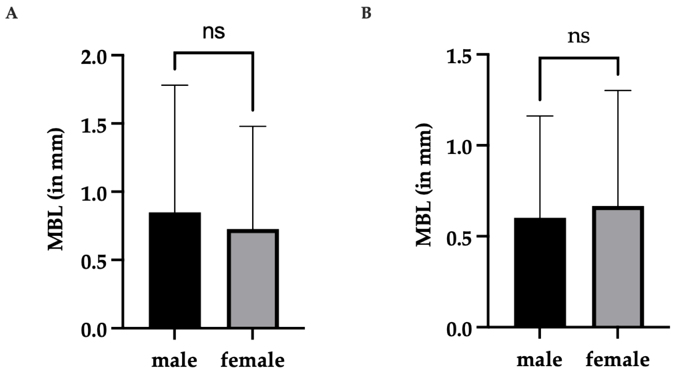
  • There are no gender-related differences in marginal bone loss around implants;
  • There is no difference in marginal bone loss around implants between age groups.

2. Materials and Methods
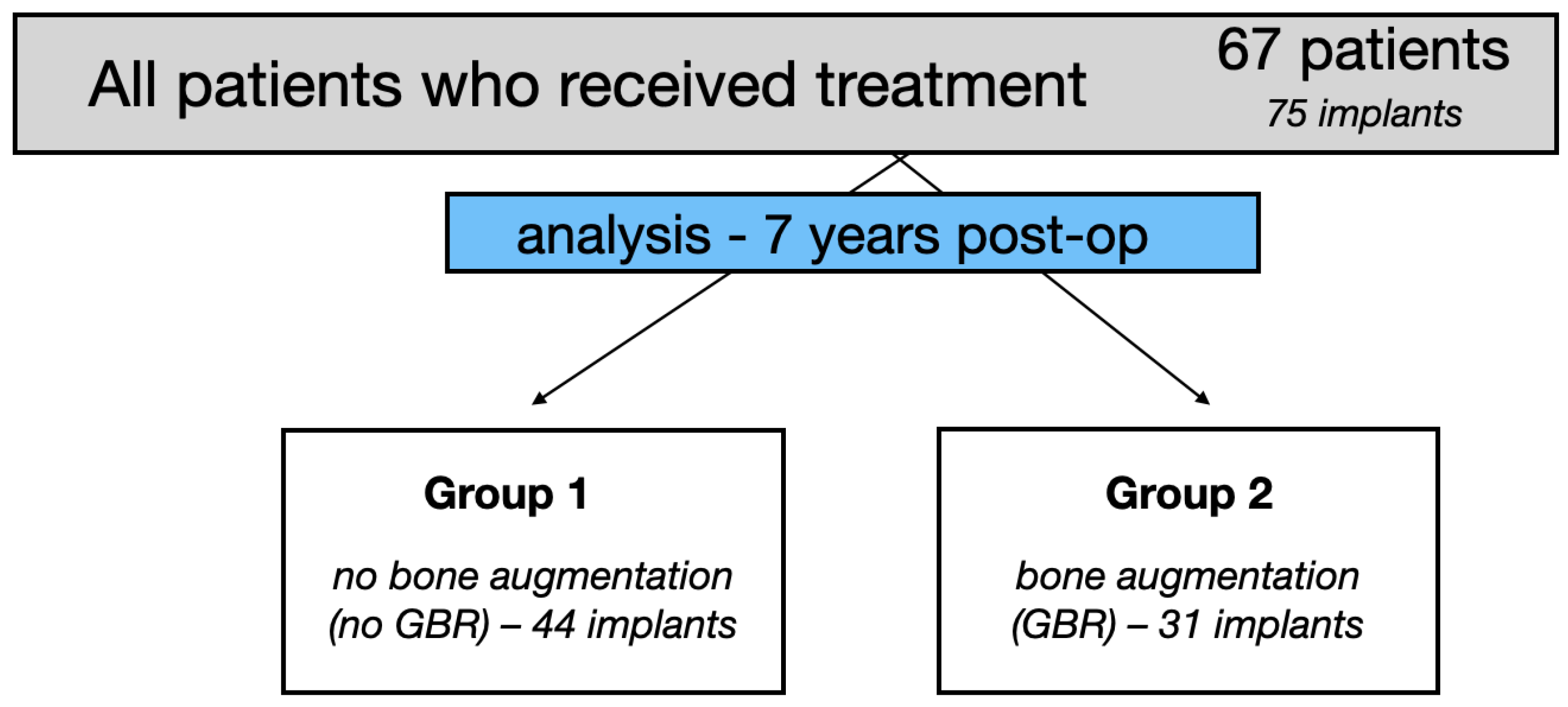
2.1. Study Design

The present study was a retrospective study aimed at evaluating the long-term effectiveness of the applied GBR procedure in correcting small peri-implant bone dehiscence. The study was performed in Wroclaw Medical University Dental Clinical and Teaching facility. Among the group of patients who received Conelog®, Camlog (Biotechnologies AG, Switzerland) implants as part of a clinical trial conducted in 2012–2014, a group of patients was selected who received implants with lateral bone contour regeneration and placed with no bone regeneration. Medical records of a 7-year follow-up were analyzed. A bioethics approval for this follow-up study was granted by a local bioethics committee of Wrocław Medical University (registration number 861/2021). The study has been conducted in full compliance with the Declaration of Helsinki. All patients gave two written consents: the first was general consent to have dental implants placed, and the other consent involved participation in the clinical study. The original clinical study analyzed changes in soft tissue thickness; detailed data from the clinical study and 12 months of follow-up can be found in our study [14]. In a 5 years follow-up period, the soft tissue augmentation with CTG (connective tissue graft) 3 months prior to implant placement was found to be the most effective method in terms of the soft tissue thickness gain—1.035 mm (SD = 0.73 mm) over the entire follow-up period (5 years) [15].
The purpose of the present study was to evaluate previously unanalyzed medical data in terms of the influence on MBL. The influence of lateral GBR procedure, gender structure, and age structure was analyzed. The prevalence of peri-implantitis was also assessed.

2.2. Clinical Data, Groups of Patients, Surgery, Implant Loading

The medical records of 67 patients (27 male, 47 female) who had 75 bone-level tapered two-piece dental implants Conelog®, Camlog (Biotechnologies AG, Switzerland). The 3.8 mm and 4.3 mm diameter implants of different lengths were used in the study. Implants were placed in the healed bone in the aesthetic area of both jaws were selected (no prior GBR allowed). Specific exclusion criteria for this group of patients were presented previously [16]; briefly: generally healthy adult patients were included, and patients with periodontal disease, noncontrolled metabolic diseases, and heavy smokers were excluded.
The implants were divided into 2 groups: G1—no bone augmentation (no GBR)—44 implants, and G2—bone augmentation (GBR)—31 implants, Figure 1. All of the patient records analyzed and included in the study contained full details of the procedure, including the depth of the implant placement and the GBR procedure used in case of dehiscence presence. Forty-four implants were inserted and completely embedded in the native bone, placed equicrestally. In 31 implants where the dehiscence (to the implant first thread) was present, the external lamina of the alveolar process was augmented during implantation with the deproteinized bovine bone mineral material (Bio-Oss®, Geistlich Pharma AG, Switzerland) and a native bilayer collagen membrane (Bio-Gide®, Geistlich Pharma AG, Switzerland) to cover the exposed implant surface on the facial aspect. For both groups, the closed healing protocol with a primary wound closure was used (Figure 1).
Implant loading took place after 6 months. Conventional impressions were taken using the polyether (PE) impression compound Impregum Penta (3M, Maplewood, MN, USA). All implants were restored on a standard abutment provided by the implant manufacturer’s Camlog (Biotechnologies AG, Switzerland) with metal-ceramic cemented single crowns with semipermanent cement (Implantlink®, Detax, Germany). A follow-up clinical appointment for all the patients included was conducted by the same investigator (AB).

2.3. Clinical Outcomes

Patient clinical evaluation included bleeding on gentle probing and biological complication such as peri-implantitis. Peri-implantitis, according to the 2017 World Workshop on the Classification of Periodontal and Peri-Implant Diseases and Conditions, involves bone levels ≥3 mm apical of the most coronal portion of the intra-osseous part of the implant together with bleeding on probing [17]. During the clinical examination, the thickness of keratinized tissue (TKT) in the surrounding implant site was examined using the Ultrasound (USG) device Pirop® (Echoson, Poland) as described previously in our study.

2.4. Marginal Bone Loss

Since all the implants in G1 were placed equicrestally, and in G2, the bone augmentation was performed to cover the first implant thread, the level of 0 was taken as the initial value for further measurements. The MBL was reported in mm and referred to millimeters of bone loss compared to the initial value.
The intraoral radiographs were done using a holder (Visualize HD®, Gendex®, USA). The radiological evaluation and X-ray measurements were done using the Gendex software (Gendex®, USA). X-ray image was calibrated using build-in calibration Gendex VixWin Platinum (Gendex®, USA). The known diameter of the dental implants was used for the calibration. The same device and software were used in all cases.

2.5. Statistical Analysis

The statistical analysis was performed using the GraphPad Prism 9 software [GraphPad Software, Inc., USA]. The data obtained in the study were tested to check the normal distribution (Shapiro–Wilk and Kolmogorov–Smirnov tests). Depending on the criteria met (normal distribution), an appropriate test was selected for further analysis. A parametric and nonparametric statistical approach was applied depending on the data. An unpaired t-test was used for parametric, and a Mann–Whitney U test for non-parametric data. All data are means ± standard deviation (SD). p < 0.05 was considered statistically significant.

3. Results

3.1. Implant Survival Rate

All of the 75 placed implants remained integrated after 7 years. This resulted in a 100% implant survival rate.

3.2. Peri-Implantitis

A total number of 4 implants met the criteria for periimplantitis [17] (bone levels ≥ 3 mm apical of the most coronal portion of the intra-osseous part of the implant together with bleeding on probing). This corresponds to 5.3% of the total implants placed in the study.
Among the G1 group with no GBR, based on clinical and radiological examination, one patient (2.27%) was diagnosed with peri-implant disease. However, in the G2 group, where the xenogeneic bone substitute was used in the GBR procedure, three patients (9.7%) were diagnosed with peri-implantitis. This indicates a three-fold increase in peri-implantitis comparing the groups without and with bone augmentation.
When dividing patients into groups by gender, 11% of male patients were diagnosed with periimplantitis and only 2.1% of female patients.
When patients were analyzed by age groups, in the youngest group (25–40 years old), a 4.35% rate of peri-implantitis was found; in the middle group (41–60 years old), this rate was 2.94%, and in the oldest group (61–72 years old) 10.5% of patients were diagnosed with peri-implantitis.
It was decided to include the entire group of patients in further analysis; however, due to the severe peri-implantitis cases found that might affect the results, especially in some groups, the results are given separately (see results all figures) for the whole group (75 implants) and for the group excluding the peri-implantitis cases (71 implants).

3.3. Marginal Bone Loss
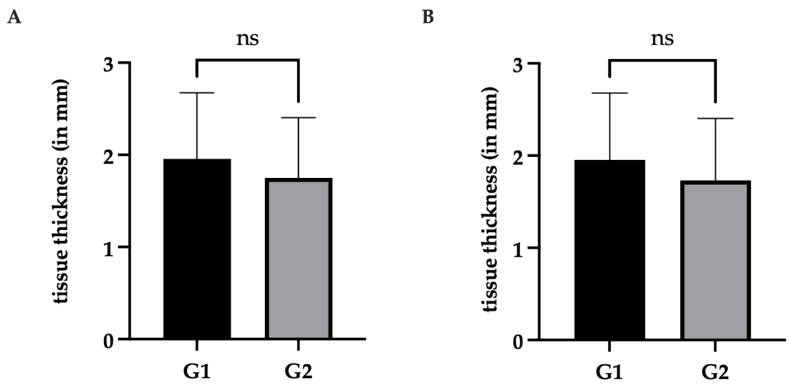
For G1 with no bone augmentation, a mean MBL = 0.63 was nominally lower than in G2, where a bone augmentation was performed, and the MBL was 0.98. However, no statistically significant differences were observed between these groups (Figure 2A, Table 1). In this case, all patients were included, including those diagnosed with peri-implantitis, and importantly, even after excluding cases of peri-implantitis, no significant differences were observed between the groups (Figure 2B).
When dividing patients by gender, no statistically significant differences were observed between men and females (MBL = 0.85mm and MBL = 0.73mm, respectively). Similarly—after excluding patients with peri-implantitis, no statistically significant differences were noted (Figure 3, Table 2).
When dividing patients by age groups (age ranges: 25–40; 41–60; 60–72), statistically significant differences were observed between the youngest (25–40 years old) and oldest group (60–72 years old) of patients (MBL = 0.85mm and MBL = 0.73mm, respectively) Figure 4, Table 3. The patient’s age given in the results is the age at the time of the 7-year follow-up; patients were younger at the time of surgery.

3.4. Soft Tissue

The mean value of the thickness of keratinized tissue (TKT) analyzed together for both G1 and G2 was 1.87 mm. The mean values of TKT in the implant area were similar in both groups. No statistically significant differences were observed between these groups, G1 (no bone augmentation) and G2 (bone augmentation), in terms of the TKT (Figure 5, Table 4). In this case, all patients were included, including those diagnosed with peri-implantitis, and importantly, even after excluding cases of peri-implantitis, no significant differences were observed between the groups (Figure 5B).
Regarding the research hypotheses:
  • The first research hypothesis was accepted. No statistically significant (p < 0.05) differences in terms of marginal bone loss were found between the groups with and without guided bone regeneration;
  • The second research hypothesis was accepted. No gender-related differences were found in marginal bone loss;
  • The third hypothesis was rejected. A statistically significant difference in marginal bone loss around the implant neck was found between the age group 25–40 and 60–72.

4. Discussion

Our aim was to evaluate the long-term effectiveness of the applied GBR procedure in correcting small peri-implant bone dehiscence using the xenogeneic deproteinized bovine bone mineral material and a xenogeneic native bilayer collagen membrane. To assess the effectiveness of treatment with the GBR method, several factors affecting long-term treatment were evaluated. Among them are the implant survival rate in augmented and nonaugmented sites, the incidence of complications (particularly peri-implantitis), and the marginal bone loss in the peri-implant area.
The implant survival rate was 100% in the 7 years of observation. However, the survival rate itself applies only to implants that have been integrated or lost and is not sufficient for a positive assessment of the treatment method.
The stability of hard and soft tissues around dental implants is believed to be a key factor for long-term implant treatment. In our study, it was found that when the guided bone regeneration with a xenogeneic bone is used for implant dehiscence treatment, it does not influence the marginal bone loss around the implant in 7 years of observation.
Taking into account the frequency of the need to perform GBR procedures presented by Cha et al. [18] who, during a retrospective evaluation of the 1512 implants, placed found that bone graft was performed in estimated that up to 50% of all dental implant procedures, among the bone grafted sites, sinus lifting with lateral approach (22.1%) and guided bone regeneration (22.7%).
An important aspect is the type of material used in the GBR procedure. Despite the widespread use of bone grafts and their substitutes, there are still limitations that remain associated with these commonly used dental surgical biomaterials. Starting with golden standard autogenous bone that needs to be obtained from intraoral and extraoral sites from the same individual. The disadvantages of autogenous bone are the lack of availability of graft tissue, associated pain, and morbidity at the site donor. Autogenous odontogenic materials are biocompatible and have the inorganic component of autogenous teeth; however, it is necessary to have and process patient teeth [19]. Then allogeneic bone provides many options for its use, ranging from guided bone regeneration of small defects to extensive reconstructions using 3D allogenic graft blocks for mandibular sagittal bone defect reconstruction, as described by Dominiak et al. [20]. Allogenic materials can be prepared in three primary forms—fresh, frozen, or freeze-dried. Fresh and frozen allograft materials possess superior osteoinductive properties, but at the same time, they carry the greatest risks risk of a host immunogenic response [21]. The further processing of allogenic material through freeze-drying can decrease the immunogenicity at the cost of decreased osteoinductive potential [21]. Animal materials are also widely used, including the deproteinized bovine bone, which is commercially available as BioOss (Bio-Oss®, Geistlich Pharma AG, Switzerland) and was used in our study. The porous structure of deproteinized bovine bone resembles the human bone and can provide mechanical support and stimulate bone healing through osteoconduction [22,23,24]. The application of the bovine bone is very wide and covers most GBR indications. However, it is important to keep in mind the biggest limitation of this material, which is exclusively osteogenic potential. Referring to the systematic review and meta-analysis from the Canellas et al. group [25], it seems that xenograft materials should be considered among the best available graft materials for preserving the alveolar process after tooth extraction. A 5-year study conducted by Ozkan et al. [26] found that sufficient quality and volume of bone allows for the predictable placement of implants in the maxillary sinus augmented with bovine bone grafts. Various combinations of biomaterial combinations have been described in the literature to maximize the advantage and minimize the disadvantage of the biomaterials described above. As an example, allogenic bone can successfully be used in combination with xenografts for guided bone regeneration (GBR) in bone augmentation procedures as described by the Urban group for the technique known by the common name “the sausage technique” where a particulated autogenous bone with an organic bovine bone-derived mineral is used [27,28]. The literature also describes the possible use of bovine xenogeneic bone in GBR combined with blood derivates such as platelet-rich plasma (PRP), platelet-rich fibrin (PRF), and concentrated growth factors CGF are used in various days of dentistry to promote healing and their combination with bone graft particles is known colloquially as a sticky bone [23,29,30].
The use of xenogeneic bone to graft increases the width of the alveolar process when used in lateral augmentation [31]. Sanz-Sanches et al. found that lateral ridge augmentation procedures with implant placement can maintain peri-implant health over time [32]. Zitzmann et al. [33], in a 5-year follow-up, demonstrated that dehisced implants, if treated with GBR (membrane + grafting material), may lead to a survival rate of implants similar to that obtained in implants completely embedded in native bone. Severi et al. [34], in the latest systematic review and meta-analysis, have found that reconstructive surgical correction of peri-implant dehiscences and fenestrations with GBR is associated with a lower probability of implant dehiscences and fenestrations persistence when compared to a repositioning of a full-thickness flap without use of reconstructive techniques. The application of the xenogeneic bone material and a xenogeneic collagen membrane is considered to be a recommended, well-documented and widely used dental surgical procedure [35,36]. We have found no statistically significant differences in MBL between no GBR G1 and GBR G2; moreover, even after excluding cases of peri-implantitis, still no significant differences were observed between the groups, and the nominal differences between the groups were even smaller.
Taking into account the characteristics of the material, it seems reasonable to recommend xenogeneic bone in the case of the treatment of small peri-implant bone defects such as dehiscence.
In our material, the incidence of peri-implantitis complications was assessed, and a total of 5.3% of patients were diagnosed with peri-implantitis. Peri-implantitis is an inflammatory process that affects both the hard and soft tissues that surround dental implants. This condition is caused by a polymicrobial aggressive biofilm that colonizes the implant and abutment surface at the peri-implant crevice level [37]. According to Diaz et al. [38], as stated in a systematic review and meta-analysis, the mean prevalence of peri-implantitis is 19.53% at the patient level. Derks et al., in a 9-year observational study of randomly selected 588 patients of the Swedish population, observed 45% of all patients with peri-implantitis (bleeding on probing/suppuration and bone loss > 0.5 mm) and a moderate/severe peri-implantitis (bleeding on probing/suppuration and bone loss > 2 mm) was diagnosed in 14.5% of patients and bone loss > 2 mm) [39]. However, the reports of the prevalence of peri-implant mucositis and peri-implantitis range are relatively wide and can be dependent on the study population, as Wada et al. found in their review the population that attends dental office for regular maintenance have a lower incidence of peri-implant disease [40]. Our result of 5.3% of patients diagnosed with peri-implantitis is significantly less than the recent available literature describes. However, it should be noted that in our study, the included patients were generally healthy, heavy smokers, and patients with uncontrolled metabolic diseases were excluded. In addition, the group of patients analyzed in our study was under regular medical care from the time the implants were inserted, which, as Wada et al. [40] point out, is not insignificant for the occurrence of peri-implant inflammation. Although peri-implantitis is thought to be caused mainly by dental plaque accumulation on the implant or abutment surface, many other risk factors have been reported, including cigarette smoking, noncontrolled diabetes, osteoporosis, history of periodontitis, presence of keratinized mucosa, occlusal overload, the surface of trans gingival abutment element, and the position of implant-abutment junction [40,41,42,43]. Our results have shown a three-fold increase in peri-implantitis incidence, comparing the groups without and with bone augmentation (no GBR G1 = 2.27%, GBR G2 = 9.7%). However, the literature does not mention GBR as a risk factor for peri-implantitis [38,44]. Similarly age structure of the patient with a diagnosed peri-implant disease, in our study, there were differences found between the age groups of patients, but there are still isolated cases of peri-implantitis that are, therefore, difficult to interpret.
Proper width and thickness of the gingival connective tissue have been proven to be one of the success criteria in dental reconstructive surgery. The mean value of the thickness of keratinized tissue (TKT) analyzed together for both G1 and G2 was 1.87 mm.
According to the literature, the influence of soft tissue remains not insignificant in terms of preserving the bone around the implant. Linkevicius [45] found that the gingival tissue thickness at the alveolar crest can significantly affect marginal bone loss (MBL) around implants. A value of 2 mm has been described in the literature as necessary to maintain the proper level of bone in the area of the implant [14,45,46,47]. Gianfilippo et al. [48], in a recent systematic review, have found that the soft tissue thickness is correlated with MBL except in cases of platform-switching implants when implants with thin tissues and screw-retained prostheses are used. In the case of our study, platform-switching implants were used, and soft tissue thickness was similar in both groups with no GBR G1 = 1.95 and GBR G2 = 1.74 with no statistically significant difference, so it can be assumed that tissue thickness did not affect the results of our comparison between G1 without augmentation and G2 with augmentation on the MBL.
The characteristics of the dental implant have an influence on the surrounding tissue behavior among the position of the implant platform in relation to the height of the alveolar crest (supracrestal, equicrestall, subcrestal); the type of connection between the implant and the superstructure, e.g., utilizing the platform switching [49,50,51]. Valles et al. [52], in a systematic review, found that platform-switching implants placed in a subcrestal position seem to have less marginal bone level changes when compared with implants placed equicrestall. However, Valles et al. [52] pointed out that significant differences were only observed in animal studies. In our study, Conelog®, Camlog bone level platform switching implants were used and placed equicrestally in all cases, so it can be assumed that the position of the implants did not affect the results of the comparison between the groups.
The limitation of the presented study is its design as a retrospective study. The long-term analysis is based on the patients’ medical records.

5. Conclusions

Within the limitations of this retrospective study, it can be concluded that the use of xenogeneic deproteinized bovine bone mineral material and a xenogeneic native bilayer collagen membrane in a GBR procedure can be recommended to correct small peri-implant bone dehiscence.

Author Contributions

Conceptualization, J.H.; methodology, J.H.; software, J.H.; formal analysis, M.D.; investigation, J.H. and A.B.; data curation, A.B.; writing—original draft preparation, J.H.; writing—review, J.H. and T.G.; visualization, J.H.; supervision, J.H. All authors have read and agreed to the published version of the manuscript.

Funding

Initial study was supported by Camlog Foundation with a grant (CF41106). This follow-up study received no external funding and was supported by the and Wroclaw Medical University SUBZ.B040.23.048.

Institutional Review Board Statement

All the patients in the original study were informed of the planned treatment, its possible consequences and had given their written consent for the procedure. The protocol of this retrospective study was approved by the Bioethics Committee of the Medical University of Wroclaw. The follow-up required a bioethics committee approval that was granted (registration number 861/2021).

Informed Consent Statement

All patients gave two written consents: the first was general consent to have dental implants placed, and the other consent involved the participation in the clinical study. The study has been conducted in full compliance with the Declaration of Helsinki.

Data Availability Statement

Data available on request.

Acknowledgments

Authors thank Monika Puzio for her involvement and participation in the first part of the study.

Conflicts of Interest

The authors declare no conflict of interest.

References

  1. Balshi, T.; Wolfinger, G.; Stein, B.; Balshi, S. A Long-term Retrospective Analysis of Survival Rates of Implants in the Mandible. Int. J. Oral Maxillofac. Implant. 2015, 30, 1348–1354. [Google Scholar] [CrossRef] [PubMed]
  2. Chappuis, V.; Buser, R.; Brägger, U.; Bornstein, M.M.; Salvi, G.E.; Buser, D. Long-Term Outcomes of Dental Implants with a Titanium Plasma-Sprayed Surface: A 20-Year Prospective Case Series Study in Partially Edentulous Patients. Clin. Implant Dent. Relat. Res. 2013, 15, 780–790. [Google Scholar] [CrossRef] [PubMed]
  3. Elgali, I.; Omar, O.; Dahlin, C.; Thomsen, P. Guided bone regeneration: Materials and biological mechanisms revisited. Eur. J. Oral Sci. 2017, 125, 315–337. [Google Scholar] [CrossRef] [PubMed]
  4. Aghaloo, T.; Misch, C.; Lin, G.-H.; Iacono, V.; Wang, H.-L. Bone Augmentation of the Edentulous Maxilla for Implant Placement: A Systematic Review. Int. J. Oral Maxillofac. Implant. 2017, 31, s19–s30. [Google Scholar] [CrossRef] [PubMed]
  5. Retzepi, M.; Donos, N. Guided Bone Regeneration: Biological principle and therapeutic applications. Clin. Oral Implant. Res. 2010, 21, 567–576. [Google Scholar] [CrossRef] [PubMed]
  6. Soldatos, N.K.; Stylianou, P.; Koidou, P.; Angelov, N.; Yukna, R.; Romanos, G.E. Limitations and options using resorbable versus nonresorbable membranes for successful guided bone regeneration. Quintessence Int. 2017, 48, 131–147. [Google Scholar] [CrossRef] [PubMed]
  7. Wickramasinghe, M.L.; Dias, G.J.; Premadasa, K.M.G.P. A novel classification of bone graft materials. J. Biomed. Mater. Res. Part B Appl. Biomater. 2022, 110, 1724–1749. [Google Scholar] [CrossRef]
  8. Kao, S.T.; Scott, D.D. A Review of Bone Substitutes. Oral Maxillofac. Surg. Clin. N. Am. 2007, 19, 513–521. [Google Scholar] [CrossRef]
  9. Sbricoli, L.; Guazzo, R.; Annunziata, M.; Gobbato, L.; Bressan, E.; Nastri, L. Selection of collagen membranes for bone regeneration: A literature review. Materials 2020, 13, 30786. [Google Scholar] [CrossRef]
  10. Ren, Y.; Fan, L.; Alkildani, S.; Liu, L.; Emmert, S.; Najman, S.; Rimashevskiy, D.; Schnettler, R.; Jung, O.; Xiong, X.; et al. Barrier Membranes for Guided Bone Regeneration (GBR): A Focus on Recent Advances in Collagen Membranes. Int. J. Mol. Sci. 2022, 23, 14987. [Google Scholar] [CrossRef]
  11. Wang, J.; Wang, L.; Zhou, Z.; Lai, H.; Xu, P.; Liao, L.; Wei, J. Biodegradable polymer membranes applied in guided bone/tissue regeneration: A review. Polymers 2016, 8, 115. [Google Scholar] [CrossRef]
  12. Testori, T.; Weinstein, T.; Scutellà, F.; Wang, H.L.; Zucchelli, G. Implant placement in the esthetic area: Criteria for positioning single and multiple implants. Periodontology 2000 2018, 77, 176–196. [Google Scholar] [CrossRef]
  13. Chiapasco, M.; Zaniboni, M. Clinical outcomes of GBR procedures to correct peri-implant dehiscences and fenestrations: A systematic review. Clin. Oral Implant. Res. 2009, 20, 113–123. [Google Scholar] [CrossRef]
  14. Puzio, M.; Hadzik, J.; Błaszczyszyn, A.; Gedrange, T.; Dominiak, M. Soft tissue augmentation around dental implants with connective tissue graft (CTG) and xenogenic collagen matrix (XCM). 1-year randomized control trail. Ann. Anat. 2020, 230, 151484. [Google Scholar] [CrossRef]
  15. Hadzik, J.; Błaszczyszyn, A.; Gedrange, T.; Dominiak, M. Soft-Tissue Augmentation around Dental Implants with a Connective Tissue Graft (CTG) and Xenogeneic Collagen Matrix (CMX)—5-Year Follow-Up. J. Clin. Med. 2023, 12, 924. [Google Scholar] [CrossRef]
  16. Puzio, M.; Błaszczyszyn, A.; Hadzik, J.; Dominiak, M. Ultrasound assessment of soft tissue augmentation around implants in the aesthetic zone using a connective tissue graft and xenogeneic collagen matrix—1-year randomised follow-up. Ann. Anat. 2018, 217, 129–141. [Google Scholar] [CrossRef]
  17. Berglundh, T.; Armitage, G.; Araujo, M.G.; Avila-Ortiz, G.; Blanco, J.; Camargo, P.M.; Chen, S.; Cochran, D.; Derks, J.; Figuero, E.; et al. Peri-implant diseases and conditions: Consensus report of workgroup 4 of the 2017 World Workshop on the Classification of Periodontal and Peri-Implant Diseases and Conditions. J. Clin. Periodontol. 2018, 45, S286–S291. [Google Scholar] [CrossRef]
  18. Cha, H.S.; Kim, J.W.; Hwang, J.H.; Ahn, K.M. Frequency of bone graft in implant surgery. Maxillofac. Plast. Reconstr. Surg. 2016, 38, 1–4. [Google Scholar] [CrossRef]
  19. Zhang, S.; Li, X.; Qi, Y.; Ma, X.; Qiao, S.; Cai, H.X.; Zhao, B.C.; Jiang, H.B.; Lee, E.S. Comparison of Autogenous Tooth Materials and Other Bone Grafts. Tissue Eng. Regen. Med. 2021, 18, 327–341. [Google Scholar] [CrossRef] [PubMed]
  20. Dominiak, M.; Dominiak, S.; Targonska, S.; Gedrange, T. Three-Dimensional Bone Block Planning for Mandibular Sagittal Bone Defect Reconstruction. J. Healthc. Eng. 2020, 2020, 8829288. [Google Scholar] [CrossRef] [PubMed]
  21. Zhao, R.; Yang, R.; Cooper, P.R.; Khurshid, Z.; Shavandi, A.; Ratnayake, J. Bone grafts and substitutes in dentistry: A review of current trends and developments. Molecules 2021, 26, 3007. [Google Scholar] [CrossRef] [PubMed]
  22. Elnayef, B.; Porta, C.; del Amo, F.; Mordini, L.; Gargallo-Albiol, J.; Hernández-Alfaro, F. The Fate of Lateral Ridge Augmentation: A Systematic Review and Meta-Analysis. Int. J. Oral Maxillofac. Implant. 2018, 33, 622–635. [Google Scholar] [CrossRef] [PubMed]
  23. Barbu, H.M.; Iancu, S.A.; Rapani, A.; Stacchi, C. Guided bone regeneration with concentrated growth factor enriched bone graft matrix (Sticky bone) vs. bone-shell technique in horizontal ridge augmentation: A retrospective study. J. Clin. Med. 2021, 10, 3953. [Google Scholar] [CrossRef] [PubMed]
  24. Jain, G.; Blaauw, D.; Chang, S. A Comparative Study of Two Bone Graft Substitutes–InterOss® Collagen and OCS-B Collagen®. J. Funct. Biomater. 2022, 13, 28. [Google Scholar] [CrossRef] [PubMed]
  25. Canellas, J.V.d.S.; Soares, B.N.; Ritto, F.G.; Vettore, M.V.; Vidigal Júnior, G.M.; Fischer, R.G.; Medeiros, P.J.D.A. What grafting materials produce greater alveolar ridge preservation after tooth extraction? A systematic review and network meta-analysis. J. Cranio-Maxillofac. Surg. 2021, 49, 1064–1071. [Google Scholar] [CrossRef]
  26. Özkan, Y.; Akoǧlu, B.; Kulak-Özkan, Y. Maxillary sinus floor augmentation using bovine bone grafts with simultaneous implant placement: A 5-year prospective follow-up study. Implant Dent. 2011, 20, 455–459. [Google Scholar] [CrossRef]
  27. Urban, I.A.; Nagursky, H.; Lozada, J.L.; Nagy, K. Horizontal Ridge Augmentation with a Collagen Membrane and a Combination of Particulated Autogenous Bone and Anorganic Bovine Bone–Derived Mineral: A Prospective Case Series in 25 Patients. Int. J. Periodontics Restor. Dent. 2013, 33, 299–307. [Google Scholar] [CrossRef]
  28. Arnal, H.M.; Angioni, C.D.; Gaultier, F.; Urbinelli, R.; Urban, I.A. Horizontal guided bone regeneration on knife-edge ridges: A retrospective case–control pilot study comparing two surgical techniques. Clin. Implant Dent. Relat. Res. 2022, 24, 211–221. [Google Scholar] [CrossRef]
  29. Selahi, D.; Spiegel, M.; Hadzik, J.; Pitułaj, A.; Michalak, F.; Kubasiewicz-ross, P.; Dominiak, M. The Appliance of A-PRF and CGF in the Treatment of Impacted Mandibular Third Molar Extraction Sockets—Narrative Review. Appl. Sci. 2023, 13, 165. [Google Scholar] [CrossRef]
  30. Pietruszka, P.; Chruścicka, I.; Duś-Ilnicka, I.; Paradowska-Stolarz, A. PRP and PRF—Subgroups and Divisions When Used in Dentistry. J. Pers. Med. 2021, 11, 944. [Google Scholar] [CrossRef]
  31. Aludden, H.C.; Mordenfeld, A.; Hallman, M.; Dahlin, C.; Jensen, T. Lateral ridge augmentation with Bio-Oss alone or Bio-Oss mixed with particulate autogenous bone graft: A systematic review. Int. J. Oral Maxillofac. Surg. 2017, 46, 1030–1038. [Google Scholar] [CrossRef]
  32. Sanz-Sánchez, I.; Carrillo de Albornoz, A.; Figuero, E.; Schwarz, F.; Jung, R.; Sanz, M.; Thoma, D. Effects of lateral bone augmentation procedures on peri-implant health or disease: A systematic review and meta-analysis. Clin. Oral Implant. Res. 2018, 29, 18–31. [Google Scholar] [CrossRef]
  33. Zitzmann, N.U.; Schärer, P.; Marinello, C.P. Long-term results of implants treated with guided bone regeneration: A 5-year prospective study. Int. J. Oral Maxillofac. Implant. 2001, 16, 355–366. [Google Scholar]
  34. Severi, M.; Simonelli, A.; Farina, R.; Tu, Y.K.; Lan, C.H.; Shih, M.C.; Trombelli, L. Effect of lateral bone augmentation procedures in correcting peri-implant bone dehiscence and fenestration defects: A systematic review and network meta-analysis. Clin. Implant Dent. Relat. Res. 2022, 24, 251–264. [Google Scholar] [CrossRef] [PubMed]
  35. Avila-Ortiz, G.; Chambrone, L.; Vignoletti, F. Effect of alveolar ridge preservation interventions following tooth extraction: A systematic review and meta-analysis. J. Clin. Periodontol. 2019, 46, 195–223. [Google Scholar] [CrossRef] [PubMed]
  36. Majzoub, J.; Ravida, A.; Starch-Jensen, T.; Tattan, M.; Suárez-López del Amo, F. The Influence of Different Grafting Materials on Alveolar Ridge Preservation: A Systematic Review. J. Oral Maxillofac. Res. 2019, 10, 10306. [Google Scholar] [CrossRef] [PubMed]
  37. Kubasiewicz-Ross, P.; Fleischer, M.; Pitułaj, A.; Hadzik, J.; Nawrot-Hadzik, I.; Bortkiewicz, O.; Dominiak, M.; Jurczyszyn, K. Evaluation of the three methods of bacterial decontamination on implants with three different surfaces. Adv. Clin. Exp. Med. 2020, 29, 177–182. [Google Scholar] [CrossRef]
  38. Diaz, P.; Gonzalo, E.; Villagra, L.J.G.; Miegimolle, B.; Suarez, M.J. What is the prevalence of peri-implantitis? A systematic review and meta-analysis. BMC Oral Health 2022, 22, 1–13. [Google Scholar] [CrossRef]
  39. Derks, J.; Schaller, D.; Håkansson, J.; Wennström, J.L.; Tomasi, C.; Berglundh, T. Effectiveness of Implant Therapy Analyzed in a Swedish Population: Prevalence of Peri-implantitis. J. Dent. Res. 2016, 95, 43–49. [Google Scholar] [CrossRef] [PubMed]
  40. Wada, M.; Mameno, T.; Otsuki, M.; Kani, M.; Tsujioka, Y.; Ikebe, K. Prevalence and risk indicators for peri-implant diseases: A literature review. Jpn. Dent. Sci. Rev. 2021, 57, 78–84. [Google Scholar] [CrossRef] [PubMed]
  41. Skorik, Y.A.; Puppi, D.; Wiessner, A.; Wassmann, T.; Wiessner, J.M.; Schubert, A.; Wiechens, B.; Hampe, T.; Bürgers, R. In Vivo Biofilm Formation on Novel PEEK, Titanium, and Zirconia Implant Abutment Materials. Int. J. Mol. Sci. 2023, 24, 1779. [Google Scholar] [CrossRef]
  42. Hadzik, J.; Kubasiewicz-Ross, P.; Gębarowski, T.; Waloszczyk, N.; Maciej, A.; Stolarczyk, A.; Gedrange, T.; Dominiak, M.; Szajna, E.; Simka, W. An Experimental Anodized Titanium Surface for Transgingival Dental Implant Elements&mdash;Preliminary Report. J. Funct. Biomater. 2023, 14, 34. [Google Scholar] [CrossRef]
  43. Hadzik, J.; Kubasiewicz-Ross, P.; Simka, W.; Gębarowski, T.; Barg, E.; Cieśla-Niechwiadowicz, A.; Szajna, A.T.; Szajna, E.; Gedrange, T.; Kozakiewicz, M.; et al. Fractal Dimension and Texture Analysis in the Assessment of Experimental Laser-Induced Periodic Surface Structures (LIPSS) Dental Implant Surface-In Vitro Study Preliminary Report. Materials 2022, 15, 82713. [Google Scholar] [CrossRef] [PubMed]
  44. Kordbacheh Changi, K.; Finkelstein, J.; Papapanou, P.N. Peri-implantitis prevalence, incidence rate, and risk factors: A study of electronic health records at a U.S. dental school. Clin. Oral Implant. Res. 2019, 30, 306–314. [Google Scholar] [CrossRef] [PubMed]
  45. Linkevicius, T.; Apse, P.; Grybauskas, S.; Puisys, A. The influence of soft tissue thickness on crestal bone changes around implants: A 1-year prospective controlled clinical trial. Int. J. Oral Maxillofac. Implant. 2009, 24, 712–719. [Google Scholar]
  46. Lissek, M.; Boeker, M.; Happe, A. How thick is the oral mucosa around implants after augmentation with different materials: A systematic review of the effectiveness of substitute matrices in comparison to connective tissue grafts. Int. J. Mol. Sci. 2020, 21, 5043. [Google Scholar] [CrossRef] [PubMed]
  47. Hadzik, J.; Kubasiewicz-Ross, P.; Nawrot-Hadzik, I.; Gedrange, T.; Pitułaj, A.; Dominiak, M. Short (6 mm) and regular dental implants in the posterior maxilla–7-years follow-up study. J. Clin. Med. 2021, 10, 940. [Google Scholar] [CrossRef]
  48. Gianfilippo, R.D.; Valente, N.A.; Toti, P.; Wang, H.L.; Barone, A. Influence of implant mucosal thickness on early bone loss: A systematic review with meta-analysis. J. Periodontal Implant Sci. 2020, 50, 209–225. [Google Scholar] [CrossRef]
  49. Hadzik, J.; Botzenhart, U.; Krawiec, M.; Gedrange, T.; Heinemann, F.; Vegh, A.; Dominiak, M. Comparative evaluation of the effectiveness of the implantation in the lateral part of the mandible between short tissue level (TE) and bone level (BL) implant systems. Ann. Anat. 2017, 213, 78–82. [Google Scholar] [CrossRef]
  50. Saleh, M.H.A.; Ravidà, A.; Suárez-López del Amo, F.; Lin, G.H.; Asa’ad, F.; Wang, H.L. The effect of implant-abutment junction position on crestal bone loss: A systematic review and meta-analysis. Clin. Implant Dent. Relat. Res. 2018, 20, 617–633. [Google Scholar] [CrossRef]
  51. Makowiecki, A.; Hadzik, J.; Błaszczyszyn, A.; Gedrange, T.; Dominiak, M. An evaluation of superhydrophilic surfaces of dental implants—A systematic review and meta-analysis. BMC Oral Health 2019, 19, 79. [Google Scholar] [CrossRef] [PubMed]
  52. Valles, C.; Rodríguez-Ciurana, X.; Clementini, M.; Baglivo, M.; Paniagua, B.; Nart, J. Influence of subcrestal implant placement compared with equicrestal position on the peri-implant hard and soft tissues around platform-switched implants: A systematic review and meta-analysis. Clin. Oral Investig. 2018, 22, 555–570. [Google Scholar] [CrossRef] [PubMed]
Figure 1. Allocation of patients, size of the groups. Guided bone regeneration (GBR) G1—no bone augmentation (no GBR)—44 implants, G2—guided bone augmentation (GBR)—31 implants.
Applsci 13 02324 g001
Figure 2. Marginal bone loss (MBL) in G1 (no bone augmentation) and G2 (bone augmentation) (A) all patients included in the study; (B) patients with peri-implantitis excluded. Error bars shown in this figure are means ± SD. The abbreviation non-significant (ns) is used for statistically nonsignificant, satistically significant at p ≤ 0.05.
Applsci 13 02324 g002
Figure 3. Marginal bone loss (MBL) for both gender groups. (A) all patients included in the study; (B) patients with peri-implantitis excluded. Error bars shown in this figure are means ± SD. The abbreviation ns is used for statistically nonsignificant, statistically significant at p ≤ 0.05.
Applsci 13 02324 g003
Figure 4. Marginal bone loss (MBL) in age groups. (A) all patients included in the study; (B) patients with peri-implantitis excluded. Error bars shown in this figure are means ± SD. The abbreviation ns is used for statistically nonsignificant, * statistically significant at p ≤ 0.05.
Applsci 13 02324 g004
Figure 5. The thickness of keratinized tissue (TKT) in G1 (no bone augmentation) and G2 (bone augmentation). (A) all patients included in the study; (B) patients with peri-implantitis excluded. Error bars shown in this figure are means ± SD. The abbreviation ns is used for statistically nonsignificant, statistically significant at p ≤ 0.05.
Applsci 13 02324 g005
Table 1. Marginal bone loss (MBL) for Group G1 and Group G2.
G1G2G1, Excluded Peri-ImplantitisG2, Excluded Peri-Implantitis
mean MBL (in mm)0.630.980.600.72
SD0.61.00.570.65
minimum0000
maximum2.123.972.172.60
Table 2. Marginal bone loss (MBL) for both gender groups. Data are means ±SD.
MaleFemaleMale Peri-Implantitis ExcludedFemale Peri-Implantitis Excluded
mean MBL (in mm)0.850.730.600.67
SD0.930.750.560.64
minimum0000
maximum3.973.51.952.58
Table 3. Marginal bone loss (MBL) in age groups. Data are means ± SD.
All Implants25–40 Years Old41–60 Years Old60–72 Years Old
mean MBL (in mm)0.600.671.13
SD0.680.800.88
minimum000
maximum2.603.973.50
Excluded peri-implantitis25–40 years old41–60 years old60–72 years old
mean MBL (in mm)0.510.570.95
SD0.540.570.67
minimum000
maximum1.952.172.58
Table 4. Thickness of keratinized tissue (TKT) in studied groups of patients. Data are means ± SD.
G1G2G1, Excluded Peri-ImplantitisG2, Excluded Peri-Implantitis
mean TKT (in mm)1.951.741.951.73
SD0.720.650.720.67
minimum0.720.450.720.45
maximum3.452.93.452.90
Disclaimer/Publisher’s Note: The statements, opinions and data contained in all publications are solely those of the individual author(s) and contributor(s) and not of MDPI and/or the editor(s). MDPI and/or the editor(s) disclaim responsibility for any injury to people or property resulting from any ideas, methods, instructions or products referred to in the content.

