Article

The Effect of Ethanol Propolis Extracts on Inhibition of Growth of Fusarium solani on Hen Eggs

by
Małgorzata Gniewosz
*,
Katarzyna Pobiega
*,
Natalia Olbryś
,
Karolina Kraśniewska
and
Alicja Synowiec
Department of Food Biotechnology and Microbiology, Institute of Food Sciences, Warsaw University of Life Sciences—SGGW (WULS—SGGW), Nowoursynowska 159, 02-776 Warsaw, Poland
*
Authors to whom correspondence should be addressed.
Appl. Sci. 2023, 13(1), 315; https://doi.org/10.3390/app13010315
Submission received: 9 December 2022 / Revised: 21 December 2022 / Accepted: 23 December 2022 / Published: 27 December 2022
(This article belongs to the Special Issue State-of-Art of Microbial Concerns in Food Safety)

Abstract

:
The inhibition of growth of Fusarium solani mold on the shells of coated table hen eggs of a ethanol extract of propolis was investigated. Hen eggs were inoculated with F. solani spores and then coated with propolis extract at a concentration of 1, 2, 3, 5 or 10% using the spray method. Hen eggs were stored at room temperature for 28 days. Weight loss and the color of coated hen eggs were checked during storage. The color of hen eggs was tested using the CIELab method. Propolis extract was found to inhibit the growth of F. solani on hen eggshells. A reduction of three log cycles in the number of molds was observed on hen eggs coated with 10% propolis extract. Coating hen eggs with propolis extract slowed down and limited weight loss. No statistically significant changes in color parameters or sensory characteristics were observed during storage of hen eggs. There was a slight smell of propolis on the shells of hen eggs. Spraying table hen eggs with propolis extract at a concentration of 10% can help to improve the microbiological safety of hen eggs in terms of anti-mold protection.

1. Introduction

Hen eggs are widely available food products and valuable in terms of their nutritional and health properties [1]. As a rule, most of the eggshells immediately after laying are microbiologically uncontaminated or contain few microorganisms [2,3]. In the place of laying eggs, they are subject to secondary pollution, which is most affected by lack of hygiene [4,5,6,7].
Bacteria and fungi contaminate hen eggs. The total number of viable microorganisms is important because it determines the safety of eggs and their shelf life. Bacteria of the genera Staphylococcus [8], Bacillus, Stenotrophomonas and Pseudomonas [9], Enterococcus and the species Escherichia coli [10,11] are very often isolated from eggshells. Fungi are also present on the surface of the shells, which are mainly responsible for the spoilage of eggs. Fungal spores can penetrate through the pores of the eggshell into the interior of the egg, and after the development of mycelium; they can cause an unpleasant odor and taste of the egg [1]. Rajmani et al. [4] isolated Aspergillus, Penicillium, Fusarium, Mucor, Rhizopus and Alternaria. Cumeras et al. [1] identified Cladosporium macrocarpum and Botrytis cinerea inside the eggs. Tomczyk et al. [12] reported the presence of toxigenic strains (Fusarium culmorum and F. equiseti) in eggs.
The natural protection of hen eggs against microorganisms is the shell covered with the cuticle and subvalvular membranes [13]. Nevertheless, eggs spoil and their quality deteriorates [14]. In order to avoid and reduce economic losses and reduce the risk to public health, modern techniques are being developed to protect table eggs against the development of microorganisms, e.g., eggshell coating, as well as disinfection using various substances and physical agents [7,15]. Good microbiological protection is provided by coating eggs with bioactive coatings containing plant essential oils and other natural substances [16,17].
Propolis is a complex plant substance whose components are derived from secretions of a resinous nature from the buds, flowers and leaves of trees, e.g., poplar, willow, elm, alder and conifers [18,19,20,21]. Propolis exhibits antimicrobial activity due to its rich chemical composition of bioactive ingredients, i.e., flavonoids, phenols, terpenoids, aliphatic acids and their esters, carboxylic acids and their esters, alcohols, polyphenols, aldehydes, and ketones [22]. Propolis owes its anti-mold properties to flavonoids [23,24]. Ethanol extract of propolis in concentrations of 1–10% is effective against food-contaminating fungi: Aspergillus versicolor, Penicillium aurantiogriseum [23], Alternaria alternata, Fusarium sp., Ulocladium sp., Botrytis cinerea, P. expansum, Colletotrichum musae [25,26], and A. niger, A. flavus, P. chrysogenum, Rhizopus stolonifer [27]. Due to its high antioxidant activity and antimicrobial properties, propolis is considered as a natural preservative [22]. Propolis extracts are added directly to food, or the surfaces of products are coated with polymer coatings containing propolis extract in their composition [22,28]. Ethanol or water extracts of propolis inhibit fungal growth in apple juice [29] and delay microbial spoilage of strawberries [30,31].
So far, propolis has been used to a small extent to reduce the microflora on the shells of hen eggs [16,32]. Nevertheless, the coating of hen eggshells with propolis reduces the total number of bacteria and aerobic mesophilic bacteria [7,33,34], and inhibits the growth of Salmonella and coliforms [35,36]. The overall quality of table eggs was improved by a coating with rice protein coatings containing propolis extract [37] and a propolis coating [38].
In our previous studies, we documented the antibacterial and antifungal activity of ethanolic propolis extract [39,40] and characterized the physical and anti-mold properties of a pullulan coating containing ethanolic propolis extract [41]. Our research also shows that the pullulan coating with propolis extract has a beneficial effect on extending the shelf life of cherry tomatoes [42] and blueberry [43].
This work consisted in examining the effect of ethanol extract of propolis on the inhibition of F. solani on hen eggshells. The study also evaluated the effect of ethanolic propolis extract on egg weight loss, color and sensory characteristics.

2. Materials and Methods

2.1. Materials

Table hen eggs of weight class M (53–63 g), light brown, uniformly stained, were selected for the study. In total, 240 eggs were used for the study. Hen eggs were purchased from a local store, where they were stored in refrigerated conditions at 4–5 °C and relative humidity RH 30–35%. Ethanol propolis extract was obtained from an ecological beekeeping farm and had the following composition: bio-propolis 10%; ethanol 70%; water 20%. Sabouraud Agar medium (SA) was purchased from BTL (Lódź, Poland). NaCl and glycerol were from Chempur (Piekary Śląskie, Poland). Fusarium solani ATCC 36031 came from the collection of pure cultures of the Department of Biotechnology and Food Microbiology of Warsaw University of Life Sciences—SGGW (WULS—SGGW, Warsaw, Poland). Mold spores were suspended in a cryoprotective medium (20% glycerol) and stored at −80 °C.

2.2. Method of Inoculation and Application of Propolis Extract on Hen Eggshells

The frozen spore suspension of F. solani ATCC 36031 was transferred to SA and incubated at 28 °C for 7 days. After cultivation, F. solani spores were washed with saline and counted in a Thoma chamber. An inoculum containing ~1 × 10⁷ spores/mL was prepared for the study. Ethanol propolis extract was dissolved in sterile distilled water. Briefly, 1 mL, 2 mL, 3 mL, 5 mL and 10 mL ethanol propolis extract was mixed with appropriately 99 mL, 98mL, 97mL, 95 mL, 90 mL of sterile distilled water. Final ethanol propolis extract concentration was 1%, 2%, 3%, 5% and 10% [37].
The hen eggs were washed under warm running water for about 2–3 min and allowed to dry on paper at room temperature. Atotal 1000 μL of mold inoculum was spread at the pole site of each egg and left for 15 min at room temperature. Then, whole eggs were coated with a solution of ethanolic propolis extract using the spray method (airbrush, PZ-270XS with a 0.5-mm nozzle, PointZero Airbrush Co., Tamarac, FL, USA). The eggs were coated thoroughly and evenly on all sides. The operation was performed twice, and then the eggs were left to dry for 1 h. Control eggs were coated in the same manner with sterile distilled water. The eggs were then placed in cardboard boxes and stored at room temperature (22 ± 2 °C, and RH 50–55%) for 28 days. Six groups of eggs were prepared, including five coated with propolis extract (E + P1, E + P2, E + P3, E + P5 and E + P10) and one control group (EC). Each group contained 10 hen eggs. The experiment was repeated three times.

2.3. Determination of the Number of Molds on Hen Eggshells

The eggs were transferred to saline and then shaken for 10 min (S-50 shaker, Ingenieurbüro CAT, M. Zipperer GmbH, Ballrechten-Dottingen, Germany). Subsequently, a decimal dilution series was prepared and transferred to two parallel Petri dishes with SA medium. The plates were incubated at 28 °C for 72 h. After incubation, grown colonies were counted using a colony counter (ProtoCOL 3—Symbiosis, Frederick, MD, USA) and converted to CFU/g. The initial number of molds (0 day) and the number after 7, 14, 21 and 28 days of storage were determined.

2.4. Preparation of Hen Eggs for Determination of Weight Loss, Color and Sensory Evaluation

Non-inoculated eggs were coated with propolis extract solutions in the same way as in Section 2.2. Ten eggs in each group were placed in cardboard boxes and stored at room temperature for 28 days. Three series of experiments were carried out.

2.5. Determination of Weight Loss of Hen’s Eggs

Each egg was weighed with an accuracy of 0.01 g on a technical balance (PS3500/C/2, Radwag, Poland). The percentage weight loss of hen eggs (%) was calculated.

2.6. Determination of the Color of Hen’s Eggs

Egg color was measured using a colorimeter (Konica Minolta, Japan) on the CIE L*a*b* scale. The values of L* (brightness), a* (green to red color), and b* (blue to yellow color) were measured. For this purpose, four points were selected in the equatorial part of the egg and one point at both poles, the “pointed” upper pole and lower “flat” pole. Then, the average values of the color parameters were calculated. Color changes (ΔE) on each measurement day were calculated from the following formula:
E = [ ( L *   L ) 2 + ( a *   a ) 2 + ( b *   b ) 2 ]

2.7. Sensory Evaluation of Hen Eggs

The sensory evaluation was carried out by a semi-qualified team of 35 WULS research workers, aged 22 to 59, who declared that they consume eggs and do not have an egg white or yolk allergy. Raw hen eggs were coated with propolis extract solutions as described in Section 2.2. Raw coated eggs were evaluated for appearance and smell. Additionally, coated eggs cooked at 100 °C for 10 min were evaluated. After the eggs were boiled, the hot water was poured off and the eggs were covered with cold water. After peeling the eggs, they were divided into particles containing egg white and yolk. The panelists assessed the appearance, smell and taste of the cooked egg. In the sensory research, the scaling method was used, thanks to which the intensity of the selected characteristics was expressed numerically on a 9-point scale: 1 = I dislike it extremely, 2 = I dislike it strongly, 3 = I dislike it moderately, 4 = I dislike it a little, 5 = I neither like nor dislike it, 6 = I like it moderately, 7 = I like it quite a bit, 8 = I like it a lot, 9 = I like it extremely [44].

2.8. Statistical Analysis

Figures are presented as the mean ± SD for each of the study groups. Statistical tests were performed using Statistica version 13 PL (TIBCO, Palo Alto, CA, USA). One-way analysis of variance (ANOVA) was used to assess the relationship. Significance of differences between mean values was assessed using Tukey’s test at the significance level of p < 0.05.

3. Results and Discussion

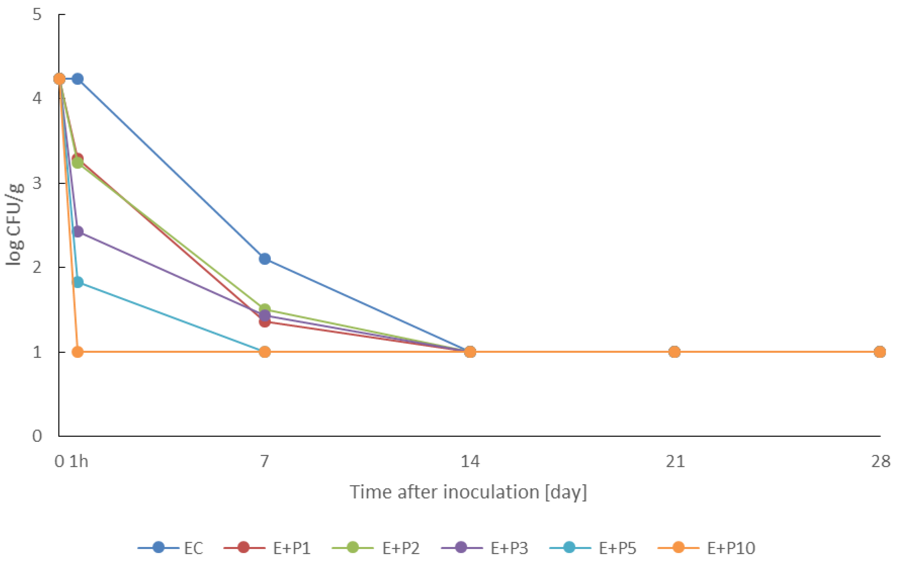
3.1. Effect of Coating Hen Eggs with Propolis Extract on Inhibition of F. solani

Changes in the number of F. solani ATCC 36031 on the shells of inoculated eggs are shown in Figure 1. The initial mold count was 4.24 log CFU/g. After 1 h in the control group (EC) the number of molds did not change, while on the coated eggs (from E + P1 to E + P10) a decrease in the number of molds was observed. The higher the concentration of propolis extract, the greater was the reduction in the number of molds on eggshells. For groups E + P1 and E + P2, the number of molds decreased by only about one log cycle. In the E + P3 group, the number of fungi decreased by 1.80 log cycles, and in the E + P5 group by 2.41 log cycles. The number of F. solani decreased to the greatest extent in the group of E + P10 eggs by 3.24 log cycles, i.e., to the detection limit of fungi, and remained at that level for 28 days.
After 7 days, the number of F. solani decreased in all groups of eggs, with groups E + P1, E + P2 and E + P3 decreasing by 2.87, 2.72 and 2.80 log cycles, respectively, from the initial number. In the case of eggs coated with 5% propolis extract, the number of molds decreased to the limit of detection, i.e., by 3.24 log cycles. No mold growth was found in group E + P10. In the control group, the number of F. solani decreased by 2.13 log cycles. After 14 days of storage, no mold growth was observed on uncoated and coated hen eggs. Our observations show that the activity against the Fusarium strain on hen eggs depends on the concentration of propolis extract. In higher concentrations, propolis extract inhibits the germination of Fusarium spores, preventing mold growth on eggshells.
Similar conclusions were reached by Temiz et al. [23], who observed complete inhibition of mycotoxic mold strains (Aspergillus versicolor and Penicillium aurantiogriseum) by 10% and partial inhibition of their growth by 5% and 1% Turkish propolis extract. Although propolis has so far been used to a small extent for the microbiological protection of table eggs, the first results are encouraging. Pires et al. [37], by coating table quail eggs with a layer of rice protein with propolis extract, reduced the number of microorganisms present on the eggshells, similarly to Ezazi et al. [36], who completely eliminated Salmonella from the surface of hen eggs by coating them with chitosan with propolis extract. The use of a coating of sweet potato starch with thyme essential oil also gave good results and reduced the growth of Salmonella on eggshell [45], and chitosan emulsion with beeswax and basil essential oil reduced the number of native bacteria on the eggshell surface [46]. A composite coating, which included cassava starch, carboxymethyl cellulose and paraffin, protects eggs against microbiological contamination [47].
Other methods of disinfection for edible and hatching eggs are also the subject of extensive research worldwide [3]. The number of microorganisms was reduced by chemical disinfection of eggs, e.g., sodium hydroxide and phenols [48,49], benzalkonium chloride with glutaraldehyde [50], electrolyzed oxidizing (EO) water [51], chlorine dioxide gas [52], silver-stabilized hydrogen peroxide [53], and neutral anolyte [54]. In addition, the effectiveness of physical and combined methods in egg disinfection has been demonstrated, e.g., ultraviolet (UV) irradiation [55], pulsed light [56], nonthermal plasma [57,58], UV light and hydrogen peroxide [59], ultrasonic waves with refrigerated temperature [60], ultrasonic waves with heat treatment [61] and high-intensity ultrasound [62]. Research has shown the effectiveness of decontamination of eggs with natural antimicrobial substances, e.g., essential oils and garlic extract [63], licorice plant extract [64], thyme essential oil [65] and nisin [66].

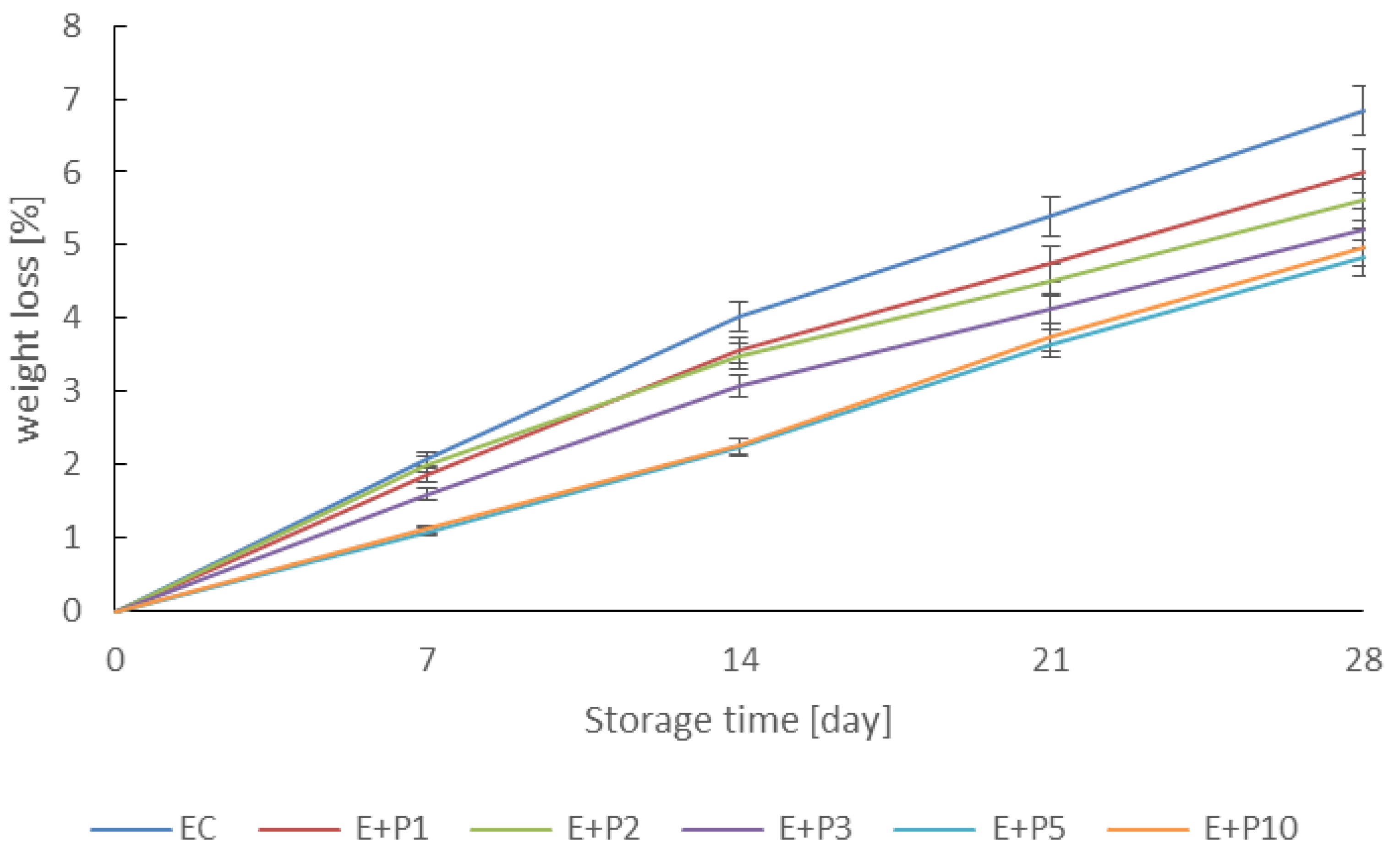
3.2. Effect of Coating with Propolis Extract on Weight Loss of Hen Eggs

Weight is an important indicator of the price and quality of a hen’s egg. Weight loss of all eggs was observed, increasing with each day of storage (Figure 2). After 28 days, the weight loss was greater in the EC group, at 6.85%, while the coated eggs had weight losses from 6.01% (E + P1) to 4.83% (E + P10). The results of this study allow us to conclude that the higher the concentration of propolis extract, the lower was the loss of egg weight, which is consistent with the studies of Aygun and Sert [33] and Akpinar et al. [15], who coated table quail eggs with solutions of propolis extract in concentrations of 5–15%. It was also found that the acceptable 3% weight loss of eggs in retail circulation [35,67] was achieved by uncoated eggs on day 10 and only on day 18 by eggs coated with 10% propolis extract, which is a very beneficial effect.
The applied propolis extract coatings on the eggs slowed down and limited the loss of egg mass. The weight loss of hen eggs is due to the loss, during storage, of water and carbon dioxide, which pass through pores in the eggshells [37]. Egg coating is a process that counteracts weight loss. To a large extent, mass losses depend on the nature of the substance coating the eggs and are the smallest when using a lipid coating, which, thanks to its hydrophobic properties, provides the best protection against water evaporation [16]. Nevertheless, polysaccharide, protein and mixed coatings also fulfill this role well. Lower weight losses were observed in eggs coated with cassava starch mixed with carboxymethyl cellulose and palm oil [68], chitosan coatings with lauric alginate ester [17], coating with shellac [69] and pullulan coating [66] compared to uncoated eggs. A similar trend was noted by Pires et al. [37], who used a coating based on rice protein with propolis.

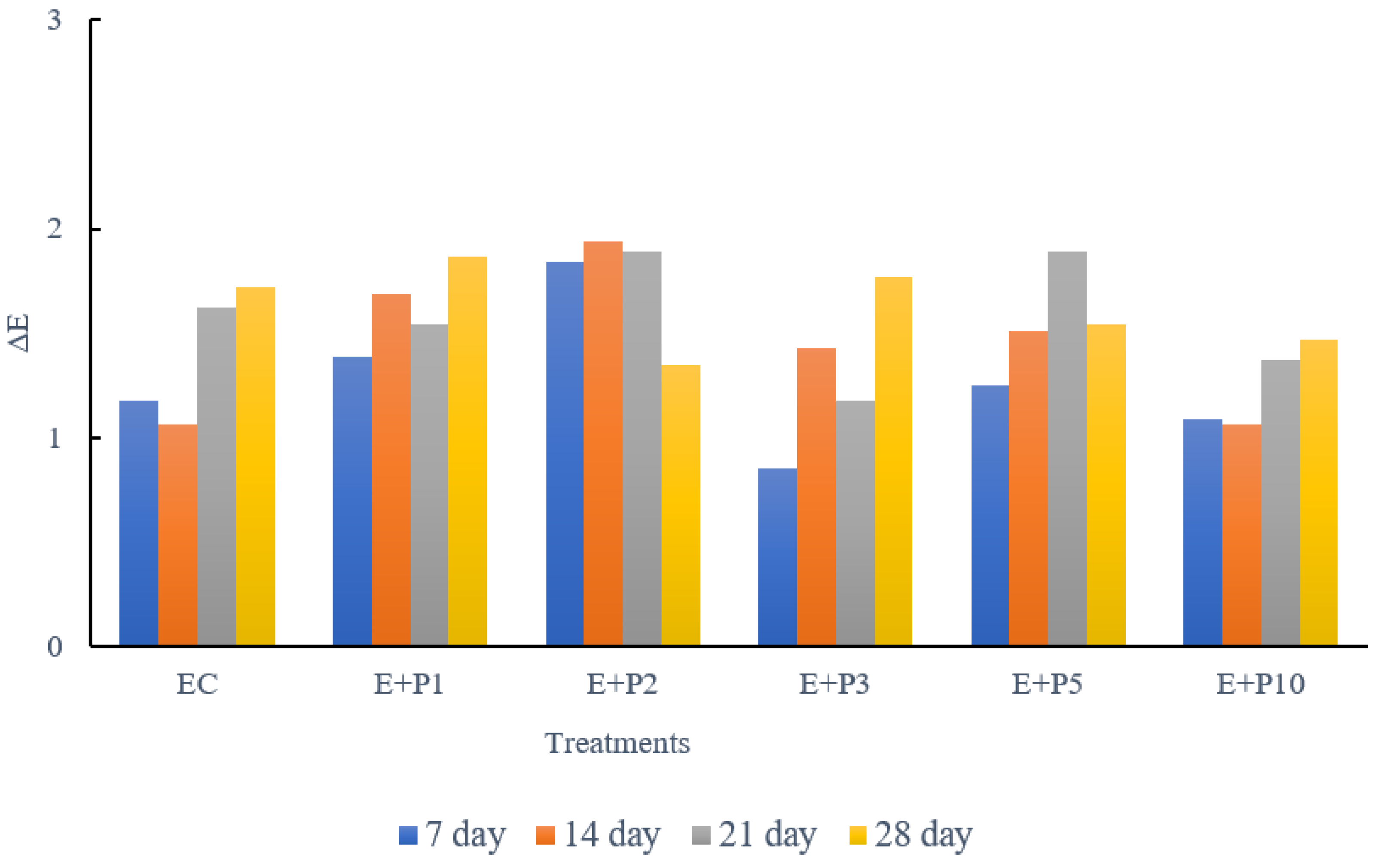
3.3. Effect of Coating with Propolis Extract on the Color of Hen’s Eggs

The color of eggshells is a genetically determined trait ranging from white to dark brown [70]. Uniform coloration of the shell largely determines consumers’ willingness to buy eggs [70,71]. Table 1, Table 2 and Table 3 shows the results of color measurements of hen eggs uncoated and coated with propolis extract at concentrations of 1, 2, 3, 5 and 10% during storage for 28 days.
The undiluted ethanol extract of propolis was dark brown in color, and the 1–10% solutions were much lighter. As reported by Pires et al. [37], propolis can change the color of eggshells due to the content of yellow pigments that darken the color. In our study, the initial L* brightness values ranged from 64.14 to 66.50 and after 28 days they did not change statistically significantly regardless of the propolis extract concentration (Table 1). Similarly, no statistically significant changes in a* values, ranging from 14.03 to 16.32 (Table 2), and b* values, in the range from 25.26 to 28.43 (Table 3), were observed during egg storage. The values of the absolute color difference ΔE of the coated eggs during the 28 days did not exceed the value of 2.0, which proves high color stability (Figure 3). In another study, a change in the color of coated eggs was found using a solution of rosemary extract [64]. Shellac-coated eggs had higher shell gloss, but ΔE values were small, below 3.0, similar to our study [72].

3.4. Sensory Evaluation of Hen Eggs Coated with Propolis Extract

Table 4 shows the sensory analysis scores of raw and cooked hen eggs coated with propolis extract. The appearance of raw coated hen eggs was highly accepted by the panelists, and the ratings ranged from 8.41 to 8.77 and did not differ statistically significantly from the assessment of the appearance of control eggs (8.86). There were lower smell ratings for raw eggs (7.00–6.83) that were coated with 3 to 10% propolis extract than for uncoated eggs and eggs coated with 1 and 2% propolis extract (8.09–8.64), but the ratings were not statistically significantly different. This proves that the perceptible delicate smell of propolis on the eggshells did not disqualify them. However, coating eggs with various substances can change the odor of the eggs, which affects the purchasing preferences of consumers. The smell of chitosan and pennyroyal essential oil on eggshells was less well received by consumers than thyme essential oil [15,65].
The appearance, smell and taste of cooked hen eggs coated with propolis extract were also assessed. The appearance of boiled eggs received average scores from 7.17 to 8.02, which were not statistically significantly different. Similarly, very similar mean egg odor and taste scores of 7.48–7.80 and 7.28–8.16, respectively, were found. It can be assumed that the propolis extract did not penetrate through the eggshell into the egg interior, and therefore it had no effect on these qualitative characteristics. On the other hand, the research results show that the propolis or chitosan and propolis coating has a beneficial effect on the preservation of the qualitative features of the egg interior due to slowing down the diffusion of gases, including CO2, through the pores in the shell, by plugging cracks in their surface [36,38].

4. Conclusions

The results of this study indicate that the coating of hen eggs with propolis extract may contribute to the improvement of the microbiological safety of hen eggs in terms of anti-mold protection. Propolis extract at concentrations of 1–10% showed an inhibitory effect on the growth of F. solani on hen eggshells, with the most effective concentration being 10%. Smaller weight losses of coated hen eggs during storage and no negative effect of coating on the color and sensory features of hen eggs were found.

Author Contributions

Conceptualization, M.G.; methodology, M.G. and K.P.; validation, K.P. and K.K.; formal analysis, N.O. and K.P.; investigation, K.P., K.K. and A.S.; data curation, M.G. and A.S.; writing—original draft preparation, M.G. and K.K.; writing—review and editing, M.G. and K.K.; visualization, M.G. and K.P.; supervision, M.G. All authors have read and agreed to the published version of the manuscript.

Funding

This research received no external funding.

Institutional Review Board Statement

Not applicable.

Informed Consent Statement

Not applicable.

Data Availability Statement

Data available from the corresponding author.

Acknowledgments

Research equipment was purchased as part of the “Food and Nutrition Centre—modernisation of the WULS campus to create a Food and Nutrition Research and Development Centre (CŻiŻ)” co-financed by the European Union from the European Regional Development Fund under the Regional Operational Programme of the Mazowieckie Voivodeship for 2014-2020 (Project No. RPMA.01.01.00-14-8276/17).

Conflicts of Interest

The authors declare no conflict of interest.

References

  1. Adeyeye, E.I. Nutritional values of the lipid composition of the free-range chicken eggs. Am. J. Agric. Biol. Sci. 2012, 3, 374–384. [Google Scholar] [CrossRef]
  2. Cumeras, R.; Aksenov, A.A.; Pasamontes, A.; Fung, A.G.; Cianchetta, A.N.; Doan, H.; Davis, R.M.; Davis, C.E. Identification of fungal metabolites from inside Gallus gallus domesticus eggshells by non-invasively detecting volatile organic compounds (VOCs). Anal. Bioanal. Chem. 2016, 408, 6649–6658. [Google Scholar] [CrossRef] [Green Version]
  3. Turdoi, M.; Borda, D. Decontamination of egg shells using ultraviolet light treatment. Poult. Sci. J. 2014, 70, 265–278. [Google Scholar] [CrossRef]
  4. Rajmani, R.S.; Singh, A.P.; Singh, P.K.; Doley, J.; Verma, S.P. Fungal contamination in eggs. J. Vet. Public Health 2011, 9, 59–61. [Google Scholar]
  5. Liu, Y.C.; Chen, T.H.; Wu, Y.C.; Lee, Y.C.; Tan, F.J. Effects of egg washing and storage temperature on the quality of eggshell cuticle and eggs. Food Chem. 2016, 211, 687–693. [Google Scholar] [CrossRef] [PubMed]
  6. Tomczyk, Ł.; Szablewski, T.; Stuper-Szablewska, K.; Nowaczewski, S.; Cegielska-Radziejewska, R. The influence of the conditions of acquisition and storage of table eggs on changes in their quality and the presence of mycobiota and Fusarium mycotoxins. Poult. Sci. 2019, 98, 2964–2971. [Google Scholar] [CrossRef] [PubMed]
  7. Alkan, S.; Ertürk, Ö.; Türker, İ. Determination of microbial activity and quality traits of eggs coated with propolis. Turk. J. Agric. Food Sci. Technol. 2020, 8, 1380–1384. [Google Scholar] [CrossRef]
  8. De Reu, K. Assessment of the vertical and horizontal aerobic bacterial infection of shell eggs. J. Neurosci. 2007, 27, 3734–3742. [Google Scholar]
  9. Liu, W.; Zhang, J.; Guo, A.; Chen, Q.; Gu, L.; Ruan, Y.; Zhang, X. The specific biological characteristics of spoilage microorganisms in eggs. LWT Food Sci. Technol. 2021, 135, 110069. [Google Scholar] [CrossRef]
  10. Vlčková, J.; Tůmová, E.; Mohamed Ketta, M.; Englmaierová, M.; Chodová, D. Effect of housing system and age of laying hens on eggshell quality, microbial contamination, and penetration of microorganisms into eggs. Czech J. Anim. Sci. 2018, 63, 51–60. [Google Scholar] [CrossRef] [Green Version]
  11. Chousalkar, K.K.; Flynn, P.; Sutherland, M.; Roberts, J.R.; Cheetham, B.F. Recovery of Salmonella and Escherichia coli from commercial egg shells and effect of translucency on bacterial penetration in eggs. Int. J. Food Microbiol. 2010, 142, 207–213. [Google Scholar] [CrossRef] [PubMed]
  12. Tomczyk, Ł.; Stępień, Ł.; Urbaniak, M.; Szablewski, T.; Cegielska-Radziejewska, R.; Stuper-Szablewska, K. Characterisation of the mycobiota on the shell surface of table eggs acquired from different egg-laying hen breeding systems. Toxins 2018, 10, 293. [Google Scholar] [CrossRef] [PubMed]
  13. Zhang, W.; Zheng, J.X.; Xu, G.Y. Toward better control of Salmonella contamination by taking advantage of the egg’s self-defense system: A Review. J. Food Sci. 2011, 76, R77. [Google Scholar] [CrossRef] [PubMed]
  14. Chousalkar, K.K.; Khan, S.; McWhorter, A.R. Microbial quality, safety and storage of eggs. Curr. Opin. Food Sci. 2020, 38, 91–95. [Google Scholar] [CrossRef]
  15. Akpinar, G.C.; Canogullari, S.; Baylan, M.; Alasahan, S.; Aygun, A. The use of propolis extract for the storage of quail eggs. J. Appl. Poult. Res. 2015, 24, 427–435. [Google Scholar] [CrossRef]
  16. Pires, P.G.; Pires, P.D.; Cardinal, K.M.; Bavaresco, C. The use of coating in eggs: A systematic review. Trends Food Sci. Technol. 2020, 106, 312–321. [Google Scholar] [CrossRef]
  17. Jin, T.Z.; Gurtler, J.B.; Li, S.Q. Development of antimicrobial coatings for improving the microbiological safety and quality of shell eggs. J. Food Prot. 2013, 76, 779–785. [Google Scholar] [CrossRef] [PubMed] [Green Version]
  18. Pellati, F.; Orlandini, G.; Pinetti, D.; Benvenuti, S. HPLC-DAD and HPLC-ESI-MS/MS methods for metabolite profiling of propolis extracts. J. Pharm. Biomed. Anal. 2011, 55, 934–948. [Google Scholar] [CrossRef]
  19. Bankova, V.; Galabov, A.S.; Antonova, D.; Vilhelmova, N.; Di Perri, B. Chemical composition of propolis extract ACF® and activity against herpes simplex virus. Phytomedicine 2014, 21, 1432–1438. [Google Scholar] [CrossRef] [PubMed]
  20. Martinotti, S.; Ranzato, E. Propolis: A new frontier for wound healing? Burn. Trauma 2015, 3, 9. [Google Scholar] [CrossRef] [PubMed] [Green Version]
  21. Bayram, N.E.; Sorkun, K.; Öz, G.C.; Salih, B.; Topcu, G. Chemical characterization of 64 propolis samples from Hakkari, Turkey. Rec. Nat. Prod. 2018, 12, 569. [Google Scholar] [CrossRef]
  22. Pobiega, K.; Kraśniewska, K.; Gniewosz, M. Application of propolis in antimicrobial and antioxidative protection of food quality–A review. Trends Food Sci. Technol. 2019, 83, 53–62. [Google Scholar] [CrossRef]
  23. Temiz, A.; Mumcu, A.Ş.; Tüylü, A.Ö.; Sorkun, K.; Salih, B. 2013: Antifungal activity of propolis samples collected from different geographical regions of Turkey against two food-related molds, Aspergillus versicolor and Penicillium aurantiogriseum. GIDA J. Food 2013, 38, 135–142. [Google Scholar]
  24. Boisard, S.; Le Ray, A.M.; Landreau, A.; Kempf, M.; Cassisa, V.; Flurin, C.; Richomme, P. Antifungal and antibacterial metabolites from a French poplar type propolis. Evid. Based Complement. Altern. Med. 2015, 2015, 319240. [Google Scholar] [CrossRef] [Green Version]
  25. Curifuta, M.; Vidal, J.; Sánchez-Venegas, J.; Contreras, A.; Salazar, L.A.; Alvear, M. The in vitro antifungal evaluation of a commercial extract of Chilean propolis against six fungi of agricultural importance. Agric. Nat. Resour. 2012, 39, 347–359. [Google Scholar] [CrossRef] [Green Version]
  26. Dudoit, A.; Mertz, C.; Chillet, M.; Cardinault, N.; Brat, P. Antifungal activity of Brazilian red propolis extract and isolation of bioactive fractions by thin-layer chromatography-bioautography. Food Chem. 2020, 327, 127060. [Google Scholar] [CrossRef] [PubMed]
  27. Vica, M.L.; Glevitzky, M.; Dumitrel, G.A.; Bostan, R.; Matei, H.V.; Kartalska, Y.; Popa, M. Qualitative characterization and antifungal activity of Romanian honey and propolis. Antibiotics 2022, 11, 1552. [Google Scholar] [CrossRef]
  28. Bankova, V.; Popova, M.; Trusheva, B. New emerging fields of application of propolis. Maced. J. Chem. Chem. Eng. 2016, 35, 1–11. [Google Scholar] [CrossRef]
  29. Silici, S.; Karaman, K. Inhibitory effect of propolis on patulin production of Penicillium expansum in apple juice. J. Food Process. Preserv. 2014, 38, 1129–1134. [Google Scholar] [CrossRef]
  30. Yang, Y.; Gong, Y.; Gao, Y.; Huang, J.; Xu, M.; Xiong, B. Study on the preservation effect of propolis on sweet cherry. IOP Conf. Ser. Earth Environ. Sci. 2020, 474, 032029. [Google Scholar] [CrossRef]
  31. Xiao, Y.; Tang, H.; Ge, C.; Luo, Y. The effects of ethanol-dissolved propolis on preservation of strawberries. AIP Conf. Proc. 2020, 2252, 020008G. [Google Scholar]
  32. Oliveira, D.S.; dos Santos, V.M.; McManus, C. Propolis: Effects on the sanitisation of hatching eggs. Worlds Poult. Sci. J. 2022, 78, 261–272. [Google Scholar] [CrossRef]
  33. Aygun, A.; Sert, D. Effects of prestorage application of propolis and storage time on eggshell microbial activity, hatchability, and chick performance in Japanese quail (Coturnix coturnix japonica) eggs. Poult. Sci. 2013, 92, 3330–3337. [Google Scholar] [CrossRef] [PubMed]
  34. Soares, A.C.B.; Brito, D.A.P.; Soares, S.C.P.; Gomes, K.S.; Saldanha, G.K.M.S.; Soares, V.S. Maintenance of quality of eggs submitted to treatment with propolis extract and sanitizers. Acta Sci. Anim. Sci. 2022, 44, e53584. [Google Scholar] [CrossRef]
  35. Pires, P.G.S.; Bavaresco, C.; Morsy, P.D.S.; Cardinal, K.M.; Leuven, A.F.R.; Andretta, I. Development of an innovative green coating to reduce egg losses. Clean. Eng. Technol. 2021, 2, 100065. [Google Scholar] [CrossRef]
  36. Ezazi, A.; Javadi, A.; Jafarizadeh-Malmiri, H.; Mirzaei, H. Development of a chitosan-propolis extract edible coating formulation based on physico-chemical attributes of hens’ eggs: Optimization and characteristics edible coating of egg using chitosan and propolis. Food Biosci. 2021, 40, 100894. [Google Scholar] [CrossRef]
  37. Pires, P.G.D.S.; Pires, P.D.D.S.; Cardinal, K.M.; Leuven, A.F.R.; Kindlein, L.; Andretta, I. Effects of rice protein coatings combined or not with propolis on shelf life of eggs. Poult. Sci. 2019, 98, 4196–4203. [Google Scholar] [CrossRef]
  38. Copur, G.; Camci, O.; Sahinler, N.; Gul, A. The effect of propolis egg shell coatings on interior egg quality. Arch. Geflügelk 2008, 72, S35–S40. [Google Scholar]
  39. Pobiega, K.; Kraśniewska, K.; Derewiaka, D.; Gniewosz, M. Comparison of the antimicrobial activity of propolis extracts obtained by means of various extraction methods. J. Food Sci. Technol. 2019, 56, 5386–5395. [Google Scholar] [CrossRef] [Green Version]
  40. Pobiega, K.; Kraśniewska, K.; Przybył, J.L.; Bączek, K.; Żubernik, J.; Witrowa-Rajchert, D.; Gniewosz, M. Growth biocontrol of foodborne pathogens and spoilage microorganisms of food by Polish propolis extracts. Molecules 2019, 24, 2965. [Google Scholar] [CrossRef] [Green Version]
  41. Gniewosz, M.; Pobiega, K.; Kraśniewska, K.; Synowiec, A.; Chaberek, M.; Galus, S. Characterization and antifungal activity of pullulan edible films enriched with propolis extract for active packaging. Foods 2022, 11, 2319. [Google Scholar] [CrossRef] [PubMed]
  42. Pobiega, K.; Przybył, J.L.; Żubernik, J.; Gniewosz, M. Prolonging the shelf life of cherry tomatoes by pullulan coating with ethanol extract of propolis during refrigerated storage. Food Bioprocess Technol. 2020, 13, 1447–1461. [Google Scholar] [CrossRef]
  43. Pobiega, K.; Igielska, M.; Włodarczyk, P.; Gniewosz, M. The use of pullulan coatings with propolis extract to extend the shelf life of blueberry (Vaccinium corymbosum) fruit. Int. J. Food Sci. Technol. 2021, 56, 1013–1020. [Google Scholar] [CrossRef]
  44. Phothisuwan, S.; Preechatiwong, W.; Matan, N. Enhancement of antibacterial activity of essential oil vapor released from a paper egg tray in combination with UV-C radiation against pathogenic bacteria on chicken eggs. J. Food Process. Preserv. 2020, 44, e14794. [Google Scholar] [CrossRef]
  45. Eddin, A.S.; Tahergorabi, R. Efficacy of sweet potato starch-based coating to improve quality and safety of hen eggs during storage. Coatings 2019, 9, 205. [Google Scholar] [CrossRef]
  46. Sun, R.; Song, G.; Zhang, H.; Chi, Y.; Ma, Y.; Li, H.; Bai, S.; Zhang, X. Effect of basil essential oil and beeswax incorporation on the physical, structural, and antibacterial properties of chitosan emulsion based coating for eggs preservation. LWT 2021, 150, 112020. [Google Scholar] [CrossRef]
  47. Rachtanapun, P.; Homsaard, N.; Kodsangma, A.; Phongthai, S.; Leksawasdi, N.; Phimolsiripol, Y.; Seesuriyachan, P.; Chaiyaso, T.; Chotinan, S.; Jantrawut, P.; et al. Effects of storage temperature on the quality of eggs coated by cassava starch blended with carboxymethyl cellulose and paraffin wax. Poult. Sci. 2021, 101, 101509. [Google Scholar] [CrossRef]
  48. Berrang, M.E.; Cox, N.A.; Frank, J.E.; Buhr, R.J.; Bailey, J.S. Hatching egg sanitisation for prevention or reduction of human enteropathogens: A review. J. Appl. Poult. Res. 2000, 9, 279–284. [Google Scholar] [CrossRef]
  49. Cox, N.A.; Berrang, M.E.; Bailey, J.S.; Stern, N.J. Bactericidal treatment of hatching eggs V: Efficiency of repetitive immersions in hydrogen peroxide or phenol to eliminate Salmonella from hatching eggs. J. Appl. Poult. Res. 2002, 11, 328–331. [Google Scholar] [CrossRef]
  50. Aksu, H.; Bostan, K.; Aydin, A.; Yildirim, M.; Keles, O. Disinfection of eggshells contaminated with Salmonella enteritidis. Med. Weter. 2006, 62, 641–643. [Google Scholar]
  51. Fasenko, G.M.; O’Dea Christopher, E.E.; McMullen, L.M. Spraying hatching eggs with electrolyzed oxidizing water reduces eggshell microbial load without compromising broiler production parameters. Poult. Sci. 2009, 88, 1121–1127. [Google Scholar] [CrossRef] [PubMed]
  52. Kim, H.; Yum, B.; Yoon, S.S.; Song, K.J.; Kim, J.R.; Myeong, D.; Chang, B.; Choe, N.H. Inactivation of Salmonella on eggshells by chlorine dioxide das. Korean J. Food Sci. Anim. 2016, 36, 100–108. [Google Scholar] [CrossRef] [PubMed]
  53. Tomczyk, Ł.; Cegielska-Radziejewska, R.; Lewko, L.; Konieczny, P. An assessment of the influence of silver stabilized hydrogen peroxide on the eggshell condition. Emir. J. Food Agric. 2018, 30, 131–136. [Google Scholar]
  54. Kozak, S.S.; Semenov, V.G. Effect of shell disinfection with Aqualyte (neutral anolyte) on inactivation of Salmonella enteritidis and quality of edible eggs. IOP Conf. Ser. Earth Environ. Sci. 2021, 935, 012039. [Google Scholar] [CrossRef]
  55. De Reu, K.; Grijspeerdt, K.; Herman, L.; Heyndrickx, M.; Uyttendaele, M.; Debevere, J.; Putirulan, F.F.; Bolder, N.M. The effect of a commercial UV disinfection system on the bacterial load of shell eggs. Lett. Appl. Microbiol. 2006, 42, 144–148. [Google Scholar] [CrossRef]
  56. Hierro, E.; Manzano, S.; Ordóñez, J.A.; de la Hoz, L.; Fernández, M. Inactivation of Salmonella enterica serovar Enteritidis on shell eggs by pulsed light technology. Int. J. Food Microbiol. 2009, 135, 125–130. [Google Scholar] [CrossRef]
  57. Lin, C.M.; Herianto, S.; Syu, S.M.; Song, C.H.; Chen, H.L.; Hou, C.Y. Applying a large-scale device using non-thermal plasma for microbial decontamination on shell eggs and its effects on the sensory characteristics. LWT 2021, 142, 111067. [Google Scholar] [CrossRef]
  58. Ragni, L.; Berardinelli, A.; Vannini, L.; Montanari, C.; Sirri, F.; Guerzoni, M.E.; Guarnieri, A. Non-thermal atmospheric gas plasma device for surface decontamination of shell eggs. J. Food Eng. 2010, 100, 125–132. [Google Scholar] [CrossRef]
  59. Wells, J.B.; Coufal, C.D.; Parker, H.M.; McDaniel, C.D. Disinfection of eggshells using ultraviolet light and hydrogen peroxide independently and in combination. Poult. Sci. 2010, 89, 2499–2505. [Google Scholar] [CrossRef]
  60. Sert, D.; Aygun, A.; Demir, M. Effects of ultrasonic treatment and storage temperature on egg quality. Poult. Sci. 2011, 90, 869–875. [Google Scholar] [CrossRef]
  61. Cabeza, M.C.; Ordóñez, J.A.; Cambero, I.; de la Hoz, L.; García, M.L. Effect of thermoultrasonication on Salmonella enterica serovar Enteritidis in distilled water and intact shell eggs. J. Food Protect. 2004, 67, 1886–1891. [Google Scholar] [CrossRef] [PubMed]
  62. Techathuvanan, C.; D’Souza, D.H. High intensity ultrasound for Salmonella Enteritidis inactivation in culture and liquid whole eggs. J. Food Sci. 2018, 83, 1733–1739. [Google Scholar] [CrossRef] [PubMed]
  63. Oliveira, G.D.S.C.; McManus, C.; dos Santos, V.M. Garlic as active principle of sanitiser for hatching eggs. Worlds Poult. Sci. J. 2022, 78, 1037–1052. [Google Scholar] [CrossRef]
  64. Abdel-Salam, A.B.; Nader, S.M.; Emam, S.R. Decontamination of eggshell contaminated with Salmonella Typhimurium using natural plant extracts. Int. J. Pharm. Res. Allied Sci. 2018, 7, 10–19. [Google Scholar]
  65. Smith, T.J.; George, D.R.; Sparagano, O.A.E.; Seal, C.; Shiel, R.S.; Guy, J.H. A pilot study into the chemical and sensorial effect of thyme and pennyroyal essential oil on hens eggs. Int. J. Food Sci. Technol. 2009, 44, 1836–1842. [Google Scholar] [CrossRef]
  66. Morsy, M.K.; Sharoba, A.M.; Khalaf, H.H.; El-Tanahy, H.H.; Cutter, C.N. Efficacy of antimicrobial pullulan-based coating to improve internal quality and shelf-life of chicken eggs during storage. J. Food Sci. 2015, 80, M1066–M1074. [Google Scholar] [CrossRef] [PubMed]
  67. FAO, Food and Agriculture Organization of United Nations. Egg marketing-a guide for the production and sale of eggs. Food and Agriculture Organization of the United Nations Agricultural Services. Bulletin 2003, 150, 29–51. [Google Scholar]
  68. Homsaard, N.; Kodsangma, A.; Jantrawut, P.; Rachtanapun, P.; Leksawasdi, N.; Phimolsiripol, Y.; Jantanasakulwong, K. Efficacy of cassava starch blending with gelling agents and palm oil coating in improving egg shelf life. Int. J. Food Sci. Technol. 2020, 56, 3655–3661. [Google Scholar] [CrossRef]
  69. Caner, C.; Yüceer, M. Efficacy of various protein-based coating on enhancing the shelf life of fresh eggs during storage. Poult. Sci. 2015, 94, 1665–1677. [Google Scholar] [CrossRef]
  70. Samullah, S.; Roberts, J.R.; Chousalkar, K. Eggshell color in brown-egg laying hens—A review. Poult. Sci. 2015, 94, 2566–2575. [Google Scholar] [CrossRef]
  71. Eddin, A.S.; Ibrahim, S.A.; Tahergorabi, R. Egg quality and safety with an overview of edible coating application for egg preservation. Food Chem. 2019, 296, 29–39. [Google Scholar] [CrossRef] [PubMed]
  72. Caner, C. The effect of edible eggshell coatings on egg quality and consumer perception. J. Sci. Food Agric. 2005, 85, 1897–1902. [Google Scholar] [CrossRef]
Figure 1. Changes in the number of F. solani on hen eggshells during 28 days after inoculation. Treatments: EC—control hen eggs, E + P1–E + P10 coated hen eggs with 1–10% with ethanol propolis extract at concentrations of 1–10%.
Applsci 13 00315 g001
Figure 2. Changes in weight loss of hen eggs coated with propolis extract during 28 storage days. Treatments: EC—control hen eggs, E + P1–E + P10 coated hen eggs with ethanol propolis extract at concentrations of 1–10%.
Applsci 13 00315 g002
Figure 3. Changes in the total color difference of hen eggs during 28 storage days. Treatments: EC—control hen eggs, E + P1–E + P10 coated hen eggs with ethanol propolis extract at concentrations of 1–10%.
Applsci 13 00315 g003
Table 1. Changes in L* parameter of the color of hen eggs coated with propolis extract at concentrations of 1, 2, 3, 5 and 10% during 28 storage days.
DayECE + P1Coating *
E + P2
E + P3E + P5E + P10
L* Parameter
066.50 ± 1.77 a65.04 ± 0.87 a64.14 ± 3.50 a64.20 ± 0.59 a65.69 ± 0.72 a65.10 ± 1.46 a
767.07 ± 1.79 a65.47 ± 0.99 a62.97 ± 3.29 a63.88 ± 2.04 a65.50 ± 0.76 a65.08 ± 1.32 a
1466.94 ± 2.58 a65.42 ± 0.51 a63.86 ± 3.13 a63.07 ± 1.93 a64.63 ± 2.96 a65.24 ± 1.26 a
2166.40 ± 2.47 a65.14 ± 0.40 a62.37 ± 2.89 a63.35 ± 2.03 a65.28 ± 0.84 a64.51 ± 1.90 a
2867.48 ± 1.96 a65.01 ± 0.60 a63.12 ± 2.79 a63.19 ± 4.12 a65.24 ± 0.82 a64.48 ± 1.91 a
* Coating: EC—control hen eggs, E + P1–E + P10 coated hen eggs with propolis extract in the concentration of 1–10%. Mean values marked with the same letter symbol (a) in the row indicate no statistically significant differences. n = 30 eggs per mean.
Table 2. Changes in a* parameter of the color of hen eggs coated with propolis extract at concentrations of 1, 2, 3, 5 and 10% during 28 storage days.
DayECE + P1Coating *
E + P2
E + P3E + P5E + P10
a* Parameter
015.45 ± 1.64 a16.32 ± 0.59 a15.93 ± 1.48 a15.83 ± 0.32 a14.03 ± 0.65 a14.17 ± 0.30 a
714.81 ± 2.04 a15.28 ± 0.97 a16.10 ± 1.74 a15.43 ± 0.82 a13.99 ± 0.62 a13.41 ± 0.79 a
1414.56 ± 2.23 a15.83 ± 0.34 a16.20 ± 1.48 a15.54 ± 0.58 a14.36 ± 0.49 a14.06 ± 0.54 a
2114.32 ± 2.13 a15.54 ± 1.34 a15.42 ± 1.60 a15.37 ± 1.08 a14.21 ± 0.67 a13.82 ± 0.86 a
2815.38 ± 1.96 a16.02 ± 1.03 a16.79 ± 1.27 a16.18 ± 1.62 a14.13 ± 0.66 a13.80 ± 0.77 a
* Coating: EC—control hen eggs, E + P1–E + P10 coated hen eggs with propolis extract in the concentration of 1–10%. Mean values marked with the same letter symbol (a) in the row indicate no statistically significant differences. n = 30 eggs per mean.
Table 3. Changes in b* parameter of the color of hen eggs coated with propolis extract at concentrations of 1, 2, 3, 5 and 10% during 28 storage days.
DayECE + P1Coating*
E + P2
E + P3E + P5E + P10
b* Parameter
025.26 ± 1.62 a26.99 ± 0.63 a27.16 ± 1.01 a28.43 ± 0.63 ab27.83 ± 3.39 a28.36 ± 1.08 a
726.07 ± 1.38 a28.24 ± 0.60 b28.57 ± 0.54 a29.11 ± 1.28 b29.06 ± 0.63 a29.14 ± 1.03 a
1425.46 ± 1.29 a27.46 ± 0.47 ab27.88 ± 0.53 a29.25 ± 0.72 b28.85 ± 0.48 a29.40 ± 0.83 a
2126.41 ± 1.58 a28.13 ± 0.71 b27.59 ± 0.90 a29.10 ± 1.02 b29.37 ± 0.38 a29.55 ± 1.06 a
2826.11 ± 1.33 a27.49 ± 0.79 ab26.93 ± 3.21 a29.84 ± 0.96 b29.30 ± 0.37 a29.64 ± 1.01 a
* Coating: EC—control hen eggs, E + P1–E + P10 coated hen eggs with propolis extract in the concentration of 1–10%. Mean values marked with the same letter symbol (a,b) in the row indicate no statistically significant differences. n = 30 eggs per mean.
Table 4. Assessment of sensory factors of hen eggs coated with propolis extract in concentrations of 1, 2, 3, 5 and 10%.
Coating *Raw Hen EggsBoiled Hen Eggs (without Shelleggs)
AppearanceShell OdorAppearanceOdorTaste
EC8.86 ± 0.31 a8.64 ± 0.64 a7.17 ± 1.03 a7.48 ± 1.44 a8.02 ± 0.96 a
E + P18.77 ± 0.39 a8.36 ± 0.77 a7.34 ± 0.97 a7.80 ± 0.66 a8.16 ± 0.72 a
E + P28.41 ± 0.97 a8.09 ± 1.00 a7.57 ± 0.92 a7.59 ± 1.18 a8.11 ± 0.67 a
E + P38.46 ± 0.31 a7.00 ± 1.65 a7.27 ± 1.55 a7.57 ± 1.17 a8.16 ± 0.70 a
E + P58.26 ± 0.97 a7.27 ± 1.70 a8.02 ± 0.83 a7.56 ± 1.62 a7.56 ± 0.98 a
E + P108.59 ± 0.63 a6.83 ± 1.76 a7.61 ± 1.11 a7.50 ± 1.52 a7.28 ± 1.26 a
* Coating: EC—control hen eggs, E + P1–E + P10 coated hen eggs with propolis extract in the concentration of 1–10%. Mean values marked with the same letter symbol (a) in the columns indicate no statistically significant differences. n = 30 eggs per mean.
Disclaimer/Publisher’s Note: The statements, opinions and data contained in all publications are solely those of the individual author(s) and contributor(s) and not of MDPI and/or the editor(s). MDPI and/or the editor(s) disclaim responsibility for any injury to people or property resulting from any ideas, methods, instructions or products referred to in the content.

