Review

Endodontic Regenerative Procedures in Necrotic Adult Teeth

by
Sara Garrido-Parada
1,
Pablo Castelo-Baz
1,*,
Nancy Feijoo-Pato
1,
José Gaviño-Orduña
2 and
Benjamín Martín-Biedma
1
1
Department of Surgery and Medical-Surgical Specialties, School of Medicine and Dentistry, University of Santiago de Compostela, 15701 Santiago de Compostela, Spain
2
Department of Odonto-Stomatology, Faculty of Medicine and Health Sciences, School of Dentistry, University of Barcelona, 08907 Barcelona, Spain
*
Author to whom correspondence should be addressed.
Appl. Sci. 2022, 12(9), 4212; https://doi.org/10.3390/app12094212
Submission received: 29 March 2022 / Revised: 17 April 2022 / Accepted: 20 April 2022 / Published: 21 April 2022
(This article belongs to the Topic State-of-the-Art Dentistry and Oral Health)

Abstract

:

Featured Application

Regarding the promising future of regenerative endodontic techniques, this review gathers the evidence available on these procedures performed in chronologically adult teeth, and proposes a clinical protocol, taking into account the evidence published (protocols carried out in the randomized clinical trials analyzed), age considerations, and the protocols published by The American Association of Endodontists and The European Society of Endodontology for immature teeth, also taking into consideration the protocols followed by the majority of the clinicians included in the review.

Abstract

There have been published regenerative endodontic protocols for treating immature teeth in young patients, but there are no clinical considerations for the adult teeth. The goal of the present review is to propose a specific clinical protocol for both mature and immature adult teeth with necrotic pulps. Research was performed from January to April of 2021. From the 539 studies identified through the initial search, 23 studies were qualified for the final analysis (3 randomized controlled trials and 20 case reports). The results in mature adult teeth indicate a success rate of 96.35 and 100% in bone healing through the randomized controlled trials and case reports, respectively; 100% in absence of clinical symptoms, and 58 and 62.5% in positive response to sensibility tests. The success rate in the case reports in teeth with open apex reported a 61.5% of root development, 100% of bone healing, 96.15% of absence of clinical symptoms, and 43.7% of positive response to sensibility tests. The current evidence is scarce but emerging, so REPs may be a promising alternative for treating adult necrotic teeth. The clinical protocol proposed is based on the evidence available and age considerations, and should be updated in the future.

1. Introduction

Regenerative endodontic procedures (from now on REPs) have been defined as ‘biologically based procedures that intend to physiologically replace damaged tooth structures, including dentine and root, as well as cells of the pulp–dentin complex’ [1]. REPs were first exclusively developed for the treatment of immature teeth, with the purpose of achieving a complete root development, increasing the root length, thickening the root wall, and accomplishing apical closure [2]. The American Association of Endodontists (AAE) [3], the European Society of Endodontology (ESE) [4], and several systematic reviews [5,6,7] agree that these techniques are an alternative with a huge potential in the treatment of immature permanent necrotic teeth, although they underline the poor quality of the available evidence and the need for further investigation [2,5,8,9,10,11,12].
These procedures can not only achieve a tooth with open apex completing its development, decreasing its susceptibility to fracture, and increasing its longevity, but also can restore tooth vitality and pulp functions thanks to the pulp tissue regeneration. Thus, sensibility, immunity, healing, and restorative properties of dental pulp would also be recovered [13], in turn reducing the chances of reinfection. Besides, it is believed that regenerated tissues may structurally be more resistant to fracture than endodontically treated teeth [14]. For this reason, the feasibility of this treatment in mature teeth should be further studied, as until now, conservative treatment for irreversible necrosis or pulpitis has been limited to conventional endodontic therapy. The high success of this technique is undeniable [15,16], with success percentages raging, according to the systematic review made by Ny et al. in 2010, from 83 to 93%, with a follow-up of 2 to 10 years [17]. Nevertheless, although the treatment would be successful, the tooth does not remain vital and, therefore, it is more susceptible to fracture and reinfection [14].
Three key elements are essential for performing REPs: stem cells, scaffolds, and activators [18]. These techniques begin with the disinfection of the root canal system, followed by the induction of bleeding in the periapical region with the purpose of obtaining a blood clot. This mass would behave as a natural scaffold for the migration of undifferentiated stem cells that come from outside the apex, mostly from the alveolar bone and periodontal ligament [3], while providing growth factors that stimulate cell differentiation and proliferation, inducing the formation of new tissue.
However, the composition and concentration of the clot is unpredictable, and the uncertainty increases with age [18,19]. When bleeding is insufficient and it does not provide an ideal blood clot, an alternative material should be used. Evidence supports the use of autologous platelet concentrates [20,21,22,23], such as platelet-rich plasma (PRP) and fibrin-rich plasma (PRF) [21], which are the most popular ones. These concentrates can be used as a scaffold, by their own or in combination with a blood clot [11].
Although a closed apex is found in most of the adult teeth, it is possible to find immature teeth that physiologically should have already completed their root development, mainly due to a dental trauma, caries, or developmental anomaly, which led to the necrosis of the dental organ [24]. In Figure 1 is an adult patient’s immature central incisor, necrotic due to a dental trauma. Because of the age of the patient, in these cases, some conditions must be considered. 1. Reduced revascularization potential. On a systemic level, angiogenesis is reduced by degenerative changes that occur in vessels and nerves over time [25]. In the pulp–dentin complex, age can imply a compromise of its vascular supply [26], as the size of the pulp chamber decreases due to the apposition of dentin, while calcifications tend to occur more frequently [27], as well as the apical narrowing due to the gradual apposition of dentin and cementum. 2. Changes in the functionality and potential of stem cells. Over the years, the ability of multipotent stem cells to replace damaged tissues decreases [20]. This could be related to the fact that the differentiation capacity of MSCs decreases over time, but not to the amount of inflow of MSCs into the root canal system in adults, because this amount does not change with age [28]. 3. Smaller diameter of the apical constriction in mature teeth. Currently, there is no consensus regarding the optimal size of the apical foramen for REP [14]. It should be as small as possible without affecting cell migration and reinnervation [29]. In 2007, Murray suggested that, in teeth with closed apexes, revascularization by inducing bleeding may require an apical instrumentation of 1 or 2 mm to allow the flow of bleeding [18]. Later, some authors proposed it as feasible to succeed apical diameters smaller than 1 mm [30] or equal to 0.5 mm [18].
REPs’ scientific evidence in adult teeth is limited. Currently, there is no specific clinical protocol for the management of these teeth. Regarding the promising future of these techniques, it is necessary to analyze the considerations that must be considered for the treatment of these teeth and the results obtained up to nowadays. Therefore, the objective of this review is to gather the available evidence on regenerative endodontic techniques in chronologically adult teeth in order to establish a clinical protocol guided to them.

2. Materials and Methods

2.1. Eligibility Criteria: PICO Question

Population, Intervention, and Outcome items of the PICO framework were used to formulate the following clinical question: What is the success rate of performing a regenerative endodontic procedure in necrotic mature and immature adult permanent teeth? (Table 1).

2.1.1. P-Population

To determine the population, the minimum age was taken as that at which each permanent tooth should, physiologically, have completed its root development (this is Nolla stage 10). When a tooth presented a range, the highest figure was taken [31]. Thus, for example, for the upper second molar, whose age at which it completes its development is between 15 and 16 years, patients who received REP treatment on that tooth, whose age was greater than or equal to 16 years, were included (Table 2).
Besides, mature adult permanent teeth without periapical radiolucent lesion were excluded, since this is the main result to be evaluated in these teeth. Regarding immature teeth, the presence or absence of periapical pathology is not an exclusion criterion (Figure 2).

2.1.2. O-Outcome

The primary, secondary, and tertiary goals evaluated must vary depending on whether the treated tooth is mature or immature (Table 3).

2.2. Search Strategy

The online search was conducted by one of the investigators (S.G.P.) in the following databases: PubMed (National Center for Biotechnology Information, US National Library of Medicine) and Cochrane (John Wiley & Sons, Ltd., London, UK), and performed between December of 2020 and April of 2021. Studies published in the last five years were included in the search with no language restriction. The following combination of key words was useful: ((regenerative) AND (endodontic)) AND (treatment OR therapy OR procedures OR technique); (((necrotic) AND (pulp)) AND (open)) AND (apex); (pulp) AND (revascularization OR revitalization OR repair OR regeneration); (tissue engineering) AND (endodontics); (stem cells OR scaffolds OR growth factor) AND (pulp) AND (tooth). Study authors were contacted privately via e-mail whenever any doubts were encountered about the study.

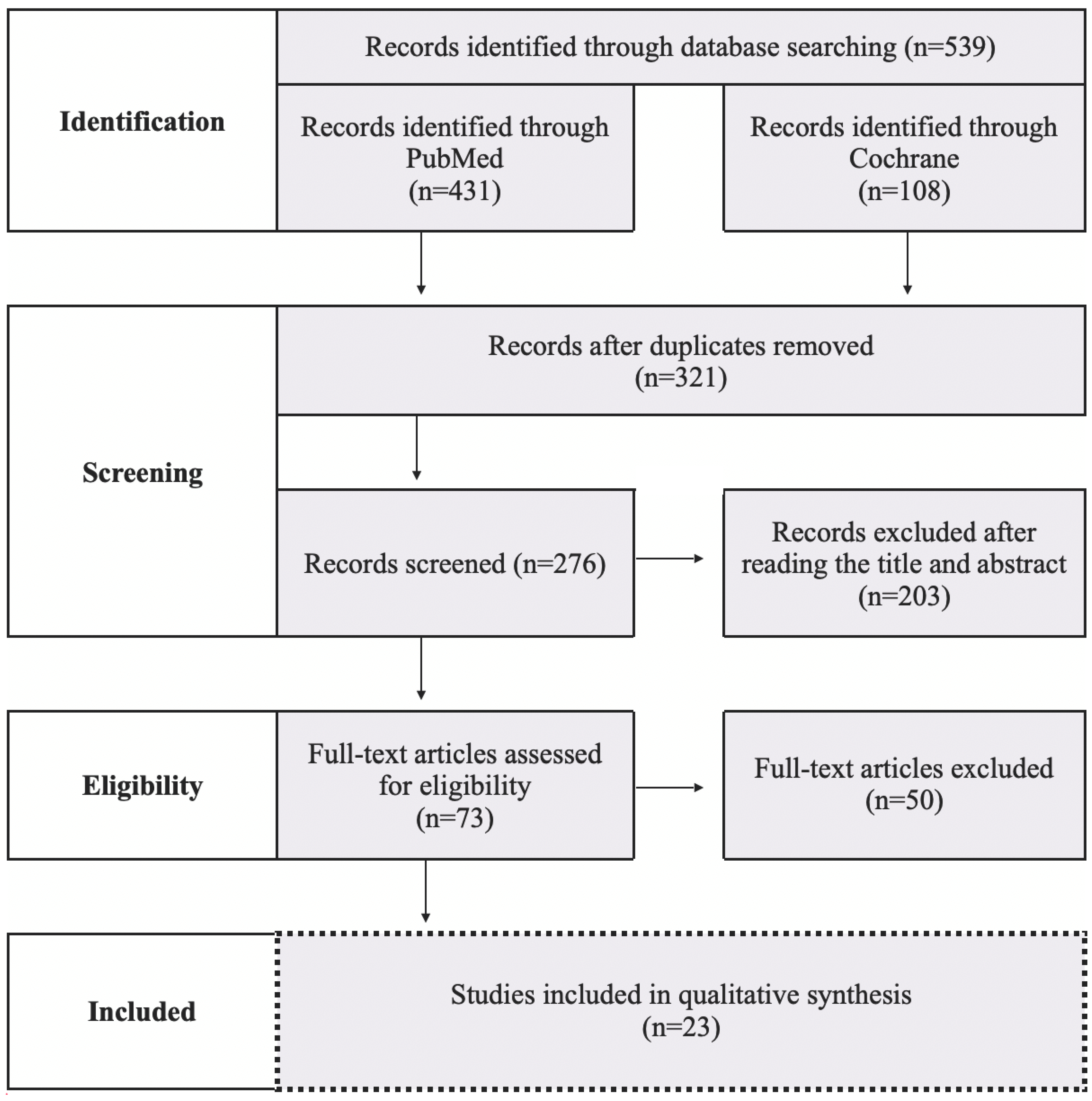
2.3. Study Selection

Searches of the two databases alongside hand-searching provided a total of 539 studies. A flow diagram of the search process was performed with the number of excluded/included articles (Figure 3). Following de-duplication and exclusion by title and abstract, 73 studies were obtained for full text screening. Eventually, 23 studies were included in the review. Search strategy and reasons for exclusion of each article are listed in Supplementary File S1, where included articles are marked in red color. Only those searches where the combination of key words showed new articles (not duplicated) were included. In the event that the full-text article was not available, the authors were personally contacted by e-mail to gain access to the full-text article.

3. Results

After the selecting process, 3 randomized clinical trials and 20 case reports were finally included. Adult teeth with closed apex constitute the population of the 3 randomized clinical trials, where REP was performed in 62 teeth from patients aged 16 to 58 years (Table 4). In addition, 5 case reports treated 8 mature teeth of 5 patients aged 12 to 21 years. (Table 5). On the other hand, no randomized clinical trials performed REP exclusively in immature adult teeth, so the available evidence for these teeth is limited to 16 case reports, where 26 teeth from 22 patients aged 11 to 25 years were treated (Table 6).
As primary, secondary, and tertiary goals vary depending on whether the treated tooth is mature or immature, the outcomes in open and closed apex teeth must be evaluated separately. Moreover, the level of evidence provided by case reports is much lower than the randomized clinical trials. For that reason, its qualitative synthesis was also analyzed separately. The treatment outcomes of the articles included in this revision are shown in Table 7, Table 8 and Table 9.
Key characteristics and population, protocol, and outcome details of the studies included in the review are available in Supplementary File S2.

4. Discussion

REPs were developed to treat immature teeth of young patients. However, according to this revision and matching the results of previous systematic reviews that analyzed these treatments in mature necrotic teeth with periapical lesions [55,56], success rates obtained in adult teeth are promising. Thus, it may be possible to extend these techniques to all teeth regardless of patient’s age. However, the clinical protocol for adult teeth should be different from the one for immature teeth, because of their different conditions with respect to young teeth: decreased revascularization capacity, lower potential of stem cells and, in general, smaller diameter of the apical constriction.
The proposed protocol, available in Supplementary File S3, is based on the ones from the AAE [57] and the ESE [4] for immature teeth, adding modifications derived from the protocols carried out in the randomized clinical trials analyzed, and considering the trend of most of the clinicians included in this review and the available evidence. Two key points of the protocol will be criticized: 1. Instrumentation and ideal apical diameter. 2. Irrigation and intracanal medication.
Regarding instrumentation, some clinicians consider that a minimal mechanical debridement is necessary to facilitate the destruction of biofilms [58], while others rely on chemical debridement using disinfectants and the use of intracanal medication to achieve the complete disinfection of the canal system [59]. From the studies reviewed, it is concluded that the majority of clinicians perform a minimal instrumentation of the canals. The instrumentation techniques and instruments used vary depending on the state of development of the tooth, predominantly reciprocating rotary instrumentation technique for the treatment of mature teeth, and manual technique with K and H files for immature teeth.
The conventional and simplest technique to achieve revascularization is by stimulating bleeding 2 mm under the apical with a manual file [57]. Previous studies have failed to achieve a consensus regarding the optimal apical diameter that better allows revascularization [14,18,29,60], although success has been reported in diameters between 0.3 to 1 mm [14]. One of the trials included in this review compares rate success through REP in mature teeth with different apical preparations (0.3 and 0.5 mm), concluding that the size of the apical diameter does not influence treatment success [33]. Therefore, although more studies are needed, it seems that revascularization can take place in teeth with apical diameters of 0.3 mm.
Regardless the apical foramen diameter, revascularization potential may be compromised due to patient’s age. Through their trials, Arslan [34] and El-Kateb [33] obtained successful results by stimulating bleeding, and most of the clinicians through case reports also performed this technique successfully. Those who did not achieve bleeding or chose platelet-rich concentrates opted for PRF, PRP, L-PRF, or the combination of both techniques.
If a REP failure occurs, other conservative approaches can be considered, whether that be a second attempt of the same technique, as the REP-retreatment successfully performed by Brogni in 2020 [44], a conventional root canal treatment in mature teeth, or the placement of a mineral trioxide aggregate apical plug in open apexes. If the conservative treatment options are dismissed or rejected by the patient, an extraction and placement of a dental implant can be considered. In young patients, implant placement should be postponed until adulthood [61], managing the tooth’s loss through a resin-bonded fixed partial denture [62].
Time-dependence relationship of pulp regeneration is one of the main challenges posed by REPs. To determine how these treatments are influenced by age, conducting randomized clinical trials in narrow age groups would be required. For example, Estefan in 2016 states that REPs can be applied in patients aged 9 to 18 years, concluding that the younger the age, the better the prognosis [29].
When an unsuccessful REP occurs in an adult patient, the cause may lie on the lower potential of its stem cells, so it is necessary to determine when it would be better to perform a cell transplant. In this review, two authors successfully performed cell transplantations using umbilical cord mesenchymal stem cells (UC-MSCs) encapsulated in platelet-poor plasma [35] and stem cells from exfoliated deciduous human teeth (SHED) [39]. Since the immunogenicity of mesenchymal stem cells is low, cell banks may become a real alternative [63]. Although these techniques are not currently available to all clinicians, with a view to the future, Nakashima estimates that cell banks will provide stem cells cryopreserved and safely transported to the dental clinic for REP [64].
One of the main unknowns is how to determine the best combination of irrigants and intracanal medication to achieve complete disinfection of the canal system while promoting, or at least not harming, tissue regeneration. To this point, EDTA should be used in all regenerative procedures [21], as it appears to enhance the liberation of dentin growth factors [59,65,66,67], which are essential as biological inductors and remain entrapped in the dentinal matrix as reservoir [68]. Besides, EDTA partially reverses the deleterious effects of NaOCl and promotes SCAP survival and differentiation [59]. The AAE recommends an irrigation with 1.5% NaOCl (20 mL/channel, 5 min) during the first session, followed by saline or EDTA (20 mL/channel, 5 min). In adult teeth, the irrigation protocol favored by most authors in the first session is irrigation with NaOCl. Regarding the concentration, the disagreement is notorious, although most clinicians lean towards 1.5%. During the second session, there is great disagreement regarding the irrigation protocol, although most clinicians apply 17% EDTA.
Despite the existence of various devices that improve the irrigant’s action, manual irrigation with a lateral opening needle is the most used technique by clinicians included in this review. It is recommended to use some activation system to maximize the viability of stem cells by achieving a bacteria-free environment. According to Castelo in 2012, continuous ultrasonic irrigation is the one that best achieves the penetration of the irrigant in lateral canals [69]. However, the possibility of extrusion into periapical tissues must be minimized. The AAE recommends negative apical aspiration for this purpose. Despite the recommendations, only two clinicians used irrigant activation devices: sonic activation [32] and negative apical aspiration [44]. Clinicians should incorporate these systems to improve canal disinfection.
As intracanal medication, the AAE recommends calcium hydroxide or a low concentration of antibiotic mixture made up of ciprofloxacin, metronidazole, and minocycline, known as triple antibiotic paste [57]. Because minocycline as an intracanal dressing may cause dentine discoloration [70], it is recommended to replace it with calcium hydroxide, double antibiotic paste composed of metronidazole and ciprofloxacin [4], or a triple antibiotic paste where minocycline is replaced with another antibiotic, either amoxicillin, clindamycin or cefaclor [3]. However, the ESE Position Statement advocates the use of calcium hydroxide instead of antibiotics, indicating that there is no evidence to support the need of using them [4]. Nevertheless, according to the literature, most dentists favor the use of triple antibiotic paste [70]. In the articles included, most clinicians favor calcium hydroxide (39% of clinicians), followed by 31% who prefer the mixture of metronidazole, ciprofloxacin, and minocycline.

5. Conclusions

Regarding the promising future of regenerative endodontic techniques, this review gathers the available evidence of these procedures performed in chronologically adult teeth. Although the current evidence is scarce, success rates are encouraging and match with the ones obtained in previous studies. Because age conditions must be taken into account, this review proposes a specific clinical protocol for REP in necrotic adult teeth based on the evidence available. We hope it may be updated in the future thanks to solid evidence.
A quality analysis in this field cannot currently be performed because of the low evidence available. To support REPs in adult teeth, new randomized clinical trials are needed. For further studies, the authors suggest considering some recommendations. Within the included studies, not all of them specified all the information the authors were intended to assess. A detailed description of the etiology and time of necrosis, preoperative variables, clinical protocol, and postoperative follow-ups should be included. Moreover, clinicians should perform sensitivity tests during the follow-ups, which was not completely fulfilled. Due to the limited evidence available, a minimum follow-up time of one year was established as inclusion criteria, which was the time chosen by the majority of clinicians. However, this follow-up is not the ideal and could lead to incorrect conclusions in the results. It is recommended to extend the follow-up time to at least 2 years [57], preferably with annual check-ups up to 5 years [4].

Supplementary Materials

The following supporting information can be downloaded at: https://www.mdpi.com/article/10.3390/app12094212/s1, File S1: Search strategy and reasons for exclusion; File S2: Key characteristics and population, protocol and outcome details of the studies included in the review; File S3: Clinical Considerations for Regenerative Endodontic Procedures in Mature and Immature Adult Teeth.

Author Contributions

Conceptualization, manuscript planning, methodology, data curation, writing and editing, creating, and formatting figures and tables, S.G.-P.; conceptualization, supervision and manuscript review, B.M.-B.; methodology, supervision and manuscript review, N.F.-P.; manuscript review J.G.-O.; manuscript review P.C.-B. All authors have read and agreed to the published version of the manuscript.

Funding

This research was supported by the Theorical-Practical International Master′s Degree in Advanced Endodontics, Restorative and Aesthetic Dentistry. University of Santiago de Compostela.

Institutional Review Board Statement

Not applicable.

Informed Consent Statement

Not applicable.

Data Availability Statement

All data related to this review is contained within the article.

Conflicts of Interest

The authors declare no conflict of interest.

References

  1. American Association of Endodontists, Glossary of Endodontic Terms (Tenth Edition). Available online: https://www.aae.org/specialty/clinical-resources/glossary-endodontic-terms/ (accessed on 15 February 2021).
  2. Lopes, L.B.; Neves, J.A.; Botelho, J.; Machado, V.; Mendes, J.J. Regenerative Endodontic Procedures: An Umbrella Review. Int. J. Environ. Res. Public Health 2021, 18, 754. [Google Scholar] [CrossRef] [PubMed]
  3. American Association of Endodontics, Colleagues for Excellence Newsletter. Regenerative Endodontics. AAE. Available online: https://f3f142zs0k2w1kg84k5p9i1o-wpengine.netdna-ssl.com/specialty/wp-content/uploads/sites/2/2017/06/ecfespring2013.pdf (accessed on 13 April 2021).
  4. Galler, K.M.; Krastl, G.; Simon, S. European Society of Endodontology position statement: Revitalization procedures. Int. Endod. J. 2016, 49, 717–723. [Google Scholar] [CrossRef] [PubMed]
  5. Duggal, M.; Tong, H.J.; Al-Ansary, M. Interventions for the endodontic management of non-vital traumatised immature permanent anterior teeth in children and adolescents: A systematic review of the evidence and guidelines of the European Academy of Paediatric Dentistry. Eur. Arch. Paediatr. Dent. 2017, 18, 139–151. [Google Scholar] [CrossRef] [PubMed]
  6. Alghamdi, F.T.; Alqurashi, A.E. Regenerative Endodontic Therapy in the Management of Immature Necrotic Permanent Dentition: A Systematic Review. Sci. World J. 2020, 2020, 7954357. [Google Scholar] [CrossRef] [PubMed]
  7. Cabral, C.S.L.; Genizelli, L.O.; Cruz, R.G.Z. Tratamento de dentes com rizogênese incompleta após procedimentos regenerativos ou de apicificação: Uma revisão sistemática de literatura. Rev. Bras. Odontol. 2016, 73, 336–339. [Google Scholar] [CrossRef] [Green Version]
  8. Shamszadeh, S.; Asgary, S.; Nosrat, A. Regenerative Endodontics: A Scientometric and Bibliometric Analysis. J. Endod. 2019, 45, 272–280. [Google Scholar] [CrossRef]
  9. Tong, H.J.; Rajan, S.; Bhujel, N. Regenerative Endodontic Therapy in the Management of Nonvital Immature Permanent Teeth: A Systematic Review-Outcome Evaluation and Meta-analysis. J. Endod. 2017, 43, 1453–1464. [Google Scholar] [CrossRef]
  10. Torabinejad, M.; Nosrat, A.; Verma, P. Regenerative Endodontic Treatment or Mineral Trioxide Aggregate Apical Plug in Teeth with Necrotic Pulps and Open Apices: A Systematic Review and Meta-analysis. J. Endod. 2017, 43, 1806–1820. [Google Scholar] [CrossRef]
  11. Digka, A.; Sakka, D.; Lyroudia, K. Histological assessment of human regenerative endodontic procedures (REP) of immature permanent teeth with necrotic pulp/apical periodontitis: A systematic review. Aust. Endod. J. 2020, 46, 140–153. [Google Scholar] [CrossRef]
  12. Wilkström, A.; Brundin, M.; Lopes, M.F. What is the best long-term treatment modality for immature permanent teeth with pulp necrosis and apical periodontitis? Eur. Arch. Paediatr. Dent. 2021, 22, 311–340. [Google Scholar] [CrossRef]
  13. Ducret, M.; Costantini, A.; Gobert, S. Fibrin-based scaffolds for dental pulp regeneration: From biology to nanotherapeutics. Eur. Cell Mater. 2021, 41, 1–14. [Google Scholar] [CrossRef] [PubMed]
  14. He, L.; Zhong, J.; Gong, Q. Treatment of Necrotic Teeth by Apical Revascularization: Meta-analysis. Sci. Rep. 2017, 7, 13941. [Google Scholar] [CrossRef] [PubMed] [Green Version]
  15. Tabassum, S.; Khan, F.R. Failure of endodontic treatment: The usual suspects. Eur. J. Dent. 2016, 10, 144–147. [Google Scholar] [CrossRef] [PubMed]
  16. Setzer, F.C.; Shah, S.B.; Kohli, M.R. Outcome of endodontic surgery: A meta-analysis of the literature--part 1: Comparison of traditional root-end surgery and endodontic microsurgery. J. Endod. 2010, 36, 1757–1765. [Google Scholar] [CrossRef] [PubMed]
  17. Imura, N.; Pinheiro, E.T.; Gomes, B.P. The outcome of endodontic treatment: A retrospective study of 2000 cases performed by a specialist. J. Endod. 2007, 33, 1278–1282. [Google Scholar] [CrossRef] [PubMed]
  18. Murray, P.E.; Garcia-Godoy, F.; Hargreaves, K.M. Regenerative endodontics: A review of current status and a call for action. J. Endod. 2007, 33, 377–390. [Google Scholar] [CrossRef]
  19. Hargreaves, K.M.; Giesler, T.; Henry, M. Regeneration potential of the young permanent tooth: What does the future hold? J. Endod. 2008, 34 (Suppl. S7), S51–S56. [Google Scholar] [CrossRef]
  20. Panda, S.; Mishra, L.; Arbildo-Vega, H.I. Effectiveness of Autologous Platelet Concentrates in Management of Young Immature Necrotic Permanent Teeth-A Systematic Review and Meta-Analysis. Cells 2020, 9, 2241. [Google Scholar] [CrossRef]
  21. Metlerska, J.; Fagogeni, I.; Nowicka, A. Efficacy of Autologous Platelet Concentrates in Regenerative Endodontic Treatment: A Systematic Review of Human Studies. J. Endod. 2019, 45, 20–30. [Google Scholar] [CrossRef]
  22. Meschi, N.; Castro, A.B.; Vandamme, K. The impact of autologous platelet concentrates on endodontic healing: A systematic review. Platelets 2016, 27, 613–633. [Google Scholar] [CrossRef]
  23. Lolato, A.; Bucchi, C.; Taschieri, S. Platelet concentrates for revitalization of immature necrotic teeth: A systematic review of the clinical studies. Platelets 2016, 27, 383–392. [Google Scholar] [CrossRef] [PubMed]
  24. Brau, C.; Aguadé, E. Tratamiento del diente con el ápice inmaduro. In Endodoncia: Técnicas Clinicas y Bases Cientificas, 3rd ed.; Elsevier: Barcelona, Spain, 2014; pp. 259–270. [Google Scholar]
  25. Lähteenvuo, J.; Rosenzweig, A. Effects of aging on angiogenesis. Circ. Res. 2012, 110, 1252–1264. [Google Scholar] [CrossRef] [PubMed] [Green Version]
  26. Morse, D.R. Age-related changes of the dental pulp complex and their relationship to systemic aging. Oral Surg. Oral Med. Oral Pathol. 1991, 72, 721–745. [Google Scholar] [CrossRef]
  27. Iezzi, I.; Pagella, P.; Mattioli-Belmonte, M. The effects of ageing on dental pulp stem cells, the tooth longevity elixir. Eur. Cell Mater. 2019, 37, 175–185. [Google Scholar] [CrossRef] [PubMed]
  28. Chrepa, V.; Henry, M.A.; Daniel, B.J. Delivery of Apical Mesenchymal Stem Cells into Root Canals of Mature Teeth. J. Dent. Res. 2015, 94, 1653–1659. [Google Scholar] [CrossRef]
  29. Estefan, B.S.; El Batouty, K.M.; Nagy, M.M. Influence of Age and Apical Diameter on the Success of Endodontic Regeneration Procedures. J. Endod. 2016, 42, 1620–1625. [Google Scholar] [CrossRef]
  30. Laureys, W.G.; Cuvelier, C.A.; Dermaut, L.R. The critical apical diameter to obtain regeneration of the pulp tissue after tooth transplantation, replantation, or regenerative endodontic treatment. J. Endod. 2013, 39, 759–763. [Google Scholar] [CrossRef]
  31. Chiego, D. Erupción y caída de los dientes. In Principios de Histología y Embriología Bucal, 4th ed.; Elsevier: Barcelona, Spain, 2014; pp. 77–91. [Google Scholar]
  32. Brizuela, C.; Meza, G.; Urrejola, D. Cell-Based Regenerative Endodontics for Treatment of Periapical Lesions: A Randomized, Controlled Phase I/II Clinical Trial. J. Dent. Res. 2020, 99, 523–529. [Google Scholar] [CrossRef]
  33. El-Kateb, N.M.; El-Backly, R.N.; Amin, W.M. Quantitative Assessment of Intracanal Regenerated Tissues after Regenerative Endodontic Procedures in Mature Teeth Using Magnetic Resonance Imaging: A Randomized Controlled Clinical Trial. J. Endod. 2020, 46, 563–574. [Google Scholar] [CrossRef]
  34. Arslan, H.; Ahmed, H.M.A.; Şahin, Y. Regenerative Endodontic Procedures in Necrotic Mature Teeth with Periapical Radiolucencies: A Preliminary Randomized Clinical Study. J. Endod. 2019, 45, 863–872. [Google Scholar] [CrossRef] [PubMed]
  35. Cordero, C.B.; Santander, G.M.; González, D.U. Allogeneic Cellular Therapy in a Mature Tooth with Apical Periodontitis and Accidental Root Perforation: A Case Report. J. Endod. 2020, 46, 1920–1927.e1. [Google Scholar] [CrossRef] [PubMed]
  36. Arslan, H.; Şahin, Y.; Topçuoğlu, H.S. Histologic Evaluation of Regenerated Tissues in the Pulp Spaces of Teeth with Mature Roots at the Time of the Regenerative Endodontic Procedures. J. Endod. 2019, 45, 1384–1389. [Google Scholar] [CrossRef] [PubMed]
  37. Nagas, E.; Uyanik, M.O.; Cehreli, Z.C. Revitalization of necrotic mature permanent incisors with apical periodontitis: A case report. Restor. Dent. Endod. 2018, 43, e31. [Google Scholar] [CrossRef] [PubMed]
  38. Saoud, T.M.; Mistry, S.; Kahler, B. Regenerative Endodontic Procedures for Traumatized Teeth after Horizontal Root Fracture, Avulsion, and Perforating Root Resorption. J. Endod. 2016, 42, 1476–1482. [Google Scholar] [CrossRef]
  39. Prasad, M.G.S.; Ramakrishna, J.; Babu, D.N. Allogeneic stem cells derived from human exfoliated deciduous teeth (SHED) for the management of periapical lesions in permanent teeth: Two case reports of a novel biologic alternative treatment. J. Dent. Res. Dent. Clin. Dent. Prospect. 2017, 11, 117–122. [Google Scholar] [CrossRef] [Green Version]
  40. Arora, A.; Bhesania, D.; Kapoor, S. A 5 years’ follow-up of root anatomy-based maturogenesis achieved in infected immature molars using regenerative techniques—A case series. J. Conserv. Dent. 2020, 23, 422–427. [Google Scholar] [CrossRef]
  41. Nivedhitha, M.S.; Jacob, B.; Ranganath, A. Concentrated Growth Factor: A Novel Platelet Concentrate for Revascularization of Immature Permanent Teeth-A Report of Two Cases. Case Rep. Dent. 2020, 2020, 1329145. [Google Scholar] [CrossRef]
  42. Kandemir, G.; Güneri, P.; Çalışkan, M.K. Regenerative Endodontic Therapy with Platelet Rich Fibrin: Case Series. J. Clin. Pediatr. Dent. 2020, 44, 15–19. [Google Scholar] [CrossRef]
  43. Nagaveni, N.B.; Poornima, P.; Mathew, M.G. A Comparative Evaluation of Revascularization Done in Traumatized Immature, Necrotic Anterior Teeth with and without Platelet-rich Fibrin: A Case Report. Int. J. Clin. Pediatr. Dent. 2020, 13, 98–102. [Google Scholar] [CrossRef]
  44. Brogni, J.K.; Vitali, F.C.; Cardoso, I.V. A second attempt at pulp revascularisation on an immature traumatised anterior tooth: A case report with two-year follow-up. Aust. Endod. J. 2021, 47, 90–96. [Google Scholar] [CrossRef]
  45. Antov, H.; Duggal, M.S.; Nazzal, H. Management of discolouration following revitalization endodontic procedures: A case series. Int. Endod. J. 2019, 52, 1660–1670. [Google Scholar] [CrossRef] [PubMed]
  46. Adhikari, H.D.; Gupta, A. Report of a case of platelet-rich fibrin-mediated revascularization of immature 12 with histopathological evaluation. J. Conserv. Dent. 2018, 21, 691–695. [Google Scholar] [CrossRef] [PubMed]
  47. Suresh, N.; Arul, B.; Kowsky, D. Successful Regenerative Endodontic Procedure of a Nonvital Immature Permanent Central Incisor Using Amniotic Membrane as a Novel Scaffold. Dent. J. 2018, 6, 36. [Google Scholar] [CrossRef] [PubMed] [Green Version]
  48. Aunmeungtong, W.; Krongbaramee, T.; Khongkhunthian, P. Endodontic Management of a Chronic Periapical Abscess in a Maxillary Central Incisor with an Immature Root Apex Using Platelet-Rich Fibrin: A Case Report. Eur. Endod. J. 2018, 3, 192–196. [Google Scholar] [CrossRef]
  49. Plascencia, H.; Díaz, M.; Moldauer, B.I. Non-Surgical Endodontic Management of Type II Dens Invaginatus with Closed and Open Apex. Iran. Endod. J. 2017, 12, 534–539. [Google Scholar] [CrossRef]
  50. Prasad, J.; de Ataide, I.N.; Chalakkal, P. Comparison between the Outcomes of Two Platelet-Rich Concentrates on Apexogenesis in Young Permanent Incisors Requiring Endodontic Retreatment. Contemp. Clin. Dent. 2018, 9 (Suppl. S1), S156–S159. [Google Scholar] [CrossRef]
  51. Pinto, N.; Harnish, A.; Cabrera, C. An Innovative Regenerative Endodontic Procedure Using Leukocyte and Platelet-rich Fibrin Associated with Apical Surgery: A Case Report. J. Endod. 2017, 43, 1828–1834. [Google Scholar] [CrossRef]
  52. Bakhtiar, H.; Esmaeili, S.; Fakhr Tabatabayi, S. Second-generation Platelet Concentrate (Platelet-rich Fibrin) as a Scaffold in Regenerative Endodontics: A Case Series. J. Endod. 2017, 43, 401–408. [Google Scholar] [CrossRef]
  53. Al-Tammami, M.F.; Al-Nazhan, S.A. Retreatment of failed regenerative endodontic of orthodontically treated immature permanent maxillary central incisor: A case report. Restor. Dent. Endod. 2017, 42, 65–71. [Google Scholar] [CrossRef] [Green Version]
  54. Gaviño Orduña, J.F.; Caviedes-Bucheli, J.; Manzanares Céspedes, M.C. Use of Platelet-rich Plasma in Endodontic Procedures in Adults: Regeneration or Repair? A Report of 3 Cases with 5 Years of Follow-up. J. Endod. 2017, 43, 1294–1301. [Google Scholar] [CrossRef]
  55. Scelza, P.; Gonçalves, F.; Caldas, I.; Nunes, F.; Lourenço, E.S.; Tavares, S.; Magno, M.; Pintor, A.; Montemezzi, P.; Edoardo, E.D.; et al. Prognosis of Regenerative Endodontic Procedures in Mature Teeth: A Systematic Review and Meta-Analysis of Clinical and Radiographic Parameters. Materials 2021, 14, 4418. [Google Scholar] [CrossRef] [PubMed]
  56. Glynis, A.; Foschi, F.; Kefalou, I.; Koletsi, D.; Tzanetakis, G.N. Regenerative Endodontic Procedures for the Treatment of Necrotic Mature Teeth with Apical Periodontitis: A Systematic Review and Meta-analysis of Randomized Controlled Trials. J. Endod. 2021, 47, 873–882. [Google Scholar] [CrossRef] [PubMed]
  57. American Association of Endodontics. AAE Clinical Considerations for a Regenerative Procedure. Available online: https://f3f142zs0k2w1kg84k5p9i1o-wpengine.netdna-ssl.com/specialty/wp-content/uploads/sites/2/2018/06/ConsiderationsForRegEndo_AsOfApril2018.pdf (accessed on 4 January 2018).
  58. Lin, J.; Zeng, Q.; Wei, X. Regenerative Endodontics Versus Apexification in Immature Permanent Teeth with Apical Periodontitis: A Prospective Randomized Controlled Study. J. Endod. 2017, 43, 1821–1827. [Google Scholar] [CrossRef] [PubMed]
  59. Diogenes, A.; Henry, M.A.; Teixeira, F.B. An update on clinical regenerative endodontics. Endod. Top. 2013, 28, 2–23. [Google Scholar] [CrossRef]
  60. Fang, Y.; Wang, X.; Zhu, J. Influence of Apical Diameter on the Outcome of Regenerative Endodontic Treatment in Teeth with Pulp Necrosis: A Review. J. Endod. 2017, 44, 414–431. [Google Scholar] [CrossRef]
  61. Oesterle, L.J.; Cronin, R.J., Jr.; Ranly, D.M. Maxillary implants and the growing patient. Int. J. Oral Maxillofac. Implant. 1993, 8, 377–387. [Google Scholar]
  62. Zitzmann, N.U.; Özcan, M.; Scherrer, S.S.; Bühler, J.M.; Weiger, R.; Krastl, G. Resin-bonded restorations: A strategy for managing anterior tooth loss in adolescence. J. Prosthet. Dent. 2015, 113, 270–276. [Google Scholar] [CrossRef] [Green Version]
  63. Iohara, K.; Utsunomiya, S.; Kohara, S.; Nakashima, M. Allogeneic transplantation of mobilized dental pulp stem cells with the mismatched dog leukocyte antigen type is safe and efficacious for total pulp regeneration. Stem Cell Res. Ther. 2018, 9, 116. [Google Scholar] [CrossRef] [Green Version]
  64. Nakashima, M.; Iohara, K.; Zayed, M. Pulp Regeneration: Current Approaches, Challenges, and Novel Rejuvenating Strategies for an Aging Population. J. Endod. 2020, 46, S135–S142. [Google Scholar] [CrossRef]
  65. Hancerliogullari, D.; Erdemir, A.; Kisa, U. The effect of different irrigation solutions and activation techniques on the expression of growth factors from dentine of extracted premolar teeth. Int. Endod. J. 2021, 54, 1915–1924. [Google Scholar] [CrossRef]
  66. Galler, K.M.; Buchalla, W.; Hiller, K.A.; Federlin, M.; Eidt, A.; Schiefersteiner, M.; Schmalz, G. Influence of root canal disinfectants on growth factor release from dentin. J. Endod. 2015, 41, 363–368. [Google Scholar] [CrossRef] [PubMed]
  67. Tavares, S.; Pintor, A.; Mourão, C.F.A.B.; Magno, M.; Montemezzi, P.; Sacco, R.; Alves, G.; Scelza, M.Z. Effect of Different Root Canal Irrigant Solutions on the Release of Dentin-Growth Factors: A Systematic Review and Meta-Analysis. Materials 2021, 14, 5829. [Google Scholar] [CrossRef] [PubMed]
  68. Jung, C.; Kim, S.; Sun, T.; Cho, Y.B.; Song, M. Pulp-dentin regeneration: Current approaches and challenges. J. Tissue Eng. 2019, 10. [Google Scholar] [CrossRef] [PubMed] [Green Version]
  69. Castelo-Baz, P.; Martín-Biedma, B.; Cantatore, G. In vitro comparison of passive and continuous ultrasonic irrigation in simulated lateral canals of extracted teeth. J. Endod. 2012, 38, 688–691. [Google Scholar] [CrossRef] [PubMed]
  70. Kahler, B.; Rossi-Fedele, G. A Review of Tooth Discoloration after Regenerative Endodontic Therapy. J. Endod. 2016, 42, 563–569. [Google Scholar] [CrossRef]
Figure 1. Maxillary right central incisor of a 23 year old female was diagnosed with pulp necrosis and chronic apical periodontitis. The patient reported a traumatic injury when she was 7 years old. Taking into account that the tooth exhibited an open apex, an REP with autologous PRP was performed. (A) Immediately periapical radiograph after REP. (B) Periapical radiograph after 1 year follow-up visit. The patient remained asymptomatic and periapical bone healing can be appreciated.
Applsci 12 04212 g001
Figure 2. Population. Mature adult teeth with periapical radiolucent lesion (A). Immature adult teeth with (B) or without (C) periapical radiolucent lesion.
Applsci 12 04212 g002
Figure 3. Flow diagram describing the study selection process.
Applsci 12 04212 g003
Table 1. Formulation of the specific PICO question.
P-PopulationI-InterventionC-ComparisonO-Outcome
Necrotic mature and immature adult permanent teeth.
The population was determined according to the tooth treated depending on the patient’s age, as is showed in Table 2.
REP (in teeth where all root canals were treated).Without comparison.Outcomes are specified in Table 3.
Follow-up time: 1 year or more.
Table 2. The study population included patients who received a REP in the corresponding tooth when aged equal or higher, as it is shown in the table [31].
Patient Age (Years)Tooth
≥9 Lower central incisor.
≥10 Upper central incisor.
Lower lateral incisor.
Lower and upper first molar.
≥11Upper lateral incisor.
≥13Lower canine.
≥14Lower and upper first premolar.
Upper second premolar.
≥15Upper canine.
Lower second premolar.
Lower second molar.
≥16Upper second molar.
≥25Lower and upper third molar.
Table 3. Outcomes evaluated in the articles included in the study, depending on the development of the treated tooth.
Primary GoalSecondary GoalTertiary Goal
Necrotic mature adult permanent teethElimination of symptoms and evidence of bony healing.Positive response to sensitivity testing.
Necrotic mature adult permanent teethIncreased root wall thickness, and increased root length. Apex closure (desirable, but perhaps not essential).Elimination of symptoms and evidence of bony healing.Positive response to sensitivity testing.
Table 4. Randomized Clinical Trials where REP in adult permanent mature teeth were performed, according to age group and number of teeth treated.
Randomized Clinical TrialAge GroupNumber of Teeth Treated
1. Brizuela 2020 [32]16–5818
2. El-Kateb 2020 [33]20–4018
3. Arslan 2019 [34]20.58 ± 2.5326
Table 5. Case reports where REP in adult permanent mature teeth were performed, according to patient’s age and tooth treated, designated following the World Dental Federation notation (FDI notation).
Case ReportAge GroupTooth Treated
1. Cordero 2020 [35]191.2
2. Arslan 2019 [36]201.1 and 1.2
3. Nagas 2018 [37]212.1 and 2.2
4. Saoud 2016 [38]161.1
5. Prasad 2017 [39]123.1 and 4.1
Table 6. Case reports where REP in adult permanent immature teeth were performed, according to patient’s age and tooth treated, designated following the World Dental Federation notation (FDI notation).
Case ReportPatient’s AgeTooth Treated
1. Arora 2020 [40]164.7
2. Nivedhitha 2020 [41]231.2
212.1
3. Kandemir 2020 [42]141.2
141.1
131.1
4. Nagaveni 2020 [43]111.2 and 2.2
5. Brogni 2020 [44]211.1
6. Antov 2019 [45]151.1
142.1
7. Adhikari 2018 [46]291.2
8. Suresh 2018 [47]181.1
9. Aunmentong 2018 [48]152.1
10. Plascencia 2017 [49]112.1
11. Prasad 2018 [50]131.1, 1.2, 2.1 and 2.2
12. Pinto 2017 [51]202.2
13. Prasad 2017 [39]121.1
14. Bakhtiar 2017 [52]181.2
15. Al-Tammami 2017 [53]121.1
16. Gaviño 2017 [54]351.1
211.1
242.2
Table 7. Success rates obtained in adult permanent mature teeth performed by REP through randomized clinical trials.
Primary OutcomeSecondary Outcome:
Positive Response to Sensitivity Testing
Evidence of Bony HealingAbsence of Symptoms
97.35% (60 of 62 teeth)100% (62 of 62 teeth)58% (36 of 62 teeth)
Table 8. Success rates obtained in adult permanent mature teeth performed by REP through case reports.
Primary OutcomeSecondary Outcome:
Positive Response to Sensitivity Testing
Evidence of Bony HealingAbsence of Symptoms
100% (8 of 8 teeth)100% (8 of 8 teeth)62.5% (5 of 8 teeth)
Table 9. Success rates obtained in adult permanent immature teeth performed by REP through randomized clinical trials.
Primary OutcomeSecondary OutcomeTertiary Outcome:
Positive Response to Sensitivity Testing
Increased Root Wall ThicknessIncreased Root LengthEvidence of Bony HealingAbsence of Symptoms
69.23% (18 of 26 teeth)69.23% (18 of 26 teeth)
Apex closure 34.61% (9 of 26 teeth)
100% (26 of 26 teeth)96.15% (25 of 26 teeth)43.75% (7 of 16 teeth)
Root development (both increased root wall thickness and length): 61.5% (16 of 26 teeth)
Publisher’s Note: MDPI stays neutral with regard to jurisdictional claims in published maps and institutional affiliations.

