
Comparative Study of Heavy Metals in Selected Medicinal Plants and Extracts, Using Energy Dispersive X-ray Fluorescence
 , , , ,
Abstract
:1. Introduction
2. Methods and Materials
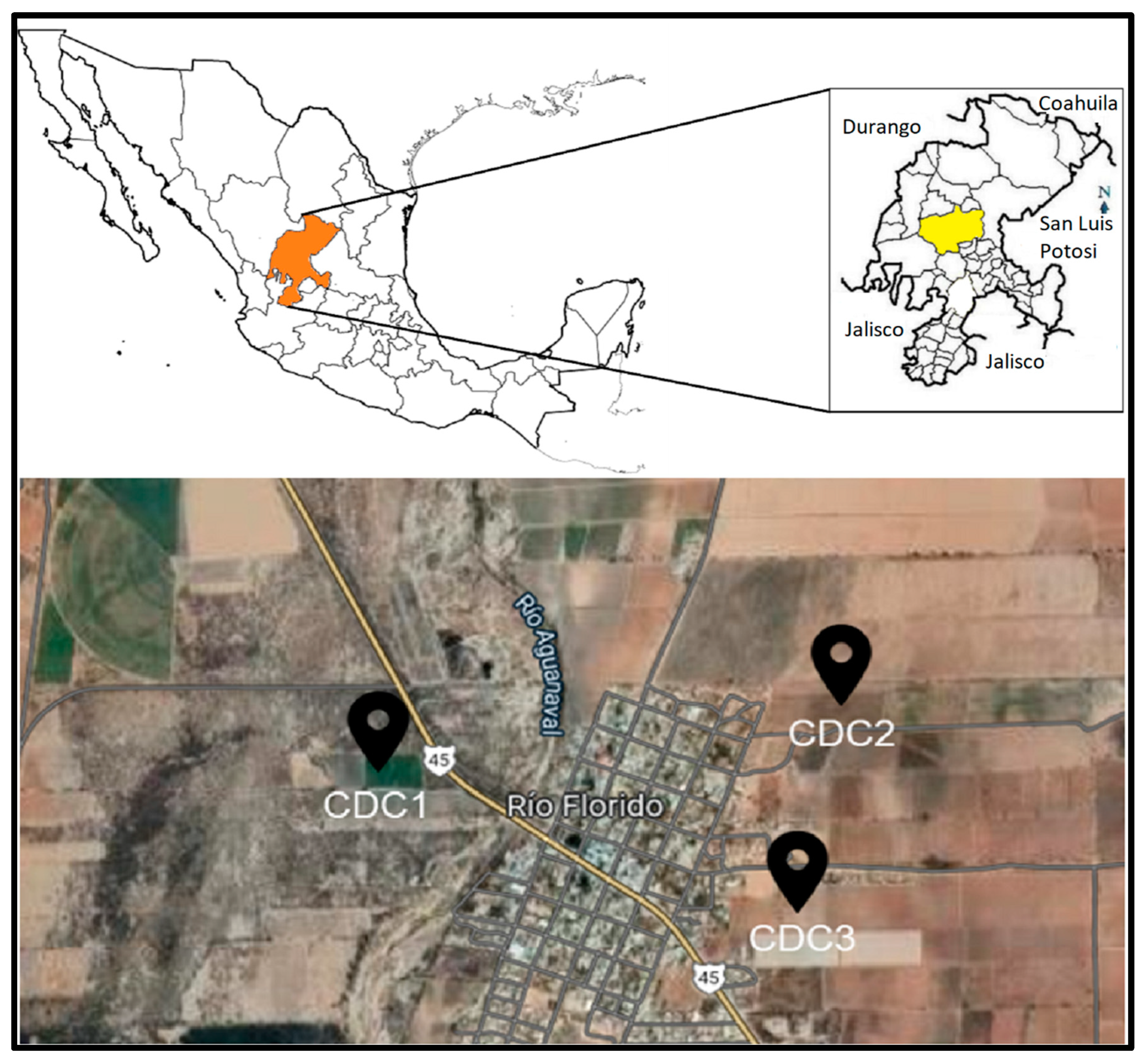
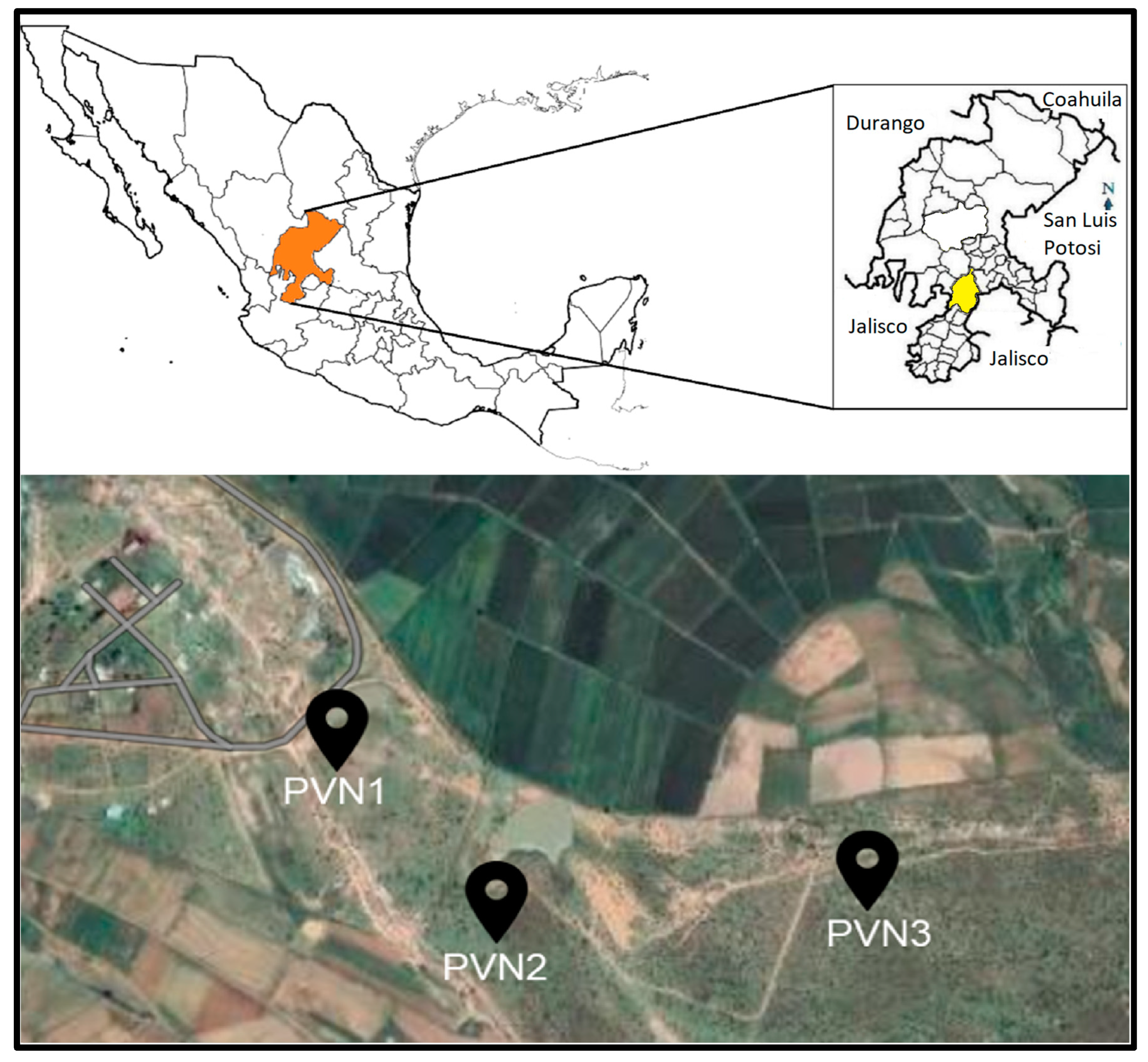
2.1. Study Site
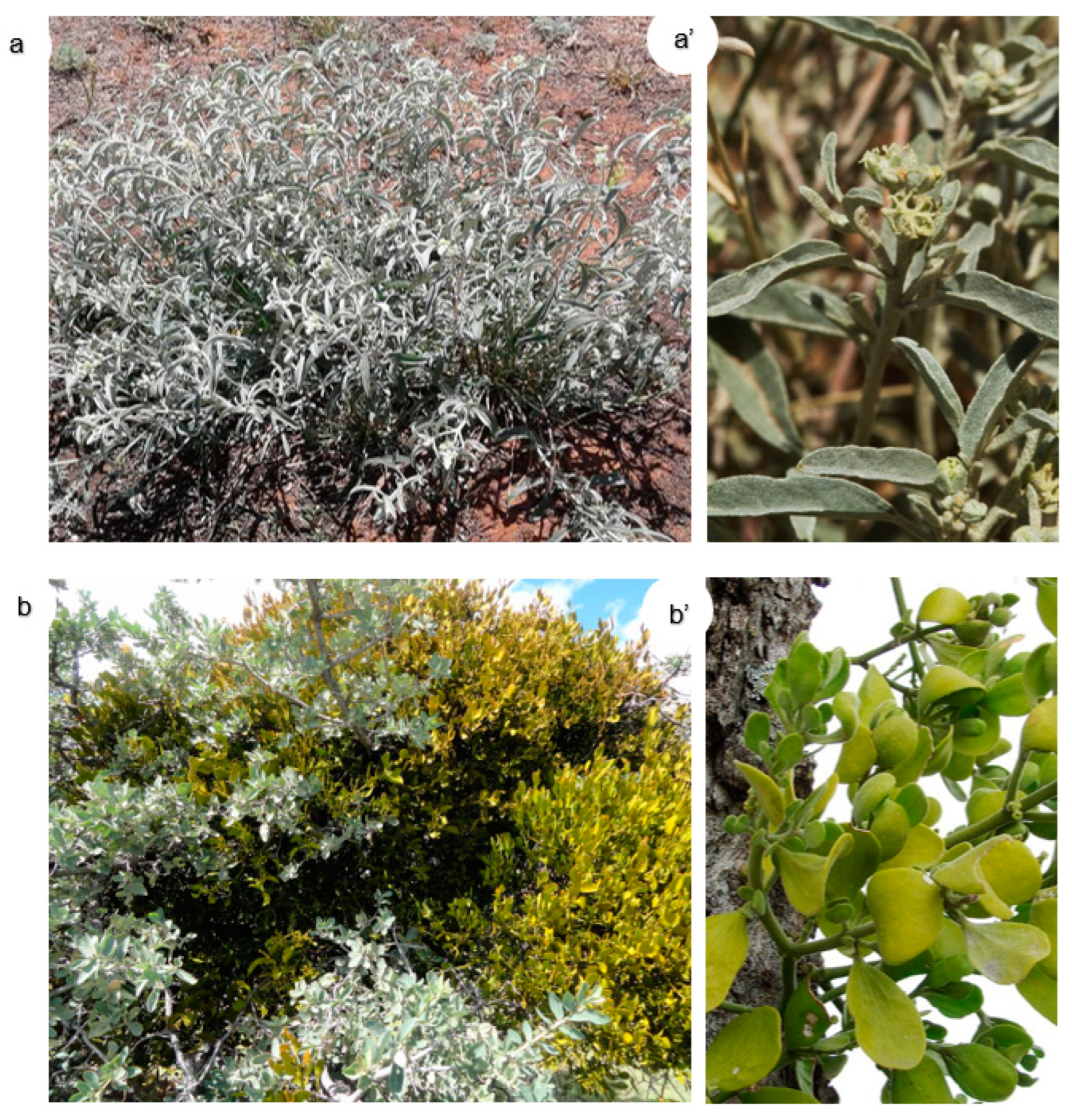
2.2. Plant Identification
2.3. Sample Preparation
2.4. Energy Dispersive X-ray Fluorescence
2.5. Quality Control
2.6. Statistical Analysis
2.7. Ethics Approval and Consent for Publication
2.8. Research and Publication Ethics
3. Results and Discussion
3.1. Elemental Analysis
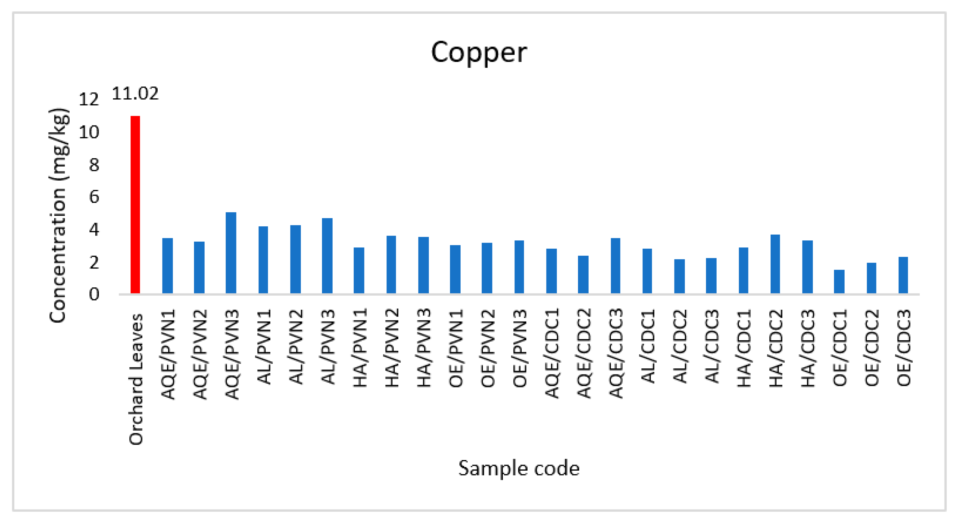
3.1.1. Copper (Cu)
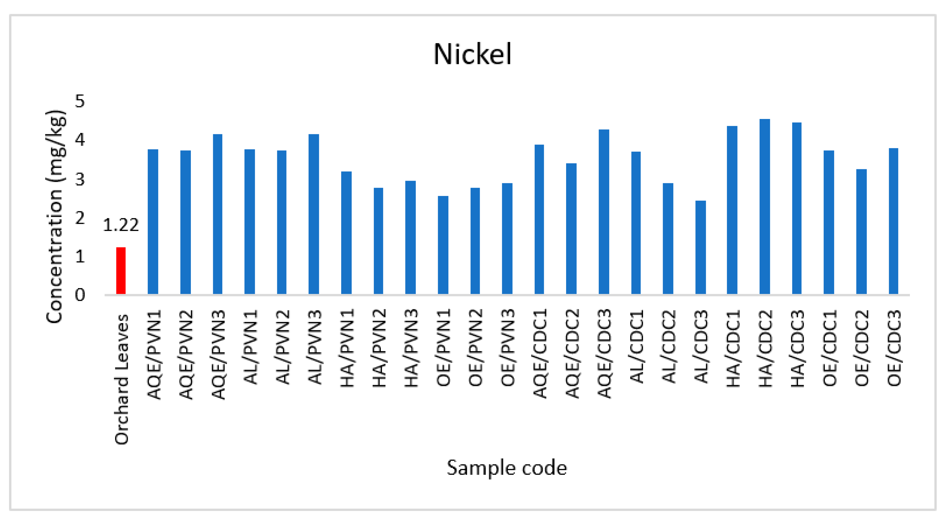
3.1.2. Nickel (Ni)
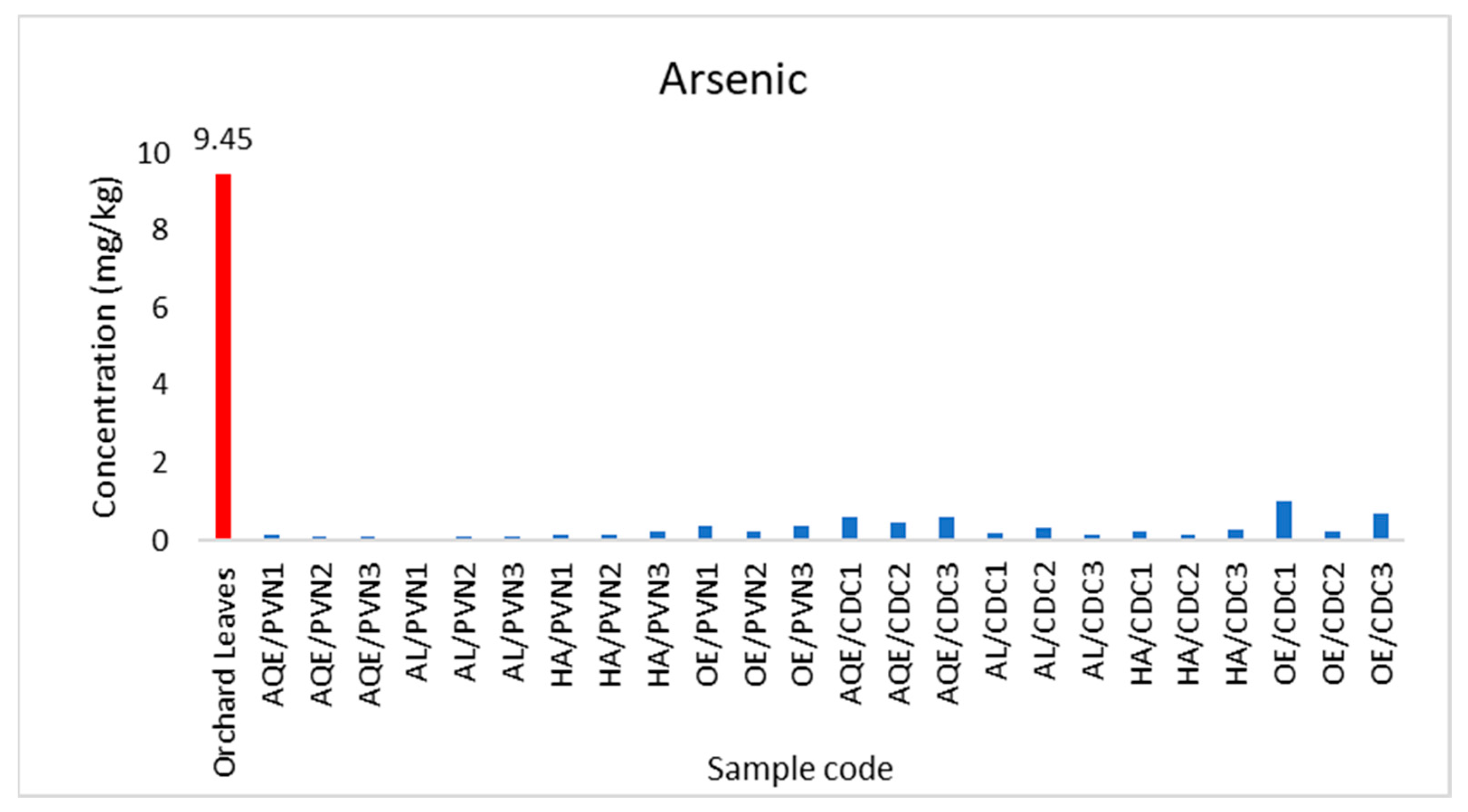
3.1.3. Arsenic (As)
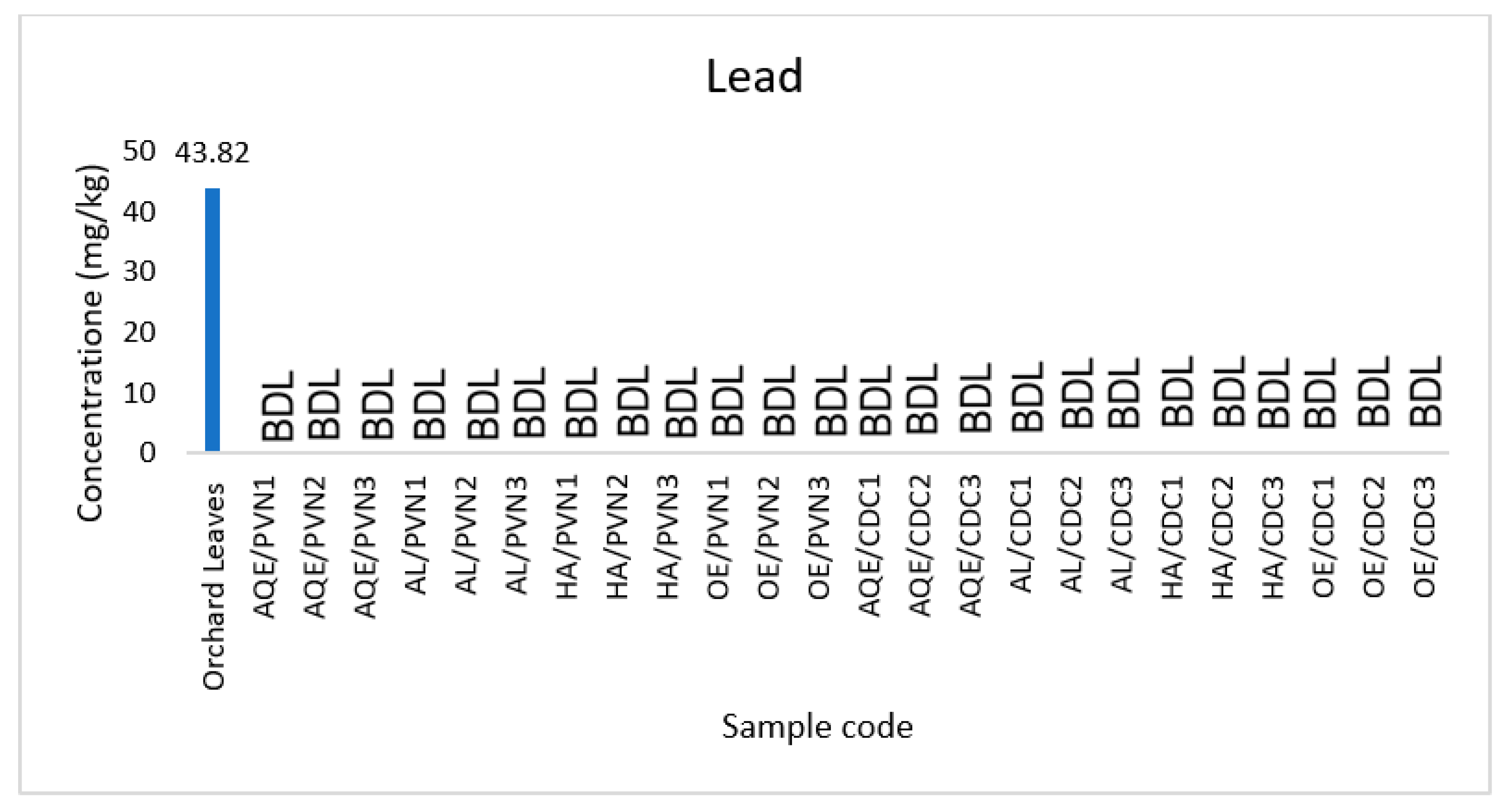
3.1.4. Lead (Pb)
3.2. Extract Analyses
4. Conclusions
Funding
Institutional Review Board Statement
Informed Consent Statement
Data Availability Statement
Conflicts of Interest
References
- Abollino, O.; Aceto, M.; Malandrino, M.; Mentasti, E.; Sarzanini, C.; Barberis, R. Distribution and mobility of metals in contaminated sites. Chemometric investigation of pollutant profiles. Environ. Pollut. 2002, 119, 177–193. [Google Scholar] [CrossRef]
- Chuparina, E.v.; Aisueva, T.S. Determination of heavy metal levels in medicinal plant Hemerocallis minor Miller by X-ray fluorescence spectrometry. Environ. Chem. Lett. 2011, 9, 19–23. [Google Scholar] [CrossRef]
- Li, W.; Niu, N.; Guo, N.; Zhou, H.; Bu, J.; Ding, A. Comparative Study on the Determination of heavy metals in Soil by XRF and ICP-MS. J. Phys. Conf. Ser. 2021, 2009, 012075. [Google Scholar] [CrossRef]
- Lucho-Constantino, C.A.; Álvarez-Suárez, M.; Beltrán-Hernández, R.I.; Prieto-García, F.; Poggi-Varaldo, H.M. A multivariate analysis of the accumulation and fractionation of major and trace elements in agricultural soils in Hidalgo State, Mexico irrigated with raw wastewater. Environ. Int. 2005, 31, 313–323. [Google Scholar] [CrossRef]
- Revenko, A.G. X-ray Fluorescence Analysis: State-of-the-Art and Trends of Development. Ind. Lab. Diagn. Mater. 2000, 66, 637–652. [Google Scholar]
- Moor, C.; Lymberopoulou, T.; Dietrich, V.J. Determination of Heavy Metals in Soils, Sediments and Geological Materials by ICP-AES and ICP-MS. Microchim. Acta 2001, 136, 123–128. [Google Scholar] [CrossRef] [Green Version]
- Stevens, D.P.; McLaughlin, M.J.; Heinrich, T. Determining Toxicity of Lead and Zinc Runoff in Soils: Salinity Effects on Metal Partitioning and on Phytotoxicity. Environ. Toxicol. Chem. 2003, 22, 3017. [Google Scholar] [CrossRef] [PubMed]
- Yan, X.; An, J.; Yin, Y.; Gao, C.; Wang, B.; Wei, S. Heavy metals uptake and translocation of typical wetland plants and their ecological effects on the coastal soil of a contaminated bay in Northeast China. Sci. Total Environ. 2022, 803, 149871. [Google Scholar] [CrossRef]
- Bowie, S.H.; Thornton, I. Environmental Geochemistry and Health: Report to the Royal Society’s British National Committee for Problems of the Environment; Springer Science & Business Media: Berlin/Heidelberg, Germany, 2012; Volume 2. [Google Scholar]
- Hooda, P. (Ed.) Trace Elements in Soils; Wiley: Hoboken, NJ, USA, 2010. [Google Scholar] [CrossRef]
- Kabata-Pendias, A. Trace Elements in Soils and Plants; CRC Press: Boca Raton, FL, USA, 2000. [Google Scholar] [CrossRef]
- Mijovilovich, A.; Morina, F.; Bokhari, S.N.; Wolff, T.; Küpper, H. Analysis of trace metal distribution in plants with lab-based microscopic X-ray fluorescence imaging. Plant Methods 2020, 16, 82. [Google Scholar] [CrossRef]
- Rodríguez Herrera, D. Occupational poisoning due to heavy metals. MEDISAN 2017, 21, 3372–3385. [Google Scholar]
- Romero, F.R.; Gutiérrez Ruíz, M. Estudio comparativo de la peligrosidad de jales en dos zonas mineras localizadas en el sur y centro de México. Bol. Soc. Geol. Mex. 2010, 62, 43–53. [Google Scholar] [CrossRef]
- Shah, A.; Niaz, A.; Ullah, N.; Rehman, A.; Akhlaq, M.; Zakir, M.; Suleman Khan, M. Comparative Study of Heavy Metals in Soil and Selected Medicinal Plants. J. Chem. 2013, 2013, 621265. [Google Scholar] [CrossRef]
- Wazir, S.M.; Saima, S.; Dasti, A.A.; Subhan, M. Ethnobotanical importance of salt range species of district Karak, Pakistan. Pak. J. Plant Sci. 2009, 13, 27–29. [Google Scholar]
- Zamir, R.; Islam, N.; Faruque, A. Comparison of Toxic Metal Concentrations in Antidiabetic Herbal Preparations (ADHPs) Available in Bangladesh Using AAS and XRF Analytical Tools. Sci. World J. 2019, 2019, 7154984. [Google Scholar] [CrossRef] [PubMed]
- Mireles, F.; Davila, J.I.; Pinedo, J.L.; Reyes, E.; Speakman, R.J.; Glascock, M.D. Assessing urban soil pollution in the cities of Zacatecas and Guadalupe, Mexico by instrumental neutron activation analysis. Microchem. J. 2012, 103, 158–164. [Google Scholar] [CrossRef]
- Pagnanelli, F.; Moscardini, E.; Giuliano, V.; Toro, L. Sequential extraction of heavy metals in river sediments of an abandoned pyrite mining area: Pollution detection and affinity series. Environ. Pollut. 2004, 132, 189–201. [Google Scholar] [CrossRef]
- Yáñez, L.; García-Nieto, E.; Rojas, E.; Carrizales, L.; Mejía, J.; Calderón, J.; Razo, I.; Díaz-Barriga, F. DNA damage in blood cells from children exposed to arsenic and lead in a mining area. Environ. Res. 2003, 93, 231–240. [Google Scholar] [CrossRef]
- Fernández Bremauntz, A.; Yarto Ramírez, M.; Castro Díaz, J. Las Sustancias Tóxicas Persistentes en México; Instituto Nacional de Ecologia: Mexico City, Mexico, 2005. [Google Scholar]
- Londoño Franco, L.F.; Londoño Muñoz, P.T.; Muñoz Garcia, F.G. Los Riesgos de Los Metales Pesados en la Salud Humana y Animal. Biotecnol. Sect. Agropecu. Agroind. 2016, 14, 145. [Google Scholar] [CrossRef]
- Markert, B. The Biological System of the Elements (BSE) for terrestrial plants (glycophytes). Sci. Total Environ. 1994, 155, 221–228. [Google Scholar] [CrossRef]
- Ekinci, N.; Ekinci, R.; Polat, R.; Budak, G. Analysis of trace elements in medicinal plants with energy dispersive X-ray fluorescence. J. Radioanal. Nucl. Chem. 2004, 260, 127–131. [Google Scholar] [CrossRef]
- Grover, J.K.; Yadav, S.; Vats, V. Medicinal plants of India with anti-diabetic potential. J. Ethnopharmacol. 2002, 81, 81–100. [Google Scholar] [CrossRef]
- Khan, I.; Ali, J.; Tullah, H. Heavy metals determination in medicinal plant Withania somnifera growing in various areas of peshawar, NWFP, Pakistan. J. Chem. Soc. Pak. 2008, 30, 69. [Google Scholar]
- Zhou, Z.-Y.; Fan, Y.-P.; Wang, M.-J. Heavy Metal Contamination in Vegetables and Their Control in China. Food Rev. Int. 2000, 16, 239–255. [Google Scholar] [CrossRef]
- Bonanno, G.; Borg, J.A.; di Martino, V. Levels of heavy metals in wetland and marine vascular plants and their biomonitoring potential: A comparative assessment. Sci. Total Environ. 2017, 576, 796–806. [Google Scholar] [CrossRef]
- Campbell, W.C. Energy-dispersive X-ray emission analysis. A. review. Analyst 1979, 104, 177. [Google Scholar] [CrossRef]
- Jyothsna, S.; Manjula, G.; Suthari, S.; Nageswara Rao, A.S. Qualitative elemental analysis of selected potential anti-asthmatic medicinal plant taxa using EDXRF technique. Heliyon 2020, 6, e03260. [Google Scholar] [CrossRef]
- Potts, P.J.; Ellis, A.T.; Kregsamer, P.; Marshall, J.; Streli, C.; West, M.; Wobrauschek, P. Atomic spectrometry update. X-ray fluorescence spectrometry. J. Anal. At. Spectrom. 2004, 19, 1397. [Google Scholar] [CrossRef]
- Carvalho, M.L.; Ferreira, J.G.; Amorim, P.; Marques, M.I.M.; Ramos, M.T. Study of heavy metals and other elements in macrophyte algae using energy-dispersive x-ray fluorescence. Environ. Toxicol. Chem. 1997, 16, 807–812. [Google Scholar] [CrossRef]
- Rindby, A. Software for energy-dispersive X-ray fluorescence. X-ray Spectrom. 1989, 18, 113–118. [Google Scholar] [CrossRef]
- World Health Organization. Quality Control Methods for Medicinal Plant Materials; World Health Organization: Geneva, Switzerland, 1998.
- World Health Organization. Directrices de la OMS Sobre Buenas Prácticas Agrícolas y de Recolección (BPAR) de Plantas Medicinales; World Health Organization: Geneva, Switzerland, 2003.
- WHO/FAO. JOINT FAO/WHO Food Standards Programme Codex Alimentarius Commission Report of the Fifty-Seventh Session of the Executive Committee of the Codex Alimentarius Commission; FAO: Rome, Italy, 2006.
- Yatkin, S.; Gerboles, M.; Borowiak, A. Evaluation of standardless EDXRF analysis for the determination of elements on PM10 loaded filters. Atmos. Environ. 2012, 54, 568–582. [Google Scholar] [CrossRef]


| Element | EDXRFS | |
|---|---|---|
| Equation [m] = cps/(mg/kg), [b] = cps | Regression | |
| Cu | y = 0.666x + 0.011 (±0.032) | 0.996 |
| Pb | y = 0.499x + 0.064 (±0.028) | 0.997 |
| As | y = 0.633x + 0.001 (±0.001) | 0.999 |
| Ni | y = 0.700x + 0.077 (±0.059) | 0.999 |
| Element (Certified SRM 1571) | Certified Value | Mean Measured Values | Recovery (%) | Accuracy (%) |
|---|---|---|---|---|
| Cu | 12 | 11.02 ± 0.70 | 91.86 | 2.14 |
| Pb | 45 | 43.82 ± 1.60 | 97.38 | 4.72 |
| As | 10 | 9.45 ± 0.48 | 94.54 | 2.23 |
| Ni | 1.3 | 1.22 ± 0.10 | 94.38 | 2.45 |
| Plant Name/Family | Part Used | Common Name | Medicinal Properties |
|---|---|---|---|
| CDC | Whole plant | Suapatle | Hypoglycemic, purgative, anti-inflammatory |
| PVN | Whole plant | Oak graft | Hypoglycemic, diuretic, lung problems |
| Metal | Limit of Detection LOD (mg/Kg) | Limit of Quantitation LOQ (mg/Kg) | Health-Criteria Levels (mg/Kg) | Tolerable Daily Intake (mg/50–60 kg bw) |
|---|---|---|---|---|
| Cu | 0.36 | 1.09 | 10 | 3 |
| Ni | 0.62 | 1.90 | 1.5 | 1.4 |
| As | 0.01 | 0.04 | 10 | 1 |
| Pb | 0.41 | 1.26 | 10 | 0.25 |
| Sample Code | Cu (mg/kg) | SD | As (mg/kg) | SD | Ni (mg/kg) | SD | Pb (mg/kg) |
|---|---|---|---|---|---|---|---|
| PVN1 | 107.59 * | 0.84 * | 11.18 * | 0.34 * | 148.38 * | 1.71 * | BDL |
| PVN2 | 100.48 * | 0.97 * | 14.79 * | 0.32 * | 150.43 * | 1.80 * | BDL |
| PVN3 | 101.56 * | 0.72 * | 14.45 * | 0.29 * | 149.7 * | 1.76 * | BDL |
| AQE/PVN1 | 3.48 | 0.29 | 0.14 | 0.04 | 3.75 | 0.13 | BDL |
| AQE/PVN2 | 3.26 | 0.27 | 0.08 | 0.02 | 3.74 | 0.12 | BDL |
| AQE/PVN3 | 5.1 | 0.24 | 0.1 | 0.04 | 4.14 | 0.13 | BDL |
| AL/PVN1 | 4.18 | 0.32 | 0.06 | 0.03 | 3.62 | 0.25 | BDL |
| AL/PVN2 | 4.25 | 0.25 | 0.07 | 0.02 | 3.14 | 0.22 | BDL |
| AL/PVN3 | 4.68 | 0.33 | 0.08 | 0.02 | 4.16 | 0.26 | BDL |
| HA/PVN1 | 2.9 | 0.36 | 0.16 | 0.02 | 3.2 | 0.38 | BDL |
| HA/PVN2 | 3.6 | 0.22 | 0.12 | 0.02 | 2.78 | 0.23 | BDL |
| HA/PVN3 | 3.57 | 0.28 | 0.2 | 0.02 | 2.96 | 0.24 | BDL |
| OE/PVN1 | 3.05 | 0.22 | 0.36 | 0.03 | 2.56 | 0.22 | BDL |
| OE/PVN2 | 3.18 | 0.18 | 0.24 | 0.05 | 2.77 | 0.23 | BDL |
| OE/PVN3 | 3.36 | 0.15 | 0.38 | 0.05 | 2.89 | 0.17 | BDL |
| WHO’s permissible limit | 10 | 10 | 1.5 | 10 |
| Sample Code | Cu (mg/kg) | SD | As (mg/kg) | SD | Ni (mg/kg) | SD | Pb (mg/kg) |
|---|---|---|---|---|---|---|---|
| CDC1 | 76.18 * | 0.21 * | 14.79 * | 0.01 * | 102.78 * | 1.84 * | BDL |
| CDC2 | 73.45 * | 0.31 * | 17.61 * | 0.01 * | 108.69 * | 1.8 * | BDL |
| CDC3 | 75.19 * | 0.28 * | 14.5 * | 0.04 * | 107.24 * | 1.73 * | BDL |
| AQE/CDC1 | 2.81 | 0.15 | 0.6 | 0.04 | 3.9 | 0.1 | BDL |
| AQE/CDC2 | 2.41 | 0.38 | 0.47 | 0.04 | 3.39 | 0.16 | BDL |
| AQE/CDC3 | 3.5 | 0.3 | 0.58 | 0.03 | 4.27 | 0.15 | BDL |
| AL/CDC1 | 2.84 | 0.39 | 0.19 | 0.01 | 3.7 | 0.24 | BDL |
| AL/CDC2 | 2.18 | 0.26 | 0.34 | 0.05 | 2.9 | 0.27 | BDL |
| AL/CDC3 | 2.26 | 0.25 | 0.15 | 0.03 | 2.44 | 0.3 | BDL |
| HA/CDC1 | 2.94 | 0.33 | 0.23 | 0.02 | 4.37 | 0.39 | BDL |
| HA/CDC2 | 3.69 | 0.27 | 0.16 | 0.02 | 4.56 | 0.26 | BDL |
| HA/CDC3 | 3.34 | 0.38 | 0.28 | 0.01 | 4.45 | 0.31 | BDL |
| OE/CDC1 | 1.55 | 0.14 | 0.99 | 0.01 | 3.74 | 0.17 | BDL |
| OE/CDC2 | 1.93 | 0.21 | 0.22 | 0.03 | 3.25 | 0.15 | BDL |
| OE/CDC3 | 2.31 | 0.18 | 0.67 | 0.04 | 3.78 | 0.2 | BDL |
| WHO’s permissible limit | 10 | 10 | 1.5 | 10 |
Publisher’s Note: MDPI stays neutral with regard to jurisdictional claims in published maps and institutional affiliations. |
© 2022 by the authors. Licensee MDPI, Basel, Switzerland. This article is an open access article distributed under the terms and conditions of the Creative Commons Attribution (CC BY) license (https://creativecommons.org/licenses/by/4.0/).
Share and Cite
Sánchez-Lara, F.; Manzanares-Acuña, E.; Badillo-Almaraz, V.; Gutiérrez-Hernández, R.; García-Aguirre, K.K.; Vargas-Díaz, M.E.; Hernández-Rangel, Á.O.; Hernández-Sánchez, K.M.; Escobar-León, M.C. Comparative Study of Heavy Metals in Selected Medicinal Plants and Extracts, Using Energy Dispersive X-ray Fluorescence. Appl. Sci. 2022, 12, 11772. https://doi.org/10.3390/app122211772
Sánchez-Lara F, Manzanares-Acuña E, Badillo-Almaraz V, Gutiérrez-Hernández R, García-Aguirre KK, Vargas-Díaz ME, Hernández-Rangel ÁO, Hernández-Sánchez KM, Escobar-León MC. Comparative Study of Heavy Metals in Selected Medicinal Plants and Extracts, Using Energy Dispersive X-ray Fluorescence. Applied Sciences. 2022; 12(22):11772. https://doi.org/10.3390/app122211772
Chicago/Turabian StyleSánchez-Lara, Fernando, Eduardo Manzanares-Acuña, Valentín Badillo-Almaraz, Rosalinda Gutiérrez-Hernández, Karol Karla García-Aguirre, María Elena Vargas-Díaz, Álvaro Omar Hernández-Rangel, Karla Mariela Hernández-Sánchez, and Martha Celia Escobar-León. 2022. "Comparative Study of Heavy Metals in Selected Medicinal Plants and Extracts, Using Energy Dispersive X-ray Fluorescence" Applied Sciences 12, no. 22: 11772. https://doi.org/10.3390/app122211772