
Antibacterial Applications of Low-Pressure Plasma on Degradation of Multidrug Resistant V. cholera
 , , , and
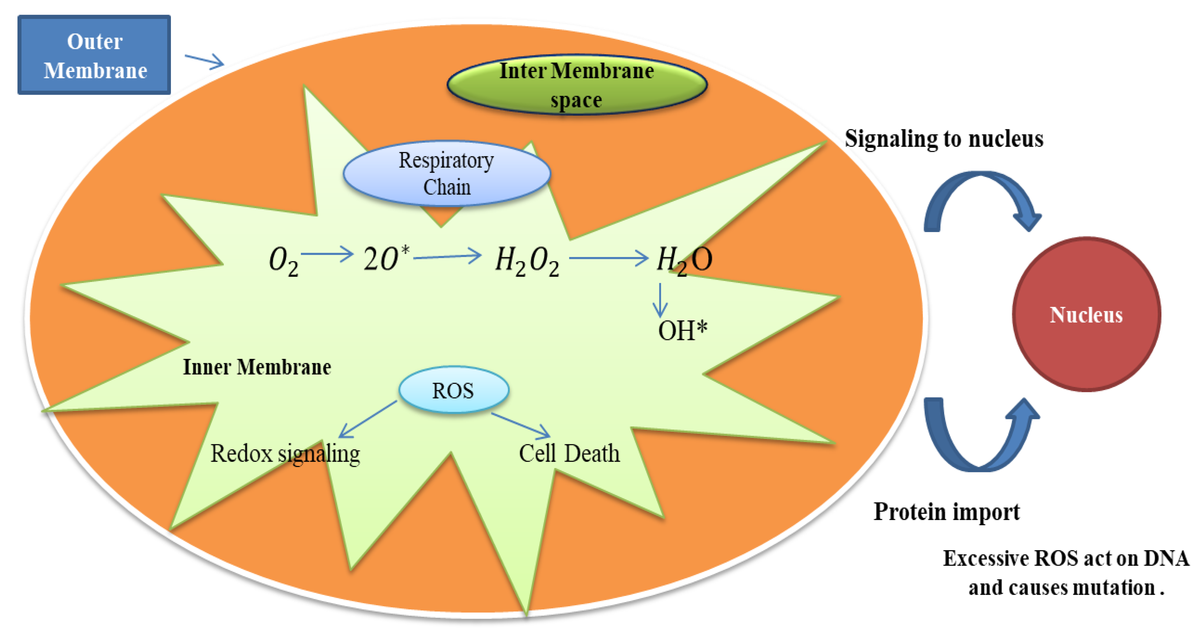
Abstract
:1. Introduction
2. Materials and Methods
2.1. Bacterial Strains Used and Growth Conditions
2.2. Bacterial Cell Survival Assay
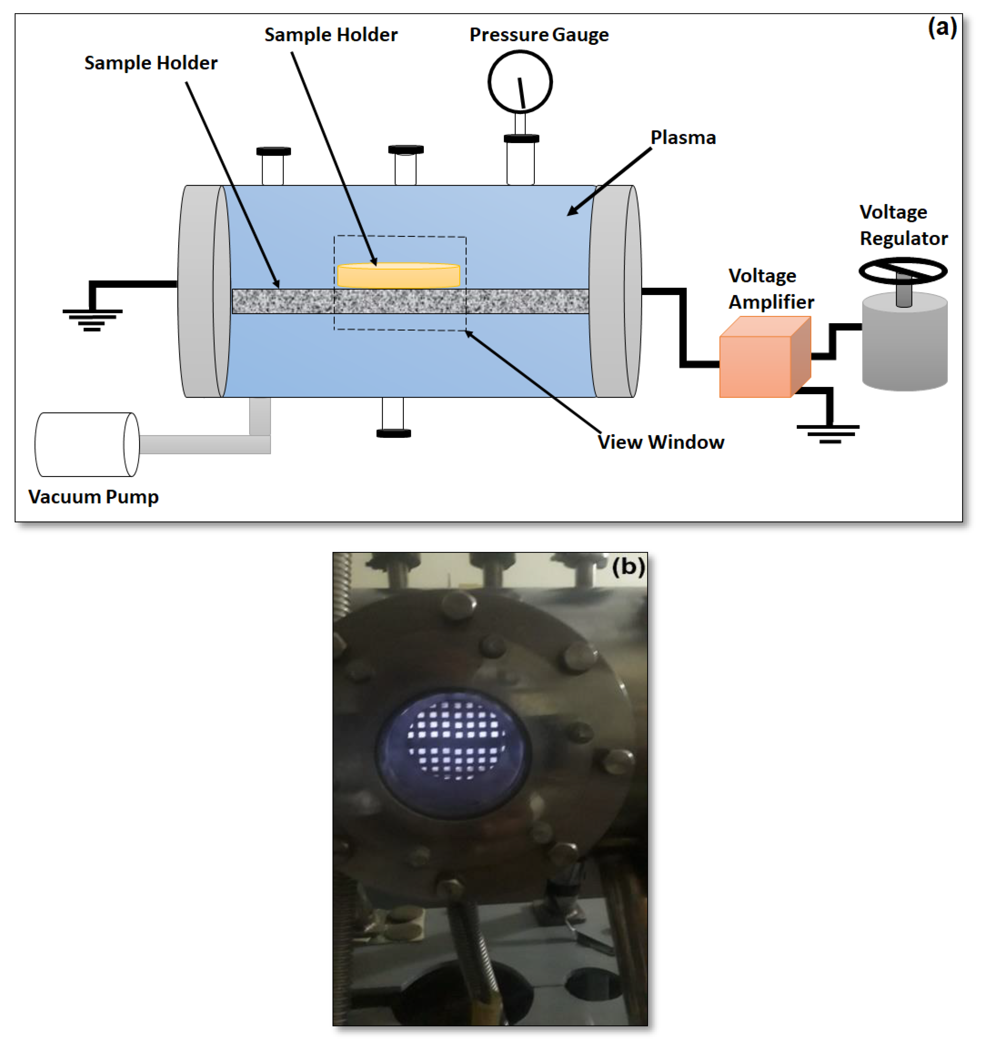
3. Experiment
4. Results and Discussion
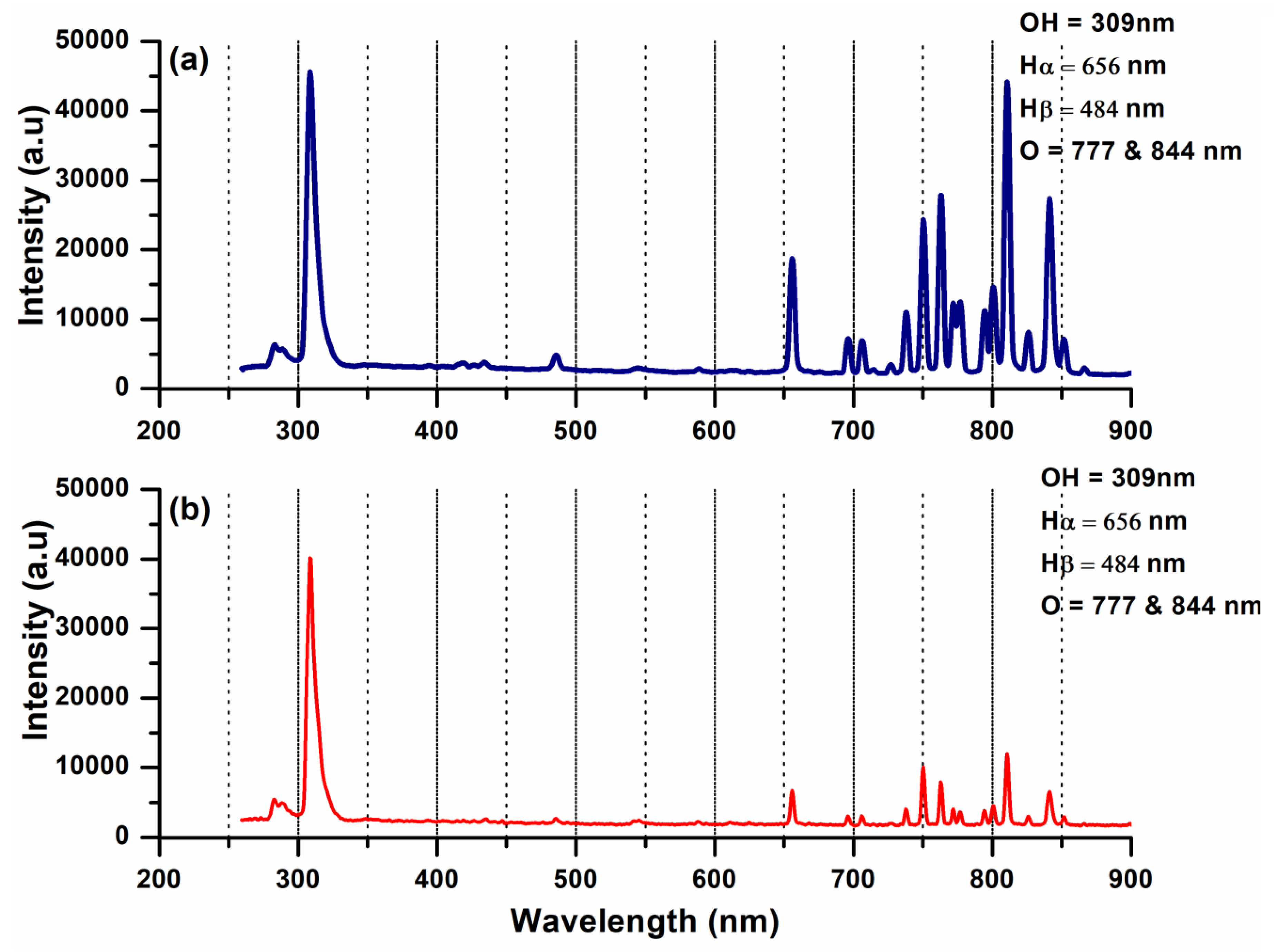
4.1. Spectral Analysis
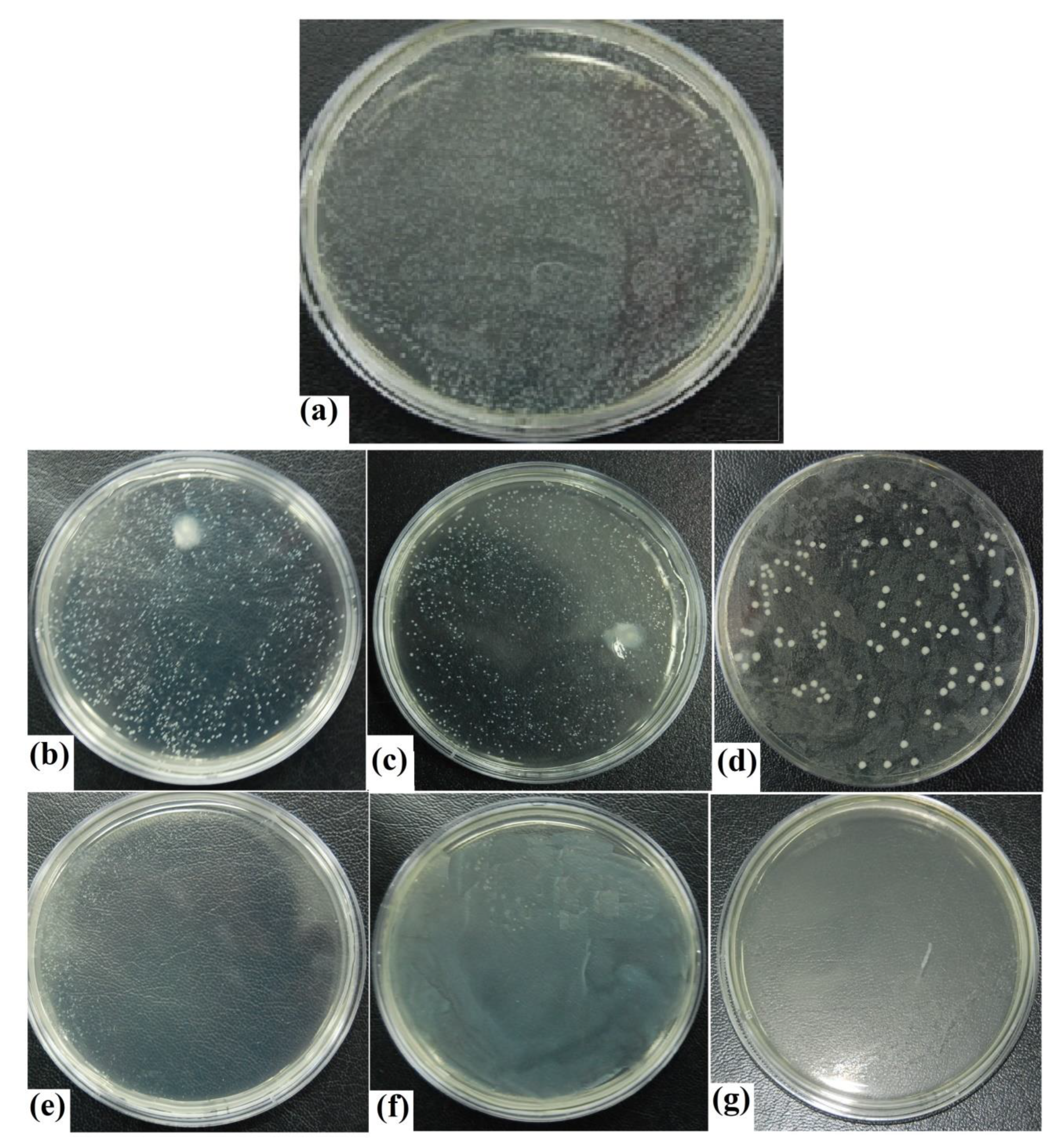
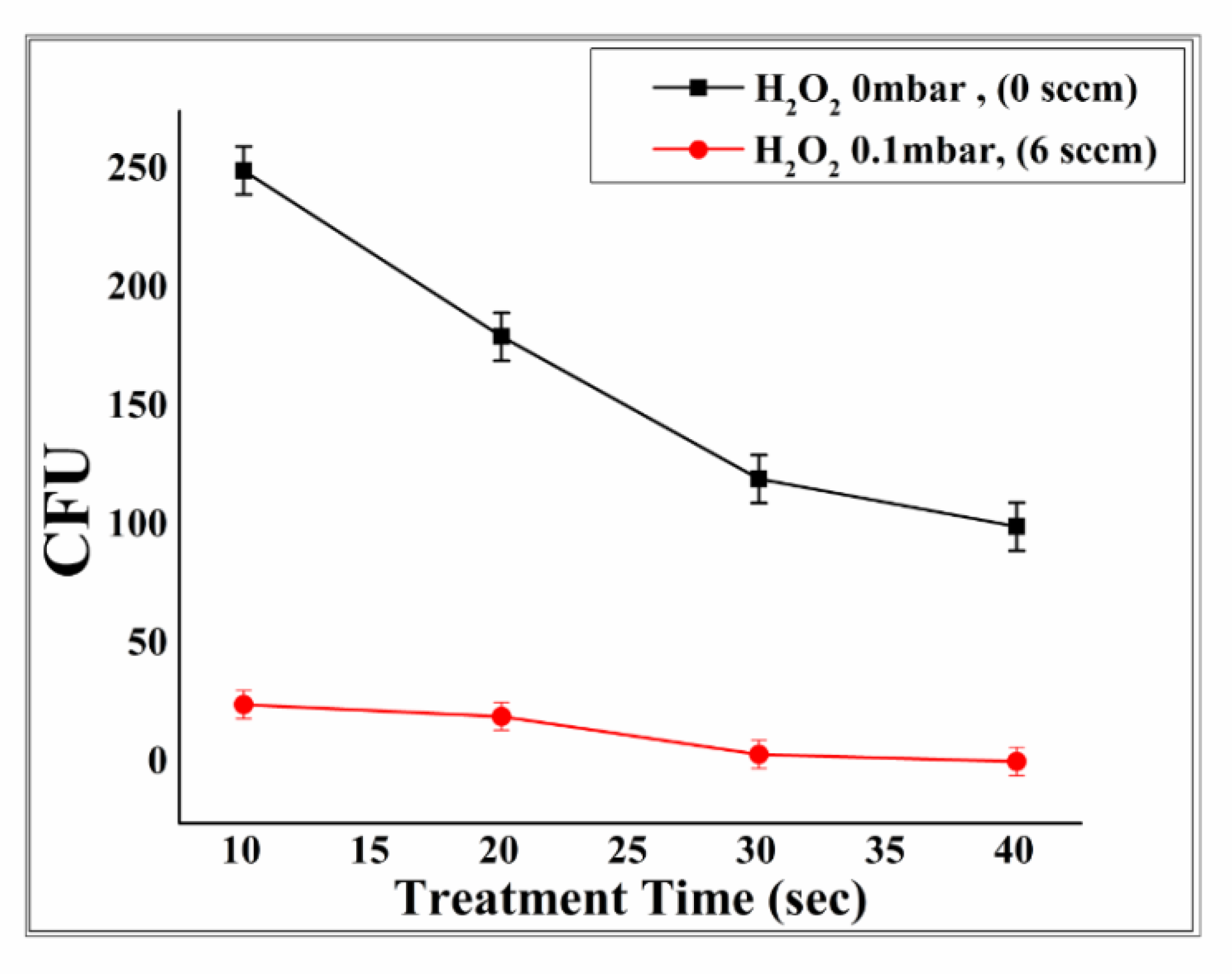
4.2. Inhabitation of Vibrio Cholera
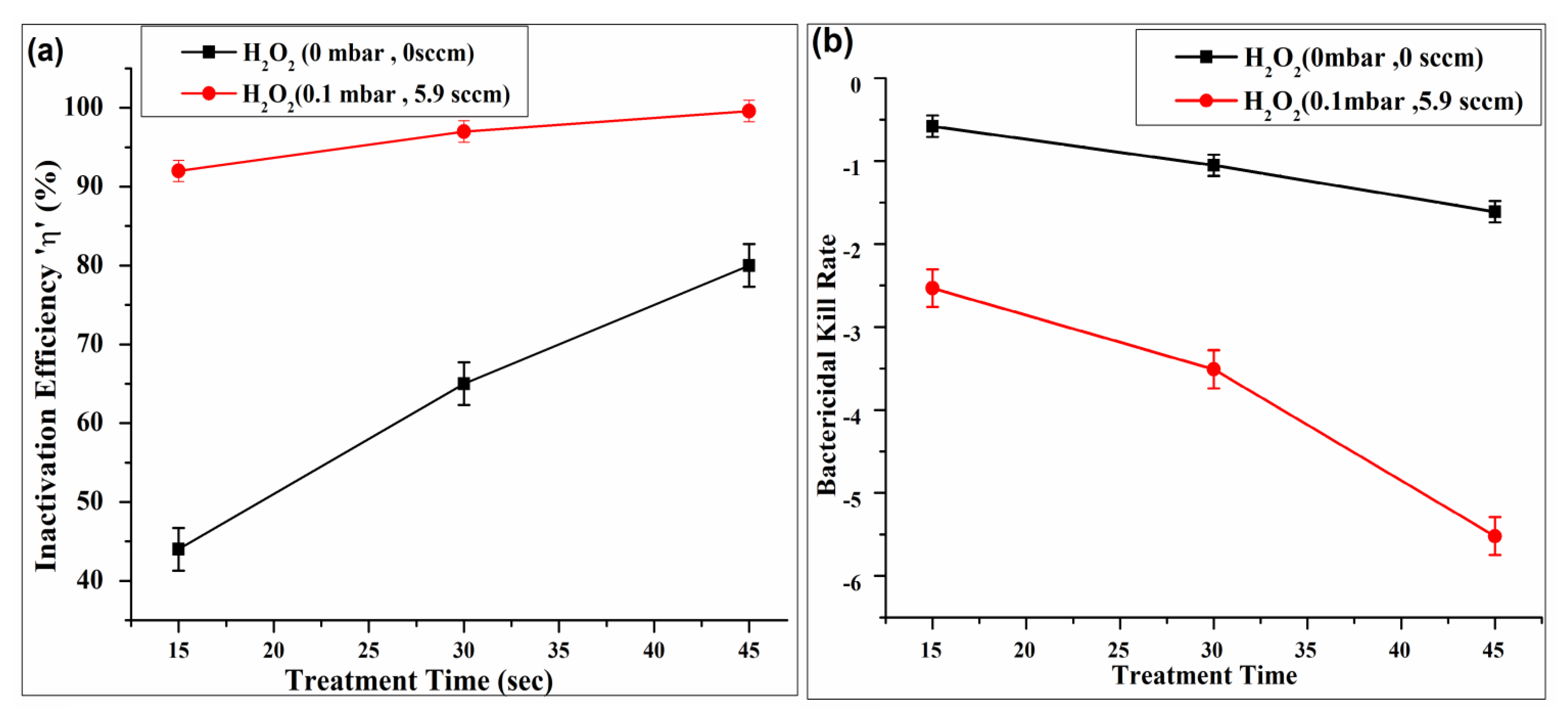
4.3. Inactivation Efficiency and Kill Rate
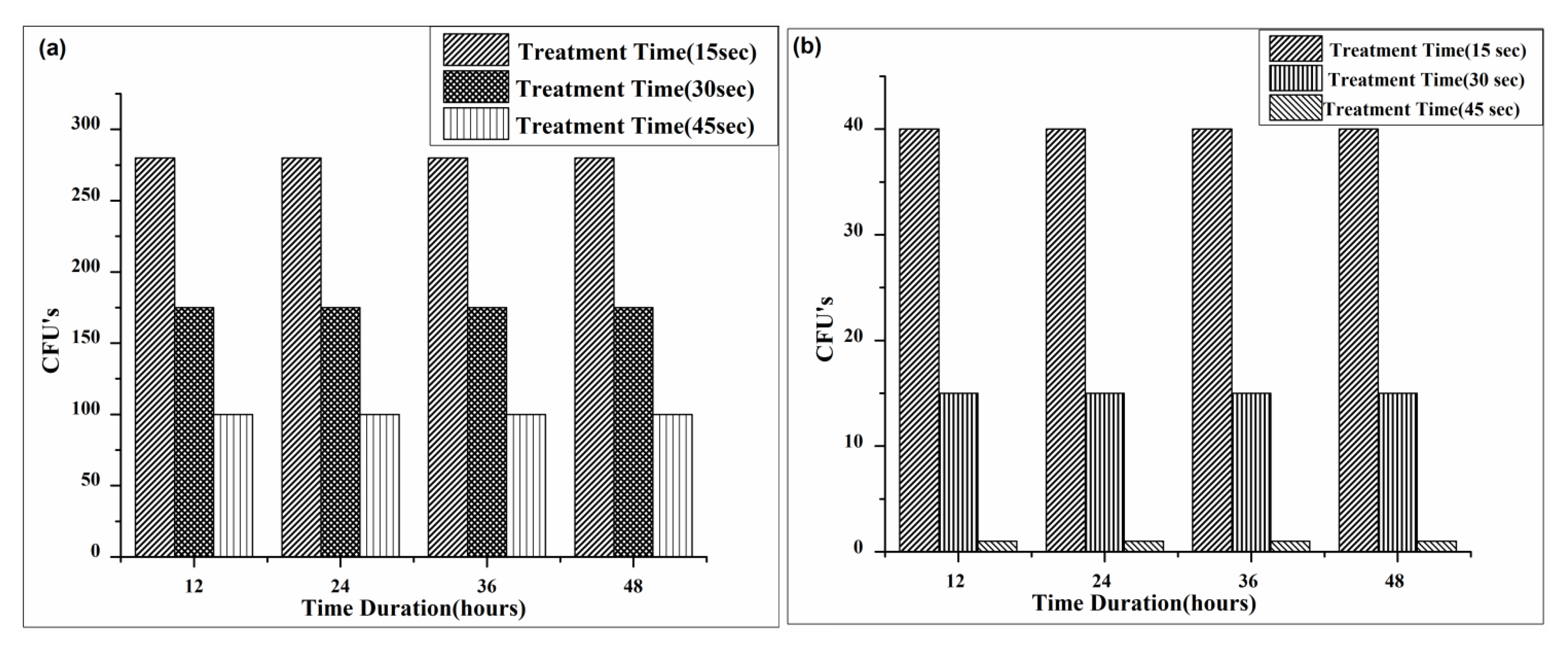
4.4. Bactericidal Retardant Effect
5. Conclusions
Author Contributions
Funding
Institutional Review Board Statement
Informed Consent Statement
Data Availability Statement
Acknowledgments
Conflicts of Interest
References
- Sakudo, A.; Yagyu, Y.; Onodera, T. Disinfection and sterilization using plasma technology: Fundamentals and future perspectives for biological applications. Int. J. Mol. Sci. 2019, 20, 5216. [Google Scholar] [CrossRef]
- Gao, L.; Shi, X.; Wu, X. Applications and challenges of low temperature plasma in pharmaceutical field. J. Pharm. Anal. 2021, 11, 28–36. [Google Scholar] [CrossRef]
- Vatansever, F.; de Melo, W.C.; Avci, P.; Vecchio, D.; Sadasivam, M.; Gupta, A.; Hamblin, M.R. Antimicrobial strategies centered around reactive oxygen species–bactericidal antibiotics, photodynamic therapy, and beyond. FEMS Microbiol. Rev. 2013, 37, 955–989. [Google Scholar] [CrossRef]
- Hirst, A.M.; Frame, F.M.; Maitland, N.J.; O’Connell, D. Low Temperature Plasma Causes Double-Strand Break DNA Damage in Primary Epithelial Cells Cultured from a Human Prostate Tumour. Plasma Sci. IEEE Trans. 2014, 42, 2740–2741. [Google Scholar] [CrossRef]
- Abdal Dayem, A.; Hossain, M.K.; Lee, S.B.; Kim, K.; Saha, S.K.; Yang, G.M.; Cho, S.G. The role of reactive oxygen species (ROS) in the biological activities of metallic nanoparticles. Int. J. Mol. Sci. 2017, 18, 120. [Google Scholar] [CrossRef]
- Lerouge, S.; Tabrizian, M.; Wertheimer, M.R.; Marchand, R.; Yahia, L. Safety of plasma-based sterilization: Surface modifications of polymeric medical devices induced by Sterrad® and Plazlyte™ processes. Bio-Med. Mater. Eng. 2002, 12, 3–13. [Google Scholar]
- Pellerin, S.; Cormier, J.M.; Richard, F.; Musiol, K.; Chapelle, J. A spectroscopic diagnostic method using UV OH band spectrum. J. Phys. D Appl. Phys. 1996, 29, 726–739. [Google Scholar] [CrossRef]
- Das, K.; Roychoudhury, A. Reactive oxygen species (ROS) and response of antioxidants as ROS-scavengers during environmental stress in plants. Front. Environ. Sci. 2014, 2, 00053. [Google Scholar] [CrossRef]
- Fridman, G.; Shereshevsky, A.; Jost, M.M.; Brooks, A.D.; Fridman, A.; Gutsol, A.; Vasilets, V.; Friedman, G. Blood Coagulation and Living Tissue Sterilization by Floating-Electrode Dielectric Barrier Discharge in Air. Plasma Chem. Plasma Process. 2007, 27, 163–176. [Google Scholar] [CrossRef]
- Ishaq, M.; Kumar, S.; Varinli, H.; Han, Z.J.; Rider, A.E.; Evans, M.D.; Murphy, A.B.; Ostrikov, K. Atmospheric gas plasma–induced ROS production activates TNF-ASK1 pathway for the induction of melanoma cancer cell apoptosis. Mol. Biol. Cell 2014, 25, 1523–1531. [Google Scholar] [CrossRef]
- Morais, D.S.; Guedes, R.M.; Lopes, M.A. Antimicrobial approaches for textiles: From research to market. Materials 2016, 21, 498. [Google Scholar] [CrossRef]
- Joshi, R.P.; Thagard, S.M. Streamer-Like Electrical Discharges in Water: Part II—Environmental Applications. Environ. Plasma 2013, 33, 17–49. [Google Scholar] [CrossRef]
- WHO. Disease Outbreak News, Cholera—Algeria; WHO: Geneva, Switzerland, 2018. [Google Scholar]
- Radha, K.R.; Dharmaraj, K.; Ranjitha, K.B.D. A comparative study on the physicochemical and bacterial analysis of drinking, borewell and sewage water in the three diferent places of Sivakasi. J. Environ. Biol. 2007, 28, 105–108. [Google Scholar]
- Calfee, M.W.; Wendling, M. Inactivation of vegetative bacterial threat agents on environmental surfaces. Sci. Total Environ. 2013, 443, 387–396. [Google Scholar] [CrossRef]
- Osei-Asare, C.; Oppong, E.E.; Apenteng, J.A.; Adi-Dako, O.; Kumadoh, D.; Akosua, A.A.; Ohemeng, K.A. Managing Vibrio cholerae with a local beverage: Preparation of an affordable ethanol based hand sanitizer. Heliyon 2020, 6, e03105. [Google Scholar] [CrossRef]
- Collivignarelli, M.C.; Abbà, A.; Miino, M.C.; Caccamo, F.M.; Torretta, V.; Rada, E.C.; Sorlini, S. Disinfection of Wastewater by UV-Based Treatment for Reuse in a Circular Economy Perspective. Where Are We at? Int. J. Environ. Res. Public Health 2021, 18, 77. [Google Scholar] [CrossRef]
- Marugán, J.; Giannakis, S.; McGuigan, K.G.; Polo-López, I. Solar disinfection as a water treatment technology. In Clean Water and Sanitation; Springer: Cham, Switzerland, 2020; pp. 1–16. [Google Scholar]
- Giannakis, S. Analogies and differences among bacterial and viral disinfection by the photo-Fenton process at neutral pH: A mini review. Environ. Sci. Pollut. Res. 2018, 25, 27676–27692. [Google Scholar] [CrossRef]
- Lerouge, S.; Wertheimer, M.R.; Yahia, L.H. Plasma sterilization: A review of parameters, mechanisms, and limitations. Plasmas Polym. 2001, 6, 175–188. [Google Scholar] [CrossRef]
- Jhonson, J.L.; Lowel, B.C.; Lloyd, S.; Mc Collough, A.K. TAT-Mediated Delivery of a DNA Repair Enzyme to Skin Cells Rapidly Initiates Repair of UV-Induced DNA Damage. J. Investig. Dermatol. 2011, 131, 753–761. [Google Scholar] [CrossRef]
- Ahmed, M.W.; Yang, J.K.; Mok, Y.S.; Lee, H.J.; Yu, Y.H. Underwater capillary discharge with air and oxygen addition. Phys. Sci. 2014, 65, 1404–1413. [Google Scholar] [CrossRef]
- Abbasi, A.; Farooq, W.; Tag-ElDin, E.S.M.; Khan, S.U.; Khan, M.I.; Guedri, K.; Galal, A.M. Heat Transport Exploration for Hybrid Nanoparticle (Cu, Fe3O4)—Based Blood Flow via Tapered Complex Wavy Curved Channel with Slip Features. Micromachines 2022, 13, 1415. [Google Scholar] [CrossRef]
- Dastani, K.; Moghimi Zand, M.; Kavand, H.; Javidi, R.; Hadi, A.; Valadkhani, Z.; Renaud, P. Effect of input voltage frequency on the distribution of electrical stresses on the cell surface based on single-cell dielectrophoresis analysis. Sci. Rep. 2020, 10, 68. [Google Scholar] [CrossRef] [Green Version]


Publisher’s Note: MDPI stays neutral with regard to jurisdictional claims in published maps and institutional affiliations. |
© 2022 by the authors. Licensee MDPI, Basel, Switzerland. This article is an open access article distributed under the terms and conditions of the Creative Commons Attribution (CC BY) license (https://creativecommons.org/licenses/by/4.0/).
Share and Cite
Manzoor, N.; Qasim, I.; Khan, M.I.; Ahmed, M.W.; Guedri, K.; Bafakeeh, O.T.; Tag-Eldin, E.S.M.; Galal, A.M. Antibacterial Applications of Low-Pressure Plasma on Degradation of Multidrug Resistant V. cholera. Appl. Sci. 2022, 12, 9737. https://doi.org/10.3390/app12199737
Manzoor N, Qasim I, Khan MI, Ahmed MW, Guedri K, Bafakeeh OT, Tag-Eldin ESM, Galal AM. Antibacterial Applications of Low-Pressure Plasma on Degradation of Multidrug Resistant V. cholera. Applied Sciences. 2022; 12(19):9737. https://doi.org/10.3390/app12199737
Chicago/Turabian StyleManzoor, Nimra, Irfan Qasim, Muhammad Ijaz Khan, Muhammad Waqar Ahmed, Kamel Guedri, Omar T. Bafakeeh, El Sayed Mohamed Tag-Eldin, and Ahmed M. Galal. 2022. "Antibacterial Applications of Low-Pressure Plasma on Degradation of Multidrug Resistant V. cholera" Applied Sciences 12, no. 19: 9737. https://doi.org/10.3390/app12199737