Next Article in Journal
Important Features Selection and Classification of Adult and Child from Handwriting Using Machine Learning Methods
Next Article in Special Issue
International Symposium on New Frontiers in Reef Coral Biotechnology (5 May 2022, Taiwan)
Previous Article in Journal
Evaluation of Indirect-Heated Microwave Thermal Desorption Treatment on Engineering Properties of Lubricant-Contaminated Soil
 
 
Correction published on 21 December 2022, see Appl. Sci. 2023, 13(1), 38.
Article

Study on the Development and Growth of Coral Larvae

1
National Museum of Marine Biology & Aquarium, Pingtung 94450, Taiwan
2
Institute of Marine Biology, National Dong Hwa University, Pingtung 944401, Taiwan
3
Department of Aquaculture, National Taiwan Ocean University, Keelung 202301, Taiwan
4
Department of Marine Biotechnology and Resources, National Sun Yat-sen University, Kaohsiung 80424, Taiwan
5
Department of Post Modern Agriculture, Mingdao University, Chang Hua 503008, Taiwan
*
Authors to whom correspondence should be addressed.
These authors contributed equally to this work.
Appl. Sci. 2022, 12(10), 5255; https://doi.org/10.3390/app12105255
Submission received: 22 March 2022 / Revised: 26 April 2022 / Accepted: 19 May 2022 / Published: 23 May 2022 / Corrected: 21 December 2022
(This article belongs to the Special Issue New Frontiers in Reef Coral Biotechnology)

Abstract

:
Studies on the early development of corals are required for academic research on coral reefs and applied reef conservation, but the interval between observations is usually weeks or months. Thus, no study has comprehensively explored the development of coral larvae after settlement. This study observed Galaxea fascicularis, Mycedium elephantotus, Pocillopora verrucosa, and Seriatopora caliendrum larvae after settlement, including their growth process and the formation of tentacles, skeletons, and polyps. The G. fascicularis and M. elephantotus polyps exhibited the skeleton-over-polyp mechanism, whereas the P. verrucosa and S. caliendrum polyps exhibited the polyp-over-skeleton mechanism. During asexual reproduction, the Symbiodiniaceae species clustered on the coenosarc, resulting in polyp development and skeletal growth. M. Elephantotus was unique in that its tentacles were umbrella-shaped, and its polyp growth and Symbiodiniaceae species performance during asexual reproduction differed from those of the other three corals. Although both P. verrucosa and S. caliendrum have branching morphologies, their vertical development stages were dissimilar. S. caliendrum relied on the mutual pushing of individuals in the colony to extend upward, whereas P. verrucosa had a center individual that developed vertically. The findings of this study can serve as a reference for future research on coral breeding, growth, and health assessments.

1. Introduction

Most studies on corals have focused on developing methods to rapidly breed corals in large numbers [1]. Some methods involve the separation of coral colonies and their individual cultivation [2]. Although this strategy can increase coral numbers and reduce costs, it would cause a decline in their genetic diversity. Studies on the breeding of corals have investigated the developmental process and its molecular effects on coral sperm, eggs, and embryos [3,4]. Moreover, a study examined the effects of different stress and stimulation levels on the developmental process of corals [5]. A previous study investigated differences in cell differentiation and gene expression before and after the larval settlement of corals [6]. However, most studies have focused on developing methods to increase the settlement rate, survival rate after settlement, and growth rate of coral larvae; investigated the effects of environmental stress on these rates; and identified techniques to cultivate, collect, and release coral larvae in large quantities [7,8]. One study designed a method to increase the number of settled coral larvae and determined appropriate growth conditions and breeding techniques for coral larvae [2].
Environmental stress results from changes in marine factors required for survival, including sea temperature, salinity, and organic salt content [9]. Environmental stress inhibits sexual reproduction in corals because corals stop producing sperms and eggs, or break down cells and recycle them to produce the energy required to resist environmental stress [10]. The fertilization rate of coral eggs is low in highly turbid water, even after the successful release of gametes by corals [11]. In addition, high sea temperatures cause abnormalities in fertilized eggs, such as those related to the development of embryos and the settlement of coral larvae [12]. Factors related to organic matter in the ocean, chemicals released from fishing boats, sea temperature, and the marine environment directly affect coral larvae [13,14]. These factors can reduce the settlement rate and survival rate after settlement of larvae. Settled coral larvae are required to resist stress and compete with other species, particularly algae, until they grow to a certain size and become tolerant [15]. The growth process of coral larvae is considerably long. Studies examining reproduction in corals have focused mainly on the survival and growth of coral larvae. The time interval required between observations can range from weeks to months [16,17]. Moreover, with this temporal resolution, the developmental process of coral larvae after settlement cannot be precisely observed.
Coral larvae enter the benthic phase after their settlement. In the benthic phase, a series of changes occur that vary among species. Under normal conditions, corals are immobile after their settlement, and their growth, development, and survival depend on the surrounding environment. However, because of climate change and global warming, the marine environment has become increasingly unfavorable for corals. Under artificial breeding conditions, the growth environment of corals can be adequately maintained by eliminating harmful environmental factors. Data on the growth phases of coral larvae are required to evaluate their health and cultivation environment conditions. Numerous studies have investigated coral larvae in the planktonic phase. However, those studies have majorly focused on the collection and settlement of coral larvae and determining factors affecting their settlement [18,19,20,21]. However, whether corals can successfully grow can be determined in the benthic phase. Thus, exploring the development of corals during the benthic phase can provide information on their survival rate, expansion rate, gene expression, and physiological expression under environmental stress.
Most studies on corals have investigated methods used to rapidly breed corals in large numbers; however, the time interval required between observations can range from weeks to months. Thus, no study has comprehensively explored the development of coral larvae after settlement. This study examined the growth phases of settled coral larvae by evaluating the polyps and skeleton as well as the formation of tentacles and new individuals. The complete developmental process of settled coral larvae was documented to provide a practical reference for studies on coral breeding, disease, and health.

2. Materials and Methods

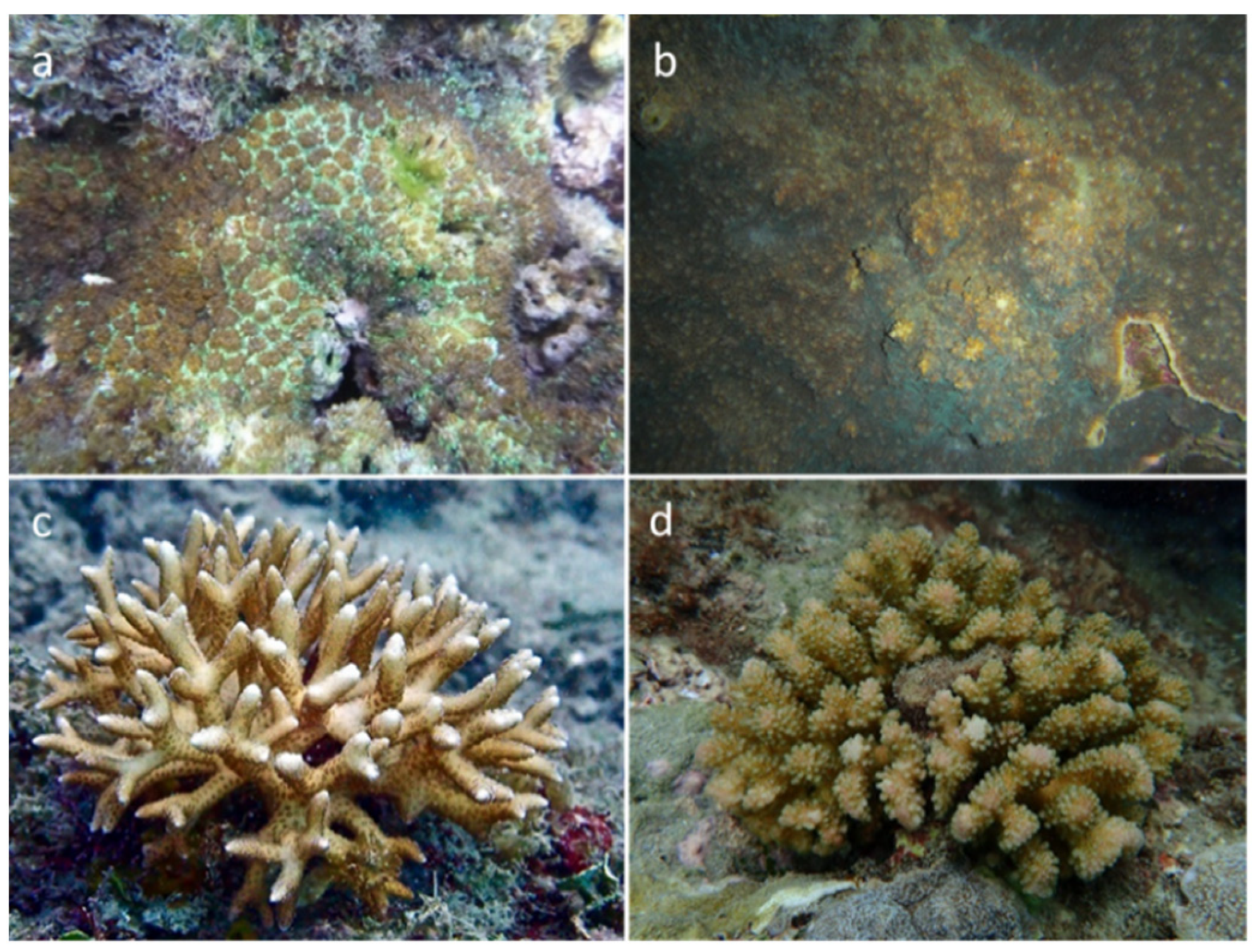
2.1. Experimental Species

Galaxea fascicularis (Figure 1a) has small polyps that are semicircular or cylindrical in shape. G. fascicularis produces colonies through asexual budding, and its coral colonies exhibit an encrusting morphology and can be brown or green. In addition, G. fascicularis reproduces sexually through spawning. Numerous studies have investigated reproduction in G. fascicularis [22]. We used the eggs of G. fascicularis in this study; these eggs were pink and were not infected by the Symbiodiniaceae species. Mycedium elephantotus (Figure 1b) colonies exhibit a laminar or encrusting morphology. M. elephantotus produces colonies through the asexual budding of polyps; the new polyps grow outward and surround the central polyp in a radial pattern. M. elephantotus is a hermaphrodite and reproduces sexually through spawning. Its eggs are light yellow and not infected by the Symbiodiniaceae species. Seriatopora caliendrum (Figure 1c) colonies can be observed as irregular branches. The branches are considerably thick, and their roots are as thick as their tips. The branches are brown or cream. S. caliendrum produces colonies through asexual budding; the new polyps grow in an orderly manner along the branches. S. caliendrum is a hermaphrodite and reproduces sexually through brooding. The coral larvae are infected by the Symbiodiniaceae species, and the Symbiodiniaceae species are evenly distributed within the larvae. Pocillopora verrucosa (Figure 1d) colonies develop as irregular branches with a plantar war structure; the thickness of the branches depends on the depth of water and currents. P. verrucosa produces colonies through asexual budding, where new polyps develop on the wart-like structure. P. verrucosa is a hermaphrodite, and it reproduces sexually through brooding. The coral larvae are infected by the Symbiodiniaceae species, and the Symbiodiniaceae species are mainly concentrated at one end of the larvae (e.g., the mouth).

2.2. Sample Collection

The sperms and eggs of wild G. fascicularis and M. elephantotus were collected during spawning from April to May in 2020 and 2021. Divers collected the samples at Kenting, Taiwan (21°56′ N, 120°44′ E). When spawning began at night, the divers collected eggs, sperms, and sea water from the sampling location in the 50-mL syringes (Terumo, Tokyo, Japan). The collected samples were rapidly transported to the Laboratory of the National Museum of Marine Biology and Aquarium for subsequent experiments. The colonies of P. verrucosa and S. caliendrum were collected, transported to the aquaria every month, and placed in customized larval collection boxes (Figure 2) [23]. When the corals released larvae, the larvae flowed out of the boxes and were collected into larval collection cups. The cups had filters on the bottom to enable sea water to flow through and carry the larvae.

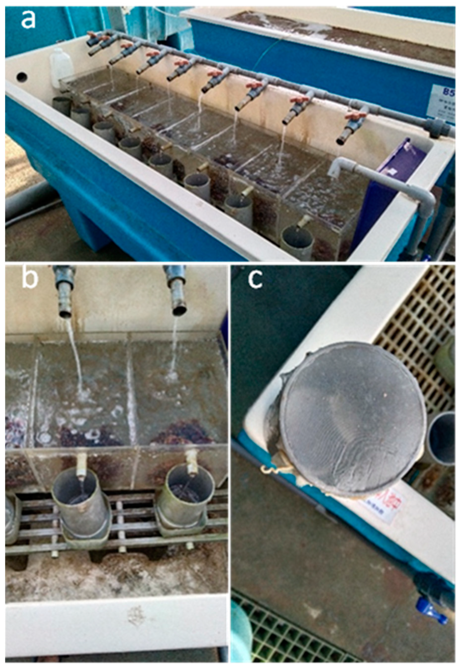
2.3. Fertilization Experiment

The sperm and egg samples of G. fascicularis and M. elephantotus were placed into beakers and evenly stirred for fertilization. The sea water samples were filtered twice using 0.4-μM filter papers and vacuum pumps. Next, 3-mL droppers were used to transfer the fertilized eggs into beakers containing fresh filtered sea water (FSW) for cleaning. Subsequently, the eggs were transferred to Petri dishes (90 × 15 mm) containing FSW, and the Petri dishes were observed under a dissecting microscope to examine the developmental process of the fertilized eggs.

2.4. Cultivation and Feeding

The embryos of G. fascicularis and M. elephantotus were transferred to Petri dishes containing FSW, and the dishes were placed at room temperature (26 °C). Because undeveloped embryos would undergo degradation and contaminate FSW, the water was changed daily. After the embryos developed into larvae and most of the larvae were settled, the Petri dishes were transferred to the aquaria and placed inside a 0.6-ton sea water tank. Furthermore, the larvae of P. verrucosa and S. caliendrum were transferred to Petri dishes and placed at room temperature. However, because the coral larvae had Symbiodiniaceae, artificial lights (ComboRay CR60, Illumagic, Tainan, Taiwan) were placed on the top of the Petri dishes during cultivation. Most of the larvae settled after approximately 1 month. The settled larvae in the Petri dishes were transferred to the aquaria and placed inside a 0.6-ton sea water tank. The Petri dishes were placed in baskets with holes under tiles to prevent the larvae from being swept away by the current generated by the wave maker. The sea water tank was installed with a cooling circulation system; sea water flowed into the circulation tank, and the motor redirected the water into the feeding tank. If the temperature of the water was considerably higher, the cooling system automatically reduced the temperature and maintained it at 27 °C on average. The tank was equipped with a wave maker to evenly stir sea water. Inlets for natural sea water and circulating water were covered with a filter bag to reduce the amount of suspended matter present in the tank water. The filter bag was cleaned weekly, and algae and sediments were eliminated as much as possible without affecting the corals.

2.5. Observation Records
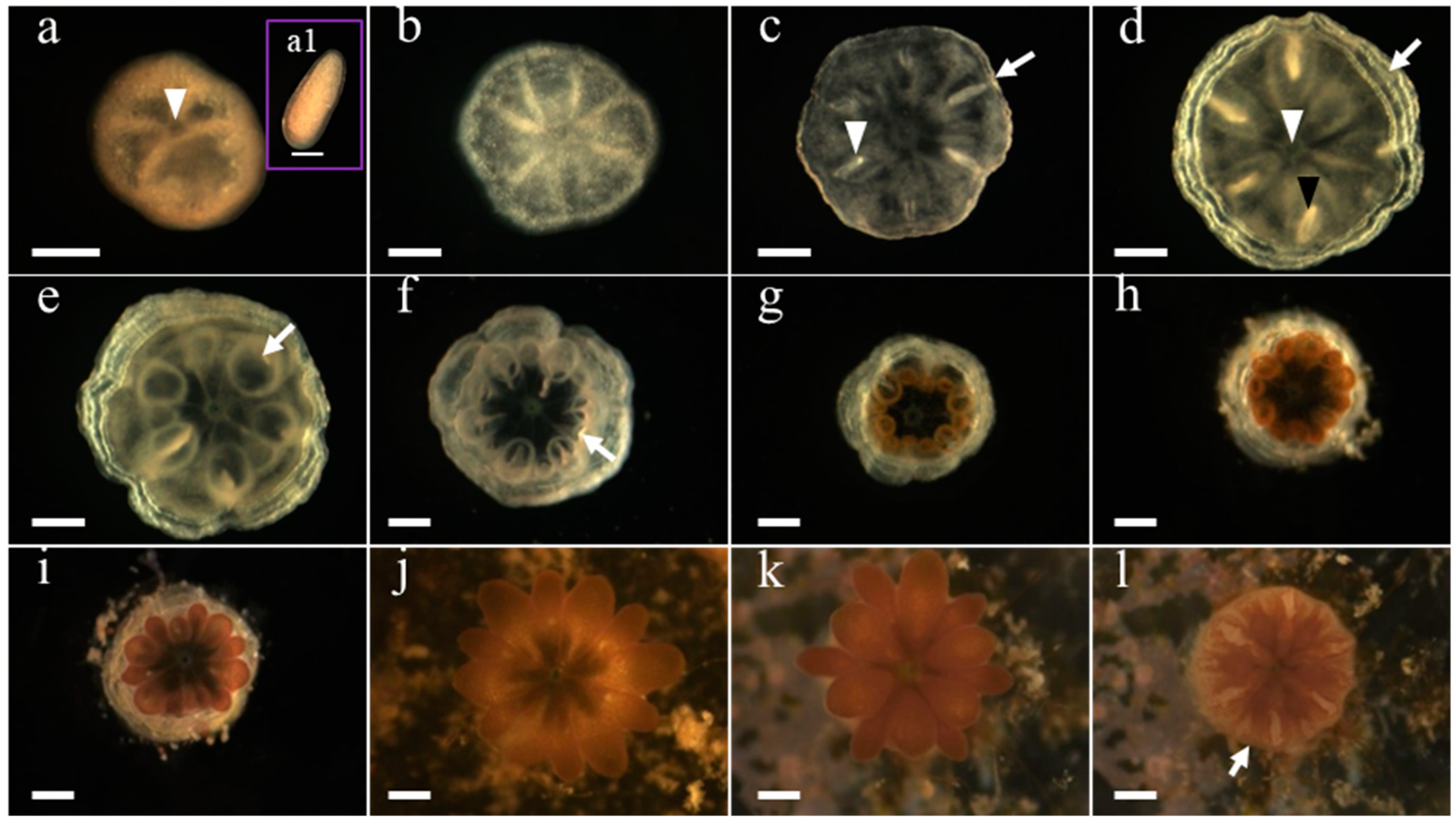
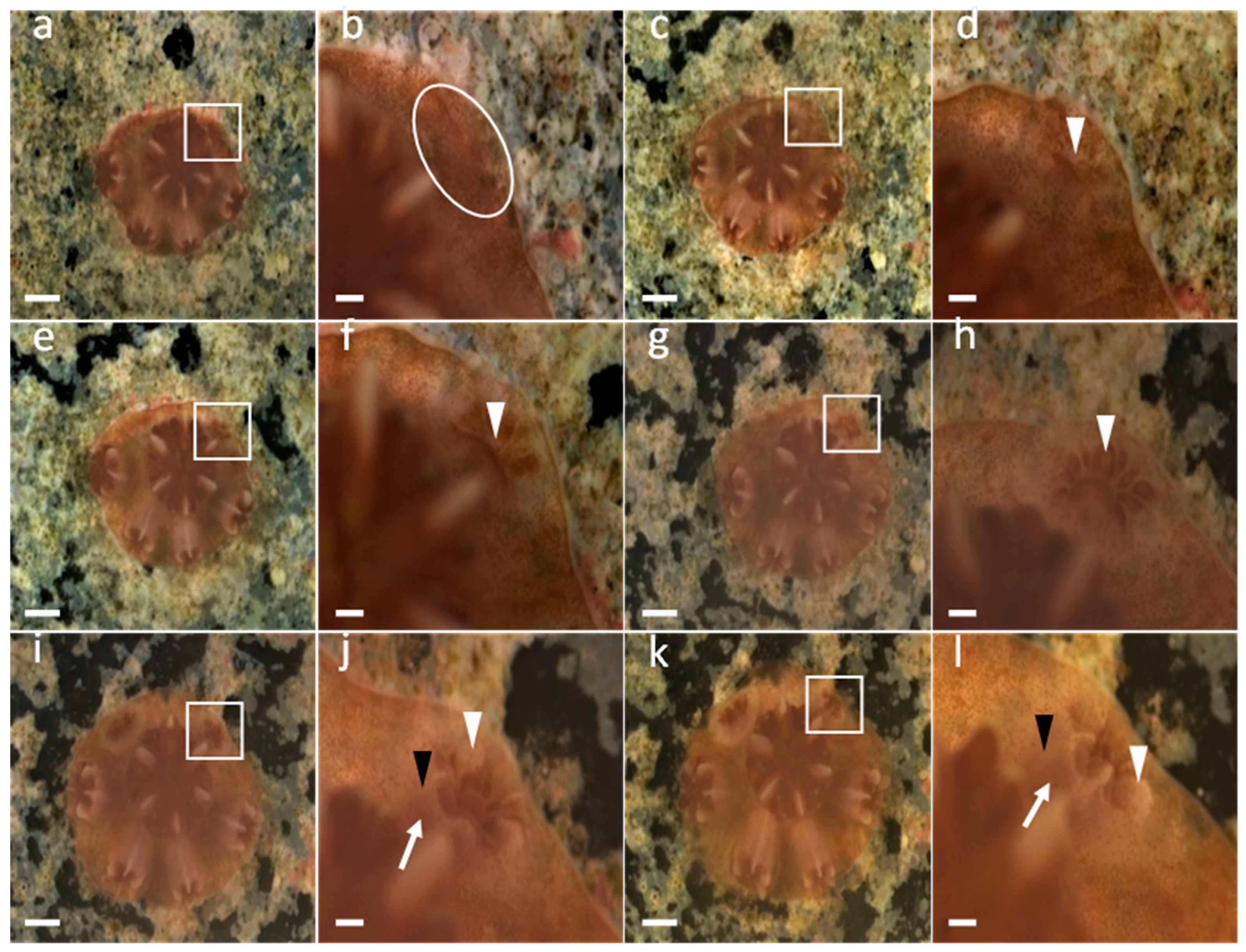
The fertilized eggs or larvae were placed in Petri dishes containing FSW and observed using a dissecting microscope (Discovery V8, SteREO; Zeiss, Jena, Germany). After the settlement of the coral larvae, they were photographed daily in the first week and then once every 2 days until 1 month. After 1 month, the corals were photographed weekly. After careful cleaning of the Petri dishes under a dissecting microscope (SZ51, Olympus, Tokyo, Japan), the corals were observed. The Petri dishes were thoroughly cleaned and photographed. When growth changes, such as infection with Symbiodiniaceae and polyp budding, were noted, the corals were photographed daily and observed under the fluorescence condition. The photographs were used to observe changes in the growth of the polyp skeleton. To facilitate the observation and photography of the skeleton, the polyps were stimulated and contracted using pointed droppers; no physical harm was caused to the polyps when droppers were used. The time required for the coral larvae to reach a certain phase was defined as the time required for at least one of the larvae to reach that phase. The following phases were observed: coral settlement, skeleton development, tentacle development, complete polyp development, and new individuals’ asexual budding. In addition, the settlement rate of the coral larvae, and the survival rate of the coral spats after settlement were calculated.

3. Results

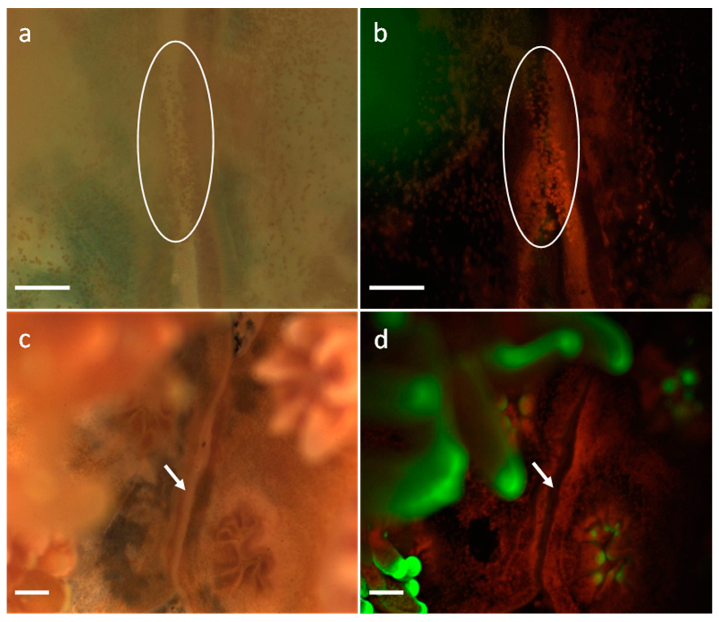
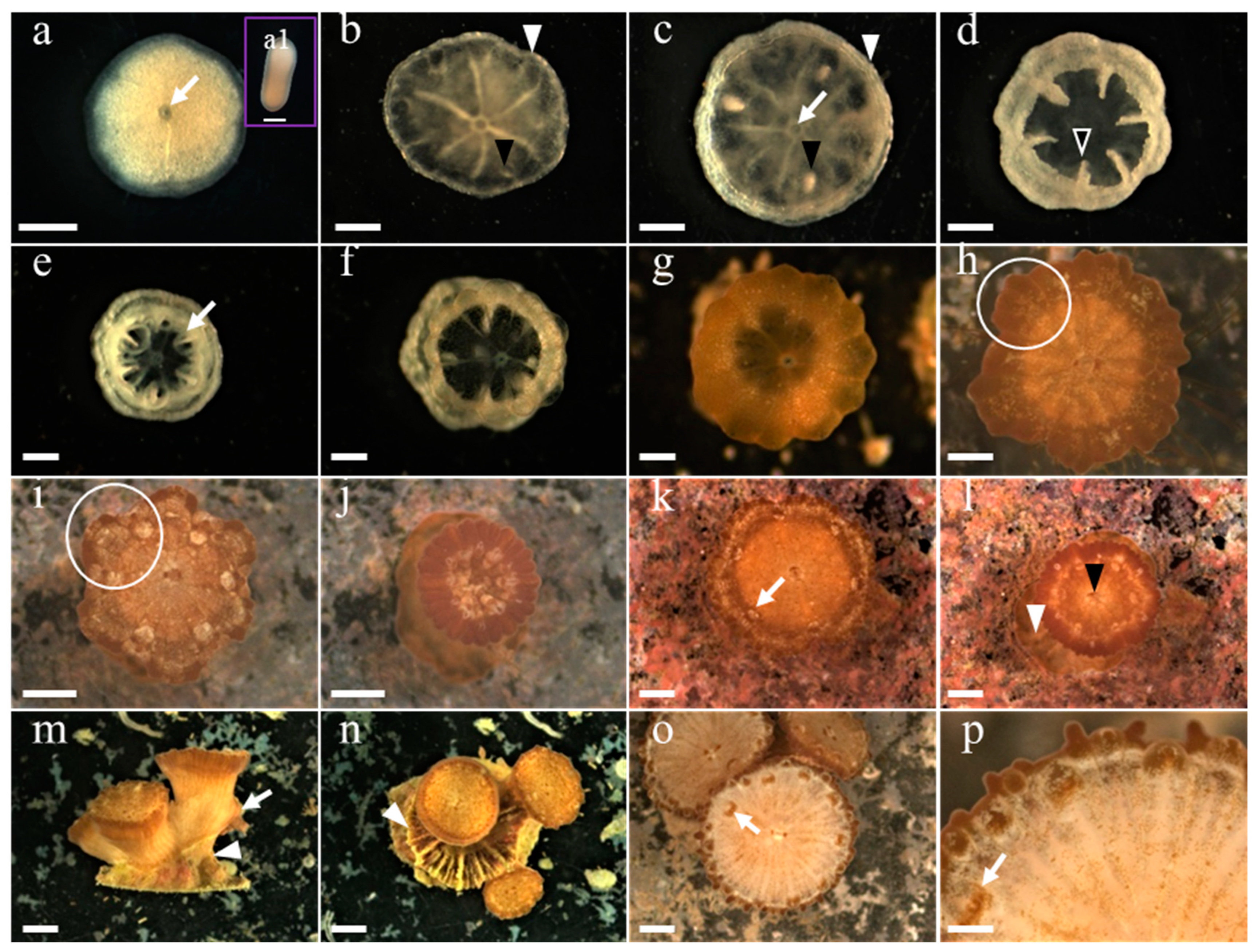
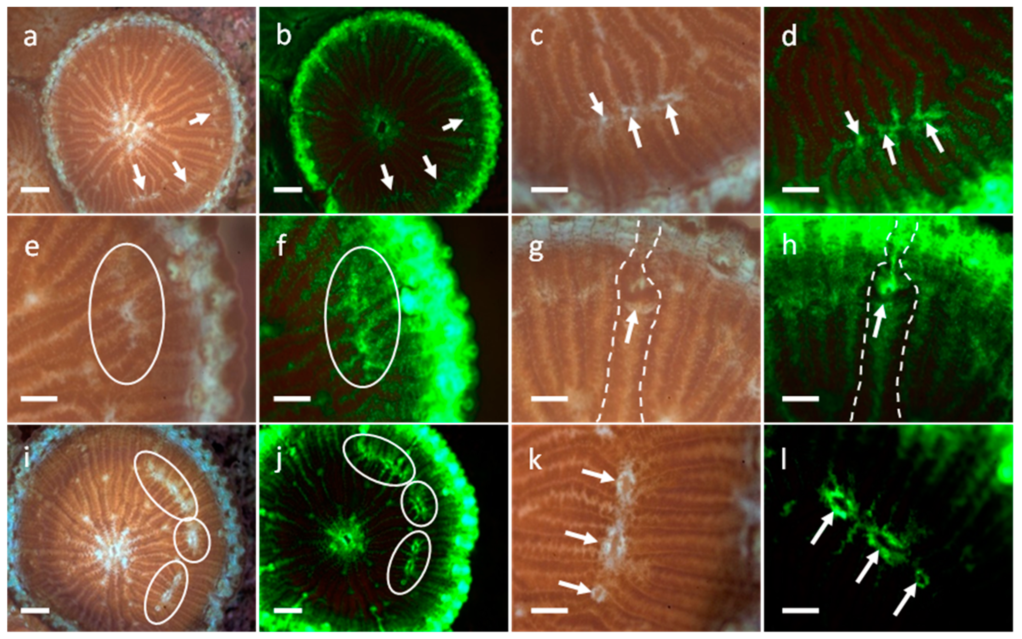
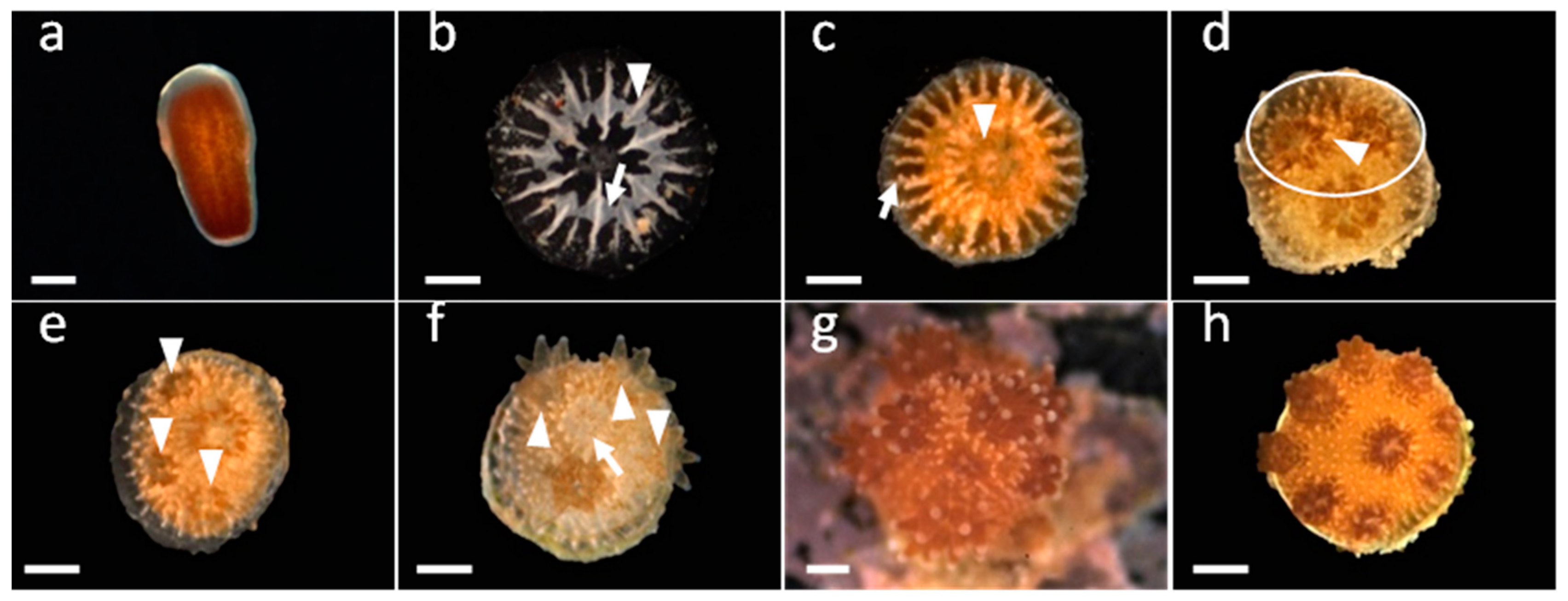
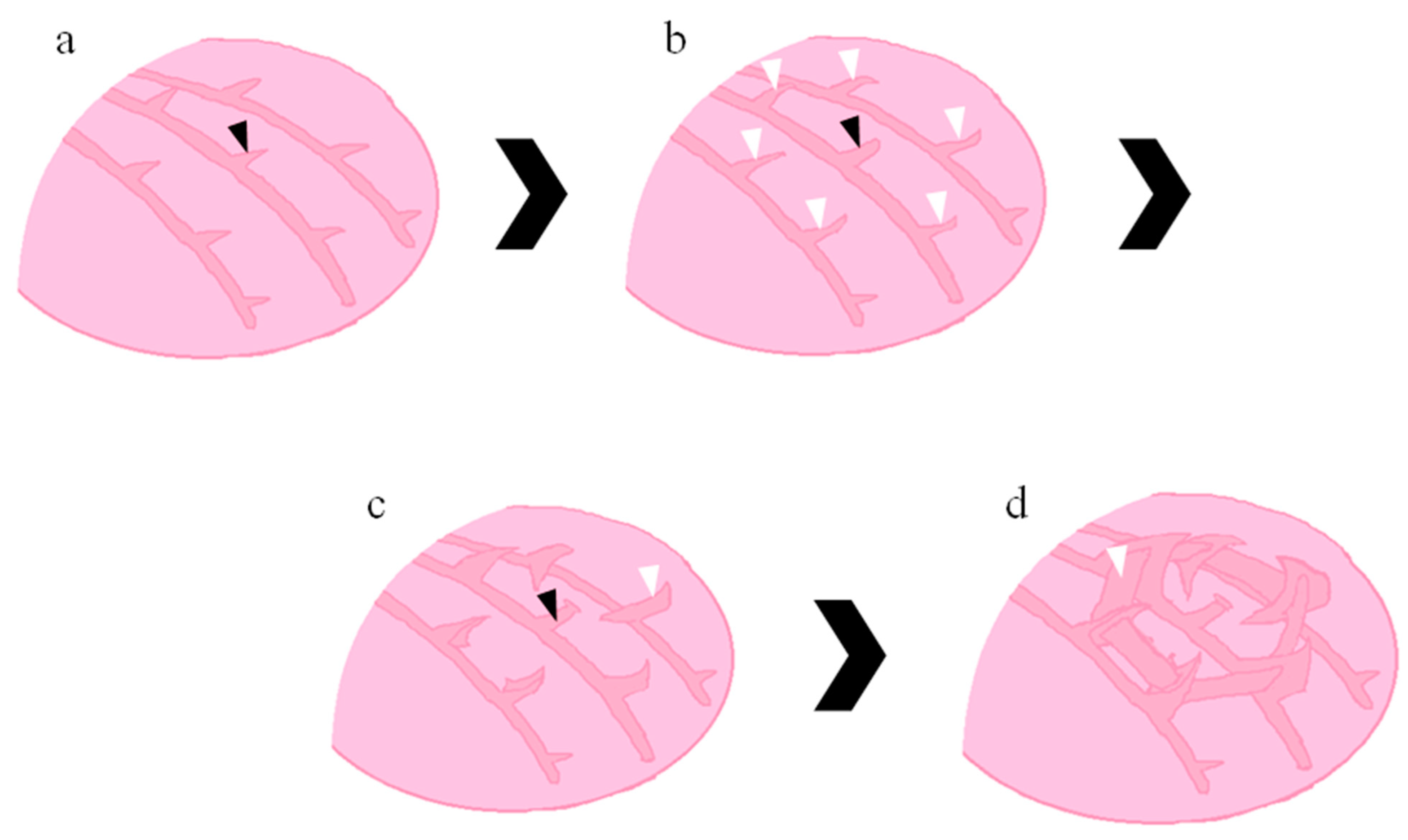
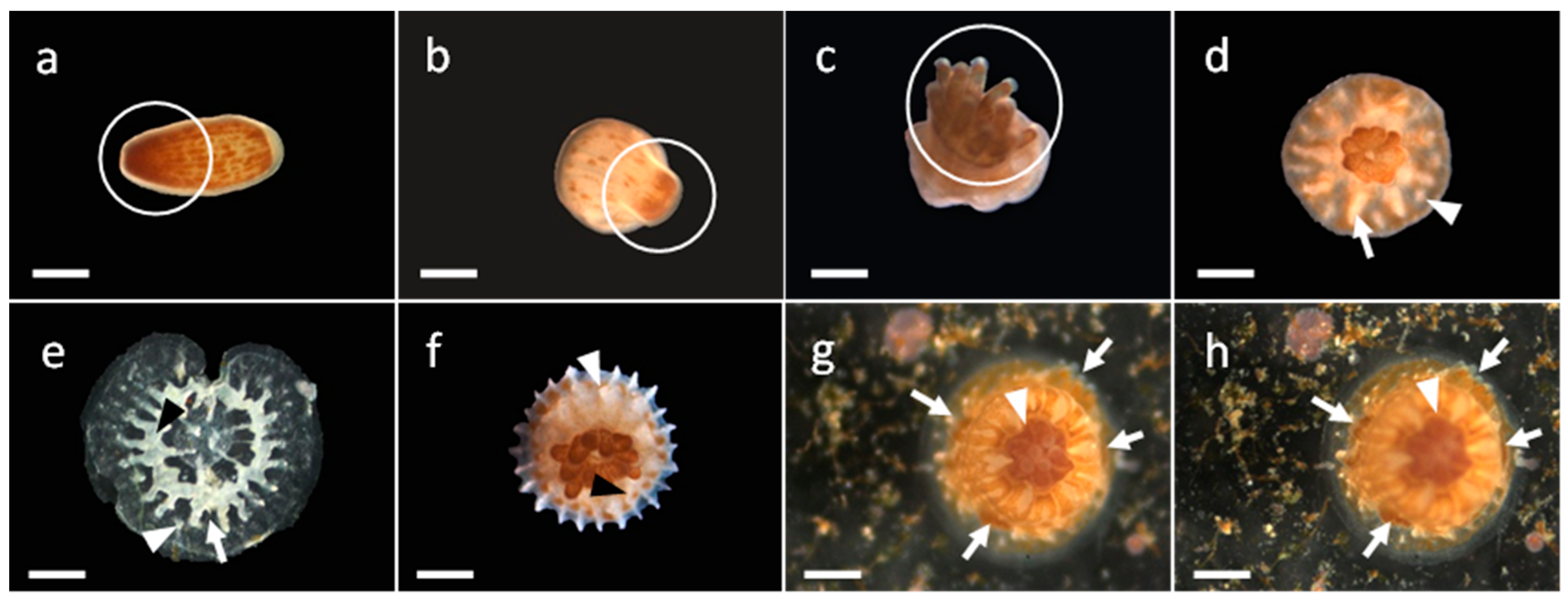
3.1. Development of G. fascicularis Spats

Figure 3 presents the development of G. fascicularis and the time required for the spats to enter each phase after settlement. After the development and maturation of the larvae, they searched for a suitable location to settle. After settlement, a colorless circular hole was observed in the center of the spat (Figure 3a; 0 day post settlement), which eventually developed into its mouth. The polyps contracted, expanded, and became flat and transparent. Over time, the interior of the polyps was divided into six sections (Figure 3b; 1 day post settlement). The polyps began to develop their skeleton from the bottom. During this phase, the development of the calcium-based basal plate began, and six septa were formed in the interior (Figure 3c; 5 days post settlement). The septa gradually grew and extended along the edge of the colony until they reached the outer epidermis; the epidermis grew along the surface of the polyps and covered them, forming the epitheca. The polyps were cylindrical and protected by the epitheca (Figure 3d; 9 days post settlement). The formation of the mouth was observed during this phase. Fluorescence microscopy revealed a high amount of green fluorescent protein around the mouth. The polyps could eat during this phase and were infected with Symbiodiniaceae; the infection could be viewed under red fluorescence. After 14 days, the polyps covered by the epitheca began to form tentacles. Initially, a few circular tentacle buds were observed on the septa (Figure 3e; 23 days post settlement). These tentacle buds became more prominent and began to protrude over time (Figure 3f; 1 month post settlement). Their roots aligned with the septa and began to protrude from the epitheca with the growth of the tentacles. In addition to the original six tentacles observed on the six septa, new tentacle buds were observed. The roots of these new tentacles formed six new small septa (Figure 3f). During this phase, Symbiodiniaceae species were concentrated into the upper sections of the infected polyp, namely the tip and tentacles (Figure 3g; 1 month post settlement). With the growth of the coral polyps, Symbiodiniaceae species present inside the polyps began to bud and caused the polyps to appear dark brown (Figure 3h; 1.5 months post settlement). The tentacles gradually extended outward (Figure 3i; 2 months post settlement) until the upper part of the polyps was completely extended and all the tentacles were spread out (Figure 3j; 2.5 months post settlement). After approximately 3 months, the coral polyps had fully developed (Figure 3k), and the polyps began to extend toward the epitheca and gradually cover it (Figure 3l).
Figure 4 presents the asexual reproduction of a G. fascicularis polyp. After approximately 3 to 4 months, the polyps expanded and small tumor-like materials began to form near the original polyps. Symbiodiniaceae species were concentrated in the tumor-like materials, and after 6 months, a few new polyps of various sizes were formed around the original coral polyps. The surrounding polyps initially exhibited small bumps (Figure 4a,b). Over time, the surrounding Symbiodiniaceae species were concentrated around the center, and the surrounding area became white because of the absence of Symbiodiniaceae species (Figure 4c,d). Furthermore, sagging regions without Symbiodiniaceae species were noted in the bumps (Figure 4e,f). The new polyps could be distinguished from the growing tentacles on the basis of the color of Symbiodiniaceae species (Figure 4g,h). Symbiodiniaceae species were concentrated near the tip of the polyps, and the basic skeleton structure began to form. After 7 months, the formation of the white circular skeleton and septa was observed (Figure 4i,j). Over time, the new polyps began to grow their skeletons, which gradually became similar to the skeletons of the original polyps (Figure 4k,l). The new polyps grew on the edges of the coral colonies. When sufficient space was available, the new polyps grew and radiated outward. Subsequently, they gradually adjusted and grew upward.
After 10 months, two G. fascicularis coral colonies with different polyps began to make contact (Figure 5). The brief fusion of the polyps was observed through fluorescence microscopy, and the polyps exchanged their internal Symbiodiniaceae species. It is also possible that the coral tissue might be damaged due to colony–colony competition (Figure 5a,b). However, after 2 days, the contact was disrupted, and no further fusion occurred (Figure 5c,d). The polyps in the two coral colonies began to compete upon contact, and instead of the expansion of the basal plate, a septum formed as a boundary between the colonies. The polyps growing near the boundary experienced growth pressure, and their skeletons curved as they grew. The coral colonies had a competitive relationship, and the boundary between them began to grow and slant toward one side.

3.2. Development of M. elephantotus Spats

Figure 6 presents the life cycle and time points for the growth events of the M. elephantotus larvae after settlement. After the development and maturation of the spats, they metamorphosed into primary polyps on the fifth day of settlement. After settlement, a colorless circular hole was observed in the center of the spat (Figure 6a; 0 day post settlement) that eventually developed into its mouth. The polyps began to divide into six sections. The calcium-based basal plate developed around the polyps, thus forming the epidermis, whereas six septa were formed in the interior (Figure 6b). The skeleton of the outer epidermis grew along the polyps until it became an epitheca that covered the polyps (Figure 6c). The septa formed inside the polyps were connected to the epitheca in a cylindrical structure (Figure 6d; 25 days post settlement). During this phase, the mouth and coelenteron were fully developed. When the polyps swallowed Symbiodiniaceae species, resulting in a symbiotic relationship, the Symbiodiniaceae species spread from the center to other parts of the polyps. Tentacles began to form after approximately 1 month (Figure 6e), and Symbiodiniaceae species were evenly distributed within the polyps (Figure 6f; 1.5 months post settlement). During this phase, 12 tentacles developed, and they were closely connected in the shape of an upright umbrella; Symbiodiniaceae species were evenly distributed among the tentacles. The bottom septa slightly protruded from the epitheca (Figure 6g; 2 months post settlement). New tentacles began to sag inward from the tips of the original tentacles and underwent budding with each tentacle splitting into three tentacles. The roots of the new tentacles formed new septa, and differences in the size of the tentacles began to appear (Figure 6h; 3 months post settlement). After approximately 3 months, the polyps were noted to have several large tentacles; upon closer inspection, we observed a few long tentacles protruding outward and a few short tentacles in the sagging regions (Figure 6i). Fluorescence microscopy revealed that fluorescent proteins were distributed along the umbrella-like surface, particularly near the tips and grooves of the tentacles. The interior portion of their skeleton developed into dense septa, and the polyps began to extend outward (Figure 6j). Over time, differences in length between the long and short tentacles gradually decreased, and the polyps became circular; brown and white stripes were noted on the polyps (Figure 6k). Because of the enlargement of the polyps, their mouths and some parts on the top sections were visible even when the tentacles were retracted. The bottom sections of the polyps expanded, and costae formed from the extension of septa (Figure 6l). After approximately 7 months, an accident occurred that severely damaged the corals. Most of the polyps died, and after first-aid treatment, only a few healthy polyps remained on the skeleton. After approximately 1 year, each remaining polyp produced three new polyps (Figure 6m,n). With the recovery of the coral colony, the new polyps exhibited the same characteristics as did the old polyps. The top sections of the new polyps had tentacles of a similar length arranged in circles, and brown stripes were observed. The new polyps were not covered by the epitheca and gradually extended outward; however, the lack of Symbiodiniaceae species caused the polyps to appear white (Figure 6o,p).
Figure 7 presents the budding of M. elephantotus and the fission of the outer polyps. After the 17th month, the top sections of the polyps exhibited a corona-like structure. The outer polyps did not exhibit any tentacles, and they appeared round and smooth (Figure 7a). The Symbiodiniaceae species left the location in which new polyps were expected to grow, thus forming a white region (Figure 7a,b); this resulted in the asexual budding of the M. elephantotus polyps. A few openings were formed in this white region, which developed into the mouths of the new polyps (Figure 7c,d). Due to the inner costae attached with the polyps closely; green fluorescence traces represented the margins of the inner costae, and the fusion of the inner costae was observed in the white region. The top sections of the corona of the outer epidermis of the polyps grew similarly to the new tentacles. Several regions on the outer epidermis grew by splitting into three regions, and nodes on the inner costae were observed after new growth (Figure 7e,f). The ends of the outer epidermis grew outward; however, new polyps did not grow. These regions became brown stripes observed on the polyps (Figure 7g,h). The presence of numerous Symbiodiniaceae species at the ends resulted in the more prominent appearance of brown stripes. The polyps in these regions continued to grow, and Symbiodiniaceae species gradually spread out. Over time, the region without Symbiodiniaceae species expanded (Figure 7i,j); the new mouths and their features became more prominent (Figure 7k,l).

3.3. Development of S. caliendrum Spats

Figure 8 presents the developmental stages and time points for the growth events of the S. caliendrum spat. S. caliendrum sexually reproduces through incubation, and its larvae are dark brown and contain numerous Symbiodiniaceae species evenly distributed throughout (Figure 8a). After the settlement of the larvae, the skeletons developed rapidly, and a few long skeletons grew from the outside of the young polyps toward the center and formed septa. Next, the two sides of the septa formed triangular laminar skeletons that were connected together; the skeletons extended to the basal plates and formed the theca (Figure 8b; 2 days post settlement). With the development of the skeletons, the tentacles formed on the polyps, and the base of the skeletons and costae resulted in the formation of short spindle-shaped skeletons that grew outward (Figure 8c and Figure 9a; 9 days post settlement). The number of the spindle-shaped skeletons increased, and they grew longer with the development and expansion of the polyps. Over time, the new polyps began to bud (Figure 8d; 1 month post settlement). Few Symbiodiniaceae species were distributed around the coenosarc, and they remained concentrated around individual spindle-shaped skeletons. With the development of the new polyps, the spindle-shaped skeletons surrounding the central spindle-shaped skeleton began to curve toward the central spindle-shaped skeletons (Figure 9b). Thus, the spindle-shaped skeletons became laminar and formed the septa for the new polyps (Figure 9c). The growth of the central spindle-shaped skeletons stagnated, and their top sections flattened, becoming the columella of the new polyps (Figure 9c). Finally, the new polyps formed tentacles, and thecae formed between the septa (Figure 9d). At this time point, the skeletons of the new polyps had fully developed. The ability of the coral colony to grow upward was correlated with the angle and density of the new polyps formed through budding. During the growth of the coral colony, the original polyps were displaced, forming new polyps (Figure 8e; 1.3 months post settlement), and the skeletons of the new polyps grew outward and caused an increase in the growth angle, thus forming bumps (Figure 8f; 1.5 months post settlement). The new polyps grew densely along the bumps, causing the bumps to extend and become branches (Figure 8g; 2.5 months post settlement). However, when space between the new polyps increased without the polyps affecting each other, the overall structure of the coral colony would become flatter; the branch structure can be explained by this growth mechanism. Additional branches were formed by the polyps growing on the ends of the branches that had small growth angles; this process prevented the originally upward-growing polyps from growing along the branches. Thus, the new polyps grew sideways and formed new branches.

3.4. Development of P. verrucosa Spats

Figure 10 presents the developmental stages and time points for the growth events of the P. verrucosa spats. P. verrucosa reproduces sexually through brooding, and its larvae contain Symbiodiniaceae species. The Symbiodiniaceae species in the larvae were distributed in patches, and most were concentrated near the end of the larvae, imparting the larvae a distinct dark-brown tail (Figure 10a). After settlement, some spats gradually metamorphosed into primary polyps, and their tails started to form tentacles. Some polyps exhibited fully developed tentacles without skeletons (Figure 10b,c; 7days post settlement). Because the settlement time of each larva differed, various developmental stages were observed within a single timeframe. Short, triangular, spindle-shaped skeletons started to form on the outside of the polyps, with the tips of the triangles pointing inward. The spindle-shaped skeletons grew toward the center and formed large triangular septa (Figure 10d). Stripe-shaped skeletons were formed at the approximate center of the septa and were connected together, forming the theca. The theca was not in contact with the basal plate; hence, several pores were present (Figure 10e). The septa curved slightly downward in a hook-shaped manner where the theca protruded from the basal plate. The basal plate formed several new outward-growing spindle-shaped skeletons (Figure 10e; 14 days post settlement). The structure where the triangular septa and basal plate were connected extended outward and formed the costae, and a few outward-growing spindle-shaped skeletons connected by stripe-shaped skeletons were formed on the structure (Figure 10f; 14 days post settlement). The young polyps grew rapidly, and after approximately 1 month, the polyps grew upward and were cylindrical. The spindle-shaped skeletons continued to grow on their basal plates, and the new polyps vertically budded near the root of the skeleton, with the original polyps as the center. (Figure 10g,h). The budding of P. verrucosa was similar to that of S. caliendrum, and the Symbiodiniaceae species were concentrated around the location at which new polyps would grow. During the development of the polyps, the nearby spindle-shaped skeletons were affected by the new polyps and curved toward them, gradually becoming septa. The septa were connected by stripe-shaped skeletons and formed the thecae. The skeletons of the new polyps grew in a manner similar to that of the original polyps. Thus, the polyps did not form additional skeletons when they budded, and the new polyps appeared to be embedded in the entire skeleton.

3.5. Settlement and Post-Settlement Survival Rates of Coral Larvae and Spats

G. fascicularis, P. verrucosa, and S. caliendrum settlement percentages and post-settlement survival rates are shown in Table 1. On the 12th day, 100% of S. caliendrum larvae had settled, while G. fascicularis and M. elephantotus were characterized by settlement rates of 46% and 28%, respectively. P. verrucosa had the lowest settlement rate. Furthermore, among the four coral species studied, G. fascicularis had the highest survival rate (29%) two months post-settlement, with P. verrucosa spat survival only 3% over this same time period.

4. Discussion

The skeleton of a coral polyp consists of the columella, septa, theca, costa, and coenostea. The costa and coenostea are part of the skeleton of the exterior of the coral polyp. This study demonstrated two skeleton growth mechanisms: the skeleton-over-polyp mechanism and the polyp-over-skeleton mechanism. The G. fascicularis and M. elephantotus polyps exhibited the skeleton-over-polyp mechanism. Their primary polyps developed laminar skeletons from the bottom sections. The skeletons surrounded the surface of the polyps and formed the epitheca. With the growth of the laminar skeletons, they connected to the epitheca and formed septa. The epitheca covered the young polyps as they grew outward, forming the theca. The P. verrucosa and S. caliendrum polyps exhibited the polyp-over-skeleton mechanism; both the types of corals appeared similar to branches. Their primary polyps developed laminar skeletons from the bottom sections. Next, horizontal laminar skeletons were formed near the center of the polyps, forming the theca. Then, spindle-shaped skeletons were formed on the laminar skeleton. P. verrucosa had compact cylindrical skeletons. The epitheca grew along the surface of the polyps and were smooth, and graduated stripes were observed. The septa lined the interior of the epitheca and protruded from the epitheca after the polyps developed tentacles. S. caliendrum had a radial appearance. Its long laminar skeletons were arranged along the basal plate, and the skeletons closer to the center were taller. New long skeletons were formed around the center, connecting to the laminar skeletons, and forming the theca. This formation process caused the theca to become a polygon. The formation of the young skeletons suggested that the coenostea of the skeletons of G. fascicularis and M. elephantotus grew slowly through continuous stacking and compaction, whereas the skeletons of P. verrucosa and S. caliendrum were connected by the thin and stripe-shaped skeleton structures. The spindle-shaped skeletons under the coenosarc of P. verrucosa formed stripe-shaped skeletons that were connected together. Therefore, the coenostea of S. caliendrum may have formed new laminar skeletons from the spindle-shaped skeletons in the same manner. The skeletons formed through this mechanism were fragile but did not undergo the slow process of stacking, enabling the structure to respond to the rapid growth of the polyps. No study has examined the live skeletons of growing corals. Similar studies include [16] that observed similar skeleton structures. However, in this study, the development of the polyps was affected by the size of the corals and was not correlated with the species. In addition, [16] did not observe live specimens and thus could not determine the effects of the development of the polyps on the formation of skeletons. Other studies on coral skeletons have mainly investigated their components and microstructures [24,25].
This study observed changes in the mouths, tentacles, and coenosarcs of the adult coral polyps. The polyps grew through mitosis. We speculated that the presence of Symbiodiniaceae species might have affected the development of the young G. fascicularis and M. elephantotus polyps. The eggs and larvae of these two corals were not infected by Symbiodiniaceae species, and they could only engage in symbiosis with the Symbiodiniaceae species after their larvae had settled and the mouths and coelenterons of their primary polyps had fully developed. The symbiosis between the corals and Symbiodiniaceae species was affected by several factors and caused the infection of Symbiodiniaceae to be probabilistic. We observed that the young corals infected by Symbiodiniaceae species developed polyps and tentacles; however, the growth of the young corals not containing Symbiodiniaceae species was stagnated. Although a few young polyps without Symbiodiniaceae species still developed tentacles, most of their tentacles remained buds, and over time, the opening on their epitheca gradually shrank. This indicated that the young polyps were shrinking, which may have been due to the corals being unable to intake nutrients owing to their underdeveloped tentacles. Thus, Symbiodiniaceae species provided most of the nutrients required for growth [26]. The young polyps of G. fascicularis and M. elephantotus developed differently from the young polyps of P. verrucosa and S. caliendrum because G. fascicularis and M. elephantotus were initially covered by the epitheca, and their polyps grew out of the epitheca and covered them (skeleton-over-polyp mechanism). Thus, the young polyps required more time to develop and expand and were protected by the epitheca. The skeletons of P. verrucosa and S. caliendrum grew under the polyps and formed coral polyps; the polyps only had a mouth (polyp-over-skeleton mechanism) because all other parts were used by the coenosarc to develop the coral colony. No study has investigated the effects of growth mechanisms on the development of young corals.
We observed that after the metamorphosis of the coral spats into primary polyps, the polyps began to split into sections. Each section had at least one tentacle and one corresponding septum, and each section of the polyp extended to the coenosarc and formed costae. Therefore, most of the costae were connected to the protruding part of the septum. The initial formation of the young skeletons of P. verrucosa and S. caliendrum indicated that the septum and costa were originally from the same skeleton, and as they grew, the theca divided the two types of skeletons. This phenomenon was more prominent in M. elephantotus. When the polyps present on the perimeter of the coral colony expanded, each polyp split and produced three new polyps. The two new adjacent polyps were connected to the original polyps, which sagged. This fission affected the performance of the costae. The costae of the original polyps developed breakpoints and dents where the polyps split. The original costae branched toward the adjacent polyps, and the central polyp began to form new costae.
The results of this study revealed that Symbiodiniaceae species present inside the corals were concentrated around the top and tentacles of the polyps. Studies have indicated that more active and continuously growing corals contained more Symbiodiniaceae species in their tentacles [27] and that the growth of these tentacles was inhibited when the polyps had a nutritional disorder [28]. In this study, the tentacle buds of G. fascicularis and M. elephantotus that were not infected with Symbiodiniaceae species remained buds until they were infected with Symbiodiniaceae species. The Symbiodiniaceae species were concentrated in the tentacle buds, and the tentacle buds began to develop into tentacles. We speculate that the polyps that did not contain Symbiodiniaceae species lacked nutrition and were inactive and that the Symbiodiniaceae species enabled the young polyps to gain sufficient nutrition and become active.
This study examined the growth of the tentacles of M. elephantotus. The tentacles grew around the circular theca and formed where the septa protruded. Unlike the long tentacles of the other three types of corals, the tentacles of M. elephantotus were fan-shaped and interconnected. The relationship between the septa and tentacles suggested that the fan-shaped polyps contained numerous Symbiodiniaceae species, which were the tentacles. However, these tentacles differed from common tentacles, which are short and lack Symbiodiniaceae species (web: Corals of the World). The corals may have regulated the distribution of their Symbiodiniaceae species, and the tentacles of the primary polyps of M. elephantotus developed large fan-shaped tentacles because they were active and contained numerous Symbiodiniaceae species. The corona skeletons that connected the top sections of the polyps supported the polyps, enabling them to use their tentacles to expand upward. This phenomenon explains why the growth of the polyps on the perimeter of the coral colony had polyp patches similar to tentacles.
When the polyps matured and expanded, they began to form new polyps. This study observed the skeletons of the three coral colonies; the budding of G. fascicularis was phaceloid, the budding of M. elephantotus was plocoid, and the budding of P. verrucosa and S. caliendrum was cerioid. The Symbiodiniaceae species were concentrated in the top sections of the coral polyps and tentacles. Therefore, the concentration of the Symbiodiniaceae species was used to determine the location of budding in the new polyps; their concentration was also used to identify the lumps of new polyps and the formation of the mouths and new tentacles. The G. fascicularis, P. verrucosa, and S. caliendrum polyps changed considerably. However, the Symbiodiniaceae species were widely dispersed in the coenosarc of M. elephantotus, and the white regions not containing the Symbiodiniaceae species formed a single or multiple mouths. This may have been caused by the corals regulating the distribution of the Symbiodiniaceae species; this phenomenon is described further in the section on the formation of new polyp skeletons.
The corals exhibited three distinct skeleton growth mechanisms. For the G. fascicularis polyps, their skeletons formed layer by layer, similar to three-dimensional printing. The Symbiodiniaceae species were highly concentrated, and they increased the activity of the new polyps, causing them to develop rapidly. With the development of the polyps, their basal plates rapidly developed the thecae and septa. Unlike the skeletons of G. fascicularis, the skeletons of P. verrucosa and S. caliendrum formed during budding from the spindle-shaped structure of the colony. The spindle-shaped skeletons grew directly under the new polyps, causing the growth of polyps to stagnate and changing the top sections of the polyps to columella. The surrounding spindle-shaped skeletons turned and grew toward the central new polyps, becoming the theca. The spindle-shaped skeletons served as scaffolds and pillars for the new polyps during budding. Because the formation of most of the skeletons was already complete at this stage, the new P. verrucosa and S. caliendrum polyps grew faster. We speculated that the ability to grow vertically is crucial for the budding of these two corals. When new polyps grew, particularly the P. verrucosa polyps, they extruded the surrounding polyps and forced the coral colony to grow upward. The extrusion could have caused the new polyps to grow in a twisted and displaced manner, resulting in the coenosarcs growing upward, as observed in the S. caliendrum polyps.
The new M. elephantotus polyps developed only their mouths until the end of the study. The skeletons of the new M. elephantotus polyps are stripe shaped. Hypotheses of the formation of stripe-shaped skeletons must account for the ability of corals to regulate the distribution of the Symbiodiniaceae species. During budding, the dispersal of the Symbiodiniaceae species caused some polyps to bleach and reduce their activity. Thus, the growth of the polyps and skeletons slowed. However, the surrounding polyps and skeletons grew normally, and this difference in growth speed caused the normal polyps to cover bleached regions in pockets. The loss of the Symbiodiniaceae species resulted in the stagnation of the growth of the tentacles of the new polyps. However, the development of the mouth was not affected by the loss of the Symbiodiniaceae species, and the formation of the mouth but not the tentacles was observed. This growth mechanism was likely caused by evolution; we hypothesized that if the new polyps contained the Symbiodiniaceae species, they would continue to grow similarly to the original polyps. The continuous growth would have negative effects on the corals, such as the skeleton snapping under the weight and the coenosarc being blocked, preventing the Symbiodiniaceae species from photosynthesizing and resulting in the death of the polyps.

5. Conclusions

Most studies on the early development of corals are required for academic research on coral reefs and applied reef conservation, but the interval between observations is usually weeks or months. Thus, no study has comprehensively explored the development of coral larvae after settlement. This study described the development of coral skeletons and polyps in detail and identified unique changes in four corals and the functions of the Symbiodiniaceae species during development. The results are valuable and can serve as reference for studies on coral taxonomy, ecological studies, coral reef conservation, and coral reef restoration.

Author Contributions

Conceptualization, C.L. and S.T.; Formal analysis, C.-M.K.; Funding acquisition, C.L., C.-Y.H. and S.T.; Methodology, C.L., C.-M.K., C.-Y.H., H.-H.L. and S.T.; Project administration, S.T.; Resources, C.L. and S.T.; Supervision, C.L.; Validation, C.-Y.H. and S.T.; Visualization, H.-H.L.; Writing—original draft, C.L. and C.-M.K.; Writing—review & editing, C.L. and S.T. All authors have read and agreed to the published version of the manuscript.

Funding

This research was funded by Ministry of Science and Technology grant number MOST 110-2313-B-291-001-MY3. And The APC was funded by Ministry of Science and Technology.

Institutional Review Board Statement

Not applicable.

Informed Consent Statement

Not applicable.

Conflicts of Interest

The authors declare no conflict of interest.

References

  1. Edwards, A.J.; Guest, J.R.; Heyward, A.J.; Villanueva, R.D.; Baria, M.V.; Bollozos, I.S.; Golbuu, Y. Direct seeding of mass-cultured coral larvae is not an effective option for reef rehabilitation. Mar. Ecol. Prog. Ser. 2015, 525, 105–116. [Google Scholar] [CrossRef]
  2. Randall, C.J.; Negri, A.P.; Quigley, K.M.; Foster, T.; Ricardo, G.F.; Webster, N.S.; Bay, L.K.; Harrison, P.L.; Babcock, R.C.; Heyward, A.J. Sexual production of corals for reef restoration in the Anthropocene. Mar. Ecol. Prog. Ser. 2020, 635, 203–232. [Google Scholar] [CrossRef]
  3. Shikina, S.; Chiu, Y.-L.; Chung, Y.-J.; Chen, C.-J.; Lee, Y.-H.; Chang, C.-F. Oocytes express an endogenous red fluorescent protein in a stony coral, Euphyllia ancora: A potential involvement in coral oogenesis. Sci. Rep. 2016, 6, 25868. [Google Scholar] [CrossRef] [PubMed]
  4. Portune, K.J.; Voolstra, C.R.; Medina, M.; Szmant, A.M. Development and heat stress-induced transcriptomic changes during embryogenesis of the scleractinian coral Acropora palmata. Mar. Genom. 2010, 3, 51–62. [Google Scholar] [CrossRef]
  5. Heyward, A.J.; Negri, A.P. Plasticity of larval pre-competency in response to temperature: Observations on multiple broadcast spawning coral species. Coral Reefs 2010, 29, 631–636. [Google Scholar] [CrossRef]
  6. Hayward, D.C.; Grasso, L.C.; Saint, R.; Miller, D.J.; Ball, E.E. The organizer in evolution–gastrulation and organizer gene expression highlight the importance of Brachyury during development of the coral, Acropora millepora. Dev. Biol. 2015, 399, 337–347. [Google Scholar] [CrossRef]
  7. Tebben, J.; Motti, C.A.; Siboni, N.; Tapiolas, D.M.; Negri, A.P.; Schupp, P.J.; Kitamura, M.; Hatta, M.; Steinberg, P.D.; Harder, T. Chemical mediation of coral larval settlement by crustose coralline algae. Sci. Rep. 2015, 5, srep10803. [Google Scholar] [CrossRef]
  8. Pollock, F.J.; Katz, S.M.; van de Water, J.A.; Davies, S.W.; Hein, M.; Torda, G.; Matz, M.V.; Beltran, V.H.; Buerger, P.; Puill-Stephan, E.; et al. Coral larvae for restoration and research: A large-scale method for rearing Acropora millepora larvae, inducing settlement, and establishing symbiosis. PeerJ 2017, 5, e3732. [Google Scholar] [CrossRef]
  9. Humanes, A.; Noonan, S.H.C.; Willis, B.L.; Fabricius, K.E.; Negri, A.P. Cumulative effects of nutrient enrichment and elevated temperature compromise the early life history stages of the coral Acropora tenuis. PLoS ONE 2016, 11, e0161616. [Google Scholar] [CrossRef]
  10. Airi, V.; Gizzi, F.; Falini, G.; Levy, O.; Dubinsky, Z.; Goffredo, S. Reproductive efficiency of a Mediterranean endemic zooxanthellate coral decreases with increasing temperature along a wide latitudinal gradient. PLoS ONE 2014, 9, e91792. [Google Scholar] [CrossRef]
  11. Ricardo, G.F.; Jones, R.J.; Negri, A.P.; Stocker, R. That sinking feeling: Suspended sediments can prevent the ascent of coral egg bundles. Sci. Rep. 2016, 6, 21567. [Google Scholar] [CrossRef] [PubMed]
  12. Randall, C.J.; Szmant, A.M. Elevated temperature affects development, survivorship, and settlement of the elkhorn coral, Acropora palmata (Lamarck 1816). Biol. Bull. 2009, 217, 269–282. [Google Scholar] [CrossRef] [PubMed]
  13. Kuffner, I.B. Effects of ultraviolet (UV) radiation on larval settlement of the reef coral Pocillopora damicornis. Mar. Ecol. Prog. Ser. 2001, 217, 251–261. [Google Scholar] [CrossRef]
  14. Knutson, S.; Downs, C.A.; Richmond, R.H. Concentrations of Irgarol in selected marinas of Oahu, Hawaii and effects on settlement of coral larval. Ecotoxicology 2012, 21, 1–8. [Google Scholar] [CrossRef] [PubMed]
  15. Doropoulos, C.; Evensen, N.R.; Gómez-Lemos, L.A.; Babcock, R.C. Density-dependent coral recruitment displays divergent responses during distinct early life-history stages. R. Soc. Open Sci. 2017, 4, 170082. [Google Scholar] [CrossRef] [PubMed]
  16. Babcock, R.C.; Baird, A.H.; Piromvaragorn, S.; Thomson, D.P.; Willis, B.L. Identification of scleractinian coral recruits from Indo-Pacific reefs. Zool. Stud. 2002, 42, 211–226. [Google Scholar]
  17. Cameron, K.A.; Harrison, P.L. Density of coral larvae can influence settlement, post-settlement colony abundance and coral cover in larval restoration. Sci. Rep. 2020, 10, 1–11. [Google Scholar] [CrossRef]
  18. Jorissen, H.; Galand, P.E.; Bonnard, I.; Meiling, S.; Raviglione, D.; Meistertzheim, A.-L.; Hédouin, L.; Banaigs, B.; Payri, C.E.; Nugues, M.M. Coral larval settlement preferences linked to crustose coralline algae with distinct chemical and microbial signatures. Sci. Rep. 2021, 11, 14610. [Google Scholar] [CrossRef]
  19. Cruz, D.W.D.; Harrison, P.L. Enhancing coral recruitment through assisted mass settlement of cultured coral larvae. PLoS ONE 2020, 15, e0242847. [Google Scholar] [CrossRef]
  20. Suzuki, G.; Okada, W.; Yasutake, Y.; Yamamoto, H.; Tanita, I.; Yamashita, H.; Hayashibara, T.; Komatsu, T.; Kanyama, T.; Inoue, M.; et al. Enhancing coral larval supply and seedling production using a special bundle collection system “coral larval cradle” for large-scale coral restoration. Restor. Ecol. 2020, 28, 1172–1182. [Google Scholar] [CrossRef]
  21. Humanes, A.; Ricardo, G.F.; Willis, B.L.; Fabricius, K.E.; Negri, A.P. Cumulative effects of suspended sediments, organic nutrients and temperature stress on early life history stages of the coral Acropora tenuis. Sci. Rep. 2017, 7, srep44101. [Google Scholar] [CrossRef] [PubMed]
  22. Keshavmurthy, S.; Hsu, C.M.; Kuo, C.Y.; Denis, V.; Leung, J.K.L.; Fontana, S.; Hsieh, H.J.; Tsai, W.S.; Su, W.C.; Chen, C.A. Larval development of fertilized “pseudo-gynodioecious” eggs suggests a sexual pattern of gynodioecy in Galaxea fascicularis (Scleractinia: Euphyllidae). Zool. Stud. 2012, 51, 143–149. [Google Scholar]
  23. Cirino, L.; Wen, Z.-H.; Hsieh, K.; Huang, C.-L.; Leong, Q.L.; Wang, L.-H.; Chen, C.-S.; Daly, J.; Tsai, S.; Lin, C. First instance of settlement by cryopreserved coral larvae in symbiotic association with dinoflagellates. Sci. Rep. 2019, 9, 18851. [Google Scholar] [CrossRef] [PubMed]
  24. Perrin, J.; Vielzeuf, D.; Ricolleau, A.; Dallaporta, H.; Valton, S.; Floquet, N. Block-by-block and layer-by-layer growth modes in coral skeletons. Am. Miner. 2015, 100, 681–695. [Google Scholar] [CrossRef]
  25. Sun, C.Y.; Stifler, C.A.; Chopdekar, R.V.; Schmidt, C.A.; Parida, G.; Schoeppler, V.; Fordyce, B.I.; Brau, J.H.; Mass, T.; Tambutté, S.; et al. From particle attachment to space-filling coral skeletons. Proc. Natl. Acad. Sci. USA 2020, 117, 30159–30170. [Google Scholar] [CrossRef] [PubMed]
  26. Fabina, N.S.; Putnam, H.M.; Franklin, E.C.; Stat, M.; Gates, R.D. Transmission mode predicts specificity and interaction patterns in coral-Symbiodinium networks. PLoS ONE 2012, 7, e44970. [Google Scholar] [CrossRef]
  27. Levy, O.; Dubinsky, Z.; Achituv, Y. Photobehavior of stony corals: Responses to light spectra and intensity. J. Exp. Biol. 2003, 206, 4041–4049. [Google Scholar] [CrossRef]
  28. Rädecker, N.; Chen, J.E.; Pogoreutz, C.; Herrera, M.; Aranda, M.; Voolstra, C.R. Nutrient stress arrests tentacle growth in the coral model Aiptasia. Symbiosis 2019, 78, 61–64. [Google Scholar] [CrossRef]
Figure 1. Coral species used in the experiment: (a) Galaxea fascicularis; (b) Mycedium elephantotus; (c) Seriatopora caliendrum; and (d) Pocillopora verrucosa.
Applsci 12 05255 g001
Figure 2. Coral larval collection system: (a) Coral collection compartments. Sea water flowed into the boxes from the top and flowed out from side drains. The corals remained in the boxes until they spawned. Coral larvae flowed out of the boxes and were carried by sea water; (b) Coral collection cups. Coral larvae flowed out of the boxes and were collected into coral collection cups through the side drains. Sea water flowed out from the bottom of the cups, and coral larvae were collected in the cups; and (c) Bottom of the coral collection cups. The bottom of the cups had filters to enable sea water to flow out.
Applsci 12 05255 g002
Figure 3. Development of G. fascicularis after settlement: (a) The larva (a1) has just settled (0 days). The triangle indicates the location of the mouth; (b) One day after settlement; (c) The skeleton begins to grow (5 days). The arrow indicates the outer epidermis, and the triangle indicates the septum; (d) The polyp begins to form skeletons (9 days) and is infected with zooxanthellae. The arrow indicates the epitheca, the white triangle indicates the mouth, and the black triangle indicates the septum; (e) The polyp begins to sprout tentacles (approximately 23 days). The arrow indicates the tentacle buds; (f) The tentacles continue to grow (approximately 1 month); however, the growth is stagnated if no zooxanthellae infect the polyp. The arrow indicates the formation of new septa; (g) The polyp is gradually infected with zooxanthellae (1 month and 5 days); (h) During budding, the zooxanthellae are concentrated near the tentacles and the top of the polyps (approximately 1.5 months); (i) The tentacles extend outward (2 months); (j) The top of the polyp fully extends (approximately 2.5 months) and starts to cover the epitheca; (k) The tentacles of the polyp extend; and (l) The tentacles of the polyp contract (approximately 3 months). The arrow indicates the expanding polyp. The scale is 200 μm.
Applsci 12 05255 g003
Figure 4. Budding of a G. fascicularis polyp: (a,b) The tumor-like material appears, and zooxanthellae begin to concentrate near the tumor-like material (approximately 7 months after settlement). The circle indicates the location of the tumor-like material; (c,d) The center of the tumor-like material sags. The triangle indicates the sagging center of the tumor-like material, where the mouth will form; (e,f) The center sags more. The triangle indicates the sagging region; (g,h) Tentacles start to sprout. The triangle indicates the location of the tentacle buds; (i,j) The skeleton gradually forms; and (k,l) The skeleton is fully formed. The white triangle indicates the septa, the black triangle indicates the costae, the arrow indicates the theca, and the rectangle indicates the location of the new polyp. The scale for (a,c,e,g,i,k) is 1000 μm, respectively. The scale for (b,d,f,g,h,j,l) is 200 μm, respectively.
Applsci 12 05255 g004
Figure 5. Contact surface of G. fascicularis in different colonies: (a) Contact surface after the initial contact on the 332nd day; (b) Fluorescence map of (a) the circle indicates the contact between the polyps of two coral colonies; (c) After the contact of the colonies for 2 days, the connected polyps separate and form a wall; and (d) Fluorescence map of (c) the arrow indicates the skeleton wall between the two colonies. The scale for (a,b) is 200 μm, respectively. The scale for (c,d) is 400 μm, respectively.
Applsci 12 05255 g005
Figure 6. Development of the M. elephantotus spat after larval settlement: (a) Larva (a1) immediately after settlement (0 days), The arrow indicates the location of the mouth; (b) The polyp separates into various sections, and the skeleton begins to form. The white triangle indicates the outer skeleton, and the black triangle indicates the location of the septa; (c) The septa and epitheca form and become prominent. The arrow indicates the location of the mouth, the white triangle indicates the epitheca, and the black triangle indicates the septa; (d) The basic skeleton of the polyp develops (25 days). The black triangle indicates the septa; (e) The tentacles begin to sprout (1.3 months). The arrow indicates tentacle buds; (f) The tentacles and top section of the polyp extend, and the polyp is infected by zooxanthellae (1.5 months); (g) The tentacles of the polyp develop, and numerous zooxanthellae are concentrated inside (2 months); (h) The tentacles begin to split and grow, and the epitheca starts to cover the polyp (3 months). The circle indicates one of the split tentacles; (i,j) The top section of the polyp grows into a flower-like shape (6 months). The circle indicates petal-like structures formed from the tentacles of various lengths. The arrow indicates the polyp growing outward; (k,l) The difference in length among the tentacles gradually decreases, and brown stripes begin to appear. The arrow indicates the location of brown stripes, the white triangle indicates the costae that extend outward, and the black triangle indicates the location of the mouth; (m,n) After the polyp is severely damaged, three new polyps grow from the original polyp (approximately 1 year). The arrow indicates the new basal plate of the new colony, and the triangle indicates the dead polyp; (o) The new polyps in the colony make contact with those from before the accident (14 months). The arrow indicates brown stripes; and (p) Close-up of the perimeter, with brown stripes on the top section of the polyp. The scale for (ag) is 200 μm, respectively. The scale for (hp) is 400 μm, respectively. The scale for (i,j) is 800 μm, respectively. The scale for (lo) is 1000 μm, respectively.
Applsci 12 05255 g006
Figure 7. Budding of M. elephantotus and fission of its outer polyps: (a) A few linear white stripes without zooxanthellae appear on the inside of the polyp in regions indicated by arrows; (b) Fluorescence map of (a); (c) Close-up of the location of the new polyp. The arrow indicates the location of the mouth of the new polyp; (d) Fluorescence map of (c) with prominent contours; (e) Close-up of a region without zooxanthellae. The area encircled by the oval shows the internal costae merging together; (f) Fluorescence map of (e) with prominent fission; (g) Close-up of a linear stripe. The arrow indicates the location of the stripe, and the dotted lines differentiate this region from other regions; (h) Fluorescence map of (g) demonstrating the growth pattern; (i) The region without zooxanthellae expands after 15 days, and the new mouth becomes more prominent. The oval indicates the region without zooxanthellae; (j) Fluorescence map of (i); and (k,l) Close-up of the new mouth after budding. The contours and holes of the mouth are more prominent than in c. The arrow indicates the location of the new mouth after budding. The scale for (a,b,i,j) is 1000 μm, respectively. The scale for (ch,k,l) is 400 μm, respectively.
Applsci 12 05255 g007
Figure 8. Changes observed during the development of the S. caliendrum spat after larval settlement: (a) S. caliendrum larva (0 days); (b) Basic skeleton structure (2 days). The triangle indicates the septa, and the arrow indicates the theca formed from the laminar skeletons; (c) The tentacles begin to sprout and form spindle-shaped skeletons. (9 days). The triangle indicates the septa, and the arrow indicates the spindle-shaped skeletons; (d) New polyp bud (1 month). The oval indicates the new polyp, and its growth angle is approximately 30°. The triangle indicates the septa formed from the spindle-shaped skeletons growing in a curve; (e) The growth angle of the new polyp increases (1.3 months). The triangle indicates the new polyp; its growth angle is approximately 45°; (f) The growth angle of the new polyp continues to increase (1.5 months). The coral colony appears similar to a hill. The triangle indicates the new polyp; its growth angle is approximately 75°. The arrow indicates the new polyp preparing to grow; its growth angle is approximately 0°; and (g) Ends of the branch-like structure of the colony (2.5 months). The new polyps have small growth angles and high density. The scale for (a) is 200 μm. The scale for (bh) is 400 μm, respectively.
Applsci 12 05255 g008
Figure 9. Changes in the spindle-shaped skeleton of the S. caliendrum polyp during budding: (a) Normal spindle-shaped skeleton on the coenosteum of the coral colony. The black triangle indicates the spindle-shaped skeleton; (b) New polyps begin to develop, and the growth of the central spindle-shaped skeleton begins to stagnate. Surrounding spindle-shaped skeletons curve toward the center. The black triangle indicates the central spindle-shaped skeleton, and the white triangle indicates the surrounding spindle-shaped skeleton; (c) The spindle-shaped skeleton transforms into the skeleton of a new polyp. The top portion of the central spindle-shaped skeleton becomes a smooth columella, and the surrounding spindle-shaped skeletons become laminar septa. The black triangle indicates the columella, and the white triangle indicates the septa; and (d) The theca develops between the septa and skeleton of the new polyp. The white triangle indicates the theca.
Applsci 12 05255 g009
Figure 10. Changes in the development of the P. verrucosa spat after larval settlement: (a) P. verrucosa larva (0 days). Most zooxanthellae are concentrated near the tail in the encircled area; (b) The larva begins to settle (7 days). The circle indicates the tail of the larva; (c) During settlement, tentacles develop from the tail of the spat. The circle indicates the tail of the larva (7 days); (d) The spat metamorphoses into the polyp and begins to develop the skeleton (7 days). The triangle indicates the spindle-shaped skeletons, and the arrow indicates the septa; (e) Basic skeleton of the polyp (14 days). The black triangle indicates the theca formed by the stripe-shaped skeletons. The white triangle indicates the recently formed spindle-shaped skeletons. The arrow indicates the hole between the theca and basal plate; (f) The spindle-shaped skeletons formed on the costae of the polyp (14 days). The spindle-shaped skeletons are connected by stripe-shaped skeletons. The white triangle indicates the spindle-shaped skeletons, and the black triangle indicates the stripe-shaped skeletons; and (g,h) The new polyp vertically buds on the cylindrical coral colony (approximately 1 month). The original polyp is at the center of the colony and continues to grow upward. The arrow indicates the new polyp, and the white triangle indicates the original polyp. The scale is 400 μm.
Applsci 12 05255 g010
Table 1. Settlement and post-settlement survival rates of coral larvae and spats Galaxea fascicularis, Mycedium elephantotus, Pocillopora verrucosa and Seriatopora caliendrum.
Galaxea fascicularis
(n = 3000)
Mycedium elephantotus
(n = 420)
Pocillopora verrucosa
(n = 797)
Seriatopora caliendrum
(n = 519)
Settlement percentage (%)46 ± 1428 ± 1414 ± 16100 ± 0
Survival rate of 2 months Post-settlement (%)29 ± 2310 ± 43 ± 115 ± 6
± represents standard deviation.
Publisher’s Note: MDPI stays neutral with regard to jurisdictional claims in published maps and institutional affiliations.

