Article

Effect of Printing Layer Thickness on the Trueness and Margin Quality of 3D-Printed Interim Dental Crowns

1
Department of Reconstructive Dentistry and Gerodontology, School of Dental Medicine, University of Bern, 3010 Bern, Switzerland
2
Department of Periodontology, Clinica Dental Rodriguez Dental Clinic, Mexico City 03100, Mexico
3
Department of Prosthodontics, Biruni University Faculty of Dentistry, 34010 Istanbul, Turkey
4
Division of Gerodontology and Removable Prosthodontics, University Clinics of Dental Medicine, University of Geneva, 1205 Geneva, Switzerland
5
Division of Biostatistics, The Ohio State University College of Public Health, Columbus, OH 43210, USA
6
Department of Restorative, Preventive and Pediatric Dentistry, School of Dental Medicine, University of Bern, 3010 Bern, Switzerland
7
Division of Restorative and Prosthetic Dentistry, The Ohio State University College of Dentistry, Columbus, OH 43210, USA
*
Author to whom correspondence should be addressed.
Appl. Sci. 2021, 11(19), 9246; https://doi.org/10.3390/app11199246
Submission received: 8 September 2021 / Revised: 29 September 2021 / Accepted: 2 October 2021 / Published: 5 October 2021
(This article belongs to the Special Issue 3D Printed Materials Dentistry)

Abstract

:
The information in the literature on the effect of printing layer thickness on interim 3D-printed crowns is limited. In the present study, the effect of layer thickness on the trueness and margin quality of 3D-printed composite resin crowns was investigated and compared with milled crowns. The crowns were printed in 3 different layer thicknesses (20, 50, and 100 μm) by using a hybrid resin based on acrylic esters with inorganic microfillers or milled from polymethylmethacrylate (PMMA) discs and digitized with an intraoral scanner (test scans). The compare tool of the 3D analysis software was used to superimpose the test scans and the computer-aided design file by using the manual alignment tool and to virtually separate the surfaces. Deviations at different surfaces on crowns were calculated by using root mean square (RMS). Margin quality of crowns was examined under a stereomicroscope and graded. The data were evaluated with one-way ANOVA and Tukey HSD tests. The layer thickness affected the trueness and margin quality of 3D-printed interim crowns. Milled crowns had higher trueness on intaglio and intaglio occlusal surfaces than 100 μm-layer thickness crowns. Milled crowns had the highest margin quality, while 20 μm and 100 μm layer thickness printed crowns had the lowest. The quality varied depending on the location of the margin.

Graphical Abstract

1. Introduction

The introduction of digital technologies into dental practice has facilitated manufacturing, as computer aided design-computer aided manufacturing (CAD-CAM) enabled the fabrication of accurate definitive restorations [1]. CAD-CAM manufacturing can be subtractive (milling) [2] or additive, and the interest has recently shifted towards additive manufacturing technologies that are also known as rapid prototyping or 3-dimensional (3D) printing [3,4,5].
3D-printing manufactures an object by building up consecutive layers [5,6] and is used for a wide range of dental applications including diagnostic and master models, surgical guides, complete dentures, occlusal splints, impression trays, implants, metal crowns, copings, and frameworks [4,7,8,9,10,11,12,13]. Printing has certain advantages over milling as less waste material is produced, multiple products with more complex geometries can be fabricated, and less energy is consumed [14,15]. Moreover, due to an increased accuracy and speed, of 3D-printing has increased its popularity in the dental field [16]. Several different 3D-printing technologies, namely stereolithography (SLA), digital light processing (DLP), material jetting (MJ), material extrusion (ME), binder jetting, powder bed fusion (PBF), sheet lamination, and direct energy deposition, are currently available for polymers [7]. However, among these technologies, the popularity of DLP, which is based on the UV light activation of the photosensitive resin [17], is increasing for the fabrication of dental prosthesis [4,18].
Interim restorations are an essential component of the prosthodontic treatment as they act as a prototype of the definitive prosthesis providing esthetics, pulp protection, tooth positional stability, and soft tissue management [19,20]. Conventional interim restorations are fabricated commonly by using polymethylmethacrylate (PMMA) due to its accessibility, ease of fabrication and repair, low cost, biocompatibility, and stability in the oral environment [19,20,21]. Even though direct fabrication of acrylic resin interim crowns is feasible [22], polymerization shrinkage, possible biologic reactions due to the residual monomers, and marginal and occlusal discrepancies can be observed [20,22,23]. The indirect fabrication of these restorations with CAD-CAM technologies led to a better internal fit [20] and marginal integrity [24], enabling successful and long-lasting restorations [20]. Both subtractive manufacturing and 3D-printing technologies are applicable for the fabrication of interim restorations [19].
Efficiency of 3D-printing is affected by the layer thickness, laser intensity, laser speed, build angle, the geometry of the supporting structures, and printing technology [14,21,25,26,27]. Layer thickness is a controllable parameter that affects the accuracy, which is defined by trueness and precision [28], of the final restoration [7]. Therefore, setting the appropriate layer thickness is crucial to achieve optimum results. In addition, layer thickness was shown to affect the printing speed and printing accuracy [16]. In general, the layer thickness of a 3D-printer based on photopolymerization ranges between 20 to 150 μm [7].
Previous studies on 3D-printed interim materials have mainly focused on the effect of printing orientation [14,18,25,26,29], while the effect of layer thickness was assessed primarily when 3D-printed dental models [16], trial dentures [30], and custom trays [13] were printed. Only one study investigated the effect of printing orientation and layer thickness on the fit of 3-unit implant-supported fixed partial dentures [31]. To the authors’ knowledge, no study has evaluated the effect of layer thickness on the accuracy of 3D-printed interim crowns. Therefore, the aim of this study was to investigate the trueness and margin quality of interim crowns printed in 3 different layer thicknesses (20, 50, and 100 μm) comparing with that of milled PMMA crowns. The null hypotheses were that (i) fabrication technique would not affect the trueness of the crowns and (ii) fabrication technique and margin location would not affect the margin quality of the restorations.

2. Materials and Methods

2.1. Model and Crown Data Acquisition

A mandibular right first molar tooth in a dentate typodont model (ANA-4, Frasaco GmbH, Tettnang, Germany) was prepared with a 1-mm-wide chamfer finish line to simulate a complete coverage crown preparation. The maxillary and mandibular typodont models and both models when in occlusion were scanned by using an intraoral scanner (Medit i500 v. 1.2.1, Medit, Seoul, Korea) that has a precision of 25 µm, according to the manufacturers’ recommended scan strategy. The scans were converted to standard tessellation language (STL) files (Figure 1). A complete-coverage restoration was designed to simulate an interim crown by using a dental design software program (Exocad Dental CAD2.2, Exocad GmbH, Darmstadt, Germany) with 30 µm cement space gap [26]. This design was saved as the reference scan STL file (RS-STL), which was then used to fabricate 3D-printed (n = 30) and milled (control) (n = 10) molar crowns.

2.2. Crown Fabrication
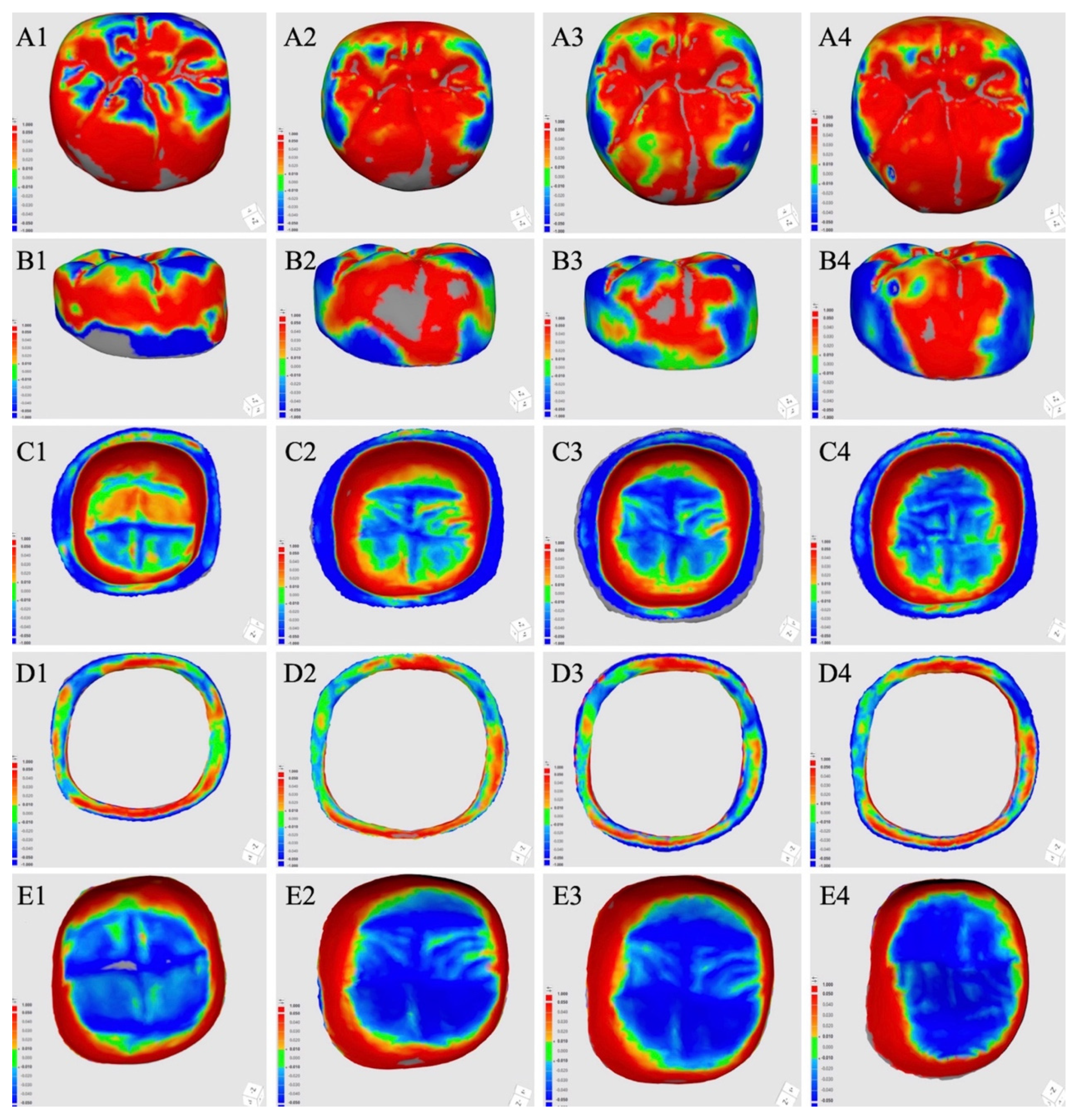
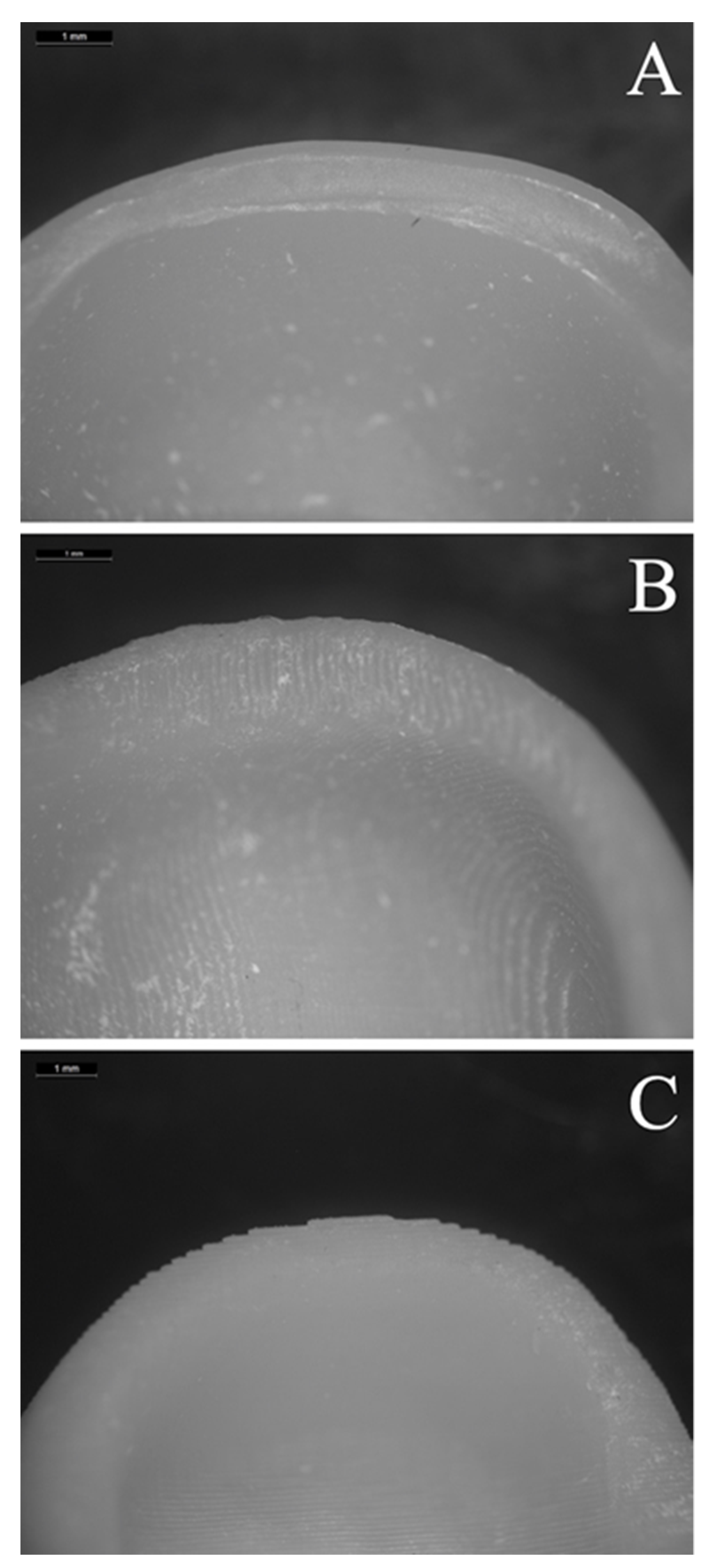
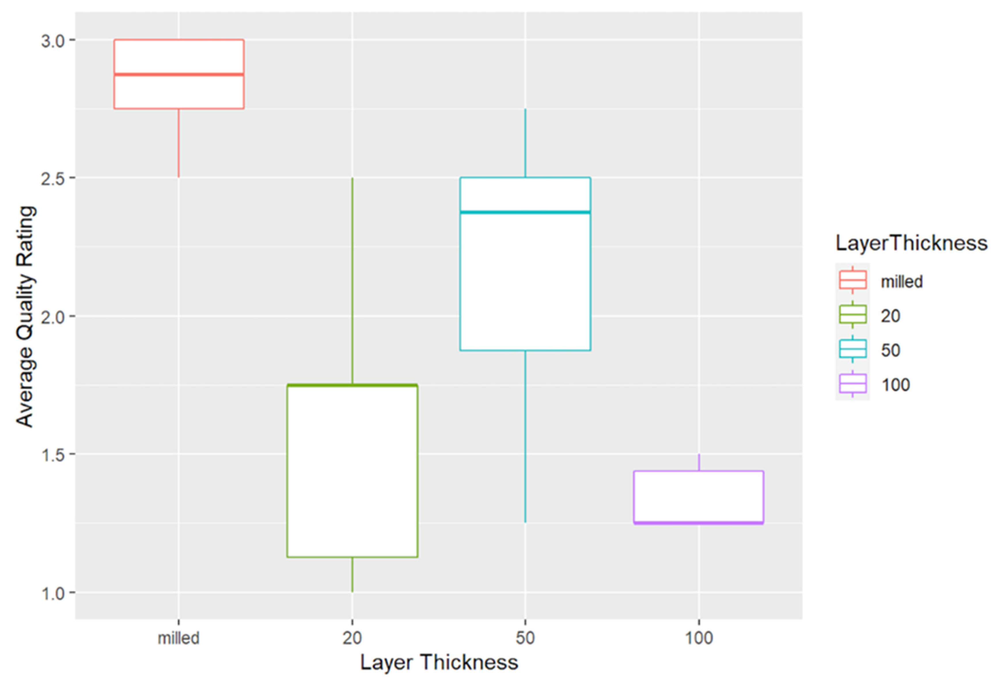
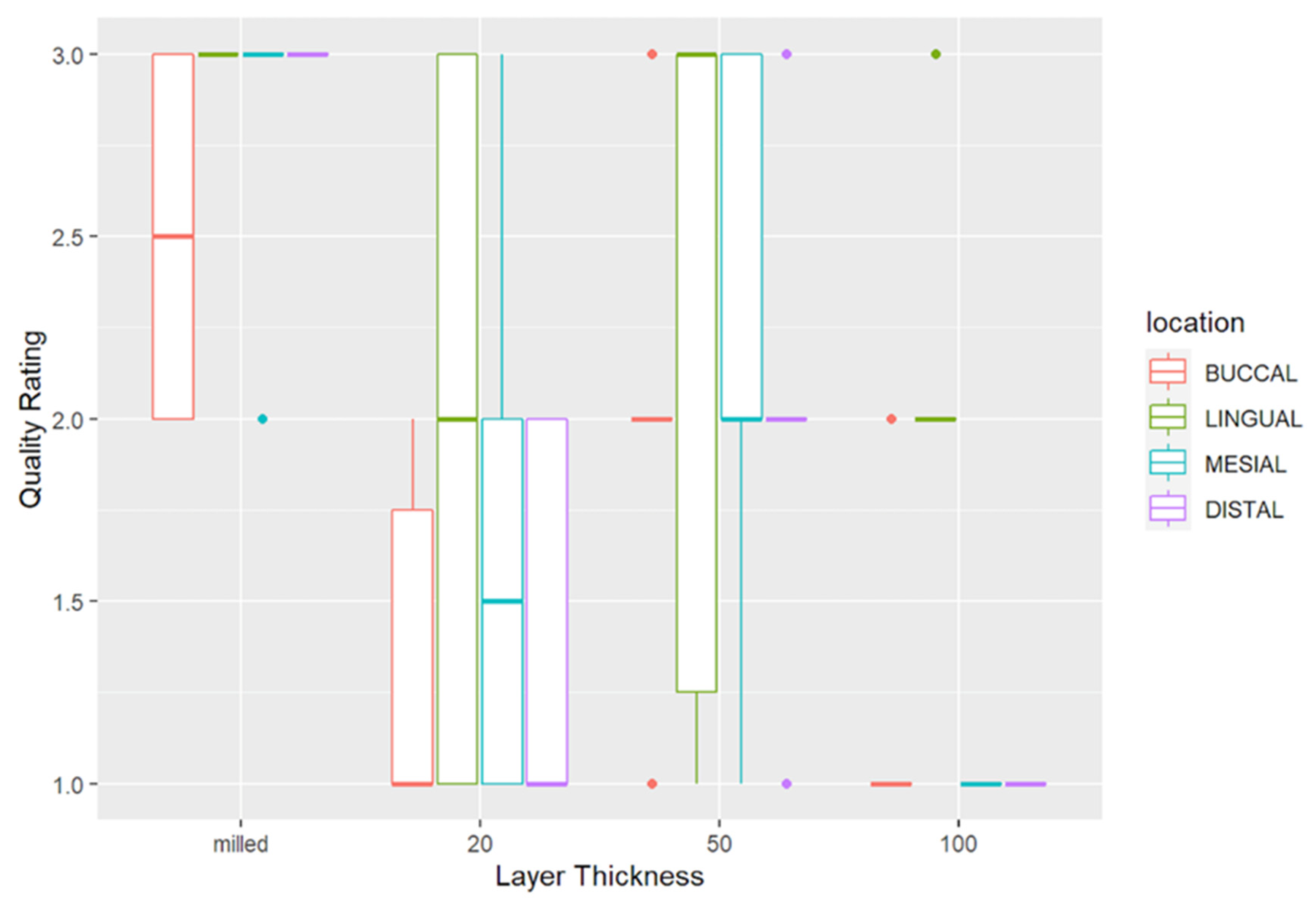
Three different layer thicknesses (20 µm, 50 µm, and 100 µm) were used to print the crowns (n = 10 per layer thickness). First, the RS-STL file of the crown was imported to DLP software (MoonRay S100, SprintRay Inc, Los Angeles, CA, USA) to arrange the build angle and support configuration. As recommended by the manufacturer of the printing resin material (Nextdent Crown and Bridge Micro Filled Hybrid-MFH, C&B; 3D systems, Soesterberg, The Netherlands), the occlusal surface of the crown was angled 45° from the print area for improved occlusal surface details. The semiautomatically created support structures were checked and supports that were automatically created on the margin area and fitting surfaces of the crowns were manually eliminated. Then, this configuration was duplicated 10 times and 10 identical crown configurations were arranged in the build platform of the DLP printer (MoonRay S100 Software, SprintRay Inc, Los Angeles, CA, USA) to print all crowns in the same configuration. This configuration was further saved for 3 different layer thicknesses to print the crowns in identical configuration, but by using different layer thicknesses. The crowns were printed with the DLP printer (MoonRay S100, SprintRay Inc, Los Angeles, CA, USA) and an interim printing resin material (N1 shade, Nextdent Crown and Bridge Micro Filled Hybrid-MFH, C&B; 3D systems, Soesterberg, The Netherlands, Lot: XH312N21) by using 20 µm, 50 µm, or 100 µm (n = 10) layer thickness. According to the manufacturer, the printing material has 100–130 MPa flexural strength, 2400–2600 MPa flexural modulus, ≤70 µg/mm3 sorption, and ≤15.5 µg/mm3 solubility. The DLP printer has UV DLP projector, LED-based light source, and 405 nm blue-violet light resin curing unit. After printing, the printed crowns were removed from the platform by using a putty knife, ultrasonically rinsed for 5 min (first 3 min then 2 min) in 96% clean alcohol solution (Alcohol isopropilico, Quimi Klean, Mexico City, Mexico) according to the manufacturers’ recommendations. After ensuring that the crowns were dry and free of alcohol residue, they were postpolymerized by using an ultraviolet polymerizing unit (SprintRay Procure Model SRP1811A, SprintRay Inc, Los Angeles, CA, USA) (405 nm LED arrays) for 30 min (Figure 2).
The support structures were cut and trimmed after cooling and the surface was gently smoothened to prevent errors during the alignment procedure.
In the milling technique, crowns were milled (Wieland Zenotec mini, V6.12.04, Wieland Dental + Technik GmbH & Co.KG, Pforzheim, Germany) from a polymethyl methacrylate (PMMA) block (A2 shade, Lot number: HL201104, Upcera, Shenzhen Upcera Dental Technology Co. Ltd., Shenzen, Guandong, China). The designed STL file was inserted in the block to mill 10 identical crowns. The support structures were cut and trimmed after milling, and the support surfaces were gently smoothened. All crown fabrication processes were performed by one operator (G. Ç).

2.3. Crown Analysis

All crowns were examined under optical magnification loupe (3.5×) to ensure that they were free from any defects and no adjustments were made on the inner surfaces of the crowns [26]. Crowns were then kept in dry and lightproof boxes to scan within 48 h after the fabrication. First, the scanner was calibrated and then the printed and milled crowns were scanned by using an intraoral scanner (Medit i500 v. 1.2.1, Medit, Seoul, Korea) by the same operator (G. Ç) and the scan files were converted to STL files (test-scan STL). During the scan, the crowns were hold with small tweezers and as the intraoral scanner utilized has a filter for colors, the parts in contact with the tweezers were scanned afterwards.
For the deviation analysis, root mean square (RMS) was used to indicate how far the deviations were from zero between the 2 different datasets [32]. A low RMS value indicated a high degree of 3D matching of the superimposed data, which translated to high trueness [33]. First, test-scan STL files and designed crown STL file (RS-STL) were imported into a software (Medit Link, Medit, Seoul, Korea). Compare tool (Medit Compare v1.1.1.61, Medit, Seoul, Korea) of the software, which has an automatic alignment tool was used for the superimposition, virtually separation of the surfaces, and RMS calculation. For the superimposition, RS-STL was moved to the reference data and test-scan STL was moved to the target data by using the alignment tool of the software. Then, manual alignment tool of the software was selected and 3 reference points; central fossa, mesial and distal triangular fossae were selected both in the reference and target data. Test-scan STL files were then superimposed over the RS-STL file (Figure 3).
To generate color maps to represent the 3D deviation, deviation display mode of the software was used. The maximum/minimum critical (nominal) values were set at +50/−50 µm with a tolerance range of +10/−10 µm, respectively [34]. After the superimposition, color-difference maps were created to compare the test scan STL file and the RS-STL for the overall RMS, which includes all surfaces of the crowns. The software automatically calculated the RMS from the color-difference maps, without the need for an additional formula. For the RMS of external, intaglio, marginal area, and intaglio occlusal surfaces of the crowns, the test-scan STL files and the RS-STL file were imported again, and these surfaces were virtually separated both in test-scan STL files and RS-STL file [34] dividing crowns into 4 different parts by using the edit mode of the software. After separation, the superimposition was done once again for each surface (external, intaglio, marginal area, and intaglio occlusal surfaces) of each crown by using automatic alignment mode of the software, and the color-difference maps were generated for those surfaces and the RMS values were automatically calculated (Figure 4). The areas with deviations exceeding the scale utilized were presented as gray by the software. However, all areas were included in the RMS calculation.
For margin quality comparison, each crown was randomly numbered by an independent individual, and then visually examined with an optical microscope (Zeiss) under ×60 magnification by a single operator (G.Ç.) who was blinded about the numbering, and a grading system from 1 to 3 was used as performed in a previous study [29]. Grade 1 crown margins indicated rough edges similar to layers. Grade 2 crown margins indicated slightly rough edges similar to waves. Grade 3 crown margins indicated smooth edges (Figure 5). Margin quality was examined at each margin location (buccal, lingual, mesial, and distal) of each crown, and the average was calculated for each printed or milled crown.

2.4. Statistical Analysis

The statistical evaluation of the data was performed by using the statistical software R. Normality assumption was verified using the Shapiro Wilks test. Difference in RMS values between fabrication technique within each area of measurement, difference in average quality rating between fabrication technique, and difference in quality rating between margin location for each fabrication technique was analyzed by using the one-way analysis of variance (ANOVA). Additionally, pairwise comparisons within the groups were analyzed with Tukey HSD Post-Hoc analysis (α = 0.05).

3. Results

One-way ANOVA results of the RMS values of each surface are presented in Table 1, and Figure 6 illustrates the RMS at each measured surface for control and different layer-thickness groups.
Significant differences were observed among fabrication techniques for intaglio and intaglio occlusal surface RMS values (p ≤ 0.026). Milled crowns presented significantly lower RMS values than 100 µm crowns at both intaglio (p = 0.025, estimated difference in means: −20.2 µm) and intaglio occlusal (p = 0.021, estimated difference in means: −10 µm) surfaces. Every other pairwise comparison for intaglio (p ≥ 0.251) and intaglio occlusal (p ≥ 0.178) surfaces were nonsignificant.
Average margin quality of the crowns showed significant differences (Figure 7); based on the 3-point scale, milled crowns presented higher quality than the others (p < 0.001 vs. 20 µm, estimated difference in means: 1.25 points; p = 0.001 vs. 50 µm, estimated difference in means: 0.68 points; and p < 0.001 vs. 100 µm, estimated difference in means: 1.53 points).
The margin quality of 50 µm crowns was higher than that of 20 µm (p = 0.006, estimated difference in means: 0.58 points) and 100 µm (p < 0.001, estimated difference in means: 0.85 points) crowns. The difference between 20 µm crowns and 100 µm crowns was nonsignificant (p = 0.348). Table 2 summarizes the p values when the Tukey HSD test was applied to analyze the effect of margin location on the margin quality within each fabrication technique.
No significant differences were found between the quality of the margins at different locations for 20 µm (p ≥ 0.121) and 50 µm (p ≥ 0.765) crowns. However, margin quality of milled (p = 0.002) and 100 µm (p < 0.001) crowns were significantly affected by the margin location. The mesial, distal, and lingual margin quality of the milled crowns was similar (p ≥ 0.885), while buccal margin quality was inferior to that at other locations (p ≤ 0.03). For the 100 µm crowns, lingual margin quality was higher compared with other locations (p < 0.001); the differences among other locations were nonsignificant (p ≥ 0.831). The margin quality in control and each layer thickness groups according to the margin location is presented in Figure 8.

4. Discussion

Significant differences in terms of trueness were found comparing the 100 µm printing layer thickness to other groups. Therefore, the first null hypothesis was rejected. The best margin quality was found in milled crowns, followed by the 50 µm layer thickness group. Accordingly, the second null hypothesis was rejected.
In terms of trueness, the present study showed that 3D-printed interim crowns, printed with a layer thickness of 20 or 50 µm, are similar to the milled interim crowns, which are currently used routinely. Similar results have been reported also for 3D-printed ceramic crowns [15,35]. The effect of the layer thickness on the trueness of 3D printed casts and complete dentures has been demonstrated in previous studies [30,36]. Those studies demonstrated the highest trueness when a layer thickness of 100 µm was used, whereas the 100 µm layer thickness resulted in the lowest trueness in the present study. The main difference between those and the present study may be due to the nature of the objects printed. Neither casts nor dentures have as thin and tapered margins as crowns have, instead, they have large plain and round surfaces [36]. When the layer thickness is too small, discrepancies can occur, especially with such thin margins [30]. The combination of thin layer thickness and thin margins could also be an explanation for the fact that the trueness seems to be slightly higher in the 50 µm group compared to the 20 µm group, although this difference was not statistically significant. It should be noted that the aforementioned scoping review on complete denture fabrication included various studies [36], which applied varying settings for the 3D-printing, and therefore, the deviations in trueness may not be attributed to the layer thickness alone. A study, which showed that layer thickness did not affect the trueness of printed models when 100 µm was applied, still recommended the use of 100 µm layer thicknesses due to the economic advantages as the printing time is shortened [37]. However, the time factor may be less important when small restorations such as crowns are fabricated. Considering the effect of the layer thickness on trueness at thin surfaces such as crown margins [30], high layer thickness may be expected to result in low trueness, which was confirmed by lower trueness and marginal quality found with the 100 µm group in the present study. Although a previous study has shown that the marginal fit of 3D-printed crowns is within a clinically acceptable range, and that there is no difference between 3D-printed and milled crowns [38], significant differences in the margin quality were found in the present study. The marginal quality in the present study was evaluated by a 3-point scale instead of absolute values, indicating worse margin quality of the 3D-printed crowns irrespective of the layer thickness. Interestingly, the margin quality was not influenced by margin location in 20 µm and 50 µm groups, but in milled and 100 µm groups. The significantly worse margin quality at the buccal aspect of the milled group may be due to the slightly narrower preparation margin on the distobuccal side. During the milling process, defects may occur at thin crown margins [39]. Hypothetically, the preparation margin could also be the reason for the better marginal quality at the lingual crown margin in the 100 µm group. The lingual aspect of the margin preparation was the most uniform, which might have allowed the crown to be fabricated accurately even with the largest layer thickness of 100 µm. A large layer thickness might have a negative effect on the margin quality, especially with varying crown margin thicknesses. The fact that the margins of the prepared tooth were not uniform at all aspects is a limitation of the present study. Future studies are necessary to determine the effect of layer thickness on trueness and margin quality.
The reference data set can be recorded with a laboratory scanner, or more precisely with a high-precision optical or tactile industrial scanner [40]. However, because such scanners cannot be used intraorally, the reference data set was taken with an intraoral scanner. The intraoral scanner used has a software that enables immediate scanning of the fabricated restoration and the analysis of the trueness of the restoration comparing with the design STL. Adequate scan accuracy with the intraoral scanner used has been demonstrated in previous studies on single crowns [41]. Generally, intraoral scanners have demonstrated sufficient accuracy especially when scanning partial arches, as executed in the present study [41]. Since the reference data set was used to fabricate all crowns, potential errors during scanning would be expected to affect all groups similarly and therefore, can be considered negligible for its effect on the comparisons aimed. The trueness was analyzed by the calculation of RMS differences between the test and reference datasets. Calculating RMS differences is the most commonly applied trueness analysis in digital dentistry [42]. However, the effect of different software programs to calculate RMS values between corresponding datasets is not clear [43,44,45]. The influence of the build angle on the fabrication of 3D-printed crowns has been shown in various studies [14,18,25,26,29]. The build angle of 45° was used to print the crowns based on the recommendation by the manufacturer. A 45° build angle is similar to 135° build angle in terms of crown orientation during the fabrication, except that the 45° angle assumes a buccal crown orientation while 135° angle assumes a lingual crown orientation. The effect of build angle on crown accuracy when DLP technology is used was previously investigated and the highest accuracy was achieved when 135° angle was used [18].
Due to the pilot nature of the present study, no sample size calculation could be performed. The sample size was based on earlier studies, focusing on the 3D-printing accuracy [14,46] and enabled the detection of statistically significant differences in terms of trueness and marginal quality. One of the main limitations of the present study is that only one material was used for the fabrication of milled and 3D-printed crowns. Since previous studies have shown that the material has a significant influence on crown accuracy [47], the present study findings are not directly applicable to other materials. Nevertheless, the present study is the first of its kind indicating the influence of layer thickness on the accuracy of 3D printed resin crowns. The quantification of the marginal gap size was not performed in the present study. Since there was no difference between the milled and 3D-printed crowns when a layer thickness of 20 µm or 50 µm was used, a major difference in terms of marginal gap size would not be expected between these groups, however, marginal gap measurements can be performed in future studies to see how milling/printing trueness translates to marginal quality. The fact that the margin quality was only assessed by one observer is another weak point. Since the observer was blinded during the evaluation, it can at least be assumed that the risk of bias (e.g., by personal preferences) was minimized [48].

5. Conclusions

Considering limitations of the present study, it can be concluded that the trueness and marginal quality of 3D-printed interim crowns was influenced by the printing layer thickness. For improved trueness and margin quality for interim crowns using the applied materials and settings, a printing layer thickness of 20 or 50 µm may be preferable over 100 µm.

Author Contributions

Conceptualization, G.Ç., A.R.C. and B.Y.; methodology, G.Ç. and A.R.C.; software, G.Ç. and M.B.D.; validation, G.Ç., M.B.D. and B.Y.; formal analysis, G.Ç. and W.-E.L.; investigation, G.Ç.; resources, A.R.C.; data curation, W.-E.L., M.B.D. and S.A.-A.; writing—original draft preparation, M.B.D. and S.A.-A.; writing—review and editing, M.B.D., S.A.-A., M.S. and B.Y.; visualization, G.Ç.; supervision, M.S. and B.Y.; project administration, G.Ç. and B.Y., funding acquisition, none. All authors have read and agreed to the published version of the manuscript.

Funding

The authors would like to thank Laboratorio Innovando (Mexico City, Mexico) for their support in milling crowns and 3D Sequence Digital Laboratorio (Mexico City, Mexico) for their support in printing machine. The materials used in the present study were self-funded.

Data Availability Statement

Data available on request. The data presented in this study are available on request from the corresponding author. The data are not publicly available due to ongoing research using the data.

Acknowledgments

The authors would like to thank Laboratorio Innovando (Mexico City, Mexico) for their support in milling crowns and 3D Sequence Digital Laboratorio (Mexico City, Mexico) for their support in printing machine. The materials used in the present study were self-funded.

Conflicts of Interest

The authors declare no conflict of interest.

References

  1. Liu, Y.; Ye, H.; Wang, Y.; Zhao, Y.; Sun, Y.; Zhou, Y. Three-dimensional analysis of internal adaptations of crowns cast from resin patterns fabricated using computer-aided design/computer-assisted manufacturing technologies. Int. J. Prosthodont. 2018, 31, 386–393. [Google Scholar] [CrossRef] [Green Version]
  2. Kessler, A.; Hickel, R.; Reymus, M. 3D printing in dentistry-state of the art. Oper. Dent. 2020, 45, 30–40. [Google Scholar] [CrossRef]
  3. Tahayeri, A.; Morgan, M.; Fugolin, A.P.; Bompolaki, D.; Athirasala, A.; Pfeifer, C.S.; Ferracane, J.L.; Bertassoni, L.E. 3D printed versus conventionally cured provisional crown and bridge dental materials. Dent. Mater. 2018, 34, 192–200. [Google Scholar] [CrossRef] [PubMed]
  4. van Noort, R. The future of dental devices is digital. Dent. Mater. 2012, 28, 3–12. [Google Scholar] [CrossRef] [PubMed]
  5. Dawood, A.; Marti Marti, B.; Sauret-Jackson, V.; Darwood, A. 3D printing in dentistry. Br. Dent. J. 2015, 219, 521–529. [Google Scholar] [CrossRef]
  6. Muta, S.; Ikeda, M.; Nikaido, T.; Sayed, M.; Sadr, A.; Suzuki, T.; Tagami, J. Chairside fabrication of provisional crowns on FDM 3D-printed PVA model. J. Prosthodont. Res. 2020, 64, 401–407. [Google Scholar] [CrossRef] [PubMed]
  7. Revilla-León, M.; Özcan, M. Additive manufacturing technologies used for processing polymers: Current status and potential application in prosthetic dentistry. J. Prosthodont. 2019, 28, 146–158. [Google Scholar] [CrossRef] [Green Version]
  8. Salmi, M.; Paloheimo, K.S.; Tuomi, J.; Ingman, T.; Mäkitie, A. A digital process for additive manufacturing of occlusal splints: A clinical pilot study. J. R. Soc. Interface. 2013, 10, 20130203. [Google Scholar] [CrossRef]
  9. Zeng, L.; Zhang, Y.; Liu, Z.; Wei, B. Effects of repeated firing on the marginal accuracy of Co-Cr copings fabricated by selective laser melting. J. Prosthet. Dent. 2015, 113, 135–139. [Google Scholar] [CrossRef]
  10. Salmi, M.; Paloheimo, K.S.; Tuomi, J.; Wolff, J.; Mäkitie, A. Accuracy of medical models made by additive manufacturing (rapid manufacturing). J. Craniomaxillofac. Surg. 2013, 41, 603–609. [Google Scholar] [CrossRef]
  11. Reyes, A.; Turkyilmaz, I.; Prihoda, T.J. Accuracy of surgical guides made from conventional and a combination of digital scanning and rapid prototyping techniques. J. Prosthet. Dent. 2015, 113, 295–303. [Google Scholar] [CrossRef] [PubMed]
  12. Alharbi, N.; Wismeijer, D.; Osman, R.B. Additive manufacturing techniques in prosthodontics: Where do we currently stand? A critical review. Int. J. Prosthodont. 2017, 30, 474–484. [Google Scholar] [CrossRef] [PubMed]
  13. Liu, Y.; Bai, W.; Cheng, X.; Tian, J.; Wei, D.; Sun, Y.; Di, P. Effects of printing layer thickness on mechanical properties of 3D-printed custom trays. J. Prosthet. Dent. 2020. [Google Scholar] [CrossRef]
  14. Shim, J.S.; Kim, J.E.; Jeong, S.H.; Choi, Y.J.; Ryu, J.J. Printing accuracy, mechanical properties, surface characteristics, and microbial adhesion of 3D-printed resins with various printing orientations. J. Prosthet. Dent. 2020, 124, 468–475. [Google Scholar] [CrossRef]
  15. Lerner, H.; Nagy, K.; Pranno, N.; Zarone, F.; Admakin, O.; Mangano, F. Trueness and precision of 3D-printed versus milled monolithic zirconia crowns: An in vitro study. J. Dent. 2021, 103792. [Google Scholar] [CrossRef]
  16. Zhang, Z.C.; Li, P.L.; Chu, F.T.; Shen, G. Influence of the three-dimensional printing technique and printing layer thickness on model accuracy. J. Orofac. Orthop. 2019, 80, 194–204. [Google Scholar] [CrossRef]
  17. Son, K.; Lee, J.H.; Lee, K.B. Comparison of intaglio surface trueness of interim dental crowns fabricated with SLA 3D printing, DLP 3D printing, and milling technologies. Healthcare 2021, 9, 983. [Google Scholar] [CrossRef] [PubMed]
  18. Osman, R.B.; Alharbi, N.; Wismeijer, D. Build angle: Does it influence the accuracy of 3D-printed dental restorations using digital light-processing technology? Int. J. Prosthodont. 2017, 30, 182–188. [Google Scholar] [CrossRef]
  19. Peng, C.C.; Chung, K.H.; Ramos, V., Jr. Assessment of the adaptation of interim crowns using different measurement techniques. J. Prosthodont. 2020, 29, 87–93. [Google Scholar] [CrossRef]
  20. Peng, C.C.; Chung, K.H.; Yau, H.T.; Ramos, V., Jr. Assessment of the internal fit and marginal integrity of interim crowns made by different manufacturing methods. J. Prosthet. Dent. 2020, 123, 514–522. [Google Scholar] [CrossRef] [PubMed]
  21. Simoneti, D.M.; Pereira-Cenci, T.; Dos Santos, M.B.F. Comparison of material properties and biofilm formation in interim single crowns obtained by 3D printing and conventional methods. J. Prosthet. Dent. 2020. [Google Scholar] [CrossRef]
  22. Mai, H.N.; Lee, K.B.; Lee, D.H. Fit of interim crowns fabricated using photopolymer-jetting 3D printing. J. Prosthet. Dent. 2017, 118, 208–215. [Google Scholar] [CrossRef] [PubMed]
  23. Kim, Y.H.; Jung, B.Y.; Han, S.S.; Woo, C.W. Accuracy evaluation of 3D printed interim prosthesis fabrication using a CBCT scanning based digital model. PLoS ONE 2020, 15, e0240508. [Google Scholar] [CrossRef]
  24. Davis, S.; O’Connell, B. The provisional crown. J. Ir. Dent. Assoc. 2004, 50, 167–172. [Google Scholar] [PubMed]
  25. Alharbi, N.; Osman, R.; Wismeijer, D. Effects of build direction on the mechanical properties of 3D-printed complete coverage interim dental restorations. J. Prosthet. Dent. 2016, 115, 760–767. [Google Scholar] [CrossRef]
  26. Alharbi, N.; Osman, R.B.; Wismeijer, D. Factors influencing the dimensional accuracy of 3d-printed full-coverage dental restorations using stereolithography technology. Int. J. Prosthodont. 2016, 29, 503–510. [Google Scholar] [CrossRef] [PubMed] [Green Version]
  27. Yoo, S.Y.; Kim, S.K.; Heo, S.J.; Koak, J.Y.; Kim, J.G. Dimensional accuracy of dental models for three-unit prostheses fabricated by various 3D printing technologies. Materials 2021, 14, 1550. [Google Scholar] [CrossRef]
  28. Çakmak, G.; Yilmaz, H.; Treviño, A.; Kökat, A.M.; Yilmaz, B. The effect of scanner type and scan body position on the accuracy of complete-arch digital implant scans. Clin. Implant. Dent. Relat. Res. 2020, 22, 533–541. [Google Scholar] [CrossRef] [PubMed]
  29. Yu, B.Y.; Son, K.; Lee, K.B. Evaluation of intaglio surface trueness and margin quality of interim crowns in accordance with the build angle of stereolithography apparatus 3-dimensional printing. J. Prosthet. Dent. 2020. [Google Scholar] [CrossRef]
  30. You, S.M.; You, S.G.; Kang, S.Y.; Bae, S.Y.; Kim, J.H. Evaluation of the accuracy (trueness and precision) of a maxillary trial denture according to the layer thickness: An in vitro study. J. Prosthet. Dent. 2021, 125, 139–145. [Google Scholar] [CrossRef]
  31. Park, G.S.; Kim, S.K.; Heo, S.J.; Koak, J.Y.; Seo, D.G. Effects of Printing Parameters on the fit of implant-supported 3D printing resin prosthetics. Materials 2019, 12, 2533. [Google Scholar] [CrossRef] [PubMed] [Green Version]
  32. Schaefer, O.; Watts, D.C.; Sigusch, B.W.; Kuepper, H.; Guentsch, A. Marginal and internal fit of pressed lithium disilicate partial crowns in vitro: A three-dimensional analysis of accuracy and reproducibility. Dent. Mater. 2012, 28, 320–326. [Google Scholar] [CrossRef] [PubMed]
  33. International Organization for Standardization. ISO-5725-2. Accuracy (Trueness and Precision) of Measurement Methods and Results-Part 2: Basic Method for the Determination of Repeatability and Reproducibility of a Standard Measurement Method; ISO: Geneva, Switzerland, 1994; Available online: http://www.iso.org/iso/store.htm (accessed on 22 December 1994).
  34. Wang, W.; Yu, H.; Liu, Y.; Jiang, X.; Gao, B. Trueness analysis of zirconia crowns fabricated with 3-dimensional printing. J. Prosthet. Dent. 2019, 121, 285–291. [Google Scholar] [CrossRef]
  35. Baumgartner, S.; Gmeiner, R.; Schönherr, J.A.; Stampfl, J. Stereolithography-based additive manufacturing of lithium disilicate glass ceramic for dental applications. Mater. Sci. Eng. C. Mater. Biol. Appl. 2020, 116, 111180. [Google Scholar] [CrossRef]
  36. Vilela Teixeira, A.B.; Dos Reis, A.C. Influence of parameters and characteristics of complete denture bases fabricated by 3D printing on evaluated properties: A scoping review. Int. J. Prosthodont. 2021. [Google Scholar] [CrossRef]
  37. Dias Resende, C.C.; Quirino Barbosa, T.A.; Moura, G.F.; Piola Rizzante, F.A.; Mendonça, G.; Zancopé, K.; Domingues das Neves, F. Cost and effectiveness of 3-dimensionally printed model using three different printing layer parameters and two resins. J. Prosthet. Dent. 2021. [Google Scholar] [CrossRef] [PubMed]
  38. Haddadi, Y.; Ranjkesh, B.; Isidor, F.; Bahrami, G. Marginal and internal fit of crowns based on additive or subtractive manufacturing. Biomater. Investig. Dent. 2021, 8, 87–91. [Google Scholar] [CrossRef]
  39. Li, R.; Chen, H.; Wang, Y.; Sun, Y. Performance of stereolithography and milling in fabricating monolithic zirconia crowns with different finish line designs. J. Mech. Behav. Biomed. Mater. 2021, 115, 104255. [Google Scholar] [CrossRef]
  40. Pan, Y.; Tsoi, J.K.H.; Lam, W.Y.H.; Pow, E.H.N. Implant framework misfit: A systematic review on assessment methods and clinical complications. Clin. Implant. Dent. Relat. Res. 2021, 23, 244–258. [Google Scholar] [CrossRef]
  41. Zimmermann, M.; Ender, A.; Mehl, A. Local accuracy of actual intraoral scanning systems for single-tooth preparations in vitro. J. Am. Dent. Assoc. 2020, 151, 127–135. [Google Scholar] [CrossRef]
  42. O’Toole, S.; Osnes, C.; Bartlett, D.; Keeling, A. Investigation into the accuracy and measurement methods of sequential 3D dental scan alignment. Dent. Mater. 2019, 35, 495–500. [Google Scholar] [CrossRef]
  43. Pellitteri, F.; Brucculeri, L.; Spedicato, G.A.; Siciliani, G.; Lombardo, L. Comparison of the accuracy of digital face scans obtained by two different scanners. Angle Orthod. 2021, 91, 641–649. [Google Scholar] [CrossRef]
  44. Peroz, S.; Spies, B.C.; Adali, U.; Beuer, F.; Wesemann, C. Measured accuracy of intraoral scanners is highly dependent on methodical factors. J. Prosthodont. Res. 2021. [Google Scholar] [CrossRef] [PubMed]
  45. Son, K.; Lee, W.S.; Lee, K.B. Effect of different software programs on the accuracy of dental scanner using three-dimensional analysis. Int. J. Environ. Res. Public Health 2021, 18, 8449. [Google Scholar] [CrossRef]
  46. Alharbi, N.; Alharbi, S.; Cuijpers, V.; Osman, R.B.; Wismeijer, D. Three-dimensional evaluation of marginal and internal fit of 3D-printed interim restorations fabricated on different finish line designs. J. Prosthodont. Res. 2018, 62, 218–226. [Google Scholar] [CrossRef] [PubMed]
  47. Papadiochou, S.; Pissiotis, A.L. Marginal adaptation and CAD-CAM technology: A systematic review of restorative material and fabrication techniques. J. Prosthet. Dent. 2018, 119, 545–551. [Google Scholar] [CrossRef] [PubMed]
  48. Day, S.J.; Altman, D.G. Statistics notes: Blinding in clinical trials and other studies. BMJ 2000, 321, 504. [Google Scholar] [CrossRef] [PubMed] [Green Version]
Figure 1. Mandibular right first molar preparation.
Applsci 11 09246 g001
Figure 2. 3D-printed interim crowns ((A): 20 µm crowns; (B): 50 µm crowns; (C): 100 µm crowns).
Applsci 11 09246 g002
Figure 3. (A): Reference points determined for the superimposition of STL files (1: Central fossa; 2: Distal triangular fossa; 3: Mesial triangular fossa), (B): Superimposition of the Test-scan STL over the RS-STL by using these points.
Applsci 11 09246 g003
Figure 4. Color maps generated by the superimposition of the milled (1), 20 µm (2), 50 µm (3), and 100 µm (4) crown meshes over reference data ((A): Overall RMS; (B): External RMS; (C): Internal RMS; (D): Marginal RMS; (E): Intaglio Occlusal RMS).
Applsci 11 09246 g004
Figure 5. Margins according to the grading system ((A): Grade 3; (B): Grade 2; (C): Grade 1).
Applsci 11 09246 g005
Figure 6. Box-plot graph of the RMS values of milled and 3D-printed interim crowns according to different surfaces.
Applsci 11 09246 g006
Figure 7. Average margin quality of all groups, evaluated by using a 3-point scale, ranging from 1 (worst marginal quality) to 3 (best marginal quality).
Applsci 11 09246 g007
Figure 8. Margin quality of groups according to margin location, evaluated by using a 3-point scale that ranged from 1 (worst marginal quality) to 3 (best marginal quality).
Applsci 11 09246 g008
Table 1. Mean RMS (µm) values ± standard deviations for milled and 3D-printed interim crowns. Different superscript lowercase letters in same column indicate significant differences among groups (p < 0.05).
Layer ThicknessOverall RMS (µm)External RMS (µm) Intaglio RMS (µm) Marginal RMS (µm) Intaglio Occlusal RMS (µm)
Control (Milled)64.5 ±10.94 a54 ±21.29 a32.6 ±15.01 a11.3 ±14.36 a31.5 ±6.92 a
20 µm56.1 ±10.7 a59.4 ±10.7 a49.9 ±12.13 ab14.9 ±9.57 a33.4 ± 2.22 ab
50 µm53.3 ±9.3 a48.5 ±13.67 a45.4 ±15.75 ab9.1 ±8.02 a34.7 ±1.83 ab
100 µm61.3 ±15.31 a62.4 ±18.21 a52.8 ±17.32 b14.6 ±9.94 a41.5 ±12.55 b
p values0.1450.2630.0260.5760.024
Table 2. p values for the pairwise comparison between different margin locations for milled and 3D-printed interim crowns. p < 0.05 indicate significant differences between locations.
Locations
Layer ThicknessBuccal-LingualBuccal-MesialBuccal-DistalLingual-MesialLingual-DistalMesial-Distal
Control (Milled)0.0050.030.0050.885>0.050.885
20 µm0.1210.7620.9880.5660.2230.914
50 µm0.7650.7650.988>0.050.9150.915
100 µm<0.0010.8310.831<0.001<0.001>0.05
Publisher’s Note: MDPI stays neutral with regard to jurisdictional claims in published maps and institutional affiliations.

