Article

Morphological and Molecular Alterations Induced by Lead in Embryos and Larvae of Danio rerio

Department of Biology, Ecology and Earth Science, University of Calabria, Via P. Bucci 4/B, 87036 Rende, Italy
*
Author to whom correspondence should be addressed.
V.C. and R.M. contributed equally to this work and are the co-first authors.
Appl. Sci. 2021, 11(16), 7464; https://doi.org/10.3390/app11167464
Submission received: 25 June 2021 / Revised: 30 July 2021 / Accepted: 11 August 2021 / Published: 13 August 2021
(This article belongs to the Special Issue Heavy Metal Toxicity: Environmental and Human Health Risk Assessment)

Abstract

:
Lead (Pb) is one of the most toxic and persistent elements and may adversely affect both humans and wildlife. Given the risks posed to humans, lead is listed among priority substances of public health importance worldwide. In fish, available studies deal with high doses, and the potential hazard of Pb at low concentrations is largely unknown. Given its well-demonstrated translational value for human toxicity research, we used zebrafish as a model species. Embryos were exposed to two environmentally relevant concentrations of lead (2.5 and 5 µg/L) from 6 h post-fertilization and analyzed after 48, 96, and 144 h. The morphological abnormality arose after 48 h, and the incidence and intensity were dose and time dependent. Spinal and tail deformities were the most frequently detected alterations. Pb also modulated the expression of genes involved in the toxicological responses (sod and mt), thus demonstrating that zebrafish’s early stages are able to mount an adaptive response. Moreover, ldh and β-catenin were significantly upregulated in all groups, whereas wnt3 expression was increased in the high concentration group. Our results confirm that zebrafish embryos and larvae are valuable early warning indicators of pollution and may play a major role in ecosystems and human health monitoring.

1. Introduction

Heavy metals are naturally found in the earth’s crust, and some are essential in biological systems at very low concentrations fig [1]. These elements are considered toxic when their presence exceeds the vital dose, and heavy metal pollution is currently considered one of the most serious environmental issues [2,3,4,5,6].
Lead, among the most common heavy metals, is ubiquitous in the environment and can be found in traces in soils, plants, and water [7,8], although it has no recognized physiological role in living organisms. For its physical and chemical properties, lead has known many applications, and has been used by humans since prehistoric eras. However, following the industrial revolution, intensive anthropogenic activities, including industrial and agricultural practices, resulted in an increased and uncontrolled discharge of lead in all environmental comparts.
Lead is considered one of the most toxic and persistent elements and adversely affect humans and wildlife. Water, food, and air are key routes for human exposure in emergent nations, but lead poisoning also occurs in developed countries such as the United States. Given the risks posed to humans, lead is listed among priority substances of public health importance worldwide [4,5,9]. It is also one of the top three toxic pollutants of environmental concern [10,11], occupying second place in the Priority List of Hazardous Substances [7,12,13,14].
Once released into the environment, lead can enter aquatic habitats due to direct input, atmospheric deposition, erosion, or agricultural and industrial wastewater. Currently, lead pollution is so pervasive that globally many aquatic bodies are contaminated [15], and lead concentration in surface waters ranges from 0.05 µg/L to 566.2 mg/L worldwide [9,16,17,18].
In fish, lead can be absorbed from water, food, or sediment, but regardless of the route of entry into the body shows a high tendency to bioaccumulation in various fish tissues (including gills, liver, skin, and muscles), depending on environmental factors, species-specific features, concentration, and time of exposure [8,19,20]. From literature, it is known that in fish, embryos and larvae are the most sensitive life stages, and they can therefore serve as an early indicator of pollution even at low levels of exposure, which also applies to Pb and other heavy metals [21,22,23]. Nevertheless, Pb-induced alterations during development are largely overlooked, and most of the available studies have been conducted on adults. Pb was reported to induce several detrimental effects in adult fish, including neurotoxicity [8,24,25], alteration of liver metabolism [26,27], oxidative stress induction [28,29,30], immunological and haematological dysfunctions [22,31,32,33,34] and alteration of gills and liver histology [32,35,36,37,38].
Here we used as model species zebrafish (Danio rerio), a well-acknowledged tool widely used in environmental and human health risk assessment, as proven by the growing number of studies using this organism in the past few decades [39]. Its small body size, short reproductive cycle, high fecundity, and overall easy maintenance and husbandry make zebrafish an ideal model for environmental studies; on the other hand, its high genetic homology with humans provide a powerful basis for human toxicity research and increases the translational value of this model [40,41,42]. Notably, the early stages of development offer a valuable tool for in vivo ecotoxicological studies and allow the extrapolation of results to other vertebrate species, including humans.
World Health Organisation (WHO) and Food and Agriculture Organisation of the United Nations (FAO) have set high standard levels of daily Pb intake as acceptable (up to 7 µg/kg body weight); however, no such guideline is given for infants and children, who are relatively more sensitive. An accurate assessment of human risk and human uptake of Pb is made difficult due to the unavailability of data. It has been recently highlighted that the potential hazard of Pb below a certain threshold dose (i.e., 100 µg/L) remains largely under-investigated, and even very low Pb concentrations, claimed as harmless, may represent a risk for human health [7,9].
Much more effort is needed to develop a biological early warning system, especially in aquatic ecosystems [9], also considering both direct Pb effects and its potential of biomagnification through the food chain.
In zebrafish embryos and larvae, available studies on Pb toxicity mainly focused on neurotoxicity, neurodevelopmental, and neurobehavior effects [40,43,44,45,46,47,48]. Surprisingly, morpho-functional investigations during the sensitive phase of development are scarce, and only a few studies have furnished an overall morphological evaluation [45,49,50,51,52,53]. Moreover, all available information on fish deals with high lead concentrations, and there is a lack of research on the adverse effects of lead at environmentally relevant concentrations [53].
On these bases, here we investigated for the first time the effects induced by two environmentally relevant concentrations of Pb (2.5 μg/L and 5 μg/L) in the zebrafish model. Moreover, starting from the assumption that fish embryos are especially susceptible to environmental contaminants during the first 24 h post-fertilization (hpf) [54], we selected for the experiment early development stages (starting from 6 hpf), evaluating the Pb toxic effects after 48, 96, and 144 h. Given the need to identify early, sensitive biomarkers of Pb exposure, we first investigated morphological alterations, recognized as one of the most straightforward tools to investigate the effects of contaminants in fish [55]. To allow an objective comparison of gross morphology deformity after Pb exposure, a morphological grading system was used.
Considering that the combination of morphological changes and transcriptional responses is recognized as valid and promising tools for studying ecotoxicological effects [23], in a second step, we also applied molecular biomarkers to have a more comprehensive overview of Pb-induced outcomes. In detail, we evaluated the expression pattern of some genes involved in the early developmental process (Wnt and β-catenin) to address the mechanism underlying the putative morphological modifications caused by Pb exposure. Since the mechanisms through which lead may exert its toxicity may be different and stage-specific, we evaluated the expression of several proteins known as valuable biomarkers of metal exposure in adult fish. To protect themselves against heavy metals, living organisms, including fish and mammals, activate several defense systems, including the metallothionein (MTs) modulation; these are low molecular weight proteins effective for detoxifying non-essential metals, and protecting cells from oxidative stress [35,56,57,58,59,60,61,62].
Incidence of oxidative stress resulting from exposure to Pb and heavy metal contamination is the most common response in both fish and mammalian models [51]; therefore, we investigated the expression of two markers of oxidative stress induction (superoxide dismutase 1-sod1 and lactate dehydrogenase-ldh) during zebrafish development. The present study aims to elucidate the role of morphological studies for assessing sub-lethal Pb toxicity and provide a set of sensitive and early informative biomarkers for environmental risk assessment.

2. Materials and Methods

2.1. Test Substance

A stock solution of lead acetate [Pb (CH3COO)2, Sigma–Aldrich Chemical Co., St. Louis, MO, USA] (1000 µg/L) was prepared using distilled water. An appropriated stock solution was diluted in aged tap water to reach the following nominal concentrations: 2.5 µg/L and 5 µg/L, also referred to as the low and the high concentration, respectively. Low and high Pb concentrations correspond to 0.011% and 0.022% of the zebrafish embryos median lethal concentration at 96 h (LC5096), respectively (Yin et al., 2017). These concentrations were chosen considering the worldwide environmental contamination of Pb in surface water and can be considered very low concentrations [9,16,17,18].

2.2. Zebrafish Husbandry and Maintenance

Healthy adults of both sexes of Danio rerio (approximately 6/8 months of age) were obtained from a local store. In the laboratory, animals were acclimatized in an aquarium containing 100 L of aged tap water for two weeks. During the acclimatization period, the animals were fed twice a day with commercial fish food and water parameters were daily monitored and kept constant (temperature 26 ± 0.5 °C, pH 7.3, conductivity 300 Ls/cm, dissolved oxygen 8 ± 1 mg/L, hardness 180 mg, 14 h light/10 h dark photoperiod).
Zebrafish embryos were obtained from the natural spawning of adult zebrafish. Briefly, adults male and females were collocated in standard breeding tanks with a 2:1 male-female ratio. The following day, embryos were collected, rinsed in water, and placed in a 26 ± 0.5 °C incubator in Petri dishes (density: 1 egg/mL) prefilled with aged tap water.
All experiments were conducted according to the European Convention for the Protection of Vertebrate Animals used for Experimental and other Scientific Purposes (Council of Europe No. 123, Strasbourg, 1985).

2.3. Exposure Conditions

According to a standard staging guideline, before the experiment started, embryos’ developmental stage was determined under a stereomicroscope by distinctive morphological features [63]. Exposure started once embryos were at 6 hpf and was performed in Petri dishes (density: 1 egg/mL) prefilled with the appropriate Pb solution or untreated aged tap water (control). During the experiments, the solutions were renewed daily, according to Roy et al. (2015). The Petri dishes were placed in a temperature- and light-controlled incubator set to the same circadian rhythm used for adult zebrafish. After hatching and the yolk sac regression, larvae were fed ad libitum with brine shrimp nauplii (Artemia salina). Two sets of experiments were conducted, and three independent replicates were performed for each.
Mortality and hatching rate—Embryo mortality and hatching rate were evaluated using a subsample of 30 embryos for each exposed group, including the control. The numbers of survived and hatched embryos were counted every day, and dead embryos were readily removed. Embryos/larvae viability (%) was assessed by observing the following parameters: egg coagulation, heartbeat, and spontaneous motility missing. In each treatment group, hatched larvae were counted, and the hatching rate (%) was determinate as follows: hatched numbers/total exposed numbers × 100.
Gross morphology, deformity incidence and quantitative real time PCR—For morphological analysis, animals (n = 90) were sampled at the selected time points: 48, 96, and 144 h corresponding to 54, 102, and 150 hpf. Prior to the morphological analyses, embryos/larvae were anesthetized in Tricaine methanesulphonate (MS-222) (200 mg/L) to inhibit movement. Observations were conducted using a stereomicroscope Optika SLX-3 equipped with a C-HP Camera (Optika Microscopes ITALY). All individuals were photographed and the obtained images were used to check the morphological malformations. Moreover, to evaluate the severity of morphological defects and allow reproducibility and comparison between the experimental and control groups, a morphological scoring system proposed by Herrmann [64] was used with some modifications. Briefly, observing the images acquired for each individual (dorsal and frontal view), the extent of each malformation was quantified by assigning a numerical value to each lesion. The grading system used was: 0 = no effect (−); 1 = weak (+); 2 = medium (++); 4 = strong (+++). The overall severity of deformities is expressed as the sum of the estimated values for each malformation. The obtained numerical scores for all experimental groups (including the control) were statistically compared. Cumulative deformity, defined as individuals with malformations among total observed individuals, and the deformity incidence, defined as the percentage of individuals with a specific malformation among total individuals, were recorded after 48, 96, and 144 h of exposure.
All photographed animals were promptly stored at -80 for subsequent real-time PCR analyses. Total RNA of all exposed groups and control group was isolated using the PureLink RNA Mini Kit (Thermo Fisher Scientific, Waltham, MA, USA) according to the manufacturer’s instructions. RNAs quantity and quality were determined by spectrophotometry using the NanoDrop One (Thermo Fisher Scientific, Waltham, MA, USA), and for the cDNA synthesis, we used the High capacity RNA to cDNA Kit (Applied Biosystems, Foster City, CA, USA). The genes analysed were: superoxide dismutase 1 (sod1, accession number NM_131294.1), metal-regulatory transcription factor 1 (mtf1 accession number NM_152981.1), wingless-type MMTV integration site family, member 3A (wnt3a, accession number NM_001007185.1), catenin beta-like 1 (ctnnb1, accession number NM_200866.1) and lactate dehydrogenase D (ldhd, accession number NM_199873.1). The samples were run in triplicate, and the output data were normalized against the glyceraldehyde-3-phosphate dehydrogenase (gapdh, accession number NM_001115114.1).
The amplification of cDNA was checked using TaqMan Gene Expression Assays. The Real-time PCR reactions (one cycle at 50 °C for 2 min, 95 °C for 10 min and 40 amplification cycles at 95 °C for 15 s, 60 °C for 1 min) were carried out, following the manufacturer’s guidelines, in a Light Cycler (Applied Biosystems StepOn, Real-Time PCR System, Foster City, CA, USA). Each 20 µL reaction contained: 2 µL of cDNA, 10 µL of master mix (TaqMan Universal Master Mix II) (Applied Biosystems), 1 µL of assay mix (TaqMan Gene Expression Assay), and 7 µL of RNase-free H2O. The reaction specificity was determined from the dissociation curve of the PCR product. Relative quantification of each gene expression level was normalized according to the average of the housekeeping gene expression levels. Relative mRNA expression of the gene was generated using the 2_DCt method [65].

2.4. Statistical Analyses

All analyses were performed using Graph Pad Prism 8.00 (GraphPad Software Inc., San Diego, CA, USA) at a significance level of 0.05. Replicates were statistically compared for all endpoints using the Mann–Whitney test. Because no significant differences were found among the three replicates (p > 0.05), data were pooled for subsequent analyses. Survival analysis, using a Log-rank (Mantel–Cox) Test, was carried out to compare the survivals in the experimental conditions through the entire exposure period, and a Kaplan–Meier survival graph was used to illustrate cumulative survival. Chi-square and Fisher’s exact tests were used to compare exposed groups to control groups with respect to hatching rate and deformity incidence. To test the level of significance of the grading system, two-way ANOVA followed by Tukey’s multiple comparisons test was performed. Significant differences in the levels of gene transcripts between Pb-treated and control groups were compared using one-way analysis of variance (one-way ANOVA) followed by Tukey’s multiple comparisons test. The assumption of normality was tested with the Shapiro–Wilk test.

3. Results

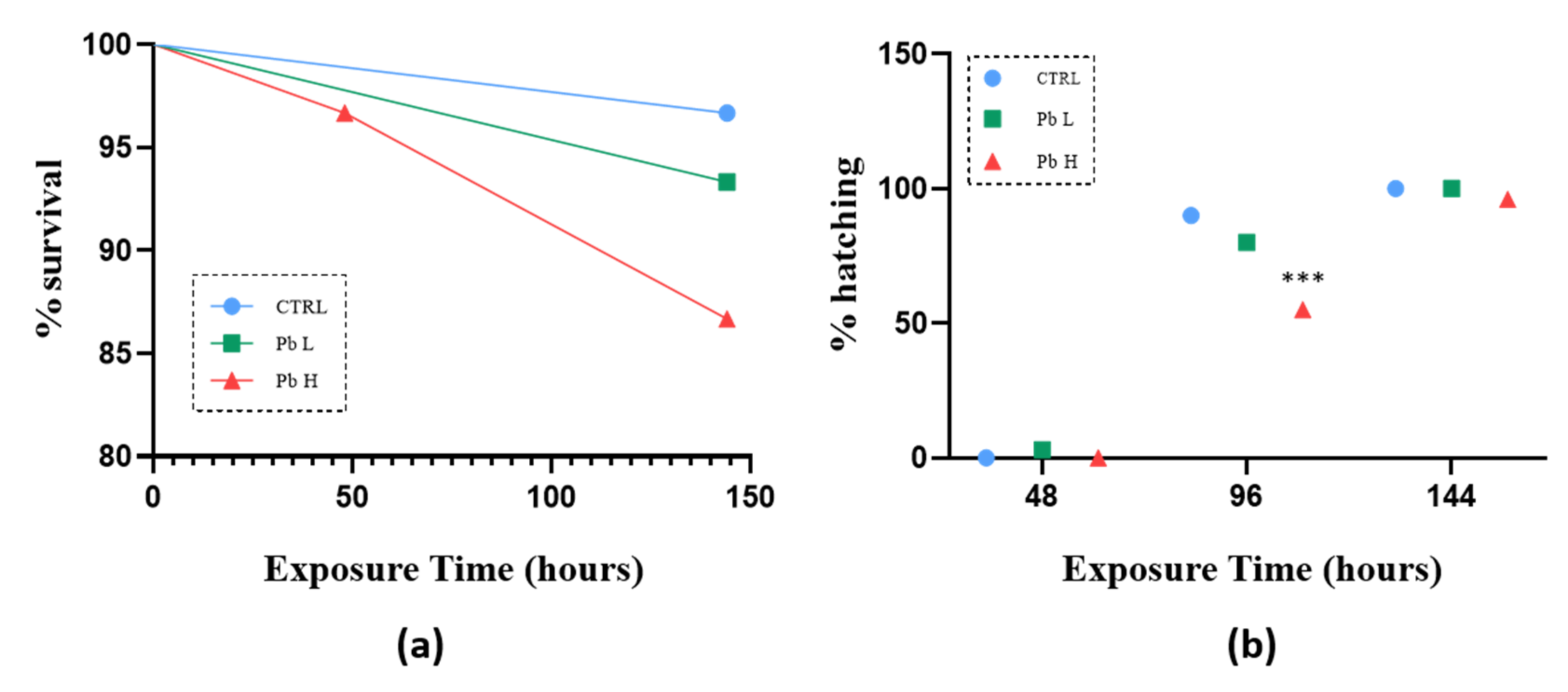
3.1. Survivor and Hatching Rate

Exposure to Pb did not affect survival in all exposed groups, and no significant differences were observed in the mortality rate compared to the control (Figure 1a). The hatching in zebrafish reared at 28 °C occurs from 48 to 72 hpf but can also take more time depending on the water temperature. Thus, time to hatch is not a staging index in this species, and development progress is independent of whether the embryo remains in the chorion or not. In our experiment, the control group hatching rate was 90% within 102 hpf (96 h from the start of the experiments), and all embryos had hatched after 150 hpf. No significant change in the hatching rate was observed in low concentration group. Animals exposed to the high Pb concentration showed a significant hatching delay compared to the control (*** p = 0.0002), and after 102 hpf (96 h of exposure), only 55.17% of individuals were hatched; however, they were swimming free out of their chorion within 150 hpf (144 h of exposure) (96.15%; Figure 1b).

3.2. Developmental Toxicity

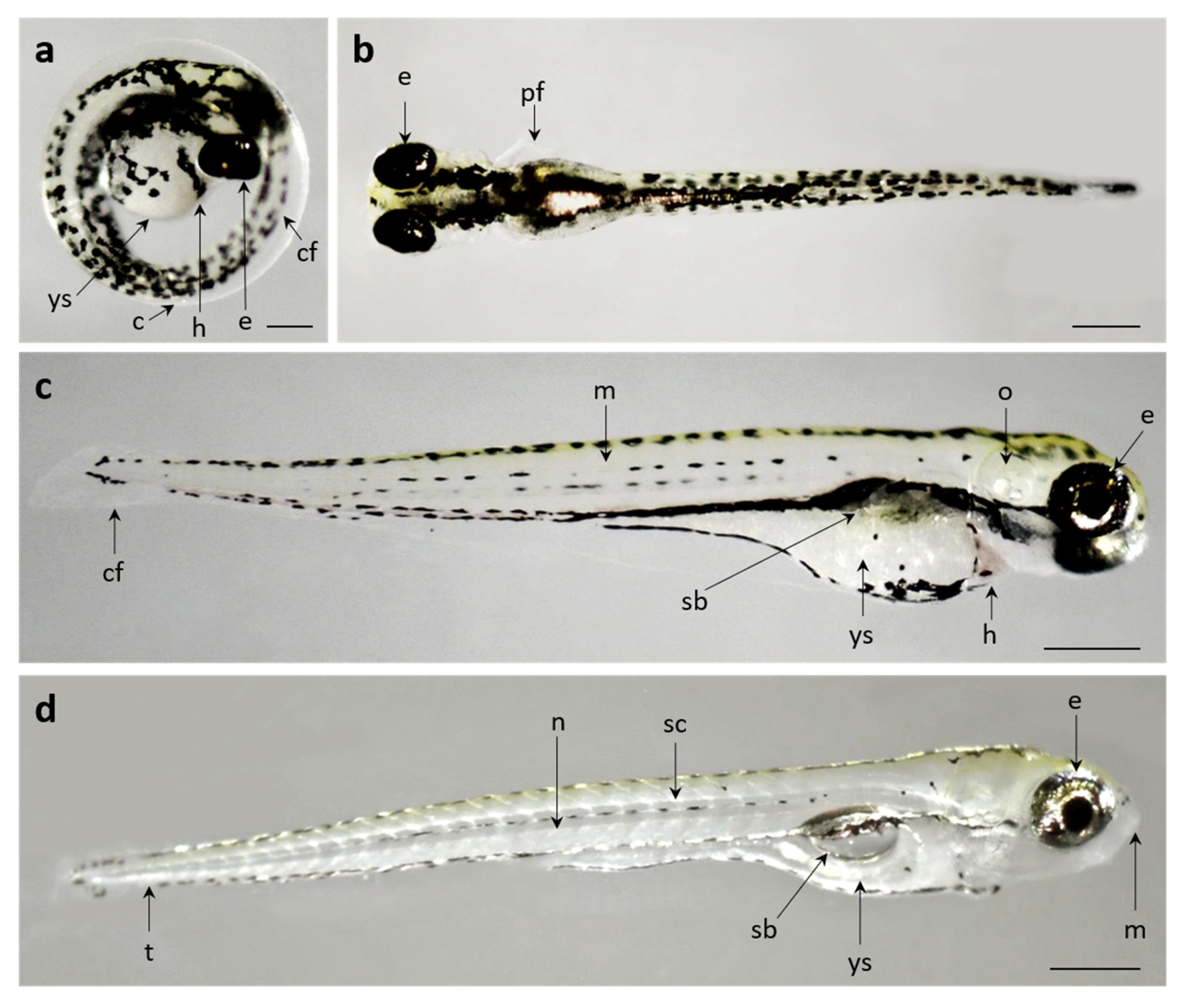
3.2.1. Control Group

To determine the developmental stage of zebrafish embryos and larvae in the present experiment, we used the data available in the literature as a reference [63,66], and no differences or discrepancies in the development of control specimens were detected. Furthermore, no individuals from the control group showed morphological abnormalities over the whole period considered.
At 54 hpf, the embryos, still enveloped within the chorion, showed high pigmentated eyes, a prominent yolk sac, and a well recognizable caudal fin. (Figure 2a). At 102 hpf, the yolk sac was still prominent, the caudal fin was further developed, and the pectoral fin was forming. Otolith-containing chambers in the otic vesicle, heart, and muscle were clearly visible. The swim bladder started to inflate and was recognizable by the accumulation of melanin, giving it a dark appearance (Figure 2b,c). At 150 hpf, the facial structure was completed, and the mouth was visible. Larvae had completely resorbed the yolk, and both the spinal cord and the notochord were well distinguishable and the swim bladder was completed and inflated (Figure 2d).

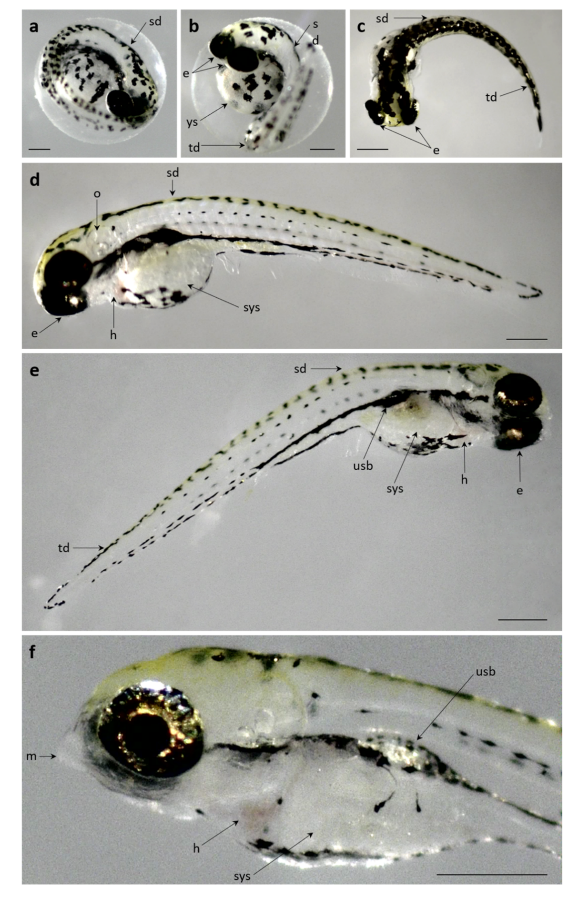
3.2.2. Pb Exposed Groups

Starting from 48 h of exposure to Pb, several types of deformities were observed in all exposed groups. Phenotypic malformations included: spinal and tail deformity, pericardial edema, and yolk swelling. As development proceeded, uninflated swim bladder was also recognizable in samples exposed to Pb.
After exposure to both low and high Pb concentrations, the severity of morphological abnormalities and the frequency of malformations (i.e., cumulative deformity) significantly increased with the increase of exposure time (**** p < 0.0001; Table 1).
In detail, after 48 h of exposure to the low Pb concentration, the first recorded alterations were spinal and tail deformity, which significantly increased compared to control (Table 1) (Figure 3a,b). Incidence of such malformation increased after 96 h of exposure when also the swelling of the yolk sac was recorded (Figure 3d). After 144 h of exposure, larvae also exhibited uninflated swim bladder (60%) (Figure 3f).
The Pb-induced deformities were dose dependent, and the overall incidence of deformities (deformity rate and score value) was significantly higher in the high-Pb exposed group (**** p < 0.0001; Table 1). Indeed, in the high concentration group, all individuals exhibited deformities at the end of the exposure period (Table 1).
After 48 h of exposure to the high Pb concentration, spinal and tail deformity incidence was significantly higher than in the control and low concentration groups (Table 1). Moreover, pericardial edema was often observed (Figure 4a,b).
After 96 h, the swelling of the yolk sac and pericardial edema were frequently observed with an incidence increased compared to low concentration (Figure 4a,b) (Figure 5c). After 144 h of exposure, all individuals showed spinal and tail deformity (Figure 4e–j). In addition, the frequency of larvae with pericardial edema (Figure 4f) and yolk sac swelling (Figure 4h,i) increased. Moreover, compared to the low concentration group, larvae with uninflated swim bladder were frequently observed (Figure 4e–j).

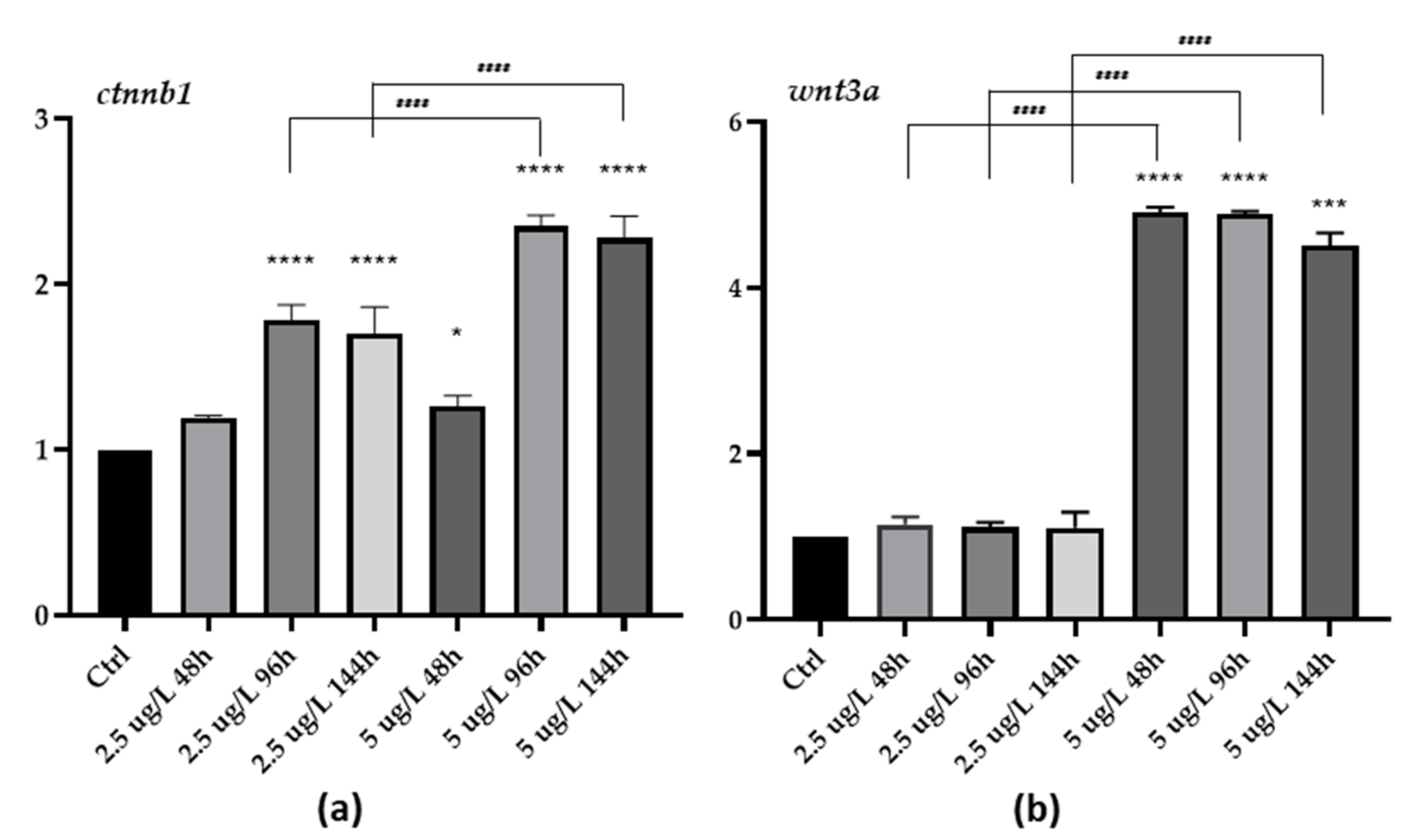
3.3. Gene Expression

Quantitative RT-PCR showed that Pb exposure induced the modulation of all analysed genes.
Metallothionein, mtf—After exposure to the low concentration of Pb, the gene was significantly upregulated compared to the control at all exposure times (* p < 0.05; Figure 5a). A similar pattern was observed in the high concentration group when the expression further increased (**** p < 0.0001; Figure 1a), reaching a peak after 96 h of exposure (**** p < 0.0001; Figure 5a).
Superoxide dismutase 1, sod1—Pb exposure resulted in a significant increase of sod1 expression in both concentration groups at all exposure time, compared to the control, also showing a dose-dependent trend (** p < 0.01; Figure 5b).
Lactate dehydrogenase, ldh—A significant upregulation of ldh expression was recorded in both concentration groups compared to the control (**** p < 0.0001; Figure 5c). The increase in gene expression was higher in the high concentration group, showing a statistically significant difference even compared to the low concentration group (#### p < 0.0001; Figure 5c).
β-catenin, ctnnb1- The low Pb concentration resulted in a significant increase in ctnnb1 expression after 96 and 144 h of exposure compared to the control group (**** p < 0.0001; Figure 6a). In the high concentration group, ctnnb1 expression significantly increased, compared to the basal condition, starting from 48 h of exposure (* p < 0.05; Figure 6a). The expression further rose after 96 and 144 h of exposure, becoming significantly relevant compared to both control and low concentration group (**** p < 0.0001; Figure 6a).
Wingless-type MMTV integration site family, wnt3a- In the low concentration group, no significant modification in wnt expression was detected. However, the expression drastically increased in the high concentration group, becoming significantly relevant compared to both the control (*** p < 0.001; Figure 6b) and the low concentration group at all exposure times (### p < 0.001; Figure 6b).

4. Discussion

Over time, several species have been used as surrogate indicators of exposure to pollutants in order to estimate the risk to human health. Still, the accuracy with which vertebrate studies predict the effects of putative toxic substances in the environment has long been a topic of interest and concern [40]. Given its well-demonstrated translational value for human toxicity research, in recent years, the use of zebrafish has undergone rapid development, becoming a prominent model in a variety of scientific disciplines, including toxicology [41]. In particular, in all vertebrates, both gene programming and development are highly conserved during the early stages of life, also allowing the maintenance of numerous morphological similarities through the evolutionary scale [40,42]. Several endpoints have been evaluated to disclose signals of lead toxicity in fish sentinel species, including zebrafish. Neurotoxicity, oxidative stress induction, and hematological alterations are the mainly reported outcomes of lead exposure in both adults and embryos [8,22,23,24,29,30,34,44,45,46,47]. However, all these literature data relate to high lead concentrations, thus leaving a gap of knowledge regarding the adverse effects of lead at environmentally relevant concentrations [9,53].
Here, we investigated the effects of lead exposure during development in zebrafish, providing evidence of the high toxicity of this heavy metal even at very low doses. To the best of our knowledge, the present study demonstrated for the first time that short-term exposure to environmentally relevant concentrations of Pb induces severe morphological modifications in the early stages of zebrafish and modulates the expression of several genes involved in the toxicological responses of fish to heavy metals. The results from the current study also confirm that embryos and larvae can act as an early warning indicator of pollution and may play a major role in environmental monitoring to protect ecosystems and human health.

4.1. Morphological Modifications

The sublethal doses of lead tested in our experiments were very low but proved to be high enough to induce severe body deformities when administered during the early life stages. We showed that morphological modifications arose from 48 h in both exposed groups, and their incidence and intensity were time dependent. The severity of injuries was significantly greater in the high concentration group at all exposure times, thus proving a strong correlation between lead dose and the incidence of morphological abnormalities; interestingly, all individuals in the high concentration group exhibited some body deformities at the end of the experimental period. Spinal and tail deformities were the most frequently detected alterations under both experimental conditions. However, animals exposed to the high Pb dose also displayed pericardial edema, which became more severe with increasing exposure time. There are few data for comparison with results presented herein; still, our observations are consistent with the few available reports on the lead-induced alterations in embryos and larvae [49,50,51,52].

4.2. Gene-Expression

Exposure to heavy metals triggers several adaptive responses, among which the induction of metal-binding proteins (MTs) involved in both transport and detoxification of metals. Stage-specific differences in the implementation of compensatory responses have been discussed several times in fish exposed to heavy metals [59]. Osman and colleagues [67] showed no significant differences in MTs’ expression after exposure to high lead concentrations in embryonic tissues of African catfish (Clarias gariepinus), suggesting that high sensitivity during early stages may be due to the failure of homeostatic mechanisms. On the contrary, the significant increase in mtf observed here clearly indicates zebrafish embryos and larvae’s ability to mount an adaptive response to lead injuries. According to Tang et al. [59], the differential activation of homeostatic mechanisms, including MT induction, may be responsible for species-specific and stage-specific differences in metal sensitivity observed in fishes. Besides, several studies reported that MTs significantly contribute to protecting the organism from oxidative stress acting as scavenging against free oxygen radicals [60,61,62].
Heavy metals are known to induce oxidative stress by stimulating the production of reactive oxygen species (ROS), considered a common toxicity mechanism driven by different environmental stressors in fish. Through their scavenging activity, antioxidant enzymes represent a protection mechanism against elevated ROS levels and play a crucial role in preventing pathological processes [68]. In zebrafish embryos, it has been previously reported that short-term exposure to high Pb concentrations induces increased ROS production and upregulation of the antioxidant enzymes [51]. We showed that two environmentally relevant concentrations of Pb induce a significant and time-dependent upregulation of sod1, a key antioxidant enzyme implicated in the first-line defense against oxidative stress [35,47,51,53,69,70]. Since Pb concentrations tested here are very low, our results reinforce the role of sod as sensitive and valuable biomarkers of lead-induced oxidative stress with important consequences thereof for the protection of aquatic life and human health. Furthermore, the increase in SOD activity observed in both embryo and larvae indicates an attempt to counterbalance increased ROS generation. However, in the early stages of development, when morphological, biochemical, and physiological development is incomplete, the overall antioxidant capacity may not fully counteract ROS production [71,72]. The imbalance between ROS formation and the antioxidant defence system can alter the normal developmental process and therefore be involved in the appearance of the morphological abnormalities observed here.
The LDH has been recognized in both toxicology and clinical chemistry as an indicator of energetic changes and a biomarker of cellular, tissue, and organ damage following exposure to xenobiotics [71,73,74]. In juveniles and adult fishes, both an increase and decrease of LDH have been observed after metal exposure [75,76,77]. Literature data on the effects of lead in the early developmental stages are scarce, and the only available study reports a decrease in LDH activity in Clarias gariepinus after exposure to three high concentrations of lead nitrate (100, 300, 500 µg/L) [74]. We do not have clear evidence regarding the mechanisms responsible for these different outcomes; nevertheless, we found in zebrafish a significant increase of ldh expression at all concentrations and exposure times. Similar results have been reported in the early life stages of several species, including zebrafish, after exposure to different pollutants, and confirmed the high energy requirement originating from chemical stress is involved in the upregulation of LDH in zebrafish [71,78,79,80].
β-catenin is a multifunctional molecule highly conserved through evolution and plays a pivotal role in animals’ developmental and homeostatic processes [81]. Depending on its binding partners and cell localization, β-catenin functions as a mediator of cell adhesion through interactions with cadherins [82] or as a downstream effector of the canonical (β-catenin dependent) wnt signaling pathway. In zebrafish, as in other vertebrates, this pathway is activated at different stages, and it is required for axial growth, neural crest cell migration, and early development of the swim bladder [83,84].
The results presented in this study show that β-catenin is significantly upregulated after exposure to lead, and the modulation is dose dependent. Instead, exposure to low doses of Pb induces a slight increase in wnt3 expression, which becomes significant in the groups exposed to high Pb concentration. The complex interactions of β-catenin with different transcription factors and the implications in many processes during animal development and tissue homeostasis have been reviewed by Valenta et al., [81], who outlined the intricate functions of this pleiotropic molecule that are only partially understood. In pathological conditions, the intracellular accumulation of free β-catenin may be caused by several molecular events which inactivate scaffold molecules [85,86]. The increased concentration of free cytoplasmic β-catenin allows for its nuclear translocation and activation of wnt/β-catenin target genes, but the mechanism through which β-catenin is translocated is not fully understood [87,88]. Although many studies have focused on the β-catenin dependent wnt pathway during embryo development, much is unknown about the hierarchical dialogues between wnt and β-catenin and the cross-talking between the canonical and the canonical noncanonical wnt pathways. Taken together, our results provide the basis for a model in which disturbance of β-catenin may represent another pathway of lead toxicity.

Author Contributions

Conceptualization, E.B. and R.M.; methodology, V.C., R.M., D.P., S.S. and A.I.M.A.; data curation, E.B., R.M., D.P., S.S. and V.C.; writing—original draft preparation, E.B., V.C. and R.M.; writing—review and editing, E.B.; project administration, E.B. All authors have read and agreed to the published version of the manuscript.

Funding

This research received no external funding.

Institutional Review Board Statement

All experiments were conducted according to European Convention for the Protection of Vertebrate Animals used for Experimental and other Scientific Purposes (Council of Europe No. 123, Strasbourg, 1985).

Data Availability Statement

The data presented in this study are available on request from the corresponding author.

Conflicts of Interest

The authors declare no conflict of interest.

References

  1. Rahman, Z.; Singh, V.P. The relative impact of toxic heavy metals (THMs) (arsenic (As), cadmium (Cd), chromium (Cr)(VI), mercury (Hg), and lead (Pb) on the total environment: An overview. Environ. Monit. Assess. 2019, 191, 1–21. [Google Scholar] [CrossRef]
  2. Pratush, A.; Kumar, A.; Hu, Z. Adverse effect of heavy metals (As, Pb, Hg, and Cr) on health and their bioremediation strategies: A review. Int. Microbiol. 2018, 21, 97–106. [Google Scholar] [CrossRef] [PubMed]
  3. Sall, M.L.; Diaw, A.K.D.; Gningue-Sall, D.; Efremova Aaron, S.; Aaron, J.J. Toxic heavy metals: Impact on the environment and human health, and treatment with conducting organic polymers, a review. Environ. Sci. Pollut. Res. 2020, 27, 29927–29942. [Google Scholar] [CrossRef]
  4. Su, C.; Lu, Y.; Johnson, A.C.; Shi, Y.; Zhang, M.; Zhang, Y.; Juergens, M.D.; Jin, X. Which metal represents the greatest risk to freshwater ecosystem in Bohai Region of China? Ecosyst. Health Sustain. 2017, 3, e01260. [Google Scholar] [CrossRef]
  5. Wu, X.; Cobbina, S.J.; Mao, G.; Xu, H.; Zhang, Z.; Yang, L. A review of toxicity and mechanisms of individual and mixtures of heavy metals in the environment. Environ. Sci. Pollut. Res. 2016, 23, 8244–8259. [Google Scholar] [CrossRef] [PubMed]
  6. Gu, Y.G.; Lin, Q.; Huang, H.H.; Wang, L.G.; Ning, J.J.; Du, F.Y. Heavy metals in fish tissues/stomach contents in four marine wild commercially valuable fish species from the western continental shelf of South China Sea. Mar. Pollut. Bull. 2017, 114, 1125–1129. [Google Scholar] [CrossRef] [PubMed]
  7. Kumar, A.; Cabral-Pinto, M.M.S.; Chaturvedi, A.K.; Shabnam, A.A.; Subrahmanyam, G.; Mondal, R.; Gupta, D.K.; Malyan, S.K.; Kumar, S.S.; Khan, S.A.; et al. Lead toxicity: Health hazards, influence on food chain, and sustainable remediation approaches. Int. J. Environ. Res. Public Health 2020, 17, 2179. [Google Scholar] [CrossRef] [PubMed] [Green Version]
  8. Lee, J.W.; Choi, H.; Hwang, U.K.; Kang, J.C.; Kang, Y.J.; Kim, K.I.; Kim, J.H. Toxic effects of lead exposure on bioaccumulation, oxidative stress, neurotoxicity, and immune responses in fish: A review. Environ. Toxicol. Phar. 2019, 68, 101–108. [Google Scholar] [CrossRef]
  9. Li, X.; Zhang, B.; Li, N.; Ji, X.; Liu, K.; Jin, M. Zebrafish neurobehavioral phenomics applied as the behavioral warning methods for fingerprinting endocrine disrupting effect by lead exposure at environmentally relevant level. Chemosphere 2019, 231, 315–325. [Google Scholar] [CrossRef] [PubMed]
  10. Ahmed, M.K.; Parvin, E.; Islam, M.M.; Akter, M.S.; Khan, S.; Al-Mamun, M.H. Lead-and cadmium-induced histopathological changes in gill, kidney and liver tissue of freshwater climbing perch Anabas testudineus (Bloch, 1792). Chem. Ecol. 2014, 30, 532–540. [Google Scholar] [CrossRef]
  11. Ahmed, Q.; Khan, D.; Elahi, N. Concentrations of heavy metals (Fe, Mn, Zn, Cd, Pb, and Cu) in muscles, liver and gills of adult Sardinella Albella (Valenciennes, 1847) from Gwadar water of Balochistan, Pakistan. FUUAST J. Biol. 2014, 4, 195–204. [Google Scholar]
  12. Regulation, E.C. No 1907/2006 Of the European Parliament and of the Council of 18 December 2006 Concerning the Registration. Evaluation, Authorisation and Restriction of Chemicals (REACH), Establishing a European Chemicals Agency. Amend. Dir. 2006, 45, 1–849. [Google Scholar]
  13. Obeng-Gyasi, E. Sources of lead exposure in various countries. Rev. Environ. Health 2019, 34, 25–34. [Google Scholar] [CrossRef]
  14. Giri, S.S.; Yun, S.; Jun, J.W.; Kim, H.J.; Kim, S.G.; Kang, J.W.; Kim, S.J.; Han, S.J.; Sukumaran, V.; Park, S.C. Therapeutic effect of intestinal autochthonous Lactobacillus reuteri P16 against waterborne lead toxicity in Cyprinus carpio. Front. Immunol. 2018, 9, 1824. [Google Scholar] [CrossRef]
  15. Jaishankar, M.; Tseten, T.; Anbalagan, N.; Mathew, B.B.; Beeregowda, K.N. Toxicity, mechanism and health effects of some heavy metals. Interdiscip. Toxicol. 2014, 7, 60–72. [Google Scholar] [CrossRef] [PubMed] [Green Version]
  16. Ali, M.M.; Ali, M.L.; Islam, M.S.; Rahman, M.Z. Preliminary assessment of heavy metals in water and sediment of Karnaphuli River, Bangladesh. Environ. Nanotechnol. Monit. Manage. 2016, 5, 27–35. [Google Scholar] [CrossRef] [Green Version]
  17. Bhuyan, M.S.; Bakar, M.A.; Rashed-Un-Nabi, M.; Senapathi, V.; Chung, S.Y.; Islam, M.S. Monitoring and assessment of heavy metal contamination in surface water and sediment of the Old Brahmaputra River, Bangladesh. Appl. Water Sci. 2019, 9, 1–13. [Google Scholar] [CrossRef] [Green Version]
  18. Dvořák, P.; Andreji, J.; Dvořáková-Líšková, Z.; Vejsada, P. Assessment of selected heavy metals pollution in water, sediments and fish in the basin Dyje, Czech Republic. Neuroendocrinol. Lett. 2014, 35, 101–109. [Google Scholar]
  19. Kim, H.J.; Mahboob, S.; Viayaraghavan, P.; Al-Ghanim, K.A.; Al-Misned, F.; Kim, Y.O.; Ahmed, Z. Determination of toxic effects of lead acetate on different sizes of zebrafish (Danio rerio) in soft and hard water. J. King Saud Univ. Sci. 2020, 32, 1390–1394. [Google Scholar] [CrossRef]
  20. Kim, J.H.; Kang, J.C. The lead accumulation and hematological findings in juvenile rock fish Sebastes schlegelii exposed to the dietary lead (II) concentrations. Ecotox. Environ. Safe. 2015, 115, 33–39. [Google Scholar] [CrossRef] [PubMed]
  21. Aldavood, S.J.; Abbott, L.C.; Evans, Z.R.; Griffin, D.J.; Lee, M.D.; Quintero-Arevalo, N.M.; Villalobos, A.R. Effect of Cadmium and Nickel Exposure on Early Development in Zebrafish (Danio rerio) embryos. Water 2020, 12, 3005. [Google Scholar] [CrossRef]
  22. Osman, A.G.; Wuertz, S.; Mekkawy, I.A.; Exner, H.J.; Kirschbaum, F. Lead induced malformations in embryos of the African catfish Clarias gariepinus (Burchell, 1822). Environ. Toxicol. 2007, 22, 375–389. [Google Scholar] [CrossRef] [PubMed]
  23. Viganò, L.; Casatta, N.; Farkas, A.; Mascolo, G.; Roscioli, C.; Stefani, F.; Vitelli, M.; Olivo, F.; Clerici, L.; Robles, P.; et al. Embryo/larval toxicity and transcriptional effects in zebrafish (Danio rerio) exposed to endocrine active riverbed sediments. Environ. Sci. Pollut. Res. 2020, 27, 10729–10747. [Google Scholar] [CrossRef] [PubMed]
  24. Kim, J.H.; Kang, J.C. Effects of sub-chronic exposure to lead (Pb) and ascorbic acid in juvenile rockfish: Antioxidant responses, MT gene expression, and neurotransmitters. Chemosphere 2017, 171, 520–527. [Google Scholar] [CrossRef]
  25. Lee, J.; Freeman, J.L. Zebrafish as a model for investigating developmental lead (Pb) neurotoxicity as a risk factor in adult neurodegenerative disease: A mini-review. Neurotoxicology 2014, 43, 57–64. [Google Scholar] [CrossRef]
  26. Xia, J.; Lu, L.; Jin, C.; Wang, S.; Zhou, J.; Ni, Y.; Fu, Z.; Jin, Y. Effects of short term lead exposure on gut microbiota and hepatic metabolism in adult zebrafish. Comp. Biochem. Physiol. C: Toxicol. Pharmacol. 2018, 209, 1–8. [Google Scholar] [CrossRef]
  27. Shampa, J.; Batham, D. Acute toxicity and biochemical studies of lead nitrate on the liver and kidney of fresh water fish Mystus cavasius. J. Glob. Biosci. 2016, 5, 4590–4597. [Google Scholar]
  28. Zhang, H.; Liu, Y.; Liu, R.; Liu, C.; Chen, Y. Molecular mechanism of lead-induced superoxide dismutase inactivation in zebrafish livers. J. Phys. Chem. B 2014, 118, 14820–14826. [Google Scholar] [CrossRef]
  29. Ishaque, A.; Ishaque, S.; Arif, A.; Abbas, H.G. Toxic effects of lead on fish and human. Biol. Clin. Sci. Res. J. 2020, 2020, e045. [Google Scholar] [CrossRef]
  30. Dai, J.; Zhang, L.; Du, X.; Zhang, P.; Li, W.; Guo, X.; Li, Y. Effect of lead on antioxidant ability and immune responses of Crucian carp. Biol. Trace Elem. Res. 2018, 186, 546–553. [Google Scholar] [CrossRef] [PubMed]
  31. Paul, N.; Chakraborty, S.; Sengupta, M. Lead toxicity on non-specific immune mechanisms of freshwater fish Channa punctatus. Aquat. Toxicol. 2014, 152, 105–112. [Google Scholar] [CrossRef] [PubMed]
  32. Martinez, C.B.R.; Nagae, M.Y.; Zaia, C.T.B.V.; Zaia, D.A.M. Acute morphological and physiological effects of lead in the neotropical fish Prochilodus lineatus. Braz. J. Biol. 2004, 64, 797–807. [Google Scholar] [CrossRef]
  33. Yin, J.; Wang, A.P.; Li, W.F.; Shi, R.; Jin, H.T.; Wei, J.F. Time-response characteristic and potential biomarker identification of heavy metal induced toxicity in zebrafish. Fish. Shellfish Immun. 2018, 72, 309–317. [Google Scholar] [CrossRef]
  34. Khodake, S.P.; Borale, R.P. Lead nitrate induced histochemical alteration in the liver of freshwater fish Mystus bleekeri (Day). Int. J. Life Sci. 2018, 6, 131–136. [Google Scholar]
  35. Macirella, R.; Sesti, S.; Bernabò, I.; Tripepi, M.; Godbert, N.; Brunelli, E. Lead toxicity in seawater teleosts: A morphofunctional and ultrastructural study on the gills of the Ornate wrasse (Thalassoma pavo L.). Aquat. Toxicol. 2019, 211, 193–201. [Google Scholar] [CrossRef]
  36. Paul, S.; Mandal, A.; Bhattacharjee, P.; Chakraborty, S.; Paul, R.; Mukhopadhyay, B.K. Evaluation of water quality and toxicity after exposure of lead nitrate in fresh water fish, major source of water pollution. Egypt. J. Aquat. Res. 2019, 45, 345–351. [Google Scholar] [CrossRef]
  37. Mokhtar, D.M.; Abd-Elhafeez, H.H. Histological changes in selected organs of Oreochromis niloticus exposed to doses of lead acetate. J. Life Sci. Biomed. 2013, 3, 256–263. [Google Scholar]
  38. Devi, R.; Banerjee, T.K. Toxicopathological impact of sub-lethal concentration of lead nitrate on the aerial respiratory organs of “murrel” Channa striata (Bloch, Pisces). Iran. J. Environ. Health Sci. Eng. 2007, 4, 249–256. [Google Scholar]
  39. Bambino, K.; Chu, J. Zebrafish in toxicology and environmental health. Curr. Top. Dev. Biol. 2017, 124, 331–367. [Google Scholar] [CrossRef] [Green Version]
  40. Shankar, P.; Dashner-Titus, E.J.; Truong, L.; Hayward, K.; Hudson, L.G.; Tanguay, R.L. Developmental toxicity in zebrafish (Danio rerio) exposed to uranium: A comparison with lead, cadmium, and iron. Environ. Pollut. 2021, 269, 116097. [Google Scholar] [CrossRef]
  41. Dai, Y.J.; Jia, Y.F.; Chen, N.; Bian, W.P.; Li, Q.K.; Ma, Y.B.; Chen, Y.L.; Pei, D.S. Zebrafish as a model system to study toxicology. Environ. Toxicol. Chem. 2014, 33, 11–17. [Google Scholar] [CrossRef] [PubMed]
  42. Padilla, S.; Glaberman, S. The zebrafish (Danio rerio) model in toxicity testing. In An. Introduction to Interdisciplinary Toxicology; from Molecules to Man, 1st ed.; Pope, C.N., Liu, J., Eds.; Academic Press: Cambridge, MA, USA, 2020; pp. 525–532. [Google Scholar] [CrossRef]
  43. Xu, X.; Weber, D.; Burge, R.; VanAmberg, K. Neurobehavioral impairments produced by developmental lead exposure persisted for generations in zebrafish (Danio rerio). Neurotoxicology 2016, 52, 176–185. [Google Scholar] [CrossRef]
  44. Sanchez, O.F.; Lee, J.; Yu King Hing, N.; Kim, S.E.; Freeman, J.L.; Yuan, C. Lead (Pb) exposure reduces global DNA methylation level by non-competitive inhibition and alteration of dnmt expression. Metallomics 2017, 9, 149–160. [Google Scholar] [CrossRef] [PubMed]
  45. Tu, H.; Fan, C.; Chen, X.; Liu, J.; Wang, B.; Huang, Z.; Zhang, Y.; Meng, X.; Zou, F. Effects of cadmium, manganese, and lead on locomotor activity and neurexin 2a expression in zebrafish. Environ. Toxicol. Chem. 2017, 36, 2147–2154. [Google Scholar] [CrossRef]
  46. Jin, M.; Ji, X.; Zhang, B.; Sheng, W.; Wang, R.; Liu, K. Synergistic effects of Pb and repeated heat pulse on developmental neurotoxicity in zebrafish. Ecotox. Environ. Safe. 2019, 172, 460–470. [Google Scholar] [CrossRef]
  47. Kataba, A.; Botha, T.L.; Nakayama, S.M.; Yohannes, Y.B.; Ikenaka, Y.; Wepener, V.; Ishizuka, M. Acute exposure to environmentally relevant lead levels induces oxidative stress and neurobehavioral alterations in larval zebrafish (Danio rerio). Aquat. Toxicol. 2020, 227, 105607. [Google Scholar] [CrossRef] [PubMed]
  48. Meyer, D.N.; Crofts, E.J.; Akemann, C.; Gurdziel, K.; Farr, R.; Baker, B.B.; Weber, D.; Baker, T.R. Developmental exposure to Pb2+ induces transgenerational changes to zebrafish brain transcriptome. Chemosphere 2020, 244, 125527. [Google Scholar] [CrossRef]
  49. Roy, N.M.; DeWolf, S.; Carneiro, B. Evaluation of the developmental toxicity of lead in the Danio rerio body. Aquat. Toxicol. 2015, 158, 138–148. [Google Scholar] [CrossRef]
  50. Tendulkar, R.; Chaudhari, K.; Fernandes, A.; Ratnaparkhi, P. An Evaluation of lead (Pb) toxicity in developing zebrafish (Danio rerio) embryos. Int. J. 2015, 3, 498–504. [Google Scholar]
  51. Yin, J.; Wang, A.P.; Li, W.F.; Shi, R.; Jin, H.T.; Wei, J.F. Sensitive biomarkers identification for differentiating Cd and Pb induced toxicity on zebrafish embryos. Environ. Toxicol. Pharm. 2017, 56, 340–349. [Google Scholar] [CrossRef] [PubMed]
  52. Jezierska, B.; Ługowska, K.; Witeska, M. The effects of heavy metals on embryonic development of fish (a review). Fish. Physiol. Biochem. 2009, 35, 625–640. [Google Scholar] [CrossRef]
  53. Park, K.; Han, E.J.; Ahn, G.; Kwak, I.S. Effects of thermal stress-induced lead (Pb) toxicity on apoptotic cell death, inflammatory response, oxidative defense, and DNA methylation in zebrafish (Danio rerio) embryos. Aquat. Toxicol. 2020, 224, 105479. [Google Scholar] [CrossRef]
  54. Rice, C.; Ghorai, J.K.; Zalewski, K.; Weber, D.N. Developmental lead exposure causes startle response deficits in zebrafish. Aquat. Toxicol. 2011, 105, 600–608. [Google Scholar] [CrossRef] [Green Version]
  55. Sfakianakis, D.G.; Renieri, E.; Kentouri, M.; Tsatsakis, A.M. Effect of heavy metals on fish larvae deformities: A review. Environ. Res. 2015, 137, 246–255. [Google Scholar] [CrossRef] [PubMed]
  56. Macirella, R.; Guardia, A.; Pellegrino, D.; Bernabò, I.; Tronci, V.; Ebbesson, L.O.; Sesti, S.; Tripepi, S.; Brunelli, E. Effects of two sublethal concentrations of mercury chloride on the morphology and metallothionein activity in the liver of Zebrafish (Danio rerio). Int. J. Mol. Sci. 2016, 17, 361. [Google Scholar] [CrossRef] [Green Version]
  57. Macirella, R.; Brunelli, E. Morphofunctional alterations in zebrafish (Danio rerio) gills after exposure to mercury chloride. Int. J. Mol. Sci. 2017, 18, 824. [Google Scholar] [CrossRef] [PubMed]
  58. Brunelli, E.; Mauceri, A.; Maisano, M.; Bernabò, I.; Giannetto, A.; De Domenico, E.; Corapi, B.; Tripepi, S.; Fasulo, S. Ultrastructural and immunohistochemical investigation on the gills of the teleost, Thalassoma pavo L., exposed to cadmium. Acta Histochem. 2011, 113, 201–213. [Google Scholar] [CrossRef] [PubMed]
  59. Tang, S.; Doering, J.A.; Sun, J.; Beitel, S.C.; Shekh, K.; Patterson, S.; Crawford, S.; Giesy, J.P.; Wiseman, S.B.; Hecker, M. Linking oxidative stress and magnitude of compensatory responses with life-stage specific differences in sensitivity of white sturgeon (Acipenser transmontanus) to copper or cadmium. Environ. Sci. Technol. 2016, 50, 9717–9726. [Google Scholar] [CrossRef]
  60. Monserrat, J.M.; Martínez, P.E.; Geracitano, L.A.; Amado, L.L.; Martins, C.M.G.; Pinho, G.L.L.; Chaves, I.S.; Ferreira-Cravo, M.; Ventura-Lima, J.; Bianchini, A. Pollution biomarkers in estuarine animals: Critical review and new perspectives. Comp. Biochem. Physiol. C 2007, 146, 221–234. [Google Scholar] [CrossRef]
  61. Ahmad, I.; Hamid, T.; Fatima, M.; Chand, H.S.; Jain, S.K.; Athar, M.; Raisuddin, S. Induction of hepatic antioxidants in freshwater catfish (Channa punctatus Bloch) is a biomarker of paper mill effluent exposure. BBA-Gen. Subj. 2000, 1523, 37–48. [Google Scholar] [CrossRef]
  62. Viarengo, A.; Burlando, B.; Cavaletto, M.; Marchi, B.; Ponzano, E.; Blasco, J. Role of metallothionein against oxidative stress in the mussel Mytilus galloprovincialis. Am. J. Physiol. Regul. Integr. 1999, 277, R1612–R1619. [Google Scholar] [CrossRef] [PubMed]
  63. Kimmel, C.B.; Ballard, W.W.; Kimmel, S.R.; Ullmann, B.; Schilling, T.F. Stages of embryonic development of the zebrafish. Dev. Dynam. 1995, 203, 253–310. [Google Scholar] [CrossRef]
  64. Herrmann, K. Teratogenic effects of retinoic acid and related substances on the early development of the zebrafish (Brachydanio rerio) as assessed by a novel scoring system. Toxicol. In Vitro 1995, 9, 267–283. [Google Scholar] [CrossRef]
  65. Livak, K.J.; Schmittgen, T.D. Analysis of relative gene expression data using real-time quantitative PCR and the 2− ΔΔCT method. Methods 2001, 25, 402–408. [Google Scholar] [CrossRef]
  66. Von Hellfeld, R.; Brotzmann, K.; Baumann, L.; Strecker, R.; Braunbeck, T. Adverse effects in the fish embryo acute toxicity (FET) test: A catalogue of unspecific morphological changes versus more specific effects in zebrafish (Danio rerio) embryos. Environ. Sci. Eur. 2020, 32, 1–18. [Google Scholar] [CrossRef]
  67. Osman, A.G.; Wuertz, S.; Mohammed-Geba, K. Lead-induced heat shock protein (HSP70) and metallothionein (MT) gene expression in the embryos of African catfish Clarias gariepinus (Burchell, 1822). Sci. Afr. 2019, 3, e00056. [Google Scholar] [CrossRef]
  68. Lukaszewicz-Hussain, A. Role of oxidative stress in organophosphate insecticide toxicity—Short review. Pestic. Biochem. Phys. 2010, 98, 145–150. [Google Scholar] [CrossRef]
  69. Slaninova, A.; Smutna, M.; Modra, H.; Svobodova, Z. A review: Oxidative stress in fish induced by pesticides. Neuroendocrinol. Lett. 2009, 30, 2. [Google Scholar]
  70. Yang, M.; Chen, J.; Zhao, J.; Meng, M. Etanercept attenuates myocardial ischemia/reperfusion injury by decreasing inflammation and oxidative stress. PLoS ONE 2014, 9, e108024. [Google Scholar] [CrossRef] [Green Version]
  71. Maharajan, K.; Muthulakshmi, S.; Nataraj, B.; Ramesh, M.; Kadirvelu, K. Toxicity assessment of pyriproxyfen in vertebrate model zebrafish embryos (Danio rerio): A multi biomarker study. Aquat. Toxicol. 2018, 196, 132–145. [Google Scholar] [CrossRef]
  72. Wu, S.; Ji, G.; Liu, J.; Zhang, S.; Gong, Y.; Shi, L. TBBPA induces developmental toxicity, oxidative stress, and apoptosis in embryos and zebrafish larvae (Danio rerio). Environ. Toxicol. 2016, 31, 1241–1249. [Google Scholar] [CrossRef] [PubMed]
  73. Quintaneiro, C.; Patrício, D.; Novais, S.C.; Soares, A.M.V.M.; Monteiro, M.S. Endocrine and physiological effects of linuron and S-metolachlor in zebrafish developing embryos. Sci. Total Environ. 2017, 586, 390–400. [Google Scholar] [CrossRef] [PubMed]
  74. Osman, A.G.; Mekkawy, I.A.; Verreth, J.; Kirschbaum, F. Effects of lead nitrate on the activity of metabolic enzymes during early developmental stages of the African catfish, Clarias gariepinus (Burchell, 1822). Fish. Physiol. Biochem. 2007, 33, 1–13. [Google Scholar] [CrossRef]
  75. Almeida, J.A.; Novelli, E.L.B.; Silva, M.D.P.; Júnior, R.A. Environmental cadmium exposure and metabolic responses of the Nile tilapia, Oreochromis niloticus. Environ. Pollut. 2001, 114, 169–175. [Google Scholar] [CrossRef]
  76. Almeida, J.A.; Diniz, Y.S.; Marques, S.F.G.; Faine, L.A.; Ribas, B.O.; Burneiko, R.C.; Novelli, E.L.B. The use of the oxidative stress responses as biomarkers in Nile tilapia (Oreochromis niloticus) exposed to in vivo cadmium contamination. Environ. Int. 2002, 27, 673–679. [Google Scholar] [CrossRef]
  77. Das, P.C.; Ayyappan, S.; Das, B.K.; Jena, J.K. Nitrite toxicity in Indian major carps: Sublethal effect on selected enzymes in fingerlings of Catla catla, Labeo rohita and Cirrhinus mrigala. Comp. Biochem. Phys. C 2004, 138, 3–10. [Google Scholar] [CrossRef]
  78. Oliveira, R.; Domingues, I.; Grisolia, C.K.; Soares, A.M. Effects of triclosan on zebrafish early-life stages and adults. Environ. Sci. Pollut. Res. 2009, 16, 679–688. [Google Scholar] [CrossRef] [PubMed]
  79. Wu, Y.; Zhou, Q. Dose-and time-related changes in aerobic metabolism, chorionic disruption, and oxidative stress in embryonic medaka (Oryzias latipes): Underlying mechanisms for silver nanoparticle developmental toxicity. Aquat. Toxicol. 2012, 124, 238–246. [Google Scholar] [CrossRef]
  80. Dar, O.I.; Sharma, S.; Singh, K.; Sharma, A.; Bhardwaj, R.; Kaur, A. Biomarkers for the toxicity of sublethal concentrations of triclosan to the early life stages of carps. Sci. Rep. 2020, 10, 1–16. [Google Scholar] [CrossRef]
  81. Valenta, T.; Hausmann, G.; Basler, K. The many faces and functions of β-catenin. EMBO J. 2012, 31, 2714–2736. [Google Scholar] [CrossRef] [Green Version]
  82. Gumbiner, B.M.; McCrea, P.D. Catenins as mediators of the cytoplasmic functions of cadherins. J. Cell Sci. 1993, 1993, 155–158. [Google Scholar] [CrossRef] [PubMed] [Green Version]
  83. Jurberg, A.D.; Aires, R.; Nóvoa, A.; Rowland, J.E.; Mallo, M. Compartment-dependent activities of Wnt3a/β-catenin signaling during vertebrate axial extension. Dev. Biol. 2014, 394, 253–263. [Google Scholar] [CrossRef] [PubMed] [Green Version]
  84. Ji, Y.; Hao, H.; Reynolds, K.; McMahon, M.; Zhou, C.J. Wnt signaling in neural crest ontogenesis and oncogenesis. Cells 2019, 8, 1173. [Google Scholar] [CrossRef] [PubMed] [Green Version]
  85. Polakis, P. The many ways of Wnt in cancer. Curr. Opin. Genet. Dev. 2007, 17, 45–51. [Google Scholar] [CrossRef] [PubMed]
  86. Lucero, O.M.; Dawson, D.W.; Moon, R.T.; Chien, A.J. A re-evaluation of the oncogenic nature of Wnt/beta-catenin signaling inmelanoma and other cancers. Curr. Oncol. Rep. 2010, 12, 314–318. [Google Scholar] [CrossRef] [Green Version]
  87. Henderson, B.R.; Fagotto, F. The ins and outs of APC and betacatenin nuclear transport. EMBO Rep. 2002, 3, 834–839. [Google Scholar] [CrossRef]
  88. Städeli, R.; Hoffmans, R.; Basler, K. Transcription under the control of nuclear Arm/β-catenin. Curr. Biol. 2006, 16, R378–R385. [Google Scholar] [CrossRef] [Green Version]
Figure 1. Survival and hatching percentage (%) of zebrafish embryos and larvae exposed to 2.5 and 5 µg/L of Pb. (a) Kaplan–Meier survival graph. Different symbols represent the treatment groups; Log-rank (Mantel–Cox) Test was used to compare the survivals during the whole exposure period; (b) hatching rate graph of zebrafish embryos. Asterisks indicate significant differences between the treated groups and the control group using Chi-square and Fisher’s exact tests (*** p < 0.0002).
Applsci 11 07464 g001
Figure 2. Gross morphology of zebrafish embryos and larvae in the basal conditions: (a) at 54 hpf, the embryo within the chorion has a prominent yolk sac and a well recognizable caudal fin. Note the high pigmentated eyes and the heart; (b) dorsal view of zebrafish larvae at 102 hpf; (c) at 102 hpf the yolk sac of the larvae is still prominent, and the swim bladder started to inflate; (d) at 150 hpf the larvae have resorbed the yolk sac, and the swim bladder is completed and inflated. C = chorion, ys = yolk sac, cf = caudal fin, e = eyes, h = heart, pf = pectoral fin, sb = swim bladder, o = otolith, m = muscle, sp = spinal cord, n = notochord, m = mouth, t = tail. All scale bars = 200 µm.
Applsci 11 07464 g002
Figure 3. Gross morphology of zebrafish embryos and larvae after exposure to 2.5 µg/L of Pb: (a,b) note the appearance of spinal and tail deformity after 48 h of exposure to Pb; (c) dorsal and (d) frontal view of spinal deformity after 96 h of exposure to Pb. Note the swelling of the yolk sac; (e,f) after 144 h of exposure to Pb, the severity of spinal and tail deformity increases. Also, the swelling of the yolk sac is evident along with the uninflated swim bladder. Sd = spinal deformity, td = tail deformity, sys = swelling of the yolk sac, usb = uninflated swim bladder, e = eye, m = mouth, o = otolith, h = heart, ys = yolk sac. All scale bars = 200 µm.
Applsci 11 07464 g003
Figure 4. Gross morphology of zebrafish embryos and larvae after exposure to 5 µg/L of Pb: (a,b) spinal and tail deformity are frequently detected after 48 h of exposure. Note the appearance of pericardial edema; (c,d) after 96 h of exposure, both yolk sac swelling and pericardial edema are commonly observed; (ej) all larvae show spinal and tail deformity after 144 h of exposure. Note pericardial edema, yolk sac swelling, and uninflated swim bladder. Sd = spinal deformity, td = tail deformity, pe = pericardial edema, sys = swelling of the yolk sac, usb = uninflated swim bladder. All scale bars = 200 µm.
Applsci 11 07464 g004
Figure 5. Gene expression in zebrafish embryos and larvae exposed to 2.5 and 5 µg/L of Pb for 48, 96, and 144 h: relative mRNA expression of metallothionein (a), superoxide dismutase 1 (b), lactate dehydrogenase (c). The bars represent the mean ± S.D. Asterisks indicate significant differences between treated groups and control using one-way ANOVA followed by Tukey’s multiple comparisons test. Hashtags indicate significant different between high concentration and low concentration groups; * p < 0.05, ** p < 0.01, *** p < 0.001, **** p < 0.0001; #### p < 0.0001.
Applsci 11 07464 g005
Figure 6. Gene expression in zebrafish embryos and larvae exposed to 2.5 and 5 µg/L of Pb for 48, 96, and 144 h: relative mRNA expression of β-catenin (a) and wingless-type MMTV integration site family (b). The bars represent the mean ± S.D. Asterisks indicate significant differences between treated groups and control using one-way ANOVA followed by Tukey’s multiple comparisons test. Hashtags indicate significant different between high concentration and low concentration groups; * p < 0.05, *** p < 0.001, **** p < 0.0001, #### p < 0.0001.
Applsci 11 07464 g006
Table 1. Frequency (%), cumulative deformity (%) and severity of morphological abnormalities in zebrafish embryos and larvae (n = 90) exposed to 2.5 and 5 µg/L of Pb for 48, 96 and 144 h.
Deformity rate (%) CTRL2.5 µg/L5 µg/L
48 h96 h144 h48 h96 h144 h48 h96 h144 h
Spinal deformity00030 (a **)50 (a ****)60 (a ****)90 (ab ****)90 (a ****)(b **)100 (a ****)(b **)
Tail deformity00020 (a *)50 (a ****)50 (a ****)90 (ab ****)90 (a ****)(b **)100 (ab ****)
Yolk sac swellig 000030 (a **)60 (a ****)040 (a ***)70 (a ****)
Pericardial edema00000020 (ab *)50 (ab ****)60 (ab ****)
Uninflated swim bladder0000060(a ****)0080 (a ****)
Cumulative deformity00060 (a ****)90 (a ****)90 (a ****)90 (a ****)(b *)90 (a ****)100 (a ****)
Score value00012 (a *)35 (a ****)58 (a ****)58 (ab ****)74 (ab ****)144 (ab ****)
a = significant difference between treated group and control group; b = significance difference between high concentration and low concentration group; * p < 0.05; ** p < 0.002; *** p < 0.001; **** p < 0.0001.
Publisher’s Note: MDPI stays neutral with regard to jurisdictional claims in published maps and institutional affiliations.

