Review

Nanoparticles—Plant Interaction: What We Know, Where We Are?

by
Ewa Kurczyńska
*,
Kamila Godel-Jędrychowska
,
Katarzyna Sala
and
Anna Milewska-Hendel
Faculty of Natural Sciences, Institute of Biology, Biotechnology and Environmental Protection, University of Silesia in Katowice, Jagiellońska 28, 40-032 Katowice, Poland
*
Author to whom correspondence should be addressed.
Appl. Sci. 2021, 11(12), 5473; https://doi.org/10.3390/app11125473
Submission received: 6 May 2021 / Revised: 5 June 2021 / Accepted: 9 June 2021 / Published: 12 June 2021
(This article belongs to the Special Issue Interaction between Nanoparticles and Plants)

Abstract

:
In recent years; the interaction of nanoparticles (NPs) with plants has been intensively studied. Therefore, more and more aspects related to both the positive and negative impact of NP on plants are well described. This article focuses on two aspects of NP interaction with plants. The first is a summary of the current knowledge on NP migration through the roots into the plant body, in particular, the role of the cell wall. The second aspect summarizes the current knowledge of the participation of the symplast, including the plasmodesmata (PD), in the movement of NP within the plant body. We highlight the gaps in our knowledge of the plant–NP interactions; paying attention to the need for future studies to explain the mechanisms that regulate the composition of the cell wall and the functioning of the PD under the influence of NP.

1. Introduction

Nanotechnology is a field of science that has experienced extremely intense growth in recent years. This is due to the enormous hopes that are placed in the use of nanotechnology in various areas of life. The father of nanotechnology should be considered to be Nobel Prize winner Richard Feynman, who in his lecture in 1959 pointed out that it is possible to obtain nanomaterials [1]. Currently, nanotechnology is one of the fastest developing scientific and industrial fields. The term nanoparticles (NP) is defined as follows: an insoluble or biopersistent and intentionally produced material that has one or more external dimensions or an internal structure, on a scale from 1 to 100 nm [2].
NP have always been present in the environment because their natural sources are active volcanoes and the soot that is formed in combustion processes, forest fires or dust storms. However, only the development of nanotechnology has contributed to the significant increase of NP in the environment because they are produced intentionally and/or as a result of technological processes such as welding, metal smelting, soldering, welding, vulcanizing, cutting a plasma, combustion engines, heating and power plants, cooking, grilling or due to the use of office laser devices. Nanomaterials (NMs) are used in many different industrial fields such as electronics, medicine, cosmetology, agriculture, the food industry or construction [3,4]. The burst of the nanotechnology industry has resulted in the accumulation of NM in the environment, but their fate in the environment is not yet fully known and understood [5,6]. An analysis of the impact of NP on living organisms cannot be compared to, for example, the effect of heavy metals that has been studied because NP are different than the basic material in the atomic structure, and their physico-chemical and biological properties are different [7]. The need to identify threats connected with developing nanotechnology is beyond dispute.
In recent years, it is more and more frequently pointed out that besides the advantages in our daily life that NM brings us, they may also cause adverse effects and these are not yet fully explored. The new branch of science, nanotoxicology, dedicated to the analysis of the adverse effects of nanoparticles on ecosystems, is intensively developed in recent years [8,9]. Analyses concern not only the degree of toxicity of NM on living organisms but also detailed studies of their uptake and movement within the plant body on different levels of organization: organs, tissues and cells.
A study of the literature data led to the conclusion that results obtained so far are inconsistent, and thus, do not allow one to make generalizations about the effects of NP on plants growth and development. It is not surprising as in this interaction there are two key “players”: 1/the NP, which vary not only in composition, morphology but also (among others) in size and surface properties and 2/different plant responses to NP, depending (among others) on the developmental stage, organs that interact with NP or growth conditions. At present, a picture emerges showing the stimulation, inhibition or no effect on growth processes under the influence of NP.
Due to the abundance of studies on the effects of NP on plants [10,11,12,13,14], knowledge of plant–NP interactions has increased significantly in recent years [6,15]. An excellent summary describing the NP influence on plants has been provided lately [16].
Aspect of uptake and movement of NP within the plant body is poorly understood and available literature data indicate the need to investigate the contribution of symplast and apoplast in this process. Key questions about the impact of NP on plants will not find answers before we get complete knowledge about how NP enter plants (including various organs and tissues) and the mechanisms involved in this process and full description of the “pathways” of the NP movement within the plant body.
NM can be divided into four categories: carbon-based NM, inorganic-based NM organic-based NM and composite-based NM [17]. Due to such diversity of NM, current article concerns only inorganic-based nanomaterials, including metal and metal oxide NP. In addition, only studies in which NP were applied to the root system are taken into consideration.
Despite the growing amount of literature data that describes the interaction of NP with plants [15,16,18] and progress in understanding this impact, there are still some aspects that need consideration. These include the role of the cell wall and symplast in NP uptake, and the role of plasmodesmata (PD) in postulated pathway for NP to travel through the plant body. Thus, there is a need to synthetically capture the analysis of both plant compartments: cell walls—as a part of apoplast and PD—as a structure participating in NP translocation within the symplast. To the best of our knowledge, the last review article systematizing the above-mentioned issues was published in 2016 [19], so it is worth taking a look at what we know is new in this field. The following questions arise: (a) what is the mode of NP action that penetrate the cell walls? (b) is there a correlation between the NP occurrence and the cell wall chemical composition? (c) are PD involved in the NP translocation within the plant body and, if so, what is the mechanism of this process?

2. Apoplast

The first barrier that NP must overcome is the cell wall, regardless of the organ of the plant (in the case of epidermis, there is also a cuticle, a layer that covers the outer periclinal walls and is composed mainly of lipid substances). The movement of molecules through the wall is limited by the pore size, defined as the space between the cell wall components within the wall matrix [20,21].

2.1. Cell Wall Pore Size

According to the definition [22], the pore diameter in the wall is determined of the Stoke’s radius of neutral hydrocolloid, which may pass through the wall. The pore size of the wall is determined for a few nanometers (1.6–4.6 nm, depending on the plant species and the stage of cell development). For hair cells of Raphanus sativus and fibers of Gossypium hirsutum calculated value is between 3.5 and 3.8 nm, 3.8 and 4 nm for cells of Acer pseudoplatanus, 4.5 and 5.2 nm for the isolated cells of palisade parenchyma leaves of Xanthium strumarium and Commelina communis [22] and for Chenopodium album the value is from 3.3 to 6.2 nm, depending on experimental conditions [23]. For Achlya bisexualis the pores in the wall were determined to be 2–3 nm [24]. Analysis of the pore size during the wood decay by brown-rot fungi showed that the size is between 1.2 and 3.8 nm [25]. In Chara coralliana the pore size was determined to be 2.1 nm [26]. Pore size for a few woody plants in swollen wood showed the values 1.59–13.8 nm [27]. For barley roots the pore size of rhizodermal cell walls was determined to be 3.2–3.8 nm [28]. The given values indicate that particles having a diameter larger than several nanometers cannot overcome the cell-wall barrier, at least in mentioned above species, tissues or cells. Many literature data have pointed that the pore size is between 2 and 20 nm, however, 20 nm value was obtained only after special wall treatment [29].

2.2. Documented Entrance of NP into Apoplast

The available literature data indicate that NP, even if their diameter is much larger than the determined average pore size, can cross the cell walls (for review see: [18,30,31,32,33,34,35]). For example, studies on wheat showed the presence of 20 nm titanium dioxide NP (TiO2-NP) in different plant tissues indicating the possibility of this NP to cross the wall barrier [36]. In soybean and alfalfa, the iron oxide (Fe3O4-NP) of about 18 nm diameter were taken up by roots, but translocation to the aerial parts was not detected [37]. For jasmine rice treated with silver NP (Ag-NP) of different diameter (20, 30–60, 70–120 and 150 nm) the uptake of NP was described [38]. It was shown that the lead sulfide NP (PbS-NP) in a size of about 20 nm can penetrate the maize roots, because they were detected in intercellular spaces and cytoplasm of the cortical cells [33]. In radish treated with cerium oxide NP (CeO2 NP; diameter 10–40 nm) the presence in plant tissues was also described indicating that even such large NP can cross the wall [32]. Treatment of Cucurbita pepo with superparamagnetic iron oxide NP (SPIONs; 12.5 nm in size) also showed that NP pass the cell wall [39]. Moreover, it was shown that zinc oxide NP (ZnO-NP) of 8 nm in diameter may enter cytoplasm of root cells of Brassica [40].
So, the question is, what mechanism allows NP to cross the cell wall barrier? It can be assumed that NP primarily cause changes in the chemical composition and physical parameters of the wall. The cell wall is the part of the plant cell structure, which is actively involved in the plant response to changing environmental conditions [41]. Modifications in the chemical composition of cell walls in response to biotic and abiotic factors are increasingly being investigated as an adaptation mechanism to altering environmental conditions [28,42,43,44,45,46,47,48,49].

2.3. Postulated Mechanism That Allows NP to Cross the Cell Wall

Chemical composition of cell wall influences its structure, including porosity. This parameter in turn determines the pore size of walls and controls the apoplastic exchange of macromolecules. Cell wall is composed of cellulose, hemicelluloses, pectins, structural proteins and phenolic compounds (such as lignin in specific walls). It is postulated that pectins and networks they form in muro are the main players in regulation of cell wall porosity [21,50,51,52,53]. Moreover, it was shown that low pH, cell-wall degrading enzymes such as PG (polygalacturonase) and EGase (endo-1,4-b-glucanase) or EDTA (ethylenediaminetetraacetic acid), all affect pectin properties and increase the porosity of a cell wall in different plant species [29,54]. Therefore, research on the penetration of NP through the wall should consider possible modifications of pectins and other wall components that may result in a changes of cell wall physical properties (including pore size) and lead to the penetration through the cell wall by the NP with a diameter larger than the specified pore size.
So far only a few reports indicate that NP may influence the physical properties of the cell wall causing enlargement of wall pores [42,43]. In the Arabidopsis root it was shown that copper oxide NP (CuO-NP) decreased the cell wall xyloglucan and esterified pectin contents [55]. Studies on radish taproot treated with lanthanum oxide NP (La2O3-NP) showed decrease of pectin content in rhizodermis and parenchyma cell walls [56]. In Oryza sativa roots treated with Ag-NP changes in pectins and hemicellulose were described [57]. In Brassica seedlings the ZnO-NP caused cell wall modifications including lignification, pectin accumulation and lignin-suberin deposition [40]. In alfalfa roots the nZVI-NP (nano zero-valent iron) mediated hydroxyl radicals, which induced cell wall loosening [58]. Salicylic acid-chitosan NP (SA-CS-NP) applied to maize triggered (among others) cell wall reinforcement by lignin deposition [59]. The potential impact of NP on the changes in the wall are indicated also by proteomic studies [60].
It is postulated that the influence of NP on the chemistry and structure of the wall may be a consequence of induced oxidative stress. It has been proved that NP in the form of quantum dots can transmit optical energy to nearby oxygen atoms resulting in the formation of free radicals and peroxides [61]. Depending on the level of the resulting oxidative stress (oxidative stress appears when reactive oxygen species (ROS) lead to imbalance between oxidative pressure and antioxidant defense system and can trigger various reactions like damage to proteins, lipids and DNA), which is closely related to the generated amount of ROS, plant can exhibit various reactions, including changes in structure and chemical composition of cell walls [40]. For example, NP may induce ROS, which can modulate the xyloglucan polymers in cell wall what leads to wall loosening (i.e., hydroxyl radicals in cell walls could cause non-enzymatic wall loosening) and in such conditions large NP can cross the cell wall [62]. Tobacco BY-2 cells treated with 30–40 nm in diameter CuO-NP showed that some NP were attached to the surface of plant cells, while some particles were transported into cytoplasm and mitochondria [63]. In grapevine treated with poly(d,l-lactide-co-glycolide) NP (PLGA-NP) the cell wall prevented the uptake of NP with diameter over 50 nm [64].
It is worth emphasizing that NP do not have to get inside plant cells to trigger a response. In case of Panicum virgatum TiO2-NP in size of 21 nm did not penetrate to the root, however, they influenced the plant growth [65]. Primary root of maize seedlings treated with the industrially produced TiO2-NP 30 nm in size also did not enter the root, but decreased the cell wall pore size from 6.6 to 3 nm and influenced plant growth [66].
The wall pore diameter is an element limiting the penetration of NP, but it should be taken into account that NP may be subjected to chemical changes. The studies shown that ZnO-NP may release Zn ions, which are absorbed by the roots with specific transporters [67]. This means that the conducted research should consider not only NP and their properties but also the possibility of chemical changes that they undergo when interacting with plants.
In summary, studies of NP movement through the cell wall should include determination of the actual value for the cell wall pore size because: (1) not for all species this value was determined, (2) cell wall pore size may vary depending on different factors (e.g., cell type, degree of development and physiological state of the cell), (3) wall texture in cultured cells is less dense and less structurally organized, contributing to increase the space size in walls [64] and (4) NP may interact with the cell wall, inducing an enlargement of the existing pores or the formation of new pores [42,62]. Understanding the mechanism of NP penetration into cells should also concern such parameters of the NP movement as: passive diffusion through cell wall pores, facilitated transport depending on surface properties, osmotic pressure and the participation of capillary action forces [19,68].
A review of the available literature indicates that further research should involve an analysis of changes in the chemical composition and structure of the cell wall. This will allow us to answer the question of what the molecular mechanism of NP influence on these wall parameters is.

3. Symplast

The symplast is a system consisting of protoplasts interconnected by PD, which are plasma membrane-lined pores that traverse the walls of adjacent cells thus connecting their cytoplasm [69,70]. In most cases, the diameter of the PD ranges from 25 to 40 nm [71], but there are also described cases where the diameter is 50 nm and even 80 nm [72]. Desmotubule (10–15 nm in diameter), a cylindrical structure formed from an appressed ER membrane in continuity with the ER of the connected cells, is located in the centre of PD [73]. Between the desmotubule and the plasma membrane there are cytoplasmic microchannels (cytoplasmic sleeve) with the diameter between 2 and 4 nm. The diffusion of molecules through PD takes place through these microchannels and is called symplasmic transport [74,75]. Changes in the number, structure and capacity of PD and PD modifications regulate the movement of substances between cells. These changes take place during growth and development of plant or due to operating biotic and abiotic factors [73]. The transport efficiency of molecules dissolved in the cytoplasm (diffusion) depends on the electrochemical gradient between adjacent cells, the cross-section of PD (understood as the diameter of the channel PD and length) and the number/density of PD at the interface of certain cells. In turn, each of the above features are subjected to physiological and/or mechanical regulating factors originating from the external environment [70]. Transport via PD is under complex and multistage control.
PD may exist in several conformations: closed/blocked, open-resting, dilated and gated [70]. During open/resting stage PD allows free exchange of substances between cells, such as ions, sugars, amino acids and proteins (on the condition that their molecular dimensions are smaller than the diameter of the transport channels of PD). In contrast, when PD is in stage called dilated, substances with larger diameter can move through PD [75]. Last conformational state of PD, termed gated, concerns the dilation of PD accompanying the movement of a targeted macromolecules such as proteins and mRNA molecules having a specific signal sequences that direct them to the PD so they can pass through them.
A detailed analysis of PD and their participation in the NP movement within the plant body is essential to understand the mechanism of NP translocation by symplast. If we assume that NP move through cytoplasm by diffusion, they should pass PD by cytoplasmic sleeve. This means that NP larger than a few nanometers cannot move through PD, at least on the mechanism of simple diffusion. Studies on Brassica with the use of ZnO-NP of 8 nm diameter led to the hypothesis that this NP can use the PD as a route within the plant symplast [40]. In the case of maize plants, PbS-NP translocation by the symplasmic route has also been suggested [33]. Studies with the fluorescently labeled mesoporous silica NP pointed that NP may translocate through PD, however such supposition was indirect as no PD were shown [76]. The results of studies on the ZnO-NP translocation in S. tabernaemontani indicate that this process occurs with the participation of symplasmic pathway, however without strong evidence of PD involvement [32].
Though it is possible that in described above cases, there is a facilitated transport through PD, which was demonstrated for many proteins, including transcription factors [75] it would be extremely important to find out if this mechanism is also involved in NP translocation.

3.1. Data Indicating a Symplasmic Translocation of NP through PD

Many literature data reported that NP can enter roots through the apoplast and then move through the symplast through PD [39,61,77,78]. Studies on Arabidopsis thaliana treated with Ag-NP of different sizes (20, 40 and 80 nm) showed that the NP were present in PD what led to the conclusion that in this movement, both, apoplast and symplast are involved [77]. Studies on rice treated with carbon NP of the size of 40–70 nm showed that apoplast and symplast are engaged in NP movement between cells of different tissues [39]. In the case of maize, it was found that PbS-NP are translocated by symplast [33]. Research on Cucurbita pepo has also shown that SPIONs in a diameter of 12.5 nm can move through symplast [39]. In wheat plants, which were exposed to Ag-NP and sulfidized silver NP (Ag2S-NP) and ionic Ag, involvement of symplasmic pathway was suggested, however, in cytoplasm also Ag ions were detected [79]. The symplasmic movement of CeO2NP about 4 nm in diameter has been also suggested for wheat plants [80,81,82]. The above research results indicate the participation of symplast in the movement of NP in plants. However, additional studies are necessary, especially in regard to the participation of PD in the translocation of NP with a larger diameter than the cytoplasmic sleeve diameter. It should be mentioned that PD are absent from the outer periclinal walls of epidermis and rhizodermis. To enter the symplasmic transport route, NP have to overcome the wall barrier and only then can move symplasmically through PD.

3.2. Possible Explanation of NP Movement through PD

How to explain the studies presenting that NP that is much larger in diameter than the cytoplasmic channels travel through PD? Literature data showing the involvement of PD in symplasmic transport of NP indicate only the diameter of PD, ignoring the aspect of the diameter of the cytoplasmic sleeve. Therefore, information about the movement of NP through the PD must be considered with caution. Certainly, in this aspect, the changes in PD conformation that may appear under the influence of NP should be analyzed. Such research has not been conducted so far.
The question arises what factors influence the size exclusion limit (SEL; size of a molecule that can move freely between neighboring cells) of PD. Many studies have shown that a number of biotic and abiotic factors influence SEL of PD (for review see [81,82]). For example, changes in the redox status of cells modulate PD permeability leading to increase of SEL. Some reports indicate the correlation between intracellular redox status and PD structure and permeability [82]. The mechanism that may allow NP to travel through PD may be related to mitochondria oxidative stress. NP, at least metal and metal oxide, may cause such stress [34]. Moreover, it was documented that the response to osmotic stress can cause PD to dilate [82].
Since NP have been found in plant organs other than those to which they were applied, there must be a pathway for them to travel through a plant. It has been postulated that after entering a plant, the NP are transported to various tissues through the xylem, which means that at some point in time, the NP must cross the cell membrane barrier in order to enter the cytoplasm and then translocate through the symplast via the PD. This means that both the apoplast and symplast systems are involved in the movement of NP. It is also possible that NP can translocate using other mechanisms such as by binding to the carrier proteins, ion channels or via endocytosis [83,84,85,86,87]. However, the exact mechanism that is associated with the uptake, translocation and accumulation of NP has not yet been fully confirmed. Whether the NP symplasmic transport occurs requires further, detailed research. At the moment, insufficient data does not enable the participation of PD in the movement of NP with the diameter larger than cytoplasmic sleeve dimensions to be confirmed or denied.

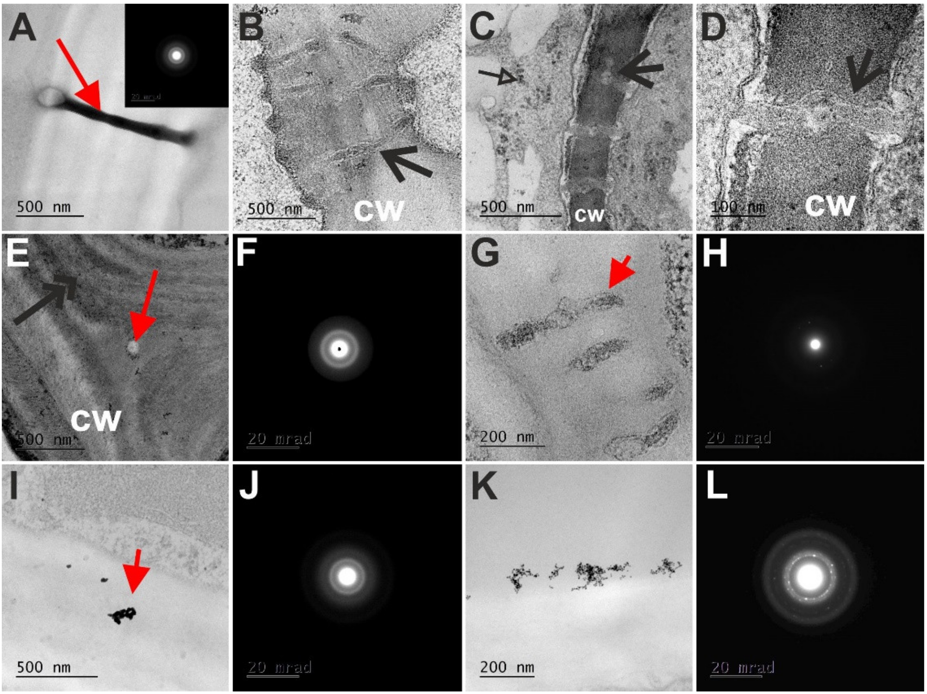
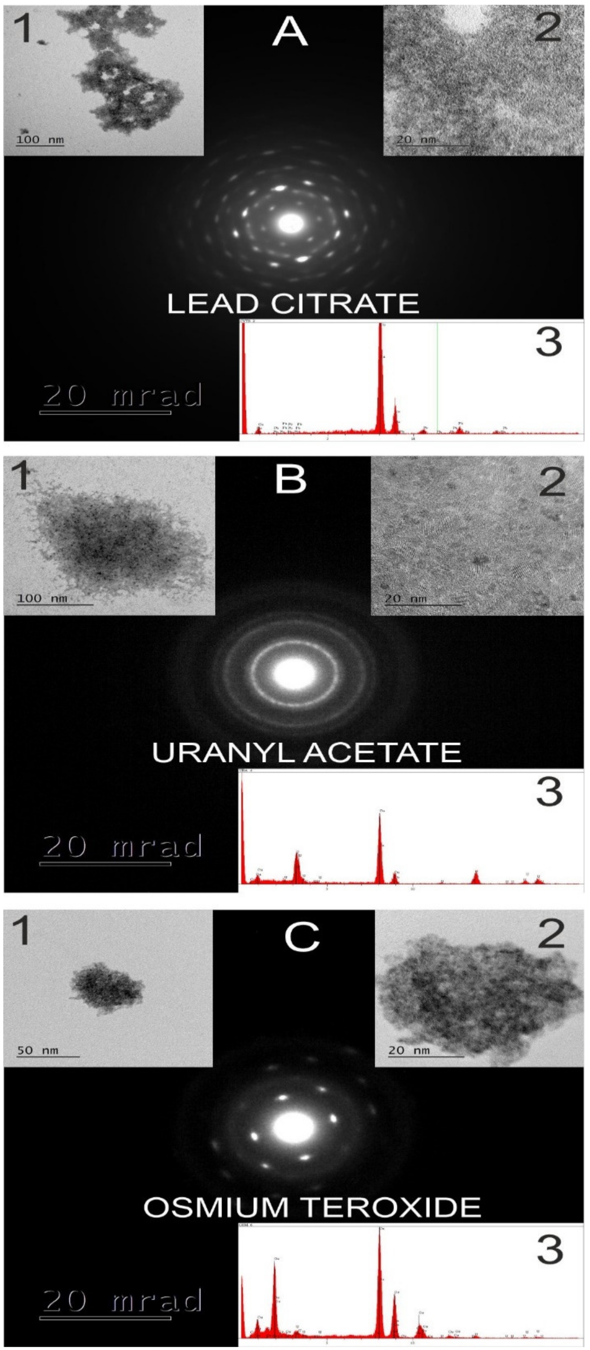
4. Transmission Electron Microscope (TEM) Analysis of the NP in the Symplast and Apoplast

Without an appropriate characterization of the chemical composition of NP, the TEM images of the NP in plant cells have to be treated with extreme caution [19,83]. For example, “rolled” PD, which are caused by the long and complicated procedure of sample preparation that can cause such an artefact, can be seen in the TEM (Figure 1A and inset). Without a specific determination concerning the diffraction pattern, chemical composition (using energy-dispersive X-ray spectroscope, EDS), the high-resolution images that can reveal the characteristic morphology/structure of the tested NP, the presence of NP in the plant material cannot be confirmed. Thus, without the above-mentioned assays, it is not possible to predict whether the PD is filled with NP, which is clearly visible in Figure 1A. In the PD electron dense spots were visible very often (Figure 1B–D,G), however, an analysis of their diffraction pattern indicated that these were not NP (Figure 1A and inset, Figure 1H). Similar caution must be taken when analyzing the cell wall after NP are applied, especially when the studies concern the primary cell wall. Namely, in such walls, the pectins are abundantly present and their appearance in a TEM is electron dense (Figure 1B,E). The middle lamella region was electron dense and the diffraction pattern confirmed that these areas in the cell walls were not NP (Figure 1E,F). Confirmation of the importance of knowledge about the chemical composition is shown in Figure 1I. The electron-dense spots look like NP (Figure 1I), but an analysis revealed that they were amorphous materials (Figure 1J). The diffraction pattern that indicates the presence of Au-NP is visible in Figure 1L, which confirms that the electron-dense spots that are visible in Figure 1K are Au-NP. The importance of such a validation was proven by Zhang et al. [32], who showed that the electron-dense NP that were seen in the plants were in fact yttrium phosphate (YbPO4) precipitates and not nano-Yb2O3.
Staining agents and buffers can produce similar artefacts, especially osmium tetroxide, cacodylate, lead citrate or uranyl acetate [83]. The “fingerprints” of the agents that were used for the sample procedure for the TEM analysis can cause misunderstandings (Figure 2). The characteristics of lead citrate (Figure 2A), uranyl acetate (Figure 2B) and osmium tetroxide (Figure 2C) are specific for these reagents and should be taken into account during studies on presence of NP in plant cells. Therefore, it is recommended that at least high resolution TEM be used to identify the crystalline structure or the distinct shape of metal-based NP.

5. Conclusions and Prospectives

Despite many years of research and an enormous amount of literature data, the translocation of NP to the cells, their movement in a plant and the mechanisms that regulate these processes, still have many unsolved problems and questions to be answered in the future. The review, which concerns the participation of the apoplast in the uptake NP and the symplast in the movement of NP in plants, led to the conclusion that further research is necessary in order to explain the mechanisms that are activated by plants under the influence of NP, which enable NP to penetrate the wall barrier and enter the postulated path of their movement through the PD. Moreover, it seems that this research should be undertaken in a comprehensive manner with the collaboration of scientists from various areas and physiologists and plant anatomists, especially in the field of PD.

Author Contributions

E.K., K.G.-J., K.S. and A.M.-H. contributed equally to this manuscript. All of the authors provided feedback on the manuscript. All authors have read and agreed to the published version of the manuscript.

Funding

This research was funded as a part of the statutory activities of the Institute of Biology, Biotechnology and Environmental Protection, Faculty of Natural Sciences, University of Silesia.

Institutional Review Board Statement

Not applicable.

Informed Consent Statement

Not applicable.

Data Availability Statement

All the required data, which are related to the current study, are embedded in this manuscript.

Conflicts of Interest

The authors declare no conflict of interest.

References

  1. Feynman, R. There’s plenty of room at the bottom. California Institute of Technology. J. Eng. Sci. 1960, 4, 23–36. [Google Scholar]
  2. Boholm, M.; Arvidsson, R. A definition framework for the terms nanomaterial and nanoparticle. NanoEthics 2016, 10, 25–40. [Google Scholar] [CrossRef]
  3. Buzea, C.; Pacheco, I. Nanomaterials and their classification. In EMR/ESR/EPR Spectroscopy for Characterization of Nanomaterials. Advanced Structured Materials; Shukla, A., Ed.; Springer: New Delhi, India, 2017; Volume 62, pp. 3–45. [Google Scholar] [CrossRef]
  4. Janković, N.Z.; Plata, D.L. Engineered nanomaterials in the context of global element cycles. Environ. Sci. Nano. 2019, 6, 2697–2711. [Google Scholar] [CrossRef] [Green Version]
  5. Arruda, S.C.C.; Silva, A.L.D.; Galazzi, R.M.; Azevedo, R.A.; Arruda, M.A.Z. Nanoparticles applied to plant science: A review. Talanta 2015, 131, 693–705. [Google Scholar] [CrossRef]
  6. Liu, W.; Zeb, A.; Lian, J.; Wu, J.; Xiong, H.; Tang, J.; Zheng, S. Interactions of Metal-based and Metal-oxide-based Nanoparticles (MBNPs and MONPs) with Crop Plants: A Critical Review of Research Progress and Prospects. Environ. Rev. 2020, 28, 294–310. [Google Scholar] [CrossRef]
  7. Nikam, A.V.; Prasad, B.L.V.; Kulkarni, A.A. Wet chemical synthesis of metal oxide nanoparticles: A review. CrystEngComm 2018, 20, 5091–5107. [Google Scholar] [CrossRef]
  8. Dietz, K.-J.; Herth, S. Plant nanotoxicology. Trends Plant Sci. 2011, 16, 583–589. [Google Scholar] [CrossRef]
  9. Millán-Chiu, B.E.; Rodriguez-Torres, M.P.; Loske, A.M. Nanotoxicology in plants. In Green Nanoparticles. Nanotechnology in the Life Sciences; Patra, J., Fraceto, L., Das, G., Campos, E., Eds.; Springer: Cham, Switzerland, 2020; pp. 43–76. [Google Scholar] [CrossRef]
  10. Khodakovskaya, M.V.; de Silva, K.; Nedosekin, D.A.; Dervishi, E.; Biris, A.S.; Shashkov, E.V.; Galanzha, E.I.; Zharov, V.P. Complex genetic, photothermal and photoacoustic analysis of nanoparticle-plant interactions. Proc. Natl. Acad. Sci. USA 2011, 108, 1028–1033. [Google Scholar] [CrossRef] [PubMed] [Green Version]
  11. Rastogi, A.; Zivcak, M.; Sytar, O.; Kalaji, H.M.; He, X.; Mbarki, S.; Brestic, M. Impact of metal and metal oxide nanoparticles on plant: A critical review. Front. Chem. 2017, 5, 78. [Google Scholar] [CrossRef] [Green Version]
  12. Hossain, Z.; Mustafa, G.; Komatsu, S. Plant responses to nanoparticle stress. Int. J. Mol. Sci. 2015, 16, 26644–26653. [Google Scholar] [CrossRef] [Green Version]
  13. Shang, H.; Guo, H.; Ma, C.; Li, C.; Chefetz, B.; Polubesova, T.; Xing, B. Maize (Zea mays L.) root exudates modify the surface chemistry of CuO nanoparticles: Altered aggregation, dissolution and toxicity. Sci. Total Environ. 2019, 690, 502–510. [Google Scholar] [CrossRef] [PubMed]
  14. Tarrahi, R.; Mahjouri, S.; Khataee, A. A review on in vivo and in vitro nanotoxicological studies in plants: A headlight for future targets. Ecotoxicol. Environ. Saf. 2021, 208, 111697. [Google Scholar] [CrossRef] [PubMed]
  15. Rajput, V.D.; Minkina, T.; Suskova, S.; Mandzhieva, S.; Tsitsuashvili, V.; Chapligin, V.; Fedorenko, A. Effects of Copper Nanoparticles (CuO NPs) on Crop Plants: A Mini Review. BioNanoScience 2018, 8, 36–42. [Google Scholar] [CrossRef]
  16. Zhou, P.; Adeel, M.; Shakoor, N.; Guo, M.; Hao, Y.; Azeem, I.; Li, M.; Liu, M.; Rui, Y. Application of Nanoparticles Alleviates Heavy Metals Stress and Promotes Plant Growth: An Overview. Nanomaterials 2021, 11, 26. [Google Scholar] [CrossRef]
  17. Jeevanandam, J.; Barhoum, A.; Chan, Y.S.; Dufresne, A.; Danquah, M.K. Review on nanoparticles and nanostructured materials: History, sources, toxicity and regulations. Beilstein J. Nanotechnol. 2018, 9, 1050–1074. [Google Scholar] [CrossRef] [PubMed] [Green Version]
  18. Sanzari, I.; Leone, A.; Ambrosone, A. Nanotechnology in plant science: To make a long story short. Front. Bioeng. Biotechnol. 2019, 7, 120. [Google Scholar] [CrossRef] [Green Version]
  19. Schwab, F.; Zhai, G.; Kern, M.; Turner, A.; Schnoor, J.L.; Wiesner, M.R. Barriers, pathways and processes for uptake, translocation and accumulation of nanomaterials in plants–Critical review. Nanotoxicology 2016, 10, 257–278. [Google Scholar] [CrossRef]
  20. Salmén, L. Micromechanical understanding of the cell-wall structure. Comptes Rendus Biol. 2004, 327, 873–880. [Google Scholar] [CrossRef]
  21. Rondeau-Mouro, C.; Defer, D.; Leboeuf, E.; Lahaye, M. Assessment of cell wall porosity in Arabidopsis thaliana by NMR spectroscopy. Int. J. Biol. Macromol. 2008, 42, 83–92. [Google Scholar] [CrossRef]
  22. Carpita, N.; Sabularsed, D.; Montezinos, D.; Delmer, D.P. Determination of the pore size of cell walls of living plant cells. Science 1979, 205, 1144–1147. [Google Scholar] [CrossRef]
  23. Fleischer, A.; O’Neill, M.A.; Ehwald, R. The pore size of non-graminaceous plant cell walls is rapidly decreased by borate ester cross-linking of the pectic polysaccharide rhamnogalacturonan II. Plant Physiol. 1999, 121, 829–838. [Google Scholar] [CrossRef] [Green Version]
  24. Money, N.P. Measurement of pore size in the hyphal cell wall of Achlya bisexualis. Exp. Mycol. 1990, 14, 234–242. [Google Scholar] [CrossRef]
  25. Flournoy, D.S.; Kirk, T.K.; Highley, T.L. Wood decay by brown-rot fungi: Changes in pore structure and cell wall volume. Holzforschung 1991, 45, 383–388. [Google Scholar] [CrossRef]
  26. Berestovsky, G.N.; Ternovsky, V.I.; Kataev, A.A. Through pore diameter in the cell wall of Chara corallina. J. Exp. Bot. 2001, 52, 1173–1177. [Google Scholar] [CrossRef]
  27. Gao, X.; Zhuang, S.; Jin, J.; Cao, P. Bound water content and pore size distribution in swollen cell walls determined by NMR technology. BioResources 2015, 10, 8208–8224. [Google Scholar] [CrossRef]
  28. Milewska-Hendel, A.; Zubko, M.; Karcz, J.; Stróż, D.; Kurczyńska, E. Fate of neutral-charged gold nanoparticles in the roots of the Hordeum vulgare L. cultivar Karat. Sci. Rep. 2017, 7, 1–13. [Google Scholar] [CrossRef] [PubMed] [Green Version]
  29. Fujino, T.; Itoh, T. Changes in pectin structure during epidermal cell elongation in pea (Pisum sativum) and its implications for cell wall architecture. Plant Cell Physiol. 1998, 39, 1315–1323. [Google Scholar] [CrossRef] [Green Version]
  30. Lv, J.; Christie, P.; Zhang, S. Uptake, translocation and transformation of metal-based nanoparticles in plants: Recent advances and methodological challenges. Environ. Sci. Nano. 2019, 6, 41–59. [Google Scholar] [CrossRef]
  31. Singh, A.; Singh, N.Á.; Afzal, S.; Singh, T.; Hussain, I. Zinc oxide nanoparticles: A review of their biological synthesis, antimicrobial activity, uptake, translocation and biotransformation in plants. J. Mater. Sci. 2018, 53, 185–201. [Google Scholar] [CrossRef]
  32. Zhang, W.; Dan, Y.; Shib, H.; Ma, X. Elucidating the mechanisms for plant uptake and in-planta speciation of cerium in radish (Raphanus sativus L.) treated with cerium oxide nanoparticles. J. Environ. Chem. Eng. 2017, 5, 572–577. [Google Scholar] [CrossRef] [Green Version]
  33. Ullah, S.; Adeel, M.; Zain, M.; Rizwan, M.; Irshad, M.K.; Jilani, G.; Rui, Y. Physiological and biochemical response of wheat (Triticum aestivum) to TiO2 nanoparticles in phosphorous amended soil: A full life cycle study. J. Environ. Manage. 2020, 263, 110365. [Google Scholar] [CrossRef]
  34. Rizwan, M.; Ali, S.; Qayyum, M.F.; Ok, Y.S.; Adrees, M.; Ibrahim, M.; Abbas, F. Effect of metal and metal oxide nanoparticles on growth and physiology of globally important food crops: A critical review. J. Hazard. Mater. 2017, 322, 2–16. [Google Scholar] [CrossRef]
  35. Lin, D.; Xing, B. Phytotoxicity of nanoparticles: Inhibition of seed germination and root growth. Environ. Pollut. 2007, 150, 243–250. [Google Scholar] [CrossRef]
  36. Abbas, Q.; Yousaf, B.; Ali, M.U.; Munir, M.A.M.; El-Naggar, A.; Rinklebe, J.; Naushad, M. Transformation pathways and fate of engineered nanoparticles (ENPs) in distinct interactive environmental compartments: A review. Environ. Int. 2020, 138, 105646. [Google Scholar] [CrossRef] [PubMed]
  37. Iannone, M.F.; Groppa, M.D.; Zawoznik, M.S.; Coral, D.F.; van Raap, M.B.F.; Benavides, M.P. Magnetite nanoparticles coated with citric acid are not phytotoxic and stimulate soybean and alfalfa growth. Ecotoxicol. Environ. Saf. 2021, 211, 111942. [Google Scholar] [CrossRef] [PubMed]
  38. Thuesombat, P.; Hannongbua, S.; Akasit, S.; Chadchawan, S. Effect of silver nanoparticles on rice (Oryza sativa L. cv. KDML 105) seed germination and seedling growth. Ecotoxicol. Environ. Saf. 2014, 104, 302–309. [Google Scholar] [CrossRef] [PubMed]
  39. Tombuloglu, H.; Slimani, Y.; Tombuloglu, G.; Korkmaz, A.D.; Baykal, A.; Almessiere, M.; Ercan, I. Impact of superparamagnetic iron oxide nanoparticles (SPIONs) and ionic iron on physiology of summer squash (Cucurbita pepo): A comparative study. Plant Physiol. Biochem. 2019, 139, 56–65. [Google Scholar] [CrossRef]
  40. Molnár, A.; Papp, M.; Kovács, D.Z.; Bélteky, P.; Oláh, D.; Feigl, G.; Szőllősi, R.; Rázga, Z.; Őrdőg, A.; Erdei, L.; et al. Nitro-oxidative signalling induced by chemically synthetized zinc oxide nanoparticles (ZnO NPs) in Brassica species. Chemosphere 2020, 251, 126419. [Google Scholar] [CrossRef]
  41. Albenne, C.; Canut, H.; Hoffmann, L.; Jamet, E. Plant Cell Wall Proteins: A Large Body of Data, but What about Runaways? Proteomes 2014, 2, 224–242. [Google Scholar] [CrossRef] [Green Version]
  42. Navarro, E.; Baun, A.; Behra, R.; Hartmann, N.B.; Filser, J.; Miao, A.J.; Sigg, L. Environmental behavior and ecotoxicity of engineered nanoparticles to algae, plants and fungi. Ecotoxicology 2008, 17, 372–386. [Google Scholar] [CrossRef] [Green Version]
  43. Kurepa, J.; Paunesku, T.; Vogt, S.; Arora, H.; Rabatic, B.M.; Lu, J.; Wanzer, M.B.; Woloschak, G.E.; Smalle, J.A. Uptake and distribution of ultrasmall anatase TiO2 Alizarin red S nanoconjugates in Arabidopsis thaliana. Nano Lett. 2010, 10, 2296–2302. [Google Scholar] [CrossRef] [PubMed] [Green Version]
  44. Jaskowiak, J.; Kwasniewska, J.; Milewska-Hendel, A.; Kurczynska, E.U.; Szurman-Zubrzycka, M.; Szarejko, I. Aluminum Alters the Histology and Pectin Cell Wall Composition of Barley Roots. Int. J. Mol. Sci. 2019, 20, 3039. [Google Scholar] [CrossRef] [PubMed] [Green Version]
  45. Mccann, M.C.; Shi, J.; Roberts, K.; Carpita, N.C. Changes in Pectin Structure and Localization during the Growth of Unadapted and Nacl-Adapted Tobacco Cells. Plant J. 1994, 5, 773–785. [Google Scholar] [CrossRef] [Green Version]
  46. Schmohl, N.; Horst, W.J. Cell wall pectin content modulates aluminium sensitivity of Zea mays (L.) cells grown in suspension culture. Plant Cell Environ. 2000, 23, 735–742. [Google Scholar] [CrossRef]
  47. Eticha, D.; Stass, A.; Horst, W.J. Localization of aluminium in the maize root apex: Can morin detect cell wall-bound aluminium? J. Exp. Bot. 2005, 56, 1351–1357. [Google Scholar] [CrossRef] [PubMed]
  48. Yang, J.L.; Li, Y.Y.; Zhang, Y.J.; Zhang, S.S.; Wu, Y.R.; Wu, P.; Zheng, S.J. Cell wall polysaccharides are specifically involved in the exclusion of aluminum from the rice root apex. Plant Physiol. 2008, 146, 602–611. [Google Scholar] [CrossRef]
  49. Milewska-Hendel, A.; Chmura, D.; Wyrwał, K.; Kurczyńska, E.U.; Kompała-Bąba, A.; Jagodziński, A.M.; Woźniak, G. Cell wall epitopes in grasses of different novel ecosystem habitats on post-industrial sites. Land Degrad. Dev. 2021, 32, 1680–1694. [Google Scholar] [CrossRef]
  50. Shi, H.; Kim, Y.; Guo, Y.; Stevenson, B.; Zhu, J.K. The Arabidopsis SOS5 locus encodes a putative cell surface adhesion protein and is required for normal cell expansion. Plant Cell. 2003, 15, 19–32. [Google Scholar] [CrossRef] [Green Version]
  51. Iraki, N.M.; Singh, N.; Bressan, R.A.; Carpita, N.C. Cell walls of tobacco cells and changes in composition associated with reduced growth upon adaptation to water and saline stress. Plant Physiol. 1989, 91, 48–53. [Google Scholar] [CrossRef] [Green Version]
  52. McNeil, M.; Darvill, A.G.; Fry, S.C.; Albersheim, P. Structure and function of the primary cell walls of plants. Ann. Rev. Biochem. 1984, 53, 625–663. [Google Scholar] [CrossRef] [PubMed]
  53. Moore, J.P.; Nguema-Ona, E.E.; Vicre-Gibouin, M.; Sorensen, I.; Willats, W.G.T.; Driouich, A. Arabinose-rich polymers as an evolutionary strategy to plasticize resurrection plant cell walls against desiccation. Planta 2013, 237, 739–754. [Google Scholar] [CrossRef] [PubMed]
  54. Zemke-White, W.L.; Clements, K.D.; Harris, P.J. Acid lysis of macroalgae by marine herbivorous fishes: Effects of acid pH on cell wall porosity. J. Exp. Mar. Biol. Ecol. 2000, 245, 57–68. [Google Scholar] [CrossRef]
  55. Nie, G.; Zhao, J.; He, R.; Tang, Y. CuO Nanoparticle Exposure Impairs the Root Tip Cell Walls of Arabidopsis thaliana Seedlings. Water Air. Soil Pollut. 2020, 231, 1–11. [Google Scholar] [CrossRef]
  56. Xiao, Z.; Yue, L.; Wang, C.H.; Chen, F.; Ding, Y.; Liu, Y.; Cao, X.; Chen, Z.; Rasmann, S.; Wang, Z. Downregulation of the photosynthetic machinery and carbon storage signaling pathways mediate La2O3 nanoparticle toxicity on radish taproot formation. J. Hazard. Mater. 2021, 411, 124971. [Google Scholar] [CrossRef]
  57. Huang, X.; Li, Y.; Chen, K.; Chen, H.; Wang, F.; Han, X.; Yuan, R. NOM mitigates the phytotoxicity of AgNPs by regulating rice physiology, root cell wall components and root morphology. Environ. Pollut. 2020, 260, 113942. [Google Scholar] [CrossRef]
  58. Kim, J.H.; Kim, D.; Seo, S.M.; Kim, D. Physiological effects of zero-valent iron nanoparticles in rhizosphere on edible crop, Medicago sativa (Alfalfa), grown in soil. Ecotoxicology 2019, 28, 869–877. [Google Scholar] [CrossRef] [PubMed]
  59. Kumaraswamy, R.V.; Kumari, S.; Choudhary, R.C.; Sharma, S.S.; Pal, A.; Raliya, R.; Saharan, V. Salicylic acid functionalized chitosan nanoparticle: A sustainable biostimulant for plant. Int. J. Biol. Macromol. 2019, 123, 59–69. [Google Scholar] [CrossRef]
  60. Singh, S.; Vishwakarma, K.; Singh, S.; Sharmab, S.; Kishor, N.; Vivek, D.; Singhe, K.; Liu, A.; Tripathi, D.K.; Chauhan, D.K. Understanding the plant and nanoparticle interface at transcriptomic and proteomic level: A concentric overview. Plant Gene. 2017, 11, 265–272. [Google Scholar] [CrossRef]
  61. Rico, C.M.; Peralta-Videa, J.R.; Gardea-Torresdey, J.L. Chemistry, Biochemistry of Nanoparticles and Their Role in Antioxidant Defense System in Plants. In Nanotechnology and Plant Sciences; Siddiqui, M., Al-Whaibi, M., Mohammad, F., Eds.; Springer International Publishing: Cham, Switzerland, 2015. [Google Scholar] [CrossRef]
  62. Ma, X.; Yan, J. Plant uptake and accumulation of engineered metallic nanoparticles from lab to field conditions. Curr. Opin. Environ. Sci. Health. 2018, 6, 16–20. [Google Scholar] [CrossRef]
  63. Dai, Y.; Zhao, J.; Liu, X.; Yu, X.; Jiang, Z.; Bu, Y.; Xing, B. Transformation and species identification of CuO nanoparticles in plant cells (Nicotiana tabacum). Environ. Sci. Nano. 2019, 6, 2724–2735. [Google Scholar] [CrossRef]
  64. Palocci, C.; Valletta, A.; Chronopoulou, L.; Donati, L.; Bramosanti, M.; Brasili, E.; Pasqua, G. Endocytic pathways involved in PLGA nanoparticle uptake by grapevine cells and role of cell wall and membrane in size selection. Plant Cell Rep. 2017, 36, 1917–1928. [Google Scholar] [CrossRef]
  65. Boykov, I.N.; Shuford, E.; Zhang, B. Nanoparticle titanium dioxide affects the growth and microRNA expression of switchgrass (Panicum virgatum). Genomics 2019, 111, 450–456. [Google Scholar] [CrossRef]
  66. Asli, S.; Neumann, P.M. Colloidal suspensions of clay or titanium dioxide nanoparticles can inhibit leaf growth and transpiration via physical effects on root water transport. Plant Cell Environ. 2009, 32, 577–584. [Google Scholar] [CrossRef]
  67. López-Moreno, M.L.; de la Rosa, G.; Hernández-Viezcas, J.Á.; Castillo-Michel, H.; Botez, C.E. Evidence of the differential biotransformation and genotoxicity of ZnO and CeO2 nanoparticles on soybean (Glycine max) plants. Environ. Sci. Technol. 2010, 44, 7315–7320. [Google Scholar] [CrossRef] [Green Version]
  68. Govea-Alcaide, E.; Masunaga, S.H.; de Souza, A.; Fajardo-Rosabal, L.; Effenberger, F.B.; Rossi, L.M.; Jardim, R.F. Tracking iron oxide nanoparticles in plant organs using magnetic measurements. J. Nanopart. Res. 2016, 18, 1–13. [Google Scholar] [CrossRef]
  69. Sevilem, I.; Miyashima, S.; Helariutta, Y. Cell-to-cell communication via plasmodesmata in vascular plants. Cell Adh. Migr. 2013, 7, 27–32. [Google Scholar] [CrossRef] [Green Version]
  70. Marzec, M.; Kurczyńska, E.U. Importance of symplasmic communication in cell differentiation. Plant Signal. Behav. 2014, 9, e27931. [Google Scholar] [CrossRef] [PubMed] [Green Version]
  71. Bell, K.; Oparka, K. Imaging plasmodesmata. Protoplasma 2011, 248, 9–25. [Google Scholar] [CrossRef]
  72. Demchenko, K.N.; Voitsekhovskaja, O.V.; Pawlowski, K. Plasmodesmata without callose and calreticulin in higher plants–open channels for fast symplastic transport? Front. Plant Sci. 2014, 5, 74. [Google Scholar] [CrossRef] [Green Version]
  73. Kragler, F. Plasmodesmata: Intercellular tunnels facilitating transport of macromolecules in plants. Cell Tissue Res. 2013, 352, 49–58. [Google Scholar] [CrossRef] [PubMed]
  74. Dashevskaya, S.; Kopito, R.B.; Friedman, R.; Elbaum, M.; Epel, B.L. Diffusion of anionic and neutral GFP derivatives through plasmodesmata in epidermal cells of Nicotiana benthamiana. Protoplasma 2008, 234, 13–23. [Google Scholar] [CrossRef] [PubMed]
  75. Burch-Smith, T.M.; Stonebloom, S.; Xu, M.; Zambryski, P.C. Plasmodesmata during development: Re-examination of the importance of primary, secondary and branched plasmodesmata structure versus function. Protoplasma 2011, 248, 61–74. [Google Scholar] [CrossRef] [Green Version]
  76. Sun, D.; Hussain, H.I.; Yi, Z.; Siegele, R.; Cresswell, T.; Kong, L.; Cahill, D.M. Uptake and cellular distribution, in four plant species of fluorescently labeled mesoporous silica nanoparticles. Plant Cell Rep. 2014, 33, 1389–1402. [Google Scholar] [CrossRef]
  77. Geisler-Lee, J.; Wang, Q.; Yao, Y.; Zhang, W.; Geisler, M.; Li, K.; Huang, Y.; Chen, Y.; Kolmakov, A.; Ma, X. Phytotoxicity, accumulation and transport of silver nanoparticles by Arabidopsis thaliana. Nanotoxicology 2013, 7, 323–337. [Google Scholar] [CrossRef] [PubMed]
  78. Zhai, G.; Walters, K.S.; Peate, D.W.; Alvarez, P.J.; Schnoor, J.L. Transport of gold nanoparticles through plasmodesmata and precipitation of gold ions in woody poplar. Environ. Sci. Technol. Lett. 2014, 1, 146–151. [Google Scholar] [CrossRef] [PubMed] [Green Version]
  79. Pradas del Real, A.E.; Vidal, V.; Carrière, M.; Castillo-Michel, H.; Levard, C.; Chaurand, P.; Sarret, G. Silver nanoparticles and wheat roots: A complex interplay. Environ. Sci. Technol. 2017, 51, 5774–5782. [Google Scholar] [CrossRef] [Green Version]
  80. Spielman-Sun, E.; Lombi, E.; Donner, E.; Howard, D.; Unrine, J.M.; Lowry, G.V. Impact of surface charge on cerium oxide nanoparticle uptake and translocation by wheat (Triticum aestivum). Environ. Sci. Technol. 2017, 51, 7361–7368. [Google Scholar] [CrossRef]
  81. Sager, R.; Lee, J.Y. Plasmodesmata in integrated cell signalling: Insights from development and environmental signals and stresses. J. Exp. Bot. 2014, 65, 6337–6358. [Google Scholar] [CrossRef] [Green Version]
  82. Schulz, A. Plasmodesmal widening accompanies the short-term increase in symplasmic phloem unloading in pea root tips under osmotic stress. Protoplasma 1995, 188, 22–37. [Google Scholar] [CrossRef]
  83. Brandenberger, C.; Clift, M.J.D.; Gehr, P.; Muhlfeld, C.; Rothen-Rutishauser, B.; Stone, V. Intracellular imaging of nanoparticles: Is it an elemental mistake to believe what you see? Part Fibre Technol. 2010, 7, 15. [Google Scholar] [CrossRef] [Green Version]
  84. Lombi, E.; Scheckel, K.G.; Kempson, I.M. In situ analysis of metal(loid)s in plants: State of the art and artefacts. Environ Exp Bot. 2011, 72, 3–17. [Google Scholar] [CrossRef]
  85. Kennedy, C.D.; Gonsalves, F.A.N. The action of divalent zinc, cadmium, mercury, copper and lead on the trans-root potential and H+ efflux of excised roots. J. Exp. Bot. 1987, 38, 800–817. [Google Scholar] [CrossRef]
  86. Da Costa, M.V.J.; Sharma, P.K. Effect of copper oxide nanoparticles on growth, morphology, photosynthesis and antioxidant response in Oryza sativa. Photosynth. 2016, 54, 110–119. [Google Scholar] [CrossRef]
  87. Shukla, P.K.; Misra, P.; Kole, C.H. Uptake, Translocation, Accumulation, Transformation and Generational Transmission of Nanoparticles in Plants. In Plant Nanotechnology; Kole, C., Kumar, D., Khodakovskaya, M., Eds.; Springer International Publishing: Cham, Switzerland, 2016; pp. 183–218. [Google Scholar]
Figure 1. High-resolution transmission electron microscope (HRTEM) images of sections from barley roots that had been treated with the Au-NP. (A) The PD in the wall. The appearance of the PD is an artefact that was caused by mistakes in the sample preparation procedure; inset—diffraction pattern that that indicates that the black color (indicated by red arrow) is not NP. (B) The PD with electron-dense material inside (indicated by black arrow), but as was proven by the diffraction pattern (see inset on (A)), the electron-dense material was not NP. (C) Cell wall traversed by the PD with electron-dense spots (indicated by black open arrow), which are not NP proved by diffraction pattern. Many electron-dense dots were present in the cytoplasm but were also not NP (black arrow). Note that the dark colouration of the cell walls resulted from the presence of pectins. (D) Similar to Figure (C), none of electron-dense particles were NP (black arrow; diffraction patterns were the same as for (A)). (E) Cross-section through the root cell walls. Note the electron-dense bands in the wall (black double arrow), which were not NP, but just a higher amount of pectins compared to the other parts of the walls (cw). Such an image is especially characteristic for the middle lamella. Electron-dense dot (indicated by red arrow) is not NP. (F) Diffraction pattern of the dot from Figure (E). (G) PD with electron-dense material inside (red arrow). (H) Diffraction pattern of the electron-dense material from the PD that indicated that this material is amorphous, thus not NP. (I) Electron-dense particles within the cell wall of barley roots (red arrow). (J) The diffraction pattern clearly supports the claim that the observed electron-dense particles were not NP. (K) Transverse section through the cell wall with many electron-dense particles. (L) The diffraction pattern indicates that the observed electron-dense particles are Au-NP.
Applsci 11 05473 g001
Figure 2. HRTEM of the electron-dense particles detected during the procedure for sample preparation for electron dense electron microscopy analysis. (A) Diffraction pattern of the lead citrate spots; inset 1—spots visible under a lower magnification, inset 2—under a higher magnification and inset 3 EDS-TEM analysis of the analyzed spots. (B) Diffraction pattern of uranyl acetate spots; inset 1—spots visible under a lower magnification, inset 2—under a higher magnification with clearly visible “fingerprints” of the uranyl acetate and inset 3—EDS-TEM analysis of the analyzed spots. (C) Diffraction pattern of the osmium tetroxide spots; inset 1—spots visible under a lower magnification, inset 2—under a higher magnification with clearly visible “fingerprints” of the osmium tetroxide and inset 3—EDS-TEM analysis of the analyzed spots.
Applsci 11 05473 g002
Publisher’s Note: MDPI stays neutral with regard to jurisdictional claims in published maps and institutional affiliations.

