Article

The ESCRT-III Protein Chmp1 Regulates Lipid Storage in the Drosophila Fat Body

Division of Science, Pennsylvania State University, Berks Campus, Reading, PA 19610, USA
*
Author to whom correspondence should be addressed.
Med. Sci. 2023, 11(1), 5; https://doi.org/10.3390/medsci11010005
Submission received: 27 November 2022 / Revised: 16 December 2022 / Accepted: 23 December 2022 / Published: 26 December 2022
(This article belongs to the Section Endocrinology and Metabolic Diseases)

Abstract

:
Defects in how excess nutrients are stored as triglycerides can result in several diseases including obesity, heart disease, and diabetes. Understanding the genes responsible for normal lipid homeostasis will help understand the pathogenesis of these diseases. RNAi screens performed in Drosophila cells identified genes involved in vesicle formation and protein sorting as important for the formation of lipid droplets; however, all of the vesicular trafficking proteins that regulate lipid storage are unknown. Here, we characterize the function of the Drosophila Charged multivesicular protein 1 (Chmp1) gene in regulating fat storage. Chmp1 is a member of the ESCRT-III complex that targets membrane localized signaling receptors to intralumenal vesicles in the multivesicular body of the endosome and then ultimately to the lysosome for degradation. When Chmp1 levels are decreased specifically in the fly fat body, triglyceride accumulates while fat-body-specific Chmp1 overexpression decreases triglycerides. Chmp1 controls triglyceride storage by regulating the number and size of fat body cells produced and not by altering food consumption or lipid metabolic enzyme gene expression. Together, these data uncover a novel function for Chmp1 in controlling lipid storage in Drosophila and supports the role of the endomembrane system in regulating metabolic homeostasis.

1. Introduction

The ability of an organism to regulate its consumption and storage of nutrients is essential for long-term survival when access to food is sporadic. However, in conditions where food is abundant, like in current Western society, excess nutrients are stored as triglycerides resulting in obesity [1]. Obesity predisposes humans to heart disease, type II diabetes and cancer, some of the leading causes of death in the United States [2]. Understanding the mechanisms of how lipid homeostasis is regulated may allow for the identification of potential therapeutic targets for this deadly disease.
The fruit fly, Drosophila melanogaster, is used as a model system to study lipid metabolism due to its similarity to humans in terms of genes and metabolic biology [3,4]. The Drosophila system has been used to perform genetic screens to identify genes important for the storage of lipids in both cultured cells and intact flies [5,6,7]. From these screens, a group of genes involved in forming the vesicular coat protein complex COPI have been implicated in the regulation of lipid storage [5,6]. Proteins that are destined to be secreted from eukaryotic cells move throughout the endomembrane system starting by being translated into the ER, then traveling to the Golgi apparatus and ultimately being secreted via exocytosis [8]. These proteins travel throughout the endomembrane system in vesicles that are formed from and targeted to different cellular compartments using many classes of proteins such as coat proteins, Rabs and SNAREs [8]. While members of the COPI coat protein complex have been shown to regulate lipid droplet formation [5,6], whether other proteins involved in the movement of cargos throughout the endomembrane system regulate lipid storage is not well understood.
Many membrane-localized signaling receptors are taken into cells through endocytosis and are recycled back to the plasma membrane when necessary. However, some signaling receptors are monoubiquitinated targeting them for endocytosis and degradation in the lysosome [9]. These monoubiquitinated receptors are localized to intralumenal vesicles (ILVs) of the endosomal compartment called the multivesicular body (MVB) [10]. Four different endosomal sorting complex required for transport (ESCRT) complexes (ESCRT-0, -I, -II, -III) are required for the localization of these signaling receptors to ILVs [9]. ESCRT-0 complex proteins function to cluster the monoubiquitinated receptors, while ESCRT-I and ESCRT-II complex proteins work together to bud the endosomal compartment membrane. ESCRT-III complex proteins then function to regulate membrane scission at a bud neck and promote ESCRT complex disassembly ultimately forming an endocytic vesicle [9]. In addition to endosomal sorting, ESCRT-III proteins are involved in a number of biological processes such as cytokinesis, neuronal pruning, nuclear envelope repair and maintenance, autophagy, viral budding and replication, and membrane shaping [11]. ESCRT proteins have also been identified as being involved in lipid droplet utilization in yeast [12]; however, whether these proteins function to regulate lipid homeostasis in a multicellular metazoan is not known.
In this study, we characterize the role of Charged multivesicular body protein 1 (Chmp1) in regulating lipid storage in Drosophila. Chmp1 is a member of the ESCRT-III complex and functions in ESCRT complex protein dissociation by binding to and recruiting the ATPase, Vps4 [13,14]. Decreasing Chmp1 levels specifically in the fat body using RNAi results in triglyceride accumulation, while fat-body-specific Chmp1 overexpression results in a lean phenotype. Chmp1 does not regulate lipid storage by affecting food consumption or the expression of lipid metabolic enzyme genes; however, fat cell size and number are altered in Chmp1-RNAi and Chmp1 overexpressing flies, providing a potential mechanism for the triglyceride phenotypes observed in these flies. These results uncover a novel role for the ESCRT-III complex protein, Chmp1, in regulating lipid homeostasis in the fly fat body.

2. Materials and Methods

2.1. Fly Genetics

The following fly strains were used in these experiments: yolk-Gal4 [15]; w[*]; UAS-Chmp1; D/TM3Ser [16]; w[*]; P{w[+mC]=UAS-GFP.S65T}Myo31DF[T2] (referred to as UAS-GFP; Bloomington #1521); y[1] v[1]; P{y[+t7.7] v[+t1.8]=TRiP.HM05117}attP2 (referred to as UAS-Chmp1-RNAi; Bloomington #28906); y[1] v[1]; P{y[+t7.7]=CaryP}attP2 (UAS-Chmp1-RNAi genetic background control; Bloomington #36303). Flies were grown on a 12 h:12 h light:dark cycle at 25 °C on cornmeal-sugar-yeast medium as described previously [17].

2.2. Protein, Triglyceride, and DNA Measurements

Approximately 1-week-old female flies were used throughout this study. Two whole flies or three dissected abdomen cuticles with fat bodies attached were homogenized in lysis buffer (140 mM NaCl, 50 mM Tris-HCl, pH 7.4, 0.1% Triton-X, and 1X protease inhibitor (ThermoFisher, Waltham, MA, USA)). Triglyceride, protein, and DNA were measured using the Infinity Triglyceride Reagent (ThermoFisher, Waltham, MA, USA), Pierce BCA Protein Assay kit (ThermoFisher, Waltham, MA, USA) and Quant-iT DNA Assay kit (Invitrogen, Waltham, MA, USA), respectively, accordingly to manufacturer’s instructions and as described previously [17,18].

2.3. CAFÉ Assay

Food consumption was measured using the capillary feeding (CAFÉ) assay [19]. Briefly, three 1-week-old female flies were housed in a vial with 1% agar as a water source. Flies were fed 5% sucrose in a Drummond 5 μL capillary tube (ThermoFisher, Waltham, MA, USA as their sole food source and the amount of sucrose consumed was measured after 24 h. Vials without flies were used to account for any sucrose evaporation.

2.4. Gene Expression Analysis

RNA isolation, DNase treatment and reverse transcription were performed on abdomen cuticles with fat bodies attached dissected from 15 one-week-old female flies as previously described [17]. qPCR reactions were generated using 1 μL of cDNA, 200 nM primers, 1 × Perfecta SYBR Green (Quanta Biosciences, Gaithersburg, MD, USA in a 25 μL reaction. qPCR was performed on a StepOnePlus instrument using the following cycling conditions: initial denaturation at 95 °C for 3 min, 40 cycles of 30 s at 95 °C, 60 s at 60 °C and 30 s at 72 °C followed by a melt curve. Expression of all genes were normalized to rp49. Primer sequences used were: Chmp1 (For 5′-GGCCTCGAACTCAACATGGA-3′ and Rev 5′-CAGAAATTCAACGGGCAGGG-3′), FASN1 (For 5′-CTGGCTGAGCAAGATTGTGTG-3′ and Rev 5′-TCGCACAACCAGAGCGTAGTA), ATPCL (For 5′-CACGACAGATTGGTCCAAGCTC-3′ and Rev 5′-CTTGCTCTTCACGTCGGCTAAC-3′), bmm (For 5′-ACGTGATCATCTCGGAGTTTG-3′ and Rev 5′-ATGGTGTTCTCGTCCAGAATG-3′), whd (For 5′-GCCAATGTGATTTCCCTGCTTC-3′ and Rev 5′-CTTTGCCCTTCAGGATTTCCTC-3′) and rp49 (For 5′-GACGCTTCAAGGGACAGTATCTG-3′ and Rev 5′-AAACGCGGTTCTGCATGAG-3′).

2.5. Statistics

The results were expressed as mean ± standard error (SE) and average values were compared between Chmp1-RNAi or Chmp1 overexpressing flies and the appropriate controls using a two-tailed Student’s t-test as calculated in Microsoft Excel. p < 0.05 was used as the cut off for statistical significance.

3. Results

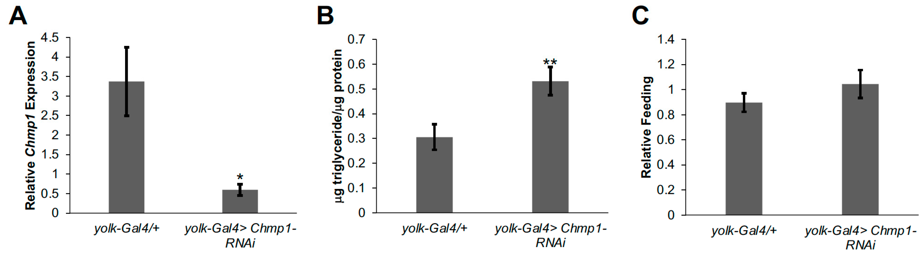
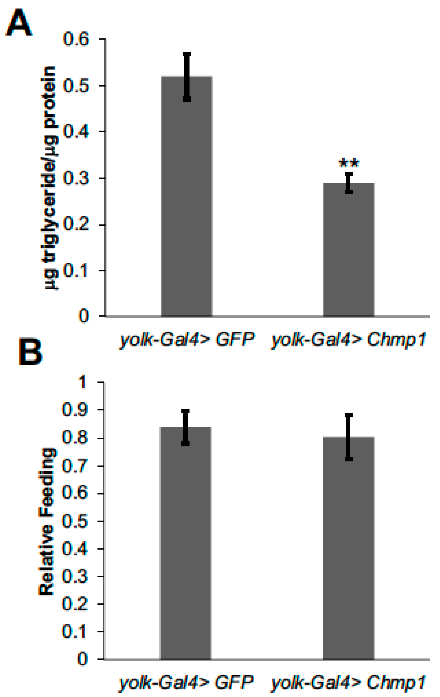
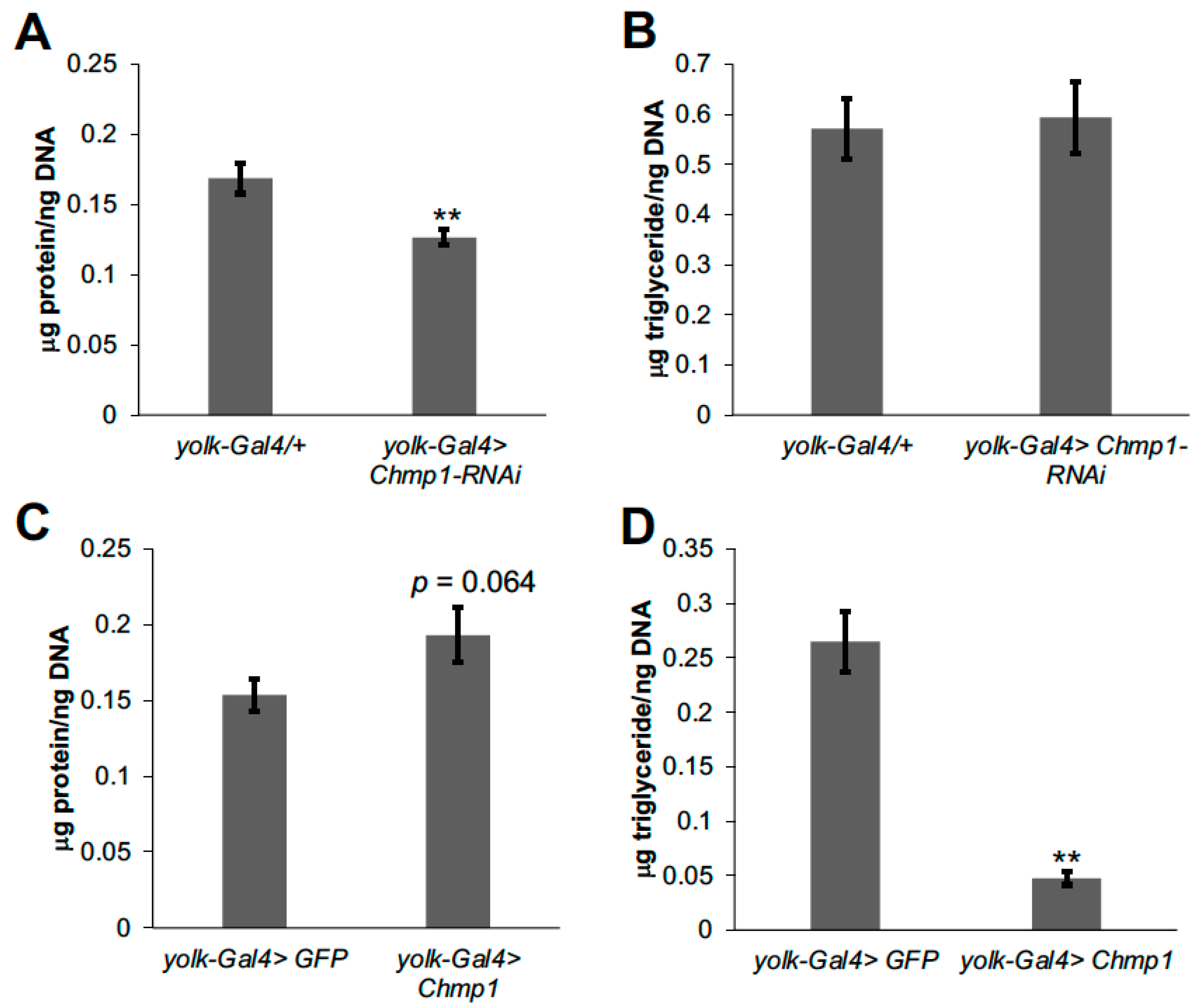
3.1. Chmp1 Acts in the Drosophila Fat Body to Regulate Triglyceride Storage

To assess the role of fat body Chmp1 in regulating lipid storage in Drosophila, we decreased Chmp1 levels by inducing RNAi towards Chmp1 specifically in the fat body using the previously described yolk-Gal4 driver [15] and UAS-Chmp1-RNAi fly line (Figure 1A; [16]). The yolk-Gal4 driver crossed to the UAS-Chmp1-RNAi genetic background stock was used as a control. Triglycerides were measured in Chmp1-RNAi flies and controls, and we observed an increase in triglyceride storage in fat-body-specific Chmp1 knockdown flies (Figure 1B). This accumulation of triglycerides was not due to increased feeding as food consumption was not altered in Chmp1-RNAi flies (Figure 1C).
To further investigate the role of Chmp1 in regulating fat body lipid storage, we overexpressed Chmp1 in the female fat body using yolk-Gal4 and the previously described UAS-Chmp1 fly line [16] and compared them to control flies expressing green fluorescent protein (GFP). Conversely to the Chmp1-RNAi flies, overexpressing Chmp1 blunts triglyceride storage (Figure 2A), and this is not due to decreased food consumption (Figure 2B). Together, the Chmp1-RNAi and Chmp1 overexpression data indicate a role of Chmp1 in limiting lipid levels in the Drosophila fat body.

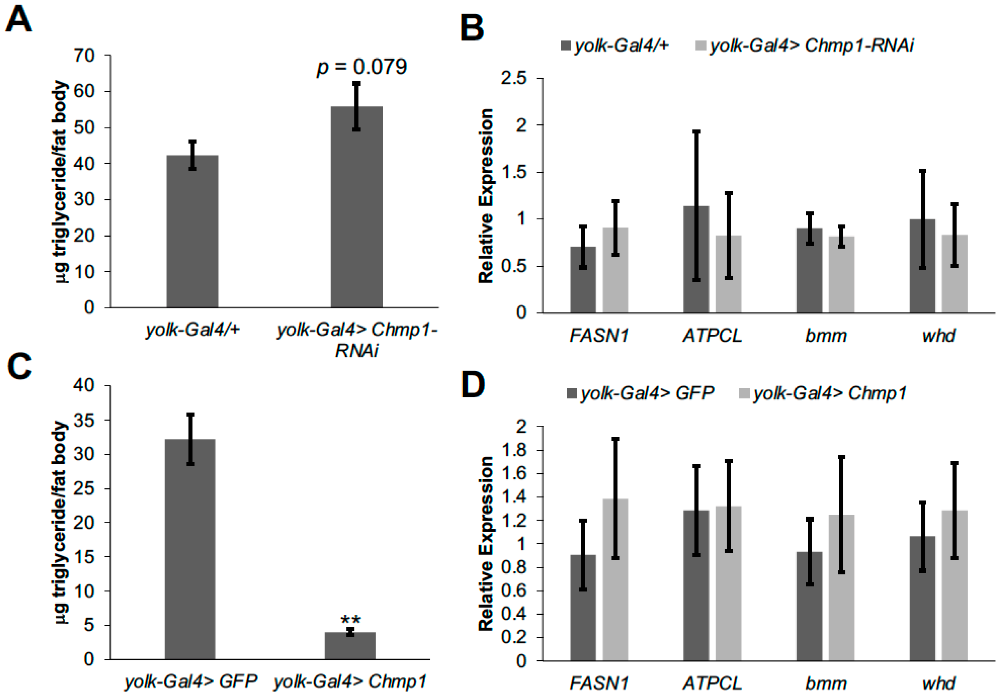
3.2. Chmp1 Does Not Alter the Expression of Lipid Metabolic Enzyme Genes in the Drosophila Fat Body

To determine whether the defects in lipid storage in Chmp1-RNAi and Chmp1 overexpressing flies were autonomous to the fat body, we dissected abdomens with fat bodies attached from these flies and measured triglyceride levels. There was a trend for an increase in triglycerides in fat bodies dissected from Chmp1-RNAi flies (Figure 3A) and there was a strong decrease in triglycerides in fat bodies dissected from Chmp1 overexpressing flies (Figure 3C), consistent with an autonomous role of Chmp1 in the fly fat body and the triglyceride storage results from whole flies. To further investigate the role of Chmp1 in regulating fat body lipid storage, we wanted to test whether the triglyceride storage defects in Chmp1-RNAi and Chmp1 overexpressing flies were due to changes in the expression of lipid metabolic enzyme genes. To address this question, we dissected abdomen cuticles with fat bodies attached, isolated RNA, and performed qPCR for two genes important for lipid synthesis (FASN1 and ATPCL) and two genes important for lipid breakdown (the lipase bmm and whd, the fly homolog of the β-oxidation gene carnitine palmitoyltransferase (CPT1)). Interestingly, the expression of each of these genes was normal in Chmp1-RNAi fat bodies (Figure 3B) and Chmp1 overexpressing fat bodies (Figure 3D) suggesting that altered expression of these lipid metabolic enzyme genes is not responsible for the triglyceride storage phenotypes observed in these flies.

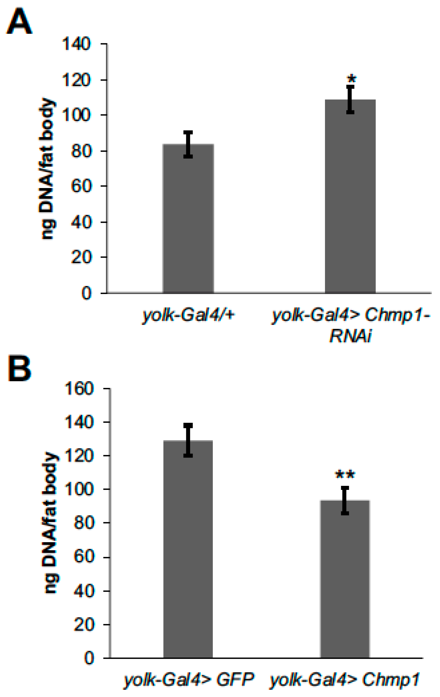
3.3. Chmp1 Regulates Fat Body Cell Number and Size and the Amount of Stored Fat Body Triglyceride

Another potential cause of the alterations in fat body lipid storage in flies with altered Chmp1 levels could be changes in the number or size, fat body cell triglyceride storage or a combination of these. To test whether Chmp1 regulates the number of fat body cells, fat body DNA levels were measured as a proxy for fat cell number as previously described [18]. Fat body DNA was increased in Chmp1-RNAi flies (Figure 4A) suggesting that the triglyceride accumulation observed in these flies could be due to more fat body cells being produced. Conversely, Chmp1 overexpressing flies had decreased total DNA content (Figure 4B) indicating that having fewer fat body cells contributes to the lean phenotype observed in these flies.
To determine whether Chmp1 can regulate fat body cell size, total protein was normalized by DNA content which has been used previously as an indication of cell size [20]. Decreasing Chmp1 in the fat body resulted in a lower protein/DNA ratio than controls (Figure 5A) and Chmp1 overexpressing fat body cells were trending towards an increased protein/DNA ratio (Figure 5C) suggesting that Chmp1 also regulates fat body cell size. Moreover, to determine whether fat body cell lipid storage was altered in Chmp1-RNAi and Chmp1 overexpressing flies, fat body triglycerides were normalized by fat body DNA. Interestingly, while Chmp1-RNAi flies had no difference in fat body triglyceride normalized by DNA (Figure 5B), Chmp1 overexpressing flies had a large decrease in triglyceride/DNA (Figure 5D). These data suggest that Chmp1 is not necessary to control fat body cell triglyceride storage but sufficient to regulate fat body cellular triglycerides. Together, these data display a function of the ESCRT-III protein, Chmp1, in the regulation of triglyceride storage and fat cell number and size in the Drosophila fat body.

4. Discussion

In this study, we have uncovered a novel role for the ESCRT-III protein, Chmp1, in controlling lipid storage in the Drosophila fat body. Our data indicate that Chmp1 functions to limit triglyceride levels as fat-body-specific Chmp1 overexpression decreases triglyceride levels and Chmp1-RNAi increases it. These results are consistent with experiments performed in the yeast Saccharomyces cerevisiae showing that mutants in ESCRT complex genes result in less lipid droplet consumption and increased triglyceride storage [12]. In addition, experiments performed in cultured Drosophila cells revealed that knocking down COPI complex genes with RNAi results in larger lipid droplets [5,6], a phenotype similar to the increased triglycerides observed in Chmp1-RNAi fat bodies, suggesting a broader role for vesicular trafficking proteins in regulating lipid homeostasis. Arf79d, a member of the Arf1 family of monomeric G proteins that function in the endomembrane system, was also shown to localize to the lipid droplet surface and alter rates of lipolysis, suggesting a role for vesicular trafficking proteins in regulating lipid breakdown [6]. Chmp1 has been shown to localize to endosomes in the developing fly wing [16], but whether Chmp1 is found on the surface of lipid droplets and plays a role in the budding of lipid-laden vesicles during lipolysis in the Drosophila fat body is not known. Additional experiments designed to visualize whether Chmp1 co-localizes with lipid droplets in Drosophila fat body cells and whether lipolysis is altered in Chmp1-RNAi or Chmp1 overexpressing fat bodies would help address this question.
We have also shown that Chmp1 controls fat body lipid storage by regulating the number and size of fat body cells. Chmp1-RNAi results in more smaller fat body cells suggesting that loss of Chmp1 results in increased cell proliferation or decreased apoptosis. Decreasing the expression of the ESCRT-III protein gene Vps4 in fly imaginal discs results in larval overgrowth, despite concomitant increases in apoptosis [21]. Since Chmp1 binds to Vps4 in the ESCRT-III complex [13,14], perhaps Chmp1 loss in the Drosophila fat body might also promote cell proliferation or growth to regulate fat body cell number and size. Measuring fat body cell proliferation and apoptosis in Chmp1-RNAi and Chmp1 overexpressing flies will test this hypothesis.
Chmp1 is also able to decrease the amount of fat stored per cell (as measured by fat body triglycerides normalized by DNA content) when it is overexpressed. This is consistent with a potential role for Chmp1 in specifically promoting lipolysis as speculated above or lipid breakdown more generally, but independently of changes in the expression of important lipid breakdown genes like bmm or whd as their expression in unchanged in Chmp1-overexpressing flies. Interestingly, Chmp1-RNAi fat bodies have normal triglyceride storage when normalized to DNA, suggesting that Chmp1 may not be limiting to regulate the amount of fat stored per cell under physiological conditions. Since Chmp1 only alters the amount of triglyceride stored in each fat cell when it is overexpressed, Chmp1 may only be sufficient to regulate fat cell lipid storage. This is consistent with experiments manipulating Chmp1 in the developing fly wing where Chmp1 overexpression revealed additional phenotypes not observed in Chmp1-RNAi wings [16]. It is possible that ESCRT complex activity is altered to different degrees in Chmp1-RNAi and Chmp1 overexpressing fat bodies causing differential regulation of the amount of fat stored per cell. It is also possible that Chmp1 overexpression might uncover additional signaling pathways regulated by Chmp1. Experiments designed to measure ESCRT complex activity in Chmp1-RNAi and Chmp1 overexpressing fat bodies would help differentiate among some of these possibilities.
One of the major functions of ESCRT proteins is to identify monoubiquitinated receptors for signaling pathways, remove them from the plasma membrane and traffic them to endosomes, ultimately leading to degradation in the lysosome [9,10]. Many ESCRT proteins in Drosophila have been shown to recognize monoubiquitinated signaling receptors such as epidermal growth factor receptor (Egfr) and Notch and target them to the multivesicular body in endosomes [22]. In ESCRT mutants in Drosophila, these receptors remain on the plasma membrane and their signaling pathways are more active compared to wildtype controls [16,22]. Consistent with this signaling receptor accumulation, decreasing Chmp1 levels in the larval fat body results in the production of small lysosomes and decreased autophagy indicating less protein degradation [23]. However, whether Egfr or Notch signaling is altered in Chmp1-RNAi or Chmp1 overexpressing fat bodies and whether this signaling may contribute to the triglyceride storage phenotypes observed here is not known. Previous studies have shown that activating the insulin receptor in the Drosophila fat body results in an increase in the number of fat cells and an increase in the amount of triglyceride stored in each fat cell [18]. Since we have shown here that Chmp1 can regulate both fat cell number and fat cell triglyceride storage, it is possible that Chmp1 may recognize a monoubiquitinated insulin receptor and decrease insulin signaling in fat body cells, altering fat cell production or fat cell lipid storage. Performing experiments measuring whether insulin signaling activity is increased in Chmp1-RNAi fat bodies and decreased in Chmp1 overexpressing fat bodies would help support this hypothesis.
Previous work from our lab has elucidated a role for Chmp1 in regulating the transport of polyamines in Drosophila using a leg imaginal disc system [24]. Polyamines are small nitrogen-containing molecules that regulate many biological processes including cell differentiation and proliferation [25,26]. In addition, polyamines have also been implicated in regulating lipid metabolism in the Drosophila fat body as well as in mammalian adipocytes [27,28]. This raises the possibility that perhaps Chmp1 may also be regulating polyamine transport in the fly fat body and this may contribute to the lipid metabolic phenotypes observed here.
In summary, we have described a novel metabolic role for the ESCRT-III protein, Chmp1, in the Drosophila fat body. Chmp1 is highly conserved from yeast to humans [10], so the results shown here could be used to increase our understanding of lipid metabolism in humans. In addition, one of the human Chmp1 homologs, Chmp1A, has been shown to be important in several cancers [29,30] potentially broadening the interest of this work. Together, these results support the expanding roles of the endomembrane system and vesicular trafficking proteins in the regulation of overall lipid homeostasis.

Author Contributions

Conceptualization, J.R.D.; methodology, J.R.D.; validation, A.M.F., K.E.L. and J.R.D.; formal analysis, A.M.F. and K.E.L.; investigation, A.M.F. and K.E.L.; writing—original draft preparation, J.R.D.; writing—review and editing, A.M.F., K.E.L. and J.R.D.; visualization, J.R.D.; supervision, J.R.D.; project administration, J.R.D.; funding acquisition, J.R.D. All authors have read and agreed to the published version of the manuscript.

Funding

This research received no external funding.

Institutional Review Board Statement

Not applicable.

Informed Consent Statement

Not applicable.

Data Availability Statement

All of the data presented here are contained within the article.

Acknowledgments

The authors would like to thank Shannon Nowotarski for helpful discussions, Ryan Bennick for technical assistance and Simon Collier for sharing his UAS-Chmp1 stocks. Stocks obtained from the Bloomington Drosophila Stock Center (NIH P40OD018537) were used in this study. This research was supported by funds from Pennsylvania State University, Berks Campus.

Conflicts of Interest

The authors declare no conflict of interest.

References

  1. Qiu, H.; Schlegel, V. Impact of nutrient overload on metabolic homeostasis. Nutr. Rev. 2018, 76, 693–707. [Google Scholar] [CrossRef] [PubMed] [Green Version]
  2. Raghupathi, W.; Raghupathi, V. An Empirical Study of Chronic Diseases in the United States: A Visual Analytics Approach. Int. J. Environ. Res. Public Health 2018, 15, 431. [Google Scholar] [CrossRef] [PubMed] [Green Version]
  3. Heier, C.; Klishch, S.; Stilbytska, O.; Semaniuk, U.; Lushchak, O. The Drosophila model to interrogate triacylglycerol biology. Biochim. Biophys. Acta Mol. Cell Biol. Lipids 2021, 1866, 158924. [Google Scholar] [CrossRef] [PubMed]
  4. Musselman, L.P.; Kuhnlein, R.P. Drosophila as a model to study obesity and metabolic disease. J. Exp. Biol. 2018, 221, jeb163881. [Google Scholar] [CrossRef] [PubMed] [Green Version]
  5. Beller, M.; Sztalryd, C.; Southall, N.; Bell, M.; Jackle, H.; Auld, D.S.; Oliver, B. COPI complex is a regulator of lipid homeostasis. PLoS Biol. 2008, 6, e292. [Google Scholar] [CrossRef] [Green Version]
  6. Guo, Y.; Walther, T.C.; Rao, M.; Stuurman, N.; Goshima, G.; Terayama, K.; Wong, J.S.; Vale, R.D.; Walter, P.; Farese, R.V. Functional genomic screen reveals genes involved in lipid-droplet formation and utilization. Nature 2008, 453, 657–661. [Google Scholar] [CrossRef] [Green Version]
  7. Pospisilik, J.A.; Schramek, D.; Schnidar, H.; Cronin, S.J.; Nehme, N.T.; Zhang, X.; Knauf, C.; Cani, P.D.; Aumayr, K.; Todoric, J.; et al. Drosophila genome-wide obesity screen reveals hedgehog as a determinant of brown versus white adipose cell fate. Cell 2010, 140, 148–160. [Google Scholar] [CrossRef] [Green Version]
  8. Beraud-Dufour, S.; Balch, W. A journey through the exocytic pathway. J. Cell Sci. 2002, 115, 1779–1780. [Google Scholar] [CrossRef]
  9. Hurley, J.H.; Hanson, P.I. Membrane budding and scission by the ESCRT machinery: It’s all in the neck. Nat. Rev. Mol. Cell Biol. 2010, 11, 556–566. [Google Scholar] [CrossRef]
  10. Michelet, X.; Djeddi, A.; Legouis, R. Developmental and cellular functions of the ESCRT machinery in pluricellular organisms. Biol. Cell 2010, 102, 191–202. [Google Scholar] [CrossRef] [PubMed]
  11. Vietri, M.; Radulovic, M.; Stenmark, H. The many functions of ESCRTs. Nat. Rev. Mol. Cell Biol. 2020, 21, 25–42. [Google Scholar] [CrossRef] [PubMed]
  12. Ouahoud, S.; Fiet, M.D.; Martinez-Montanes, F.; Ejsing, C.S.; Kuss, O.; Roden, M.; Markgraf, D.F. Lipid droplet consumption is functionally coupled to vacuole homeostasis independent of lipophagy. J. Cell Sci. 2018, 131, jcs213876. [Google Scholar] [CrossRef] [PubMed] [Green Version]
  13. Nickerson, D.P.; West, M.; Odorizzi, G. Did2 coordinates Vps4-mediated dissociation of ESCRT-III from endosomes. J. Cell Biol. 2006, 175, 715–720. [Google Scholar] [CrossRef] [PubMed]
  14. Obita, T.; Saksena, S.; Ghazi-Tabatabai, S.; Gill, D.J.; Perisic, O.; Emr, S.D.; Williams, R.L. Structural basis for selective recognition of ESCRT-III by the AAA ATPase Vps4. Nature 2007, 449, 735–739. [Google Scholar] [CrossRef]
  15. Georgel, P.; Naitza, S.; Kappler, C.; Ferrandon, D.; Zachary, D.; Swimmer, C.; Kopczynski, C.; Duyk, G.; Reichhart, J.M.; Hoffmann, J.A. Drosophila immune deficiency (IMD) is a death domain protein that activates antibacterial defense and can promote apoptosis. Dev. Cell 2001, 1, 503–514. [Google Scholar] [CrossRef] [Green Version]
  16. Valentine, M.; Hogan, J.; Collier, S. The Drosophila Chmp1 protein determines wing cell fate through regulation of epidermal growth factor receptor signaling. Dev. Dyn. 2014, 243, 977–987. [Google Scholar] [CrossRef]
  17. Bennick, R.A.; Nagengast, A.A.; DiAngelo, J.R. The SR proteins SF2 and RBP1 regulate triglyceride storage in the fat body of Drosophila. Biochem. Biophys. Res. Commun. 2019, 516, 928–933. [Google Scholar] [CrossRef]
  18. DiAngelo, J.R.; Birnbaum, M.J. Regulation of fat cell mass by insulin in Drosophila melanogaster. Mol. Cell. Biol. 2009, 29, 6341–6352. [Google Scholar] [CrossRef] [Green Version]
  19. Ja, W.W.; Carvalho, G.B.; Mak, E.M.; de la Rosa, N.N.; Fang, A.Y.; Liong, J.C.; Brummel, T.; Benzer, S. Prandiology of Drosophila and the CAFE assay. Proc. Natl. Acad. Sci. USA 2007, 104, 8253–8256. [Google Scholar] [CrossRef] [Green Version]
  20. Marguerat, S.; Bahler, J. Coordinating genome expression with cell size. Trends Genet. 2012, 28, 560–565. [Google Scholar] [CrossRef]
  21. Rodahl, L.M.; Haglund, K.; Sem-Jacobsen, C.; Wendler, F.; Vincent, J.P.; Lindmo, K.; Rusten, T.E.; Stenmark, H. Disruption of Vps4 and JNK function in Drosophila causes tumour growth. PLoS ONE 2009, 4, e4354. [Google Scholar] [CrossRef] [PubMed] [Green Version]
  22. Vaccari, T.; Rusten, T.E.; Menut, L.; Nezis, I.P.; Brech, A.; Stenmark, H.; Bilder, D. Comparative analysis of ESCRT-I, ESCRT-II and ESCRT-III function in Drosophila by efficient isolation of ESCRT mutants. J. Cell Sci. 2009, 122, 2413–2423. [Google Scholar] [CrossRef] [PubMed] [Green Version]
  23. Jacomin, A.C.; Fauvarque, M.O.; Taillebourg, E. A functional endosomal pathway is necessary for lysosome biogenesis in Drosophila. BMC Cell Biol. 2016, 17, 36. [Google Scholar] [CrossRef] [Green Version]
  24. Stump, C.L.; Casero, R.A.; Phanstiel, O.; DiAngelo, J.R.; Nowotarski, S.L. Elucidating the Role of Chmp1 Overexpression in the Transport of Polyamines in Drosophila melanogaster. Med Sci. 2022, 10, 45. [Google Scholar] [CrossRef] [PubMed]
  25. Miller-Fleming, L.; Olin-Sandoval, V.; Campbell, K.; Ralser, M. Remaining Mysteries of Molecular Biology: The Role of Polyamines in the Cell. J. Mol. Biol. 2015, 427, 3389–3406. [Google Scholar] [CrossRef] [PubMed]
  26. Pegg, A.E. Functions of Polyamines in Mammals. J. Biol. Chem. 2016, 291, 14904–14912. [Google Scholar] [CrossRef] [Green Version]
  27. Morales, T.S.; Avis, E.C.; Paskowski, E.K.; Shabar, H.; Nowotarski, S.L.; DiAngelo, J.R. The Role of Spermidine Synthase (SpdS) and Spermine Synthase (Sms) in Regulating Triglyceride Storage in Drosophila. Med. Sci. 2021, 9, 27. [Google Scholar] [CrossRef]
  28. Yuan, F.; Zhang, L.; Cao, Y.; Gao, W.; Zhao, C.; Fang, Y.; Zahedi, K.; Soleimani, M.; Lu, X.; Fang, Z.; et al. Spermidine/spermine N1-acetyltransferase-mediated polyamine catabolism regulates beige adipocyte biogenesis. Metabolism 2018, 85, 298–304. [Google Scholar] [CrossRef]
  29. Li, J.; Belogortseva, N.; Porter, D.; Park, M. Chmp1A functions as a novel tumor suppressor gene in human embryonic kidney and ductal pancreatic tumor cells. Cell Cycle 2008, 7, 2886–2893. [Google Scholar] [CrossRef]
  30. You, Z.; Xin, Y.; Liu, Y.; Sun, J.; Zhou, G.; Gao, H.; Xu, P.; Chen, Y.; Chen, G.; Zhang, L.; et al. Chmp1A acts as a tumor suppressor gene that inhibits proliferation of renal cell carcinoma. Cancer Lett. 2012, 319, 190–196. [Google Scholar] [CrossRef]
Figure 1. Decreasing Chmp1 expression in the Drosophila fat body results in triglyceride accumulation. (A) Fat body Chmp1 expression, (B) μg triglyceride/μg protein levels and (C) food consumption over 24 h were measured in 1-week-old yolk-Gal4>Chmp1-RNAi females and compared to yolk-Gal4/+ controls. Bars indicate mean ± standard error. *, p < 0.05, **, p < 0.01 by Student’s t test.
Medsci 11 00005 g001
Figure 2. Overexpressing Chmp1 in the Drosophila fat body blunts triglyceride storage. (A) μg triglyceride/μg protein levels and (B) food consumption over 24 h were measured in 1-week-old yolk-Gal4>Chmp1 females and compared to yolk-Gal4>GFP controls. Bars indicate mean ± standard error. ** p < 0.01 by Student’s t test.
Medsci 11 00005 g002
Figure 3. Fat body Chmp1 regulates triglyceride storage but not by altering lipid metabolic enzyme gene expression. μg triglyceride/μg protein levels were measured in cuticles with fat bodies attached dissected from abdomens of 1-week-old female (A) yolk-Gal4>Chmp1-RNAi or (C) yolk-Gal4>Chmp1 flies and compared to those dissected from (A) yolk-Gal4/+ or (C) yolk-Gal4>GFP controls. qPCR was performed for FASN1, ATPCL, bmm, and whd in cuticles with fat bodies attached dissected from abdomens of 1-week-old female (B) yolk-Gal4>Chmp1-RNAi or (D) yolk-Gal4>Chmp1 flies and compared to those dissected from (B) yolk-Gal4/+ or (D) yolk-Gal4>GFP controls. Expression of all genes was normalized to rp49. Bars indicate mean ± standard error. **, p < 0.01 by Student’s t test.
Medsci 11 00005 g003
Figure 4. Fat body Chmp1 regulates fat cell number. Fat body DNA content was measured in 1-week-old female (A) yolk-Gal4>Chmp1-RNAi flies and yolk-Gal4/+ controls and (B) yolk-Gal4>Chmp1 flies and yolk-Gal4>GFP controls. Bars indicate mean ± standard error. *, p < 0.05, **, p < 0.01 by Student’s t test.
Medsci 11 00005 g004
Figure 5. Fat body Chmp1 regulates fat cell size. Total fat body protein normalized to DNA content was measured in 1-week-old female (A) yolk-Gal4>Chmp1-RNAi or (C) yolk-Gal4>Chmp1 flies and compared to (A) yolk-Gal4/+ or (C) yolk-Gal4>GFP controls. Fat body triglycerides normalized by total DNA were measured in 1-week-old female (B) yolk-Gal4>Chmp1-RNAi or (D) yolk-Gal4>Chmp1 flies and compared to (B) yolk-Gal4/+ or (D) yolk-Gal4>GFP controls. Bars indicate mean ± standard error. **, p < 0.01 by Student’s t test.
Medsci 11 00005 g005
Disclaimer/Publisher’s Note: The statements, opinions and data contained in all publications are solely those of the individual author(s) and contributor(s) and not of MDPI and/or the editor(s). MDPI and/or the editor(s) disclaim responsibility for any injury to people or property resulting from any ideas, methods, instructions or products referred to in the content.

