Article

Exploring Efflux as a Mechanism of Reduced Susceptibility towards Biocides and Fluoroquinolones in Staphylococcus pseudintermedius

1
Global Health and Tropical Medicine, GHTM, Instituto de Higiene e Medicina Tropical, IHMT, Universidade Nova de Lisboa, UNL, Rua da Junqueira 100, 1349-008 Lisbon, Portugal
2
CIISA, Centre of Interdisciplinary Research in Animal Health, Faculty of Veterinary Medicine, University of Lisbon, Avenida da Universidade Técnica, 1300-477 Lisbon, Portugal
3
GeneVet, Laboratório de Diagnóstico Molecular Veterinário, Rua Quinta da Nora Loja 3B, 2790-140 Carnaxide, Portugal
*
Author to whom correspondence should be addressed.
Animals 2023, 13(7), 1270; https://doi.org/10.3390/ani13071270
Submission received: 28 February 2023 / Revised: 27 March 2023 / Accepted: 30 March 2023 / Published: 6 April 2023

Abstract

:

Simple Summary

Staphylococcus pseudintermedius is the main bacterial agent of skin and soft tissue infections in companion animals. The rising antimicrobial resistance in this species is a public health concern. Efflux activity is a resistance mechanism poorly characterized for this bacterium. This study aimed to evaluate efflux as contributor of biocide and fluoroquinolone resistance in S. pseudintermedius. Determination and application of cut-off values detected a non-wild type population against the biocide tetraphenylphosphonium bromide, linked to increased efflux activity. Further characterization of this efflux activity demonstrated that it is strain-specific and glucose-dependent. Fluoroquinolone resistance was mainly related to target mutations, which may be masking the contribution of efflux. This study highlights the relevance of efflux-mediated resistance in S. pseudintermedius, particularly to biocides, and provides a methodological basis for further studies on the efflux activity on this important veterinary pathogen.

Abstract

Staphylococcus pseudintermedius is the main bacterial cause of skin and soft tissue infections (SSTIs) in companion animals, particularly dogs. The emergence of methicillin-resistant S. pseudintermedius (MRSP) strains, frequently with multidrug resistance phenotypes is a public health concern. This study aimed to evaluate efflux, a resistance mechanism still poorly characterized in S. pseudintermedius, as a contributor to biocide and fluoroquinolone resistance. Susceptibility to the efflux pump substrates ethidium bromide (EtBr), tetraphenylphosphonium bromide (TPP) and ciprofloxacin (CIP) was evaluated by minimum inhibitory concentration (MIC) determination for 155 SSTIs-related S. pseudintermedius in companion animals. EtBr and TPP MIC distributions were analyzed to estimate cut-off (COWT) values. The effect of the efflux inhibitors (EIs) thioridazine and verapamil was assessed upon MICs and fluorometric EtBr accumulation assays, performed with/without glucose and/or EIs. This approach detected a non-wild type population towards TPP with increased efflux, showed to be strain-specific and glucose-dependent. Resistance to fluoroquinolones was mainly linked to target gene mutations, yet a contribution of efflux on CIP resistance levels could not be ruled out. In sum, this study highlights the relevance of efflux-mediated resistance in clinical S. pseudintermedius, particularly to biocides, and provides a methodological basis for further studies on the efflux activity on this important pathogen of companion animals.

1. Introduction

Staphylococcus pseudintermedius is a colonizer of the skin of companion animals, particularly dogs [1,2]. However, it is also an opportunistic agent, causing several infections, mostly skin and soft tissue infections (SSTIs), accounting for up to 92% of canine pyoderma cases [3,4]. The standard procedure to treat SSTIs caused by S. pseudintermedius relies on topical and/or systemic therapeutics [4,5], depending on the extension and severity of the infection [6,7]. Because these are often recurrent infections, animals are subjected to multiple and prolonged antimicrobial treatments [8,9], promoting the selection and dissemination of resistant strains with severe consequences for infection management.
Similarly to Staphylococcus aureus, the emergence of methicillin-resistant S. pseudintermedius (MRSP) strains, frequently associated with multidrug resistance (MDR) phenotypes is a public health concern [10]. One approach to mitigate the rising antibiotic resistance in this pathogen include the use of biocides on topical therapy [6,7], since these are effective and show low rates of emergence of resistance [4,11,12].
In recent years, the zoonotic potential of S. pseudintermedius has gained interest. Although human infections caused by S. pseudintermedius are rare [13], they can become severe, including prosthetic infections, endocarditis and bacteremia [1,13,14]. Risk factors associated with these infections include advanced age, implants, skin and wound infections and close contact with companion animals [1,15], the latter suggesting zoonotic transmission [2]. The rising presence of companion animals in households and the inherent close contact between them and family members increase the chances of zoonotic transmission. It also increases the changes of horizontal genetic transfer of antimicrobial resistance genes between S. pseudintermedius and other staphylococci (or vice-versa) that cause infections in humans, namely S. aureus [1,16]. The exchange of genetic material between staphylococcal species may contribute to the escalation of antimicrobial resistance and the limitation of antimicrobials available for human and animal health.
Efflux is a first-line bacterial defense mechanism against antimicrobials that is now recognized to play a major role in the development of MDR phenotypes [17,18,19,20,21]. Efflux-mediated resistance has been well characterized in S. aureus, with more than 30 chromosomal or plasmid-encoded efflux pumps (EPs) described [18]. The most well studied is NorA, a chromosomally encoded MDR EP, which can extrude a variety of antimicrobials. Overexpression of its coding gene, norA, is related to fluoroquinolone resistance and decreased susceptibility to biocides (quaternary ammonium compounds, chlorhexidine, tetraphenylphosphonium bromide) and dyes such as ethidium bromide in S. aureus [18,22], in S. epidermidis [23] and more recently, in S. pseudintermedius [24]. Still, efflux-mediated resistance is scarcely characterized in S. pseudintermedius. In fact, to date, few chromosomal MDR efflux pumps have been described in this pathogen and only NorA has been partially characterized [13,24,25]. Other efflux systems, such as the plasmid-encoded MDR EPs QacA/B and Smr may also contribute to decreased susceptibility towards biocides and dyes in staphylococci [18,26]. As such, many issues remain unaddressed regarding efflux-mediated resistance in S. pseudintermedius and its association with antimicrobial resistance.
In a recent report on S. pseudintermedius causing SSTIs in companion animals in Lisbon, Portugal, we documented a high burden of antimicrobial resistance towards first and second-line antimicrobials for SSTIs management, including fluoroquinolones [27]. In that previous study, we report a high frequency of MRSP (31.0%) and MDR (45.2%) strains, as well as fluoroquinolone resistant strains [27]. In the present work, we further analyze these 155 S. pseudintermedius to evaluate efflux as a contributor to biocide and fluoroquinolone resistance in S. pseudintermedius causing SSTIs in companion animals.

2. Materials and Methods

2.1. Bacterial Strains

The study collection comprised 155 SSTIs-related S. pseudintermedius isolated from companion animals (dogs = 141, cats = 3, rabbit = 1) in Lisbon district, Portugal and was recently described by Morais et al. [27]. The main phenotypic characteristics of this collection is summarized in the Supplementary Data (Table S1). In the study by Morais et al., methicillin resistance was assessed by PCR amplification of mecA gene complemented with oxacillin susceptibility testing by disk diffusion, susceptibility to the fluoroquinolones enrofloxacin, ciprofloxacin, pradofloxacin and moxifloxacin was evaluated by disk diffusion [27,28,29], and mutations in the quinolone resistance determining region (QRDR) in target genes were screened by sequencing [27]. Strains which presented resistance to at least one antimicrobial of at least three classes of antimicrobials were considered multidrug resistant.
S. pseudintermedius DSM21284T, S. aureus ATCC25923TM and S. aureus ATCC25923_EtBr [30] were used as control strains for all assays. All strains were grown in tryptic soy broth (TSB) (OxoidTM, Hampshire, UK) with shaking or in tryptic soy broth agar (TSA) (OxoidTM, Hampshire, UK) at 37 °C.

2.2. Antibiotic, Biocides and Efflux Inhibitors

Ethidium bromide (EtBr), tetraphenylphosphonium bromide (TPP), verapamil (VER) and thioridazine (TZ) were purchased from Sigma-Aldrich (St. Louis, MO, USA). Ciprofloxacin (CIP) was purchased from Fischer Scientific (New Hampshire, NH, USA). All the solutions were prepared in deionized water on the day of the experiment.

2.3. Determination of Minimum Inhibitory Concentrations

Minimum inhibitory concentrations (MICs) of EtBr and TPP were determined by the two-fold broth microdilution method with cation-adjusted Mueller-Hinton broth (CAMHB, Oxoid™) [31]. MICs of CIP were also determined by the two-fold broth microdilution method and evaluated according to the CLSI breakpoints [28].
Briefly, overnight cultures were resuspended in CAMHB to achieve, a cellular suspension corresponding to McFarland 0.5 and aliquoted in 96-well plates with two-fold dilutions of the compound to be tested. The MIC values correspond to the lowest concentration of antimicrobial that presented no visible growth after 18-hour incubation at 37 °C. To evaluate the effect of efflux inhibitors on the MIC values, parallel cultures were tested in media containing varying concentrations of the antimicrobial in the absence and presence of the efflux inhibitors TZ and VER at the sub-inhibitory concentrations of 12.5 mg/L and 400 mg/L, respectively. All assays were done in duplicate, and in case of doubt, in triplicate.

2.4. Determination of Cut-Off (COWT) Values

The analysis of EtBr and TPP MICs distributions were used to estimate cut-off values (COWT). The COWT values allow to differentiate wild type populations (WT, absence of acquired resistance mechanism(s) with phenotypic expression) from non-wild type populations (NWT, presence of acquired resistance mechanism(s) with phenotypic expression) [32]. The COWT corresponds to the highest MIC value presented by the WT population, and it is expressed as WT ≤ X mg/L. The NWT population corresponds to strains with MICs > COWT [32].
The Iterative Statistical Method was used to determine the COWT value applying the ECOFFinder datasheet available at http://www.eucast.org/mic_distributions_and_ecoffs/ (accessed on 15 February 2023). This method uses a nonlinear least squares regression for subsets of a log2-normal distribution of cumulative counts of MIC data to estimate the number of strains in each subset, the log2 values of the mean MIC and associated standard deviation (SD), as well as an optimal fit to the estimated MIC distribution of the WT population [33]. The log2 values of the mean MIC and SD are used to determine the cut-off value at 99% of the WT population [33].

2.5. Detection of Efflux Activity by Real Time Fluorometry

The real time fluorometric assay detects efflux activity by monitoring the accumulation and extrusion of EtBr, a broad substrate of efflux pumps, due to different intensities of EtBr fluorescence inside and outside the cell [17,19,20,23,30,34].
EtBr accumulation assays were performed with increasing concentrations of EtBr, in the absence/presence of 0.4% glucose (Sigma-Aldrich).
Briefly, cultures were grown overnight at 37 °C and 180 rpm in TSB and transferred to a new medium. These fresh cultures were grown until an OD600 of 0.6, centrifuged and washed twice with phosphate buffer saline (PBS). Solutions (0.1 mL) were prepared containing different concentrations of EtBr (0–3 mg/L) with or without glucose 0.8% and cellular suspension (final OD600 = 0.3).
The EtBr accumulation assays were conducted in a Rotor-gene 3000TM (Corbett Research, Mortlake, Australia), in which EtBr fluorescence was read at 530/585 nm, at the end of each 60 s cycle during 60 min, at 37 °C [34].
EtBr accumulation assays were also performed in the absence/presence of EIs, TZ and VER at sub-inhibitory concentrations (12.5 mg/L and 400 mg/L, respectively). These assays were applied as described above, except for the use of a single EtBr concentration, the EtBr equilibrium concentration, which corresponds to the equilibrium of influx and efflux of EtBr [35]. Data collected in the previous assays in this study established EtBr equilibrium concentrations for the different strains, as follows: 0.25 mg/L (DSM21284T, TPPWT and TPPNWT subgroup 3) and 0.5 mg/L (TPPNWT subgroup 2). The TPPNWT subgroup 1 were not included in these assays.
We also determined the relative final fluorescence (RFF), a parameter that evaluates the efflux inhibitory capacity of a particular compound. This parameter is calculated through the acquired fluorescence at minute 60, in the absence and presence of EIs [35], as follows:
RFF = Fluorescence 60   min EtBr + inhibitor Fluorescence 60   min EtBr Fluorescence 60   min EtBr

2.6. Detection of Plasmid-Encoded Efflux Pump Genes

Internal fragments of qacA/B and smr genes were screened using total DNA solutions (1:10) previously extracted by the boiling method [27]. The control DNAs and primers are described in Supplementary Data (Table S2) [36,37,38]. The PCR reaction mixtures (25 µL) contained: 1× Taq buffer (NZYTech, Lisbon, Portugal), 1.75 mM MgCl2, 0.2 mM of dNTPs, 0.4 µM of each primer and 0.75 U of Taq II Polimerase (NZYTech, Lisbon, Portugal), and were conducted on a thermocycler Biometra Uno II or Biometra T Personal (Analytik Jena AG, Jena, Germany). The amplification conditions were as follows: DNA was denatured at 95 °C for 4 min, followed by 35 cycles of denaturation of 1 min (qacA/B) or 30 s (smr), annealing at 40 °C for 45 s (qacA/B) or 48 °C for 30 s (smr), extension at 72 °C for 1 min (qacA/B) or 30 s (smr), followed by a step of final extension at 72 °C for 5 min.
The amplification products were visualized by electrophoresis on 1% agarose gels.

2.7. Statistical Analysis

The possible statistical relations between the MICEtBr/MICTPP ranges and methicillin resistance phenotype (MRSP and MSSP) were analyzed with SPSS program v.26 (IBM Corp., Armonk, NY, USA) for Windows. The statistical test applied was the non-parameter Mann-Whitney-Wilcoxon test. Differences were considered statistically significant when p < 0.05.

3. Results

3.1. Relation between Reduced Susceptibility to TPP and EtBr and Efflux Activity

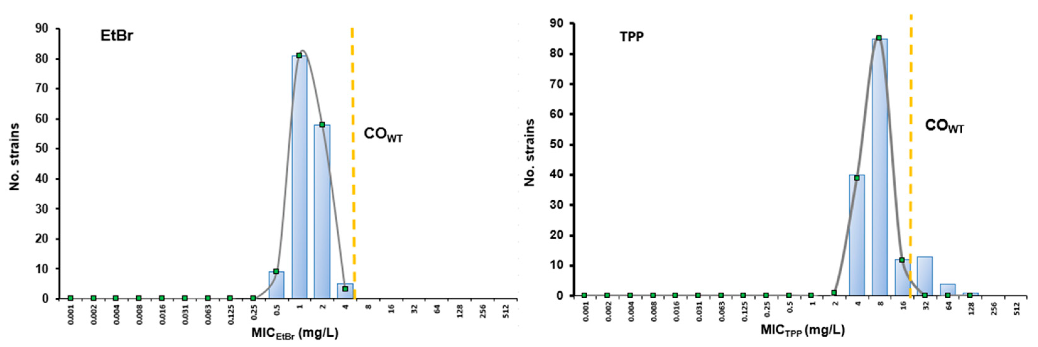
3.1.1. Susceptibility Profiling towards EtBr and TPP

Susceptibility to EtBr and TPP was evaluated by MIC determination for all the 155 S. pseudintermedius clinical strains in study (Figure 1), revealing a different MIC range for each compound. For EtBr, MICs varied between 0.5–4 mg/L, while for TPP, the MICs ranged between 4–128 mg/L. Considering the profile towards methicillin, the methicillin-susceptible strains (MSSP) presented MICEtBr between 0.5–2 mg/L, while the MRSP strains displayed MICEtBr up to 4 mg/L. However, these differences were not statistically significant (p = 0.11).
Regarding TPP, MSSP isolates showed lower MIC values, mostly up to 8 mg/L (except for 3 strains) when compared to MRSP strains (4–128 mg/L) (Figure 1). This was a significant difference (p < 0.01), indicating that MRSP generally presents lower susceptibility towards TPP than MSSP strains.

3.1.2. Cut-Off Values (COWT) and Identification of S. pseudintermedius NWT Populations for EtBr and TPP

The COWT value corresponds to the highest MIC value presented by a population devoid of resistance mechanisms with phenotypic expression to a particular compound, named wild-type (WT) population [32]. As such, the determination of this parameter permits to distinguish microorganisms that can potentially carry resistance mechanism(s) with phenotypic expression to that compound, nominated as NWT strains [32]. These NWT strains are characterized by MIC values above the COWT. This approach was used to screen for S. pseudintermedius strains with decreased susceptibility levels towards EtBr and/or TPP, potentially associated with efflux. The MICEtBr and MICTPP distributions of the entire collection were used to estimate COWT values by the iterative statistical method [33] using the ECOFFinder program (Figure 2). We determined COWT values of 4 mg/L and 16 mg/L for EtBr and TPP, respectively. Therefore, the NWT populations would correspond to those strains with an MICEtBr > 4 mg/L or MICTPP > 16 mg/L.
The results presented in Figure 2 and Table 1 indicate the absence of an NWT population for EtBr and the presence of an NWT population against TPP. This TPPNWT population was constituted by 18 clinical strains (11.6%), 17 of which were MRSP with MDR phenotypes. This TPPNWT population was characterized by significantly higher MICTPP (p < 0.001) and MICEtBr (p = 0.043) values than the TPPWT population. These results indicated the presence of a mechanism of resistance towards TPP, potentially efflux, in these 18 strains.

3.1.3. Assessment of Efflux Activity in S. pseudintermedius Strains

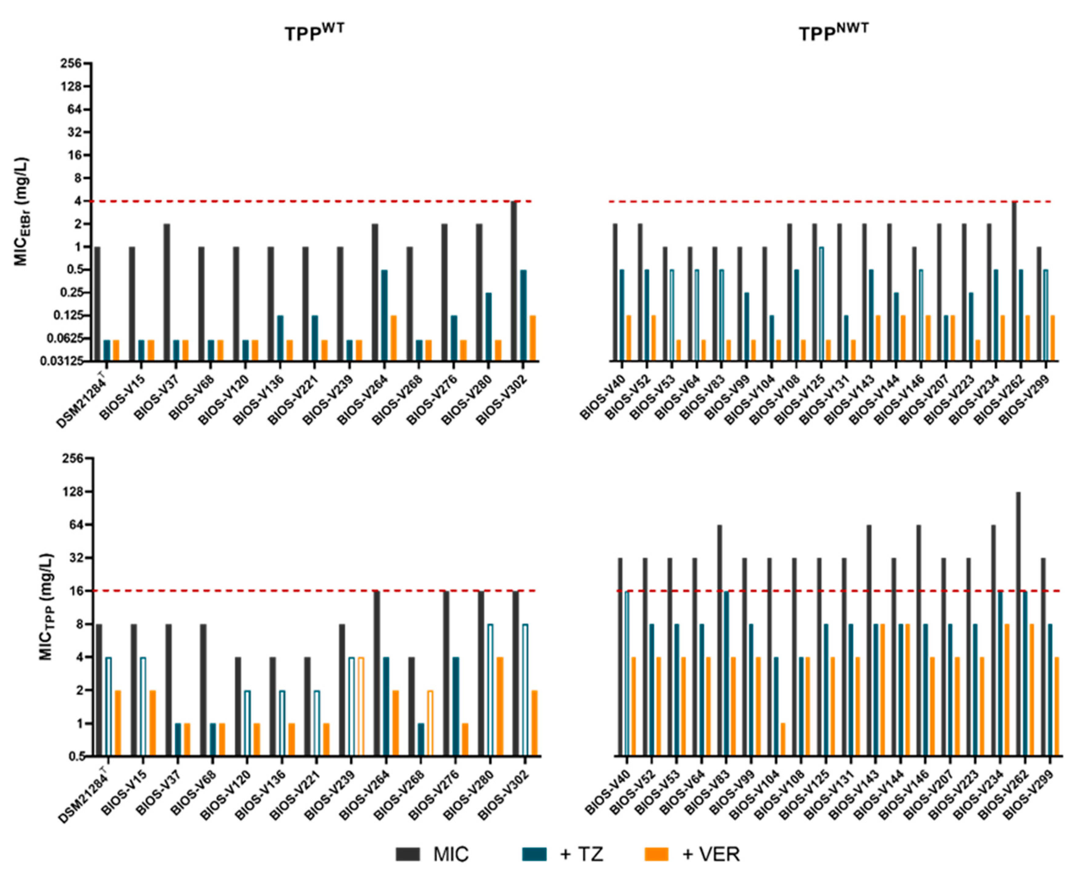
Effect of Efflux Inhibitors (EIs) on MICEtBr and MICTPP

The effect of the EIs TZ and VER on the susceptibility levels of EtBr and TPP was evaluated in a subset of 70 clinical strains and strain DSM21284T (Table 2). This subset included the 18 TPPNWT strains, as well as 52 strains from the WT population (TPPWT). These 52 strains from the WT population, which included 22 MRSP and 30 MSSP strains, were selected according to their susceptibility towards ciprofloxacin, methicillin and other antimicrobials (multidrug resistance), previously determined [27]. We considered that efflux inhibitory activity by an EI corresponded to a reduction of the MICEtBr or MICTPP to, at least, a quarter of its original value in the presence of that EI [18,19,39].
Overall, both EIs (TZ and VER), reduced the MICEtBr and MICTPP values in a scale of 2–32× of its original value (Table 2). Significant MICTPP and MICEtBr reductions (≥4-fold) were detected amongst the TPPWT and TPPNWT populations in the presence of the two EIs. However, this effect was particularly striking for the TPPNWT population, for which the EIs reduced MICTPP to values below the S. pseudintermedius COWT for TPP (Figure 3). From the two EIs, VER promoted a more pronounced effect, also for a higher number of strains. These data reinforce efflux as the mechanism conferring reduced susceptibility to TPP in the TPPNWT population.

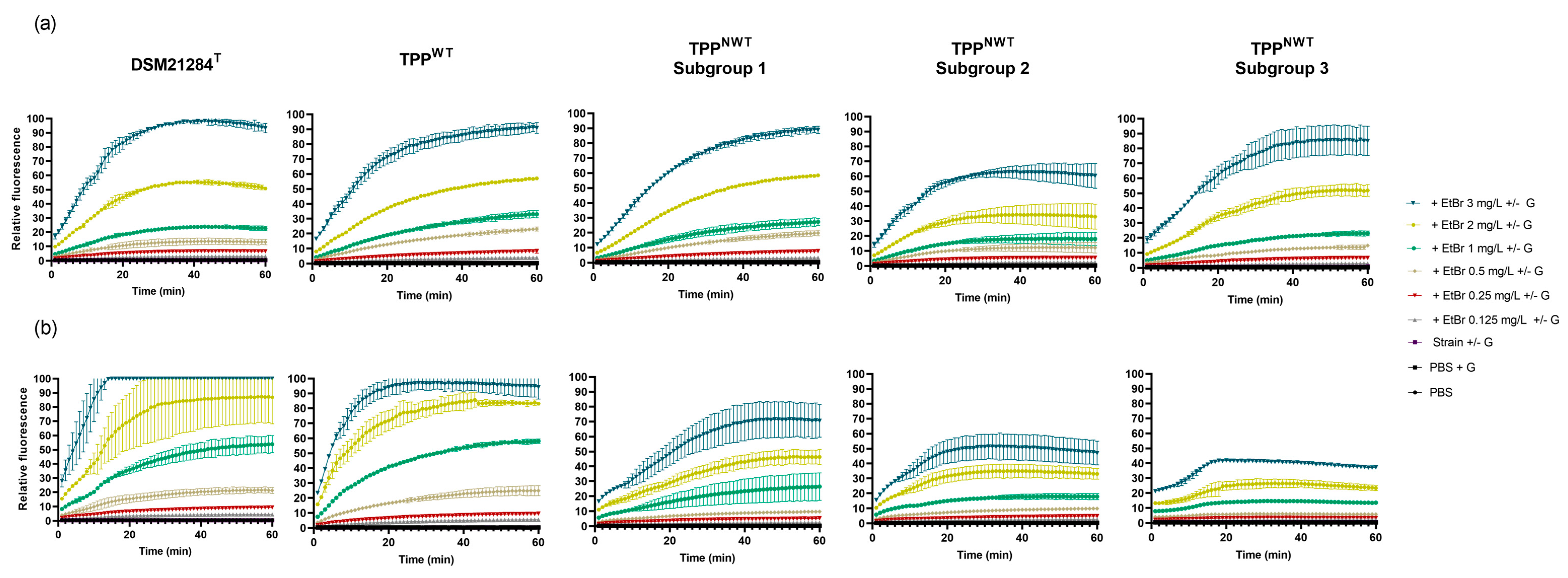
Detection and Analysis of Efflux Activity by Real-Time Fluorometry

To confirm the increased efflux activity of the TPPNWT population, the intracellular accumulation of EtBr, a broad range substrate of most bacterial MDR EPs was evaluated by real-time fluorometry.
Since this methodology has been scarcely applied to S. pseudintermedius, it required the optimization of experimental conditions, which was carried out with strain DSM21284T. This strain accumulated EtBr (increasing fluorescence) proportionally to the increasing EtBr concentrations tested, reaching almost 100% of the fluorescence signal with the highest EtBr concentration tested (3 mg/L) (Figure 4a). In the presence of glucose, an energy source of efflux systems, the EtBr accumulation in this strain was slightly promoted (Figure 4b). The optimized assay was then applied to a group of representative clinical strains (n = 11), which included eight TPPNWT and three TPPWT strains. These strains were selected based on their highest MICTPP and MICEtBr values, highest inhibitory effect of EIs, as well as genotypic (clonal lineage and agr type) and phenotypic traits (MRSP/MSSP, resistance to fluoroquinolones) previously determined [27].
Analysis of the EtBr accumulation profile of the selected strains revealed different accumulation patterns (Figure 4). The TPPWT strains BIOS-V37, BIOS-V268 and BIOS-V276 accumulated EtBr similarly to DSM21284T, suggesting absence of increased efflux. As for TPPNWT strains, the EtBr accumulation pattern was heterogeneous, and these strains were subdivided into three subgroups (Figure 4). Subgroup 1, constituted by strains BIOS-V83, BIOS-V223 and BIOS-V146, accumulated EtBr in a manner similar to DSM21284T and TPPWT strains in the absence of glucose, whereas with glucose accumulated less EtBr. Subgroup 2, formed by strains BIOS-V104, BIOS-V143 and BIOS-V234, showed reduced EtBr accumulation either in the absence or presence of glucose, in comparison to DSM21284T and TPPWT strains. Finally, subgroup 3 (strains BIOS-V99 and BIOS-V262), showed, in the absence of glucose, an accumulation pattern similar to TPPWT and TPPNWT subgroup 1 strains. In the presence of glucose, EtBr accumulated at very low values for all concentrations tested.
Since lower levels of EtBr accumulation indicated higher efflux of this molecule, these results indicated that TPPNWT strains, particularly those of subgroups 2 and 3, have higher efflux activity than TPPWT strains and DSM21284T.
To further support these findings, EtBr accumulation assays were also conducted in the absence/presence of EIs for six S. pseudintermedius clinical strains; five TPPNWT assigned to subgroup 2 or 3 and one TPPWT strain, as well as DSM21284T. The TPPNWT subgroup 1 strains were not analyzed since they presented lower efflux activity in comparison to the other subgroups. To compare the effect of the EIs TZ and VER on EtBr accumulation, we determined the relative final fluorescence (RFF) value, a parameter that reflects the inhibitory efflux capacity of each EI (Table 3). RFF values higher than 1 indicate efflux inhibition [35].
The RFF values were calculated in the absence and presence of glucose and supported the previous findings of increased efflux activity in TPPNWT strains from subgroup 2 and 3—Table 3. This is reflected by the lower RFF values obtained for strains with basal efflux activity (DSM21284T and TPPWT). On the other hand, strains with augmented efflux (TPPNWT) were more affected by EIs. This inhibitory effect was particularly notorious in the presence of glucose, a condition of optimal efflux activity. The two EIs presented different efflux inhibitory capacities; higher for VER on all strains except one (BIOS-V99, subgroup 3), lower for TZ, with significant inhibitory effect for a single strain, assigned to TPPNWT subgroup 3 (BIOS-V262). These results confirmed the higher efflux activity of TPPNWT strains, which was inhibited by EIs, particularly verapamil.

3.1.4. Screening of Plasmid-Encoded Efflux Pump Genes qacA/B and smr

The genes qacA/B and smr encode, respectively, the efflux pumps QacA/B and Smr, which are involved in decreased susceptibility towards biocides, like TPP, and dyes, such as EtBr [18,19,26]. Therefore, we assessed their presence by PCR, to infer on their potential contribution towards the increased efflux activity detected in TPPNWT population, as well as in the remaining strains. No strains were found to carry either qacA/B or smr in the entire collection (n = 155), which indicates that the detected efflux activity originates from other efflux systems, namely chromosomally-encoded MDR EPs.

3.2. Relation between Resistance to Fluoroquinolones and Efflux Activity

Effect of Efflux Inhibitors on Ciprofloxacin (CIP) Susceptibility Levels

Fluoroquinolones are also substrates of S. aureus native MDR efflux systems [17,18,19]. This observation was recently extended for a reference S. pseudintermedius strain [24]. Thus, we aimed to assess the contribution of efflux to fluoroquinolone resistance. A previous characterization of the antimicrobial susceptibility profiles of the study collection by disk diffusion assays revealed that 24.8% of the 155 strains (n = 38) were resistant or intermediate to ciprofloxacin. Susceptibility to ciprofloxacin was re-evaluated by MIC determination, which agree with the disk diffusion susceptibility profiles for all strains but one, categorized as susceptible or intermediate, by disk diffusion or MIC, respectively (Table 2) [28]. The MICCIP values ranged between 0.06 and 64 mg/L for the entire collection. The selected 70 clinical strains (Table 2) presented different susceptibility phenotypes towards ciprofloxacin, namely 31 resistant strains (MICCIP > 4 mg/L), two intermediate (MICCIP = 2 mg/L) and 37 susceptible strains (MICCIP ≤ 1 mg/L). Of these, the 18 TPPNWT strains included 17 ciprofloxacin resistant strains (MICCIP of 32–64 mg/L) and a single susceptible strain (MICCIP = 0.25 mg/L). In staphylococci, resistance to fluoroquinolones is mainly mediated by mutations in the QRDR of target genes grlA/B and/or gyrA/B [42], which can hinder the assessment of the efflux contribution to phenotypes.
The EIs TZ and VER revealed a mild effect upon MICCIP values for the TPPNWT strains, resulting mostly in two-fold MIC reductions (Table 2). However, the EIs effect may be masked by the presence of the double mutation S80I (GrlA)/S84L (GyrA) carried by most of these strains, which are often associated with MICCIP > 32 mg/L [43,44]. The same mild effect of EIs was observed for most TPPWT strains, with only four strains showing a 4-fold MICCIP reduction. Interestingly, these strains harbored no QRDR mutations or a single mutation in GrlA (S80I) and may represent a stage when resistance results from a balance between efflux and mutation acquisition.

4. Discussion

S. pseudintermedius is the main bacterial agent of pyoderma in companion animals, particularly in dogs [3,4] and the emergence of MRSP strains, often associated with MDR phenotypes, is a public health concern [10]. This study aimed to characterize efflux activity, a resistance mechanism still poorly characterized for this species, as a contributor to biocide and fluoroquinolone resistance.

4.1. Contribution of Efflux to Reduced Susceptibility to Biocides, EtBr and Resistance to Fluoroquinolones in S. pseudintermedius

The study initiated with the assessment of TPP and EtBr susceptibility levels for all 155 S. pseudintermedius in the collection and the determination of COWT to screen for NWT populations towards for these two compounds.
For EtBr, the range of MIC values (0.5–4 mg/L) is similar to the one previously described for S. pseudintermedius causing different infections in companion animals, except for one isolate with an MICEtBr of 32 mg/L [45]. The range of MICEtBr values found in this study for S. pseudintermedius is lower when compared to the ones described for S. aureus or S. epidermidis. In S. aureus, MICEtBr have been reported to vary between 2–32 mg/L in human [17] or veterinary clinical isolates [46]. For S. epidermidis, a higher MICEtBr range (1–128 mg/L) has been described for veterinary clinical isolates [47]. For all these staphylococcal species, isolates with high MICEtBr values (>8 mg/L) were mostly associated to the presence of the EP genes qacA/B or smr genes [17,45,46,47]. The S. pseudintermedius COWT value for EtBr established in this study, 4 mg/L, corresponded to an absence of a NWT population against EtBr, most likely due to the absence of the qacA/B or smr genes in this collection.
A higher MIC range was observed for TPP (4–128 mg/L) and MRSP strains were more prone to present higher MICTPP values (p < 0.001), ie, lower susceptibility to TPP. To the best of our knowledge, this is the first proposal of a COWT of S. pseudintermedius for TPP (16 mg/L), which is close to that already proposed for S. aureus, 32 mg/L [46]. The application of the COWT for TPP identified an NWT population against this biocide. Interestingly, this population was mostly constituted by MRSP-MDR strains, from the ST71-agrIII lineage [27,40]. The predominance of the MRSP ST71 strains in the NWT population is relevant, considering that this lineage continues to be one of the most predominant in Portugal and other European countries and is associated with a high burden of antimicrobial resistance [2,48,49,50]. As for EtBr, reduced susceptibility to TPP has also been linked to the presence of plasmid-encoded EP genes, like qacA/B or smr [46] or native MDR efflux systems, such as NorA. Since this collection was devoid of plasmid-encoded EP genes, the resistance mechanism present in the TPPNWT population should be associated with an increased activity of chromosomally-encoded efflux systems.
We then proceeded to the evaluation and characterization of the S. pseudintermedius efflux activity by two independent methods: MIC determination in the presence of efflux inhibitors and real time fluorometry [19]. Efflux inhibitors reduced MICEtBr and MICTPP values for both TPPWT and TPPNWT populations. However, the EIs, particularly verapamil, had a higher impact on the MICTPP values of the TPPNWT population, decreasing the MICTPP to values below the COWT established for S. pseudintermedius, i.e., for values corresponding to the wild type population for TPP. Therefore, these results confirm the presence of increased efflux activity in the TPPNWT population and its possible association with reduced susceptibility to this biocide.
These findings were corroborated by real time fluorometry. Fluorometric assays were carried out in absence and presence of glucose, which, in strains with higher efflux leads to decreased EtBr accumulation (i.e., greater EtBr efflux). This is due to the metabolization of glucose, which energizes efflux systems [19,34]. Strains with basal efflux activity, as DSM21284T and strains of the TPPWT population, accumulate EtBr significantly, a trait augmented in the presence of the source of energy, an unexpected finding. On the other hand, strains with increased efflux activity, as the TPPNWT population, showed lower EtBr accumulation in the presence of glucose. These overall results indicate that for S. pseudintermedius, efflux depends on glucose as an energy source. This glucose-dependent efflux activity was already described for other staphylococci, namely S. aureus and S. epidermidis [17,19,23,30,51]. In support of these observations, the effect of EIs on EtBr accumulation was only observed in the presence of glucose. This behavior reveals the need to energize S. pseudintermedius efflux systems to observe the EIs effect. This hypothesis is supported by the calculated RFF values, which are only significant in the presence of glucose, particularly for verapamil. Our data also indicate that verapamil is an effective efflux inhibitor in S. pseudintermedius, similarly to what has been described for S. aureus and S. epidermidis [23,52].
Despite these observations, increased efflux was not detected for all TPPNWT strains tested. Lack of complete agreement between the two approaches used to evaluate efflux activity may be due to differing experimental conditions, like time of exposure to the substrate (18 hours vs. 1 hour). Another possible explanation may be different extrusion efficiency rates for EtBr and TPP by the efflux pump(s) involved, as the result of the substrate binding sites established in the pump(s) as already been postulated for the S. aureus NorA [53,54].
Increased efflux can lead to cross-resistance to different chemical compounds, provided they are substrates of the same EPs [55]. This response has been observed in S. aureus, as its MDR EPs have several substrates, including fluoroquinolones [18,56]. In particular, studies from our group have demonstrated that exposure of S. aureus to a biocide or EtBr results in the emergence of fluoroquinolone resistance mediated solely by efflux [20,30]. Fluoroquinolones are prescribed as second tier for the treatment of canine SSTIs and their utilization should be limited [6,57]. Nevertheless, this antibiotic class is the second most prescribed, after beta-lactams, for the treatment of infections in companion animals in European countries, including Portugal [58]. The misuse of fluoroquinolones has promoted an increase of antimicrobial resistance in Portugal and worldwide [12,50,59,60]. In S. pseudintermedius, resistance mechanisms against fluoroquinolones include increased efflux activity and/or mutations in QRDR of the target genes, grlA and gyrA [41,44]. A previous characterization of the collection in study had revealed a significant rate (~25%) of fluoroquinolone resistance and presence of target gene mutations. In this work, we correlated this previous susceptibility data with MICCIP values. The presence of the double mutations GrlA:Ser80Ile and GyrA:Ser84Leu, was associated with a high level of resistance (MICCIP ≥ 32 mg/L). Other QRDR double mutations patterns were detected for resistant strains with lower MICCIP values (4–8 mg/L). These data suggest that fluoroquinolone resistance in S. pseudintermedius is mainly due to target mutations of QRDR region of grlA/gyrA as described by Loiacono and colleagues [61]. The QRDR mutations herein reported have been already associated with fluoroquinolone resistance in S. pseudintermedius [43,44,62,63], and are known to convey high MICCIP values, similar to the ones found in this work [41,63,64]. This fact prevented the assessment of a potential effect of EIs upon MICCIP values. Yet, a significant EI effect was found for a few strains with a susceptible or intermediate phenotype that carried a single GrlA mutation and could represent an intermediary stage in the development of fluoroquinolone resistance. The data gathered herein for ciprofloxacin may be extrapolated for other fluoroquinolones. Descloux et. al. have reported that the presence of GrlA:Ser80Ile reduces susceptibility to enrofloxacin and Onuma et al. also reported that the presence of GrlA:Asp84Gly leads to a decreased susceptibility towards ofloxacin and enrofloxacin and resistance to levofloxacin [41,63]. Altogether, these data emphasize that the misuse of fluoroquinolones can play a role in increasing resistance in S. pseudintermedius and other staphylococci [20,59].

4.2. Misuse of TPP May Promote Emergence of Efflux-Mediated Resistance

Biocides are widely used to treat and prevent infections caused by S. pseudintermedius, given the increasing number of infections caused by antimicrobial resistant strains. In veterinary medicine, biocides are prescribed in combination or individual therapy, depending on the severity of the lesions [4,5].
TPP is a cationic biocide with several applications, including in medicated dressings for skin infections and for domestic surfaces disinfection. This compound acts due to its ability to prevent bacteria fixation [65,66,67] and it is structurally similar to quaternary ammonium compounds, hence having the same action mechanism of disrupting cell membranes [67,68]. When TPP was introduced, it presented an important antimicrobial activity against S. aureus, but later revealed to be a substrate of several staphylococcal EPs associated with high MICTPP [18,22,69,70].
The reduced susceptibility towards TPP presented by S. pseudintermedius could be related to incorrect use as an antiseptic, or an ineffective cleaning of surfaces and objects in contact with companion animals, which allow S. pseudintermedius to be exposed to sub-lethal concentrations of TPP. This exposure can promote an over-expression of MDR efflux pumps, resulting in decreased susceptibility to substrates of these pumps, such TPP and other biocides and effluxable antimicrobials like fluoroquinolones [55]. The transfer of these strains between companion animals and surfaces (or vice-versa), can contribute to their dissemination [7,71,72].
Although the decreased susceptibility to biocides used in veterinary practice may be considered not clinically relevant, since the in-use concentrations applied are much higher [16,45,73], the data obtained in this work highlight the potential decrease in susceptibility mediated by efflux and the need to use biocides prudently to prevent the spread of S. pseudintermedius strains with reduced susceptibility, as already described in S. aureus [18,19,20,74,75,76].

5. Conclusions

This study highlights the importance and the need of characterizing efflux in S. pseudintermedius as a resistance mechanism to fluoroquinolones and biocides. The use of these antimicrobial agents in veterinary therapy is put at risk by the decreased susceptibility linked to efflux, as illustrated in this work, which demonstrates that prudence is required in the use of these agents.
Surveillance of the emergence of strains with increased efflux activity, using methodological approaches such as the ones optimized and applied in this study will be crucial for the control of infections caused by this important pathogen of companion animals.

Supplementary Materials

The following supporting information can be downloaded at: https://www.mdpi.com/article/10.3390/ani13071270/s1, Table S1: Distribution of MRSP, MSSP and MDR phenotypes among the S. pseudintermedius study collection (n = 155); Table S2: Control strains used for the detection of plasmid-encoded efflux pump genes; Table S3: Primers used in this study.

Author Contributions

Conceptualization, S.S.C. and I.C.; methodology, S.S.C. and I.C.; validation, S.S.C., P.A. and I.C.; formal analysis, M.L., P.A. and B.R.; investigation, M.L., C.M., B.R., S.S.C. and I.C.; resources, S.S.C., C.P. and I.C.; data curation, M.L.; writing—original draft preparation, M.L., S.S.C. and I.C.; writing—review and editing, M.L., C.M., B.R., S.S.C., P.A., C.P. and I.C.; visualization, M.L., S.S.C. and I.C.; supervision, S.S.C. and I.C.; project administration, I.C.; funding acquisition, S.S.C. and I.C. All authors have read and agreed to the published version of the manuscript.

Funding

This work was supported by Project BIOSAFE funded by FEDER through the Programa Operacional Factores de Competitividade—COMPETE, by the Fundação para a Ciência e a Tecnologia (FCT, Portugal)—Grant LISBOA-01-0145-FEDER-030713, PTDC/CAL-EST/30713/2017 and by FCT through grants UI/BD/151061/2021 (C.M) as well as funds to GHMT (UID/04413/2020) and the CIISA Project (UID/CVT/00276/2020).

Institutional Review Board Statement

Not applicable. This study did not involve manipulation of companion animals; all S. pseudintermedius strains studied have been previously collected by diagnostic laboratories.

Informed Consent Statement

Not applicable.

Data Availability Statement

All relevant data have been provided in the paper. Raw data can also be provided by the authors upon reasonable request.

Conflicts of Interest

The authors declare no conflict of interest.

References

  1. Bhooshan, S.; Negi, V.; Khatri, P. Staphylococcus pseudintermedius: An undocumented, emerging pathogen in humans. GMS Hyg. Infect. Control. 2020, 15, 1–11. [Google Scholar] [CrossRef]
  2. Ruiz-Ripa, L.; Simón, C.; Ceballos, S.; Ortega, C. S. pseudintermedius and S. aureus lineages with transmission ability circulate as causative agents of infections in pets for years. BMC Vet. Res. 2021, 17, 42. [Google Scholar] [CrossRef] [PubMed]
  3. Nielsen, S.S.; Bicout, D.J.; Calistri, P.; Canali, E.; Drewe, J.A.; Garin-Bastuji, B.; Rojas, J.L.G.; Gortázar, C.; Herskin, M.; Michel, V.; et al. Assessment of listing and categorization of animal diseases within the framework of the Animal Health Law (Regulation (EU) No 2016/429): Antimicrobial-resistant Staphylococcus pseudintermedius in dogs and cats. EFSA J. Eur. Food Saf. Auth. 2022, 20, e07310. [Google Scholar] [CrossRef]
  4. Lynch, S.A.; Helbig, K.J. The complex diseases of Staphylococcus pseudintermedius in canines: Where to next? Vet. Sci. 2021, 8, 11. [Google Scholar] [CrossRef]
  5. Frank, L.; Loeffler, A. Meticillin-resistant Staphylococcus pseudintermedius: Clinical challenge and treatment options. Vet. Dermatol. 2012, 23, 283–291. [Google Scholar] [CrossRef]
  6. Hillier, A.; Lloyd, D.H.; Weese, J.; Blondeau, J.; Boothe, D.; Breitschwerdt, E.; Guardabassi, L.; Papich, M.J.; Rankin, S.; Turnidge, J.T.; et al. Guidelines for the diagnosis and antimicrobial therapy of canine superficial bacterial folliculitis (Antimicrobial Guidelines Working Group of the International Society for Companion Animal Infectious Diseases). Vet. Dermatol. 2014, 25, 163-e43. [Google Scholar] [CrossRef] [PubMed]
  7. Morris, D.O.; Loeffler, A.; Davis, M.F.; Guardabassi, L.; Weese, J.S. Recommendations for approaches to meticillin-resistant staphylococcal infections of small animals: Diagnosis, therapeutic considerations and preventative measures. Vet. Dermatol. 2017, 28, 304-e69. [Google Scholar] [CrossRef] [PubMed] [Green Version]
  8. Beck, K.M.; Waisglass, S.E.; Dick, H.L.N.; Weese, J.S. Prevalence of meticillin-resistant Staphylococcus pseudintermedius (MRSP) from skin and carriage sites of dogs after treatment of their meticillin-resistant or meticillin-sensitive staphylococcal pyoderma. Vet. Dermatol. 2012, 23, 369–375. [Google Scholar] [CrossRef]
  9. Bierowiec, K.; Miszczak, M.; Korzeniowska-Kowal, A.; Wzorek, A.; Płókarz, D.; Gamian, A. Epidemiology of Staphylococcus pseudintermedius in cats in Poland. Sci. Rep. 2021, 11, 18898. [Google Scholar] [CrossRef]
  10. Pomba, C.; Rantala, M.; Greko, C.; Baptiste, K.E.; Catry, B.; van Duijkeren, E.; Mateus, A.; Moreno, M.A.; Pyorala, S.; Ruzauskas, M.; et al. Public health risk of antimicrobial resistance transfer from companion animals. J. Antimicrob. Chemother. 2017, 72, 957–968. [Google Scholar] [CrossRef]
  11. Loeffler, A.; Lloyd, D.H. What has changed in canine pyoderma? A narrative review. Vet. J. 2018, 235, 73–82. [Google Scholar] [CrossRef] [PubMed] [Green Version]
  12. Feßler, A.; Scholtzek, A.; Schug, A.; Kohn, B.; Weingart, C.; Schink, A.; Bethe, A.; Lübke-Becker, A.; Schwarz, S. Antimicrobial and biocide resistance among feline and canine Staphylococcus aureus and Staphylococcus pseudintermedius isolates from diagnostic submissions. Antibiotics 2022, 11, 127. [Google Scholar] [CrossRef] [PubMed]
  13. Gagetti, P.; Rosato, R.R.; Rosato, A.E. Staphylococcus pseudintermedius’s PBP4 is directly associated with the dissociated oxacillin and cefoxitin phenotype. Antibiotics 2021, 10, 1299. [Google Scholar] [CrossRef] [PubMed]
  14. Borjesson, S.; Gómez-Sanz, E.; Ekstrom, K.; Torres, C.; Gronlund, U. Staphylococcus pseudintermedius can be misdiagnosed as Staphylococcus aureus in humans with dog bite wounds. Eur. J. Clin. Microbiol. Infect. Dis. 2015, 34, 839–844. [Google Scholar] [CrossRef] [PubMed]
  15. Somayajin, R.; Rubin, J.E.; Priyantha, M.; Church, D. Exploring Staphylococcus pseudintermedius: An emerging zoonotic pathogen? Future Microbiol. 2016, 11, 1371–1374. [Google Scholar] [CrossRef] [Green Version]
  16. Frosini, S.M.; Bond, R.; McCarthy, A.J.; Feudi, C.; Schwarz, S.; Lindsay, J.A.; Loeffler, A. Genes on the move: In vitro transduction of antimicrobial resistance genes between human and canine Staphylococcal pathogens. Microorganisms 2020, 8, 2031. [Google Scholar] [CrossRef]
  17. Costa, S.S.; Falcão, C.; Viveiros, M.; Machado, D.; Martins, M.; Melo-Cristino, J.; Amaral, L.; Couto, I. Exploring the contribution of efflux on the resistance to fluoroquinolones in clinical isolates of Staphylococcus aureus. BMC Microbiol. 2011, 11, 241. [Google Scholar] [CrossRef] [Green Version]
  18. Costa, S.S.; Viveiros, M.; Amaral, L.; Couto, I. Multidrug Efflux Pumps in Staphylococcus aureus: An update. Open Microbiol. J. 2013, 7, 59–71. [Google Scholar] [CrossRef] [Green Version]
  19. Costa, S.S.; Junqueira, E.; Palma, C.; Viveiros, M.; Melo-Cristino, J.; Amaral, L.; Couto, I. Resistance to antimicrobials mediated by efflux pumps in Staphylococcus aureus. Antibiotics 2013, 2, 83–99. [Google Scholar] [CrossRef] [Green Version]
  20. Costa, S.S.; Viveiros, M.; Rosato, A.; Melo-Cristino, J.; Couto, I. Impact of efflux in the development of multidrug resistance phenotypes in Staphylococcus aureus. BMC Microbiol. 2015, 15, 232. [Google Scholar] [CrossRef] [Green Version]
  21. Blair, J.M.A.; Piddock, L.J.V. How to measure export via bacterial multidrug resistance efflux pumps. ASM 2016, 7, e00840-16. [Google Scholar] [CrossRef] [PubMed] [Green Version]
  22. DeMarco, C.E.; Cushing, L.A.; Frempong-Manso, E.; Seo, S.M.; Jaravaza, T.A.A.; Kaatz, G.W. Efflux-related resistance to norfloxacin, dyes, and biocides in bloodstream isolates of Staphylococcus aureus. Antimicrob. Agents Chemother. 2007, 51, 3235–3239. [Google Scholar] [CrossRef] [Green Version]
  23. Costa, S.S.; Viveiros, M.; Pomba, C.; Couto, I. Active antimicrobial efflux in Staphylococcus epidermidis: Building up of resistance to fluoroquinolones and biocides in a major opportunistic pathogen. J. Antimicrob. Chemother. 2018, 73, 320–324. [Google Scholar] [CrossRef] [PubMed] [Green Version]
  24. Rampacci, E.; Felicetii, T.; Pietrella, D.; Sabatini, S.; Passamonti, F. Drug efflux transporters in Staphylococcus pseudintermedius: In silico prediction and characterization of resistance. JAC 2022, 77, 3283–3290. [Google Scholar] [CrossRef]
  25. Couto, N.; Belas, A.; Oliveira, M.; Almeida, P.; Clemente, C.; Pomba, C. Comparative RNA-seq-based transcriptome analysis of the virulence characteristics of methicillin-resistant and -susceptible Staphylococcus pseudintermedius strains isolated from small animals. Antimicrob. Agents. Chemother. 2016, 60, 962–967. [Google Scholar] [CrossRef] [Green Version]
  26. Worthing, K.A.; Marcus, A.; Abraham, S.; Trott, D.J.; Norris, J.M. Qac genes and biocide tolerance in clinical veterinary methicillin-resistant and methicillin-susceptible Staphylococcus aureus and Staphylococcus pseudintermedius. Vet. Microbiol. 2018, 216, 153–158. [Google Scholar] [CrossRef]
  27. Morais, C.; Costa, S.S.; Leal, M.; Ramos, B.; Andrade, M.; Ferreira, C.; Abrantes, P.; Pomba, C.; Couto, I. Genetic diversity and antimicrobial resistance profiles of Staphylococcus pseudintermedius associated to skin and soft-tissue infections in companion animals in Lisbon, Portugal. Front. Microbiol. 2023; in press. [Google Scholar] [CrossRef]
  28. Clinical and Laboratory Standards Institute (CLSI). Performance Standards for Antimicrobial Susceptibility Testing. CLSI Supplement M100, 32nd ed.; Clinical and Laboratory Standards Institute: Wayne, PA, USA, 2022. [Google Scholar]
  29. Clinical and Laboratory Standards Institute (VET01S CLSI). Performance Standards for Antimicrobial Disk and Dilution Susceptibility Tests for Bacteria Isolated from Animals. CLSI Supplement VET01S, 5th ed.; Clinical and Laboratory Standards Institute: Wayne, PA, USA, 2020. [Google Scholar]
  30. Couto, I.; Costa, S.S.; Viveiros, M.; Martins, M.; Amaral, L. Efflux-mediated response of Staphylococcus aureus exposed to ethidium bromide. J. Antimicrob. Chemother. 2008, 62, 504–513. [Google Scholar] [CrossRef] [Green Version]
  31. European Committee on Antimicrobial Susceptibility Testing (EUCAST). Media Preparation for Disk Diffusion Testing and for Determination of MIC Values by the Broth Microdilution Method, Version 7.0; European Committee on Antimicrobial Susceptibility Testing (EUCAST): Växjö, Sweden, 2022. [Google Scholar]
  32. European Committee on Antimicrobial Susceptibility Testing (EUCAST). Standard Operating Procedure. MIC Distributions and the Setting of Epidemiological Cutoff (ECOFF) Values, EUCAST SOP 10.2 2021. Available online: http://www.eucast.org (accessed on 15 February 2023).
  33. Turnidge, J.; Kahlmeter, G.; Kronvall, G. Statistical characterisation of bacterial wildtype MIC value distributions and the determination of epidemiological cut-off values. Clin. Microbiol. Infect. 2006, 12, 418–425. [Google Scholar] [CrossRef] [PubMed]
  34. Viveiros, M.; Martins, M.; Couto, I.; Rodrigues, L.; Spengler, G.; Martins, A. New methods for the identification of efflux mediated MDR bacteria, genetic assessment of regulators and efflux pump constituents, characterization of efflux systems and screening for inhibitors of efflux pumps. Curr. Drugs Targets 2008, 9, 760–778. [Google Scholar] [CrossRef]
  35. Costa, S.S.; Lopes, E.; Azzali, E.; Machado, D.; Coelho, T.; Silva, P.; Viveiros, M.; Pieroni, M.; Couto, I. An experimental model for the rapid screening of compounds with potential use against Mycobacteria. Assay Drug Dev. Technol. 2016, 14, 524–534. [Google Scholar] [CrossRef]
  36. Costa, S.S.; Palma, C.; Kadlec, K.; Fessler, A.T.; Viveiros, M.; Melo-Cristino, J.; Schwarz, S.; Couto, I. Plasmid-borne antimicrobial resistance of Staphylococcus aureus isolated in a hospital in Lisbon, Portugal. Microb. Drug Resist. 2016, 22, 617–626. [Google Scholar] [CrossRef] [PubMed]
  37. Anthonisen, I.L.; Sunde, M.; Steinum, T.M.; Sidhu, M.S.; Sorum, H. Organization of the antiseptic resistance gene qacA and Tn552-related-lactamase genes in multidrug- resistant Staphylococcus haemolyticus strains of animal and human origins. Antimicrob. Agents. Chemother. 2002, 46, 3606–3612. [Google Scholar] [CrossRef] [Green Version]
  38. Bjorland, J.; Sunde, M.; Waage, S. Plasmid-borne smr gene causes resistance to quaternary ammonium compounds in bovine Staphylococcus aureus. J. Clin. Microbiol. 2001, 39, 3999–4004. [Google Scholar] [CrossRef] [PubMed] [Green Version]
  39. Martins, M.; McCusker, M.; Viveiros, M.; Couto, I.; Fanning, S.; Pagès, J.M. A Simple Method for Assessment of MDR Bacteria for Over-Expressed Efflux Pumps. Open Microbiol. J. 2013, 7, 72–82. [Google Scholar] [CrossRef]
  40. Andrade, M.; Oliveira, K.; Morais, C.; Abrantes, P.; Pomba, C.; Rosato, A.E.; Couto, I.; Costa, S.S. Virulence potential of biofilm producing Staphylococcus pseudintermedius, Staphylococcus aureus and Staphylococcus coagulans causing skin infections in companion animals. Antibiotics 2022, 11, 1339. [Google Scholar] [CrossRef]
  41. Descloux, S.; Rossano, A.; Perreten, V. Characterization of new Staphylococcal Cassette Chromosome mec (SCCmec) and topoisomerase genes in fluoroquinolone- and methicillin-Resistant Staphylococcus pseudintermedius. J. Clin. Microbiol. 2008, 46, 1818–1823. [Google Scholar] [CrossRef] [PubMed] [Green Version]
  42. Hooper, D. Emerging mechanisms of fluoroquinolones resistance. Emerg Infect. Dis. 2001, 7, 337–341. [Google Scholar] [CrossRef] [PubMed] [Green Version]
  43. Kizerwetter-Świda, M.; Chrobak-Chmiel, D.; Rzewuska, M.; Binek, M. Resistance of canine methicillin-resistant Staphylococcus pseudintermedius strains to pradofloxacin. J. Vet. Diagnostic. Investig. 2016, 28, 514–518. [Google Scholar] [CrossRef] [Green Version]
  44. Azzariti, S.; Bond, R.; Loeffler, A.; Zendri, F.; Timofte, D.; Chang, Y.; Pelligand, L. Investigation of in vitro susceptibility and resistance mechanisms in skin pathogens: Perspectives for fluoroquinolone therapy in canine pyoderma. Antibiotics 2022, 11, 1204. [Google Scholar] [CrossRef]
  45. Couto, N.; Belas, A.; Couto, I.; Perreten, V.; Pomba, C. Genetic relatedness, antimicrobial and biocide susceptibility comparative analysis of methicillin-resistant and -susceptible Staphylococcus pseudintermedius from Portugal. Microb. Drug Resist. 2013, 20, 364–371. [Google Scholar] [CrossRef] [Green Version]
  46. Ribeiro, R. Resistência a Antimicrobianos em Staphylococcus aureus Isolados de Animais de Companhia. Master’s Thesis, Instituto de Higiene e Medicina Tropical, Lisbon, Portugal, 2019. [Google Scholar]
  47. Costa, S.S.; Rosa, M.; Rodrigues, A.C.; Holtreman, F.; Viveiros, M.; Pomba, C.; Couto, I. Emergence of non-wild-type populations towards biocides and antibiotic resistance in Staphylococcus epidermidis colonizing veterinary staff and students. ePoster Mini-Oral Session. In Proceedings of the 28th European Congress on Clinical Microbiology and Infectious Diseases (ECCMID), Madrid, Spain, 21–24 April 2018. [Google Scholar]
  48. Bergot, M.; Martins-Simões, P.; Kilian, H.; Châtre, P.; Worthing, K.; Norris, J.; Madec, J.; Laurent, F.; Haenni, M. Evolution of the population structure of Staphylococcus pseudintermedius in France. Front. Microbiol. 2021, 9, 3055. [Google Scholar] [CrossRef] [PubMed]
  49. Rynhoud, H.; Forde, M.; Beatson, S.; Abraham, S.; Meler, E.; Magalhães, R.; Gibson, J. Molecular epidemiology of clinical and colonizing methicillin-resistant Staphylococcus isolates in companion animals. Front. Vet. Sci. 2021, 8, 620491. [Google Scholar] [CrossRef] [PubMed]
  50. Silva, V.; Oliveira, A.; Manageiro, V.; Caniça, M.; Contente, D.; Capita, R.; Alonso-Calleja, C.; Carvalho, I.; Capelo, J.L.; Igrejas, G.; et al. Clonal diversity and antimicrobial resistance of methicillin-resistant Staphylococcus pseudintermedius isolated from canine pyoderma. Microorganisms 2021, 9, 482. [Google Scholar] [CrossRef] [PubMed]
  51. Tambat, R.; Jangra, M.; Mahey, N.; Chandal, N.; Kaur, M.; Chaudhary, S.; Verma, D.K.; Thakur, K.G.; Raje, M.; Jachak, S.; et al. Microbe-derived indole metabolite demonstrates potent multidrug efflux pump inhibition in Staphylococcus aureus. Front. Microbiol. 2019, 10, 2153. [Google Scholar] [CrossRef] [PubMed]
  52. Baiomy, A.; Shaker, G.; Abbas, H. Sensitizing multi drug resistant Staphylococcus aureus isolated from surgical site infections to antimicrobials by efflux pumps inhibitors. Afr. Health Sci. 2020, 20, 1632–1645. [Google Scholar] [CrossRef] [PubMed]
  53. Bhaskar, B.V.; Babu, T.M.C.; Reddy, N.V.; Rajendra, W. Homology modeling, molecular dynamics, and virtual screening of NorA efflux pump inhibitors of Staphylococcus aureus. Drug Des. Devel. Ther. 2016, 10, 3237–3252. [Google Scholar] [CrossRef] [PubMed] [Green Version]
  54. Zárate, S.G.; Morales, P.; Świderek, K.; Bolanos-Garcia, V.M.; Bastida, A. Molecular modeling approach to identify novel inhibitors of the major facilitator superfamily of efflux pump transporters. Antibiotics 2019, 8, 25. [Google Scholar] [CrossRef] [PubMed] [Green Version]
  55. Buffet-Bataillon, S.; Tattevin, P.; Maillard, J.Y.; Bonnaure-Mallet, M.; Jolivet Gougeon, A. Efflux pump induction by quaternary ammonium compounds and fluoroquinolone resistance in bacteria. Future Microbiol. 2016, 11, 81–92. [Google Scholar] [CrossRef] [PubMed]
  56. Lekshmi, M.; Ammini, P.; Adjei, J.; Sanford, L. Modulation of antimicrobial efflux pumps of the major facilitator superfamily in Staphylococcus aureus. AIMS Microbiol. 2018, 4, 1–18. [Google Scholar] [CrossRef] [PubMed]
  57. Beco, L.; Guaguère, E.; Lorente Méndez, C.; Noli, C.; Nuttall, T.; Vroom, M. Suggested guidelines for using systemic antimicrobials in bacterial skin infections: Part 2-antimicrobial choice, treatment regimens and compliance. Vet. Rec. 2013, 172, 156–160. [Google Scholar] [CrossRef] [PubMed] [Green Version]
  58. European Medicines Agency (EMA). Sales of Veterinary Antimicrobial Agents in 31 European Countries in 2021; European Medicines Agency (EMA): Amesterdam, The Netherlands, 2022.
  59. Oliveira, A.; Devesa, J.S.P.; Hill, P.B.; Silva, V.; Poeta, P. Treatment of selected canine dermatological conditions in Portugal—A research survey. J. Vet. Res. 2018, 62, 563–570. [Google Scholar] [CrossRef] [PubMed] [Green Version]
  60. Gómez-Poveda, B.; Moreno, M. Antimicrobial prescriptions for dogs in the Capital of Spain. Front. Vet. Sci. 2018, 5, 309. [Google Scholar] [CrossRef] [PubMed] [Green Version]
  61. Loiacono, M.; Martino, P.A.; Albonico, F.; Dell’Orco, F.; Ferretti, M.; Zanzani, S.; Mortarino, M. High-resolution melting analysis of gyrA codon 84 and grlA codon 80 mutations conferring resistance to fluoroquinolones in Staphylococcus pseudintermedius isolates from canine clinical samples. J. Vet. Diagn. Investig. 2017, 29, 711–715. [Google Scholar] [CrossRef] [PubMed] [Green Version]
  62. Kadlec, K.; Schwarz, S. Antimicrobial resistance of Staphylococcus pseudintermedius. Vet. Dermatol. 2012, 23, 276-e55. [Google Scholar] [CrossRef]
  63. Onuma, K.; Tanabe, T.; Sato, H. Antimicrobial resistance of Staphylococcus pseudintermedius isolates from healthy dogs and dogs affected with pyoderma in Japan. Vet. Dermatol. 2012, 23, 17-e5. [Google Scholar] [CrossRef]
  64. Awji, E.G.; Tassew, D.D.; Lee, J.S.; Lee, S.J.; Choi, M.J.; Reza, M.A.; Rhee, M.H.; Kim, T.H.; Park, S.C. Comparative mutant prevention concentration and mechanism of resistance to veterinary fluoroquinolones in Staphylococcus pseudintermedius. Vet. Dermatol. 2012, 23, 376-e69. [Google Scholar] [CrossRef]
  65. Moore, L.E.; Ledder, R.G.; Gilbert, P.; McBain, A.J. In vitro study of the effect of cationic biocides on bacterial population dynamics and susceptibility. Appl. Environ. Microbiol. 2008, 74, 4825–4834. [Google Scholar] [CrossRef] [Green Version]
  66. Martín-Rodríguez, A.J.; Babarro, J.M.F.; Lahoz, F.; Sansón, M.; Martín, V.S.; Norte, M.; Fernandéz, J.J. From broad-spectrum biocides to quorum sensing disruptors and mussel repellents: Antifouling profile of alkyl triphenylphosphonium salts. PLoS ONE 2015, 10, e0123652. [Google Scholar] [CrossRef] [Green Version]
  67. McDonnell, G. Antisepsis, Disinfection, and Sterilization: Types, Action, and Resistance, 2nd ed.; ASM Press: Washigton, DC, USA, 2017; pp. 167–183. [Google Scholar]
  68. Lei, Q.; Lai, X.; Zhang, Y.; Li, Z.; Li, R.; Zhang, W.; Ao, N.; Zhang, H. PEGylated Bis-Quaternary Triphenyl-Phosphonium Tosylate allows for balanced antibacterial activity and cytotoxicity. Appl. Bio Mater. 2020, 3, 6400–6407. [Google Scholar] [CrossRef]
  69. Kanazawa, A.; Ikeda, T.; Endo, T. A novel approach to mode of action of cationic biocides: Morphological effect on antibacterial activity. J. Appl. Bacteriol. 1995, 78, 55–60. [Google Scholar] [CrossRef]
  70. Kaatz, G.; Seo, S.; O’Brien, L.; Wahiduzzaman, M.; Foster, T.J. Evidence for the existence of a multidrug efflux transporter distinct from NorA in Staphylococcus aureus. Antimicrob. Agents. Chemother. 2000, 44, 1404–1406. [Google Scholar] [CrossRef] [PubMed] [Green Version]
  71. Perkins, A.; Sellon, D.; Gay, J.; Lofgren, E.; Moore, D.; Jones, L.; Davis, M.A. Prevalence of methicillin-resistant Staphylococcus pseudintermedius on hand-contact and animal-contact surfaces in companion animal community hospitals. Can. Vet. J. La Rev. Vét. Can. 2020, 61, 613–620. [Google Scholar]
  72. Murphy, C.; Reid-Smith, R.; Boerlin, P.; Weese, J.S.; Prescott, J.; Janecko, N.; Hassard, L.; McEwen, S.A. Escherichia coli and selected veterinary and zoonotic pathogens isolated from environmental sites in companion animal veterinary hospitals in southern Ontario. Can. Vet. J. 2010, 51, 963–972. [Google Scholar]
  73. Walker, M.A.; Singh, A.; Gibson, T.W.; Rousseau, J.; Weese, J.S. Presence of Qac genes in clinical isolates of methicillin-resistant and methicillin-susceptible Staphylococcus pseudintermedius and their impact on chlorhexidine digluconate susceptibility. Vet. Surg. 2020, 49, 971–976. [Google Scholar] [CrossRef] [PubMed]
  74. Vali, L.; Davies, S.E.; Lai, L.L.G.; Dave, J.; Amyes, S.G.B. Frequency of biocide resistance genes, antibiotic resistance and the effect of chlorhexidine exposure on clinical methicillin-resistant Staphylococcus aureus isolates. J. Antimicrob. Chemother. 2008, 61, 524–532. [Google Scholar] [CrossRef]
  75. Valentine, B.K.; Dew, W.; Yu, A.; Weese, J.S. In vitro evaluation of topical biocide and antimicrobial susceptibility of Staphylococcus pseudintermedius from dogs. Vet. Dermatol. 2012, 23, 493-e95. [Google Scholar] [CrossRef] [PubMed]
  76. Clark, S.M.; Loeffler, A.; Bond, R. Susceptibility in vitro of canine methicillin-resistant and -susceptible staphylococcal isolates to fusidic acid, chlorhexidine and miconazole: Opportunities for topical therapy of canine superficial pyoderma. J. Antimicrob. Chemother. 2015, 70, 2048–2052. [Google Scholar] [CrossRef] [Green Version]
Figure 1. Distribution of MIC values of EtBr and TPP for the entire S. pseudintermedius collection (n = 155), according to methicillin resistance profile. Grey values indicate the number of strains. MSSP: methicillin-susceptible S. pseudintermedius; MRSP: methicillin-resistant S. pseudintermedius.
Animals 13 01270 g001
Figure 2. MIC distributions and cut-off (COWT) values of S. pseudintermedius for ethidium bromide (EtBr, left) and tetraphenylphosphonium bromide (TPP, right). The COWT values were estimated by the iterative statistical method. The blue columns correspond to the MIC values determined for all strains, whereas the grey lines indicate the MIC distribution for the estimated wild type (WT) population. The dashed yellow line indicates the calculated COWT value, that corresponds to the highest MIC value for the estimated WT population (COWT,EtBr = 4 mg/L, COWT,TPP = 16 mg/L).
Animals 13 01270 g002
Figure 3. Effect of efflux inhibitors TZ and VER on MICEtBr and MICTPP in the TPPWT and TPPNWT population. The filled blue and orange bars represent significant reductions (≥4×) of MICs in the presence of EIs. The red dashed lines indicate the COWT value determined for each compound.
Animals 13 01270 g003
Figure 4. Fluorometric assays of EtBr accumulation by representative S. pseudintermedius strains, in (a) absence and (b) presence of 0.4% glucose (G). Data are shown for a representative strain of TPPWT population (BIOS−V37) and each subgroup of TPPNWT population (Subgroup 1: BIOS−V83; Subgroup 2: BIOS−V143; Subgroup 3: BIOS−V262). Data correspond to the average and standard deviation of 2–3 assays. PBS: phosphate buffer saline.
Animals 13 01270 g004
Table 1. Cut-off (COWT) values of S. pseudintermedius for EtBr and TPP. The COWT values and estimated wild-type (WT) and non-wild type (NWT) populations were determined based on MIC distributions.
COWT 99%SD
(log2)
WT PopulationNWT Population
X ≤ COWTNo. Strains (%)X > COWTNo. Strains (%)
EtBr4 mg/L0.56≤4 mg/L155 (100%)>4 mg0 (0%)
TPP16 mg/L0.52≤16 mg/L137 (88.4%)>16 mg/L18 (11.6%)
SD: Standard deviation; WT: Wild-type; NWT: Non-wild type.
Table 2. Main characteristics of S. pseudintermedius strains selected for assays with EIs. Bold values indicate the MIC reduction in the presence of EI to, at least, a quarter of its original MIC value.
CategoryStrainCharacteristics 1,2Mutations in QRDR 1MIC (mg/L)
MRSP
MSSP
MDRFQST-agrGrlAGyrATPP+TZ+VEREtBr+TZ+VERCIP+TZ+VER
Type strainDSM21284TMSSPMDRFQSST63-agrIV 3--8421<0.06<0.060.1250.1250.125
TPPNWTBIOS-V40MRSPMDRFQRST71-agrIIIS80IS84L3216420.50.125643264
BIOS-V52MRSPMDRFQRST71-agrIIIS80IS84L328420.50.125643264
BIOS-V53MRSPMDRFQRST71-agrIIIS80IS84L328410.5<0.06646432
BIOS-V64MRSPMDRFQRST71-agrIIIS80IS84L328410.5<0.06646464
BIOS-V83MRSPMDRFQRST71-agrIIIS80IS84L6416410.5<0.06643264
BIOS-V99MRSPMDRFQRST71-agrIIIS80IS84L328410.25<0.06643264
BIOS-V104MRSPMDRFQRST71-agrIIIS80IS84L324110.125<0.06323232
BIOS-V108MRSPMDRFQRST71-agrIIIS80IS84L324420.5<0.06323232
BIOS-V125MRSPMDRFQRST71-agrIIIS80IS84L328421<0.06643264
BIOS-V131MRSPMDRFQRST71-agrIIIS80IS84L328420.125<0.06646464
BIOS-V143MRSPMDRFQRST71-agrIIIS80IS84L648820.50.125643232
BIOS-V144MRSPMDRFQRST71-agrIIIS80IS84L328820.250.125643232
BIOS-V146MRSPMDRFQRST71-agrIIIS80IS84L648410.50.125643264
BIOS-V207MRSPMDRFQRST71-agrIIIS80IS84L328420.1250.125646464
BIOS-V223MRSPMDRFQRST71-agrIIIS80IS84L328420.5<0.06643264
BIOS-V234MSSPNoFQSST2194-agrIII--6416820.50.1250.250.1250.125
BIOS-V262MRSPMDRFQRST118-agrIIS80IS84L12816840.50.125643264
BIOS-V299MRSPMDRFQRST71-agrIIIS80IS84L328410.50.12532------
TPPWTBIOS-V7MRSPMDRFQSST157-agrIV--4------10.060.03111
BIOS-V10MSSPNoFQSST2095-agrIV--84410.250.1250.060.060.06
BIOS-V11MSSPNoFQSST2054-agrIII--44---10.1250.030.060.060.06
BIOS-V15MSSPNoFQS---/agrIII--84220.125<0.060.1250.1250.125
BIOS-V25MRSPMDRFQSST157-agrIV--4------20.1250.510.50.5
BIOS-V26MRSPNoFQSST157-agrIV--8840.50.060.030.250.1250.125
BIOS-V29MRSPNoFQSST2055-agrI--84410.030.030.1250.1250.125
BIOS-V37MSSPNoFQS---/agrIV--8112<0.06<0.060.250.1250.125
BIOS-V39MSSPMDRFQSST2096-agrIIIS80I-84420.1250.0310.250.5
BIOS-V48MSSPNoFQS---/agrIII--4------0.50.1250.00150.250.060.06
TPPWTBIOS-V65MSSPNoFQS---/agrIV--84420.1250.030.250.250.25
BIOS-V68MSSPMDRFQSST2097-agrIV------8111<0.06<0.060.50.50.5
BIOS-V79MSSPMDRFQSST2098-agrI------88---10.1250.030.1250.1250.125
BIOS-V84MSSPMDRFQRST2099-agrIVD84GS84L8------10.060.06888
BIOS-V90MSSPNoFQSST2100-agrIII------88410.1250.060.250.1250.25
BIOS-V96MSSPMDRFQSST2101-agrIVS80R-44210.060.0610.50.5
BIOS-V97MRSPMDRFQRST71-agrIIIS80IS84L164410.250.125643232
BIOS-V101MSSPMDRFQRST2102-agrIIS80RD83N84410.060.06222
BIOS-V103MSSPNoFQS---/agrIIS80R-88810.50.510.50.5
BIOS-V105MRSPMDRFQRST2056-agrIIS80IS84L84410.1250.06643232
BIOS-V106MSSPNoFQS---/agrIII--4------10.250.060.250.250.25
BIOS-V116MSSPMDRFQS---/agrIVS80R-4------1------10.50.5
BIOS-V119MRSPMDRFQSST2057-agrIII--4440.50.060.0150.250.250.25
BIOS-V120MSSPNoFQSST2103-agrIII------4211<0.060.030.060.060.06
BIOS-V122MRSPMDRFQSST2104-agrIVS80I-88410.1250.0620.51
BIOS-V127MRSPMDRFQSST157-agrIVS80I-1616820.250.03111
BIOS-V136MSSPMDRFQSST2058-agrIII--42110.1250.030.250.1250.25
BIOS-V164MSSPMDRFQSST1350-agrIV--168810.1250.060.1250.060.06
BIOS-V175MSSPMDRFQSST555-agrIV------42---10.060.030.060.060.06
BIOS-V176MSSPNoFQS---/agrIIS80I-8------20.50.12510.50.5
BIOS-V179MRSPMDRFQRST2059-agrIIID84NS84L88810.060.125424
BIOS-V190MSSPMDRFQSST2106-agrI------8840.50.030.030.060.060.06
BIOS-V211MSSPNoFQSST2107-agrIV------8------10.250.060.250.1250.25
BIOS-V212MSSPNoFQSST1183-agrIVS80I-88420.250.060.1250.060.06
BIOS-V213MRSPMDRFQRST422-agrIIIS80IS84L8------10.1250.125643264
BIOS-V217MRSPMDRFQSST2060-agrIV--84410.060.030.250.1250.125
BIOS-V218MSSPMDRFQSST241-agrIII--88810.250.1250.250.060.25
BIOS-V224MRSPMDRFQRST45-agrIIS80IS84L84210.1250.125646464
BIOS-V225MSSPNoFQSST455-agrIII--84410.50.1250.1250.1250.125
BIOS-V227MRSPMDRFQRST551-agrIIIS80IS84L84410.1250.06323232
BIOS-V228MRSPMDRFQRST45-agrIIS80IS84L8------10.1250.125643232
BIOS-V230MSSPNoFQS---/agrIV------84410.060.030.060.060.06
BIOS-V231MSSPNoFQS---/agrIV--88420.250.1250.1250.060.125
BIOS-V240MRSPMDRFQRST2061-agrIIIS80IS84L888410.125643264
BIOS-V242MSSPMDRFQSST2108-agrIIS80I-84420.060.03110.5
BIOS-V264MRSPMDRFQRST71-agrIIIS80IS84L1642410.25643264
TPPWTBIOS-V268MSSPMDRFQS---/agrII--4121<0.06<0.060.250.1250.125
BIOS-V270MRSPMDRFQRST25-agrIIIS80IS84L84210.250.125321632
BIOS-V276MRSPMDRFQSST497-agrIIID84G-164120.125<0.0610.50.5
BIOS-V280MRSPMDRFQRST71-agrIIIS80IS84L168440.25<0.06643264
BIOS-V292MRSPMDRFQRST45-agrIIS80IS84L84410.1250.125323232
BIOS-V302MRSPMDRFQRST71-agrIIIS80IS84L168240.50.125643264
MSSP: Methicillin-susceptible S. pseudintermedius; MRSP: Methicillin-resistant S. pseudintermedius; MDR: Multidrug resistant; TPP: tetraphenylphosphonium bromide; EtBr: Ethidium bromide; CIP: Ciprofloxacin; TZ: Thioridazine; VER: Verapamil; WT: Wild type; NWT: Non-wild type; FQS: Fluoroquinolone susceptible; FQR: Fluoroquinolone resistant; QRDR: Quinolone resistance determining region; -: No mutation; D: Aspartate; G: Glycine; S: Serine; I: Isoleucine; L: Leucine; N: Asparagine; R: Arginine; ---: Not determined. 1 The main phenotypic and genotypic characteristics of the collection were previously described [27], as summarized in Section 2 and Supplementary Table S1; 2 Ref. [40]; 3 Ref. [41].
Table 3. RFF values for DSM21284T, TPPWT and TPPNWT strains, determined from EtBr accumulation assays in the presence of efflux inhibitors. The RFF values are presented as the average ± standard deviation of independent assays. Bold values highlight RFF values superior to 1, which indicate efflux inhibition.
CategoryStrainRelative Final Fluorescence (RFF)
−Glucose
+TZ
−Glucose
+VER
+Glucose
+TZ
+Glucose
+VER
Type strainDSM21284T−0.24 ± 0.000.54 ± 0.030.61 ± 0.111.05 ± 0.11
TPPWTBIOS-V37−0.37 ± 0.02−0.09 ± 0.140.65 ± 0.161.18 ± 0.14
TPPNWT
Subgroup 2
BIOS-V1040.00 ± 0.081.12 ± 0.410.93 ± 0.161.81 ± 0.17
BIOS-V143−0.13 ± 0.050.12 ± 0.050.82 ± 0.401.36 ± 0.68
BIOS-V234−0.38 ± 0.11−0.23 ± 0.040.89 ± 0.141.59 ± 0.03
TPPNWT
Subgroup 3
BIOS-V99−0.18 ± 0.130.63 ± 0.200.30 ± 0.090.76 ± 0.33
BIOS-V262−0.40 ± 0.110.77 ± 0.661.80 ± 0.302.07 ± 0.04
WT: Wild type; NWT: Non-wild type; TZ: Thioridazine; VER: Verapamil.
Disclaimer/Publisher’s Note: The statements, opinions and data contained in all publications are solely those of the individual author(s) and contributor(s) and not of MDPI and/or the editor(s). MDPI and/or the editor(s) disclaim responsibility for any injury to people or property resulting from any ideas, methods, instructions or products referred to in the content.

