In Vitro Activity of Farnesol against Malassezia pachydermatis Isolates from Otitis Externa Cases in Dogs


 , ,
, ,
Abstract
:Simple Summary
Abstract
1. Introduction
2. Materials and Methods
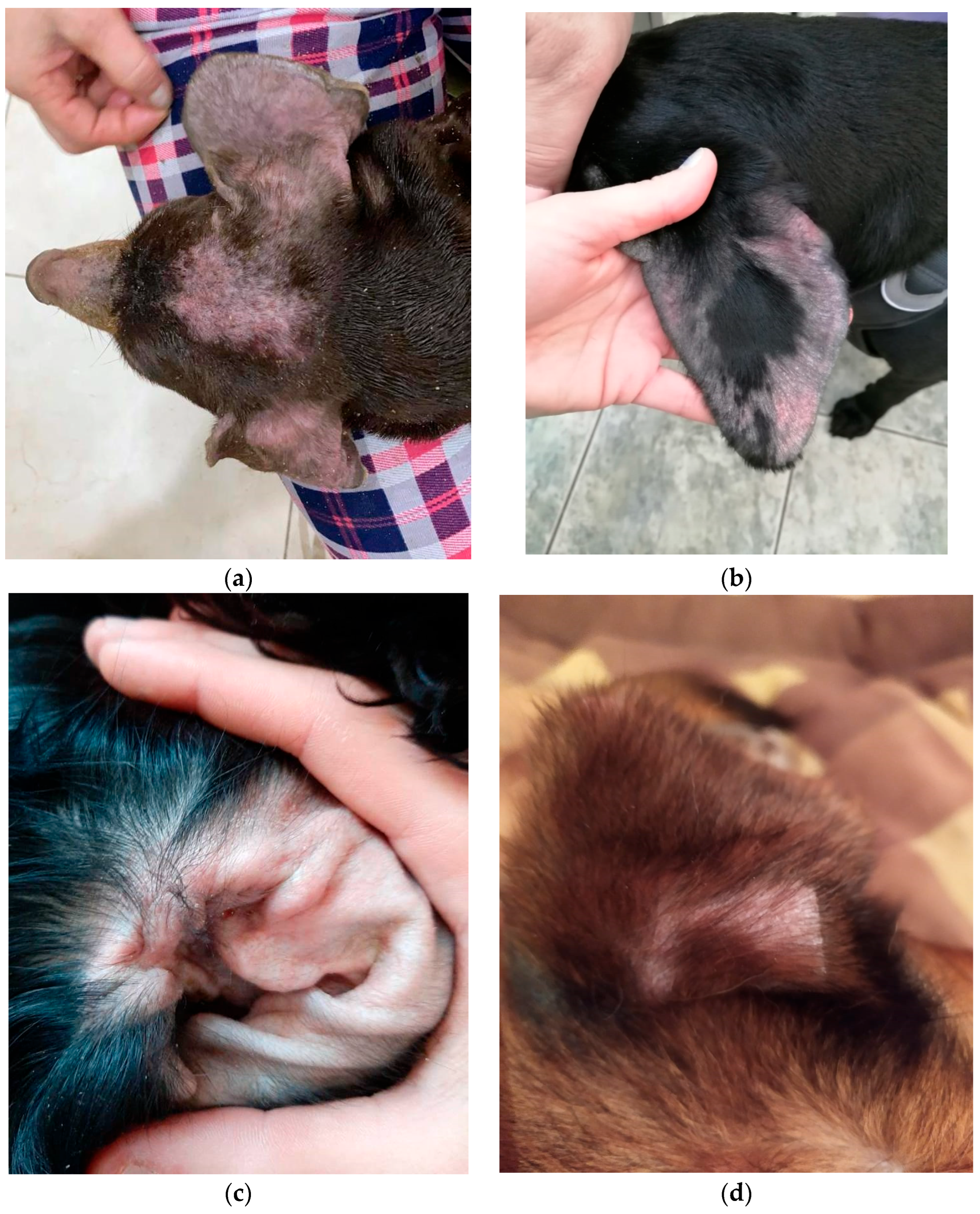
2.1. Animals
2.2. Strains
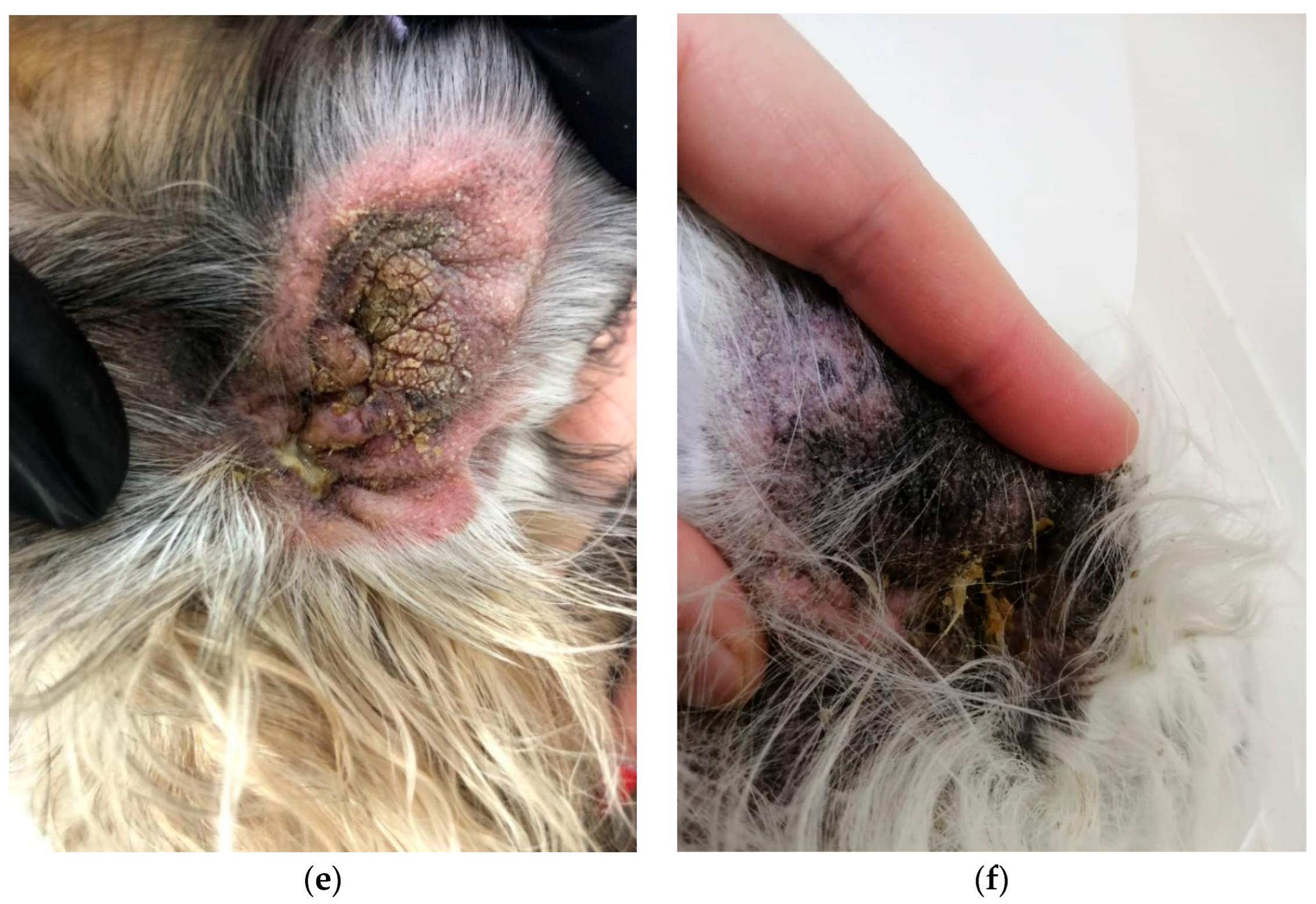
2.3. Reagents
2.4. Densitometric Indicators of Microbial Biofilms
2.5. Preparation of M. pachydermatis Cultures and Assessment of Their Susceptibility to Antimycotics
2.6. M. pachydermatis Biofilms Processing with Farnesol
- An amount of 100 µL SDB in each of the 12 holes of the first row, A.
- An amount of 100 µL of Far was added at an initial concentration of 400 µM to the second well of the first row. The first hole was left as a control. In the second hole, the volume was 200 µL, and the concentration of Farnesol was 200 µM. By successive transfer of 100 µL of the solution from the second well to the third, from the third to the fourth…, etc., the concentration of Far was reduced by half each time.
- Then, in each well of the first row, starting from the first, 100 µL of M. pachydermatis culture in SDB was added at a concentration of 4 units (McFarland).
2.7. Statistical Analysis
3. Results
3.1. Densitometric Indicators of Microbial Biofilms and Their Susceptibility to Antimycotics
- 1st group: weak biofilm producer—sensitive to antifungal drugs: Malassezia pachydermatis C13;
- 2nd group: weak biofilm producer—intermediate resistance to antifungal drugs: Malassezia pachydermatis C9;
- 3rd group: weak biofilm producer—resistant to antifungal drugs: no isolates;
- 4th group: moderate biofilm producer—sensitive to antifungal drugs: Malassezia pachydermatis C6, 7, 8, 12, 14, 16, 18, 22, 24, 26, 28;
- 5th group: moderate biofilm producer—intermediate resistance to antifungal drugs: Malassezia pachydermatis C1, 2, 4, 5, 10, 11, 15, 17, 19, 20, 21, 25, 29, 30;
- 6th group: moderate biofilm producer—resistant to antifungal drugs: no isolates;
- 7th group: strong biofilm producer—sensitive to antifungal drugs: no isolates;
- 8th group: strong biofilm producer—intermediate resistance to antifungal drugs: Malassezia pachydermatis C3, 27;
- 9th group: strong biofilm producer—resistant to antifungal drugs: Malassezia pachydermatis C23.
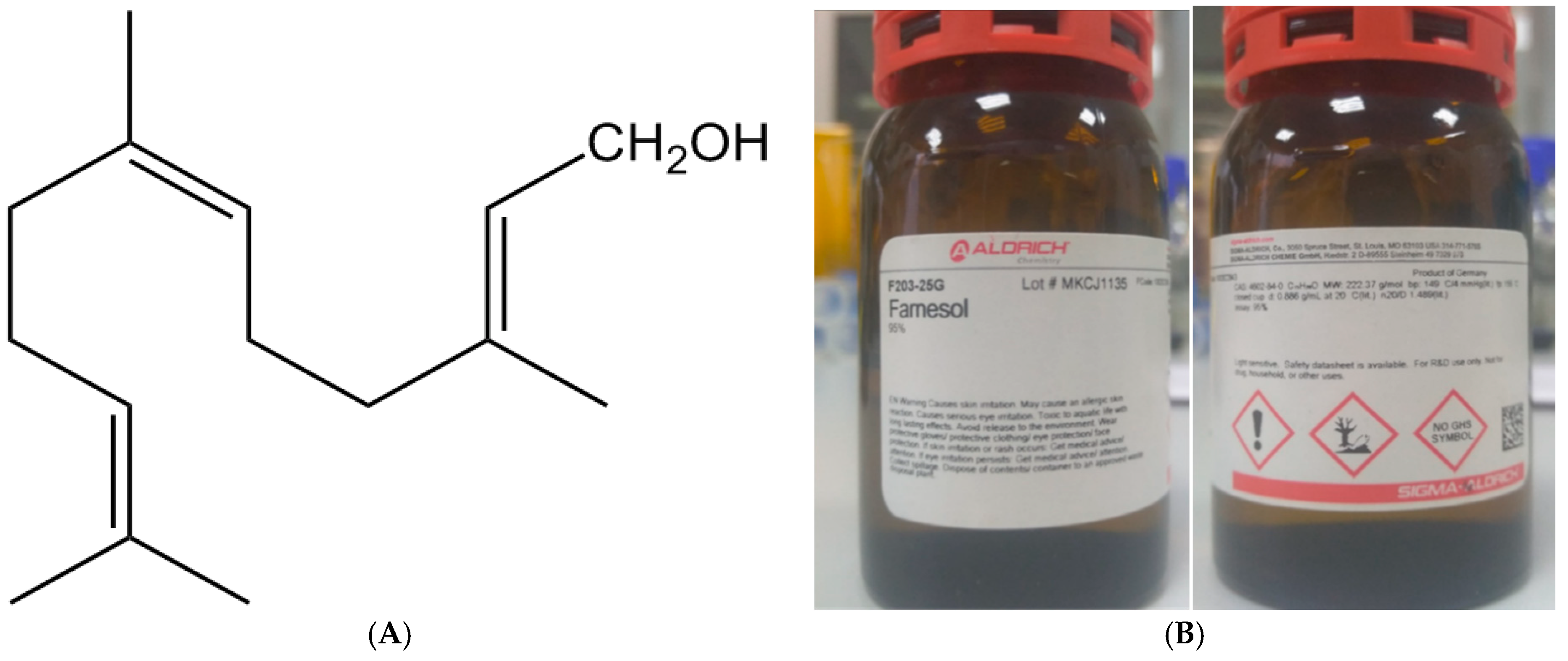
3.2. BY Malassezia pachydermatis Biofilm Inhibition by Farnesol
4. Discussion
5. Conclusions
Author Contributions
Funding
Institutional Review Board Statement
Informed Consent Statement
Data Availability Statement
Acknowledgments
Conflicts of Interest
References
- Bajwa, J. Malassezia species and its significance in canine skin disease. Can. Vet. J. 2023, 64, 87–90. [Google Scholar]
- Cafarchia, C.; Gallo, S.; Capelli, G.; Otranto, D. Occurrence and Population Size of Malassezia spp. in the External Ear Canal of Dogs and Cats Both Healthy and with Otitis. Mycopathologia 2005, 160, 143–149. [Google Scholar] [CrossRef]
- Dizotti, C.E.; Coutinho, S.D.A. Isolation of Malassezia pachydermatis and M. sympodialis from the external ear canal of cats with and without otitis externa. Acta Vet. Hung. 2007, 55, 471–477. [Google Scholar] [CrossRef] [PubMed] [Green Version]
- Crespo, M.J.; Abarca, M.L.; Cabañes, F.J. Occurrence of Malassezia spp. in the external ear canals of dogs and cats with and without otitis externa. Med. Mycol. 2002, 40, 115–121. [Google Scholar] [CrossRef] [Green Version]
- Čonková, E.; Proškovcová, M.; Váczi, P.; Malinovská, Z. In Vitro Biofilm Formation by Malassezia pachydermatis Isolates and Its Susceptibility to Azole Antifungals. J. Fungi 2022, 15, 1209. [Google Scholar] [CrossRef]
- Weiler, C.B.; de Jesus, F.P.K.; Nardi, G.H.; Loreto, E.S.; Santurio, J.M.; Dall, S.; Alves, S.H. Susceptibility variation of Malassezia pachydermatis to antifungal agents according to isolate source. Braz. J. Microbiol. 2013, 44, 174–178. [Google Scholar] [CrossRef] [PubMed]
- Nardoni, S.; Corazza, M.; Mancianti, F. Diagnostic and clinical features of animal malasseziosis. Parassitologia 2008, 50, 81–83. [Google Scholar] [PubMed]
- Carrillo-Muñoz, A.J.; Rojas, F.; Tur-Tur, C.; de Los Ángeles Sosa, M.; Diez, G.O.; Espada, C.M.; Payá, M.J.; Giusiano, G. In vitro antifungal activity of topical and systemic antifungal drugs against Malassezia species. Mycoses 2013, 56, 571–575. [Google Scholar] [CrossRef]
- Rudenko, P.; Vatnikov, Y.; Sachivkina, N.; Rudenko, A.; Kulikov, E.; Lutsay, V.; Notina, E.; Bykova, I.; Petrov, A.; Drukovskiy, S.; et al. Search for Promising Strains of Probiotic Microbiota Isolated from Different Biotopes of Healthy Cats for Use in the Control of Surgical Infections. Pathogens 2021, 10, 667. [Google Scholar] [CrossRef] [PubMed]
- Lyskova, P.; Vydrzalova, M.; Mazurova, J. Identification and Antimicrobial Susceptibility of Bacteria and Yeasts Isolated from Healthy Dogs and Dogs with Otitis Externa. J. Vet. Med. A Physiol. Pathol. Clin. Med. 2007, 54, 559–563. [Google Scholar] [CrossRef]
- Bond, R. Superficial veterinary mycoses. Clin. Dermatol. 2010, 28, 226–236. [Google Scholar] [CrossRef] [PubMed]
- Rosser, E.J. Causes of otitis externa. Vet. Clin. N. Am. Small Anim. Pract. 2004, 34, 459–468. [Google Scholar] [CrossRef] [PubMed]
- Morris, D.O. Medical therapy of otitis externa and otitis media. Vet. Clin. N. Am. Small Anim. Pract. 2004, 34, 541–555. [Google Scholar] [CrossRef] [PubMed]
- Brito, E.H.S.; Fontenelle, R.O.S.; Brilhante, R.S.N.; Cordeiro, R.A.; Júnior, F.A.S.; Monteiro, A.J. Phenotypic characterisation and in vitro antifungal sensitivity of Candida spp. and Malassezia pachydermatis strains from dogs. Vet. J. 2007, 174, 147–153. [Google Scholar] [CrossRef]
- Chen, T.-A.; Hill, P.B. The biology of Malassezia organisms and their ability to induce immune responses and skin disease. Vet. Dermatol. 2005, 16, 4–26. [Google Scholar] [CrossRef]
- Bond, R.; Ferguson, E.A.; Curtis, C.F.; Craig, J.M.; Lloyd, D.H. Factors associated with elevated cutaneous Malassezia pachydermatis populations in dogs with pruritic skin disease. J. Small Anim. Pract. 1996, 37, 103–107. [Google Scholar] [CrossRef]
- Cafarchia, C.; Gallo, S.; Romito, D.; Capelli, G.; Chermette, R.; Guillot, J.; Otranto, D. Frequency, Body Distribution, and Population Size of Malassezia Species in Healthy Dogs and in Dogs with Localized Cutaneous Lesions. J. Vet. Diagn. Investig. 2005, 17, 316–322. [Google Scholar] [CrossRef] [Green Version]
- Sachivkina, N.; Lenchenko, E.; Marakhova, A. Study of the formation of Candida albicans and Escherichia coli biofilms. Farmaciya 2019, 68, 26–30. [Google Scholar] [CrossRef]
- Rudenko, P.; Sachivkina, N.; Vatnikov, Y.; Shabunin, S.; Engashev, S.; Kontsevaya, S.; Karamyan, A.; Bokov, D.; Kuznetsova, O.; Vasilieva, E. Role of microorganisms isolated from cows with mastitis in Moscow region in biofilm formation. Vet. World 2021, 14, 40–48. [Google Scholar] [CrossRef]
- Sachivkina, N.P.; Lenchenko, E.M.; Mannapova, R.T.; Strizhakov, A.A.; Romanova, E.V.; Lukina, D.M. Candida Biofilm Modeling: Past and Present. Farmaciya 2019, 68, 18–22. [Google Scholar] [CrossRef]
- Lenchenko, E.; Blumenkrants, D.; Sachivkina, N.; Shadrova, N.; Ibragimova, A. Morphological and adhesive properties of Klebsiella pneumoniae biofilms. Vet. World 2020, 13, 197–200. [Google Scholar] [CrossRef] [Green Version]
- Sachivkina, N.; Lenchenko, E.; Strizakov, A.; Zimina, V.; Gnesdilova, L.; Gavrilov, V.; Byakhova, V.; Germanova, S.; Zharov, A.; Molchanova, M. The Evaluation of formation of biomembrane by microscopic Fungi of the Candida Genus. Int. J. Pharm. Res. 2018, 10, 738–744. [Google Scholar]
- Cannizzo, F.T.; Eraso, E.; Ezkurra, P.A.; Villar-Vidal, M.; Bollo, E.; Castellá, G.; Cabañes, F.J.; Vidotto, V.; Quindós, G. Biofilm development by clinical isolates of Malassezia pachydermatis. Med. Mycol. 2007, 45, 357–361. [Google Scholar] [CrossRef] [Green Version]
- Yang, H.O.; Cho, Y.-J.; Lee, J.M.; Kim, K.-D. Transcriptional Interplay between Malassezia restricta and Staphylococcus Species Co-Existing in the Skin Environment. J. Microbiol. Biotechnol. 2023, 33, 319–328. [Google Scholar] [CrossRef] [PubMed]
- Lee, K.; Zhang, I.; Kyman, S.; Kask, O.; Cope, E.K. Co-infection of Malassezia sympodialis With Bacterial Pathobionts Pseudomonas aeruginosa or Staphylococcus aureus Leads to Distinct Sinonasal Inflammatory Responses in a Murine Acute Sinusitis Model. Front. Cell. Infect. Microbiol. 2020, 10, 472. [Google Scholar] [CrossRef]
- Guillot, J.; Bond, R. Malassezia pachydermatis: A review. Med. Mycol. 1999, 37, 295–306. [Google Scholar] [CrossRef] [PubMed] [Green Version]
- Park, M.; Park, S.; Jung, W.H. Skin Commensal Fungus Malassezia and Its Lipases. J. Microbiol. Biotechnol. 2021, 31, 637–644. [Google Scholar] [CrossRef] [PubMed]
- Sachivkina, N.; Podoprigora, I.; Bokov, D. Morphological characteristics of Candida albicans, Candida krusei, Candida guilliermondii, and Candida glabrata biofilms, and response to farnesol. Vet. World 2021, 14, 1608–1614. [Google Scholar] [CrossRef] [PubMed]
- Langford, M.L.; Kenneth, S.H.; Nickerson, W.; Atkin, A.L. Activity and toxicity of farnesol towards Candida albicans are dependent on growth conditions. Antimicrob. Agents Chemother. 2010, 54, 940–942. [Google Scholar] [CrossRef] [PubMed] [Green Version]
- Sachivkina, N.; Lenchenko, E.; Blumenkrants, D.; Ibragimova, A.; Bazarkina, O. Effects of farnesol and lyticase on the formation of Candida albicans biofilm. Vet. World 2020, 13, 1030–1036. [Google Scholar] [CrossRef] [PubMed]
- Vatnikov, Y.; Donnik, I.; Kulikov, E.; Karamyan, A.; Sachivkina, N.; Rudenko, P.; Tumanyan, A.; Khairova, N.; Romanova, E.; Gurina, R.; et al. Research on the antibacterial and antimycotic effect of the Phyto preparation Farnesol on biofilm-forming microorganisms in veterinary medicine. Int. J. Pharm. Res. 2020, 12, 1481–1492. [Google Scholar]
- Sachivkina, N.; Karamyan, A.; Kuznetsova, O.; Ibragimova, A.; Ebzeeva, A.; Mussa, R.; Akimenkova, A. The use of extracts Tilia cordata flowers and Tripleurospermum inodorum flowers against Candida albicans biofilms. FEBS Open Bio 2021, 11, 288. [Google Scholar]
- Hoes, N.P.M.; van den Broek, J.; Vroom, M.W. The efficacy of a novel topical spray composed of sodium benzoate, alcohol and botanical oils for the treatment of Malassezia dermatitis in dogs—A split body, randomised, blinded study. Vet. Dermatol. 2022, 33, 398–401. [Google Scholar] [CrossRef] [PubMed]
- Gómez-García, M.; Madrigal, I.; Puente, H.; Mencía-Ares, Ó.; Argüello, H.; Carvajal, A.; Fregeneda-Grandes, J.M. In vitro activity of essential oils against microbial isolates from otitis externa cases in dogs. Nat. Prod. Res. 2022, 36, 4552–4556. [Google Scholar] [CrossRef]
- Sim, J.X.F.; Khazandi, M.; Chan, W.Y.; Trott, D.J.; Deo, P. Antimicrobial activity of thyme oil, oregano oil, thymol and carvacrol against sensitive and resistant microbial isolates from dogs with otitis externa. Vet. Dermatol. 2019, 30, 524-e159. [Google Scholar] [CrossRef] [PubMed]
- Chan, W.Y.; Hickey, E.E.; Khazandi, M.; Page, S.W.; Trott, D.J.; Hill, P.B. In vitro antimicrobial activity of narasin against common clinical isolates associated with canine otitis externa. Vet. Dermatol. 2018, 29, 149-e57. [Google Scholar] [CrossRef] [PubMed]
- Arora, P.; Nainwal, L.M.; Jain, S. Essential oils as Potential Source of Anti-dandruff Agents: A Review. Comb. Chem. High Throughput Screen. 2022, 25, 1411–1426. [Google Scholar] [CrossRef]
- Luqman, S.; Kumari, K.U.; Yadav, N.P. Promising essential oils/plant extracts in the prevention and treatment of dandruff pathogenesis. Curr. Top. Med. Chem. 2022, 22, 1104–1133. [Google Scholar] [CrossRef]
- Honnavar, P.; Ghosh, A.; Paul, S.; Shankarnarayan, S.; Singh, P.; Dogra, S.; Chakrabarti, A.; Rudramurthy, S. Identification of Malassezia species by MALDI-TOF MS after expansion of database. Diagn. Microbiol. Infect. Dis. 2018, 92, 118–123. [Google Scholar] [CrossRef]
- Sachivkina, N.; Kravtsov, E.G.; Vasilyeva, E.A.; Anokhina, I.V.; Dalin, M.V. Study of antimycotic activity of lyticase. Bull. Exp. Biol. Med. 2009, 148, 214–216. [Google Scholar] [CrossRef]
- Sachivkina, N.P.; Senyagin, A.N.; Podoprigora, I.V.; Brown, D.G.; Vissarionova, V.V. Modulating the antifungal activity of antimycotic drugs with farnesol. Drug Dev. Regist. Razrab. I Regist. Lek. Sredstv 2021, 10, 162–168. [Google Scholar] [CrossRef]
- Sachivkina, N.; Vasilieva, E.; Lenchenko, E.; Kuznetsova, O.; Karamyan, A.; Ibragimova, A.; Zhabo, N.; Molchanova, M. Reduction in Pathogenicity in Yeast-like Fungi by Farnesol in Quail Model. Animals 2022, 12, 489. [Google Scholar] [CrossRef] [PubMed]
- Sachivkina, N.; Senyagin, A.; Podoprigora, I.; Vasilieva, E.; Kuznetsova, O.; Karamyan, A.; Ibragimova, A.; Zhabo, N.; Molchanova, M. Enhancement of the antifungal activity of some antimycotics by farnesol and reduction of Candida albicans pathogenicity in a quail model experiment. Vet. World 2022, 15, 848–854. [Google Scholar] [CrossRef]
- Kolecka, A.; Khayhan, K.; Arabatzis, M.; Velegraki, A.; Kostrzewa, M.; Andersson, A.; Scheynius, A.; Cafarchia, C.; Iatta, R.; Montagna, M.; et al. Efficient identification of Malassezia yeasts by matrix-assisted laser desorption ionization-time of flight mass spectrometry (MALDI-TOF MS). Br. J. Dermatol. 2014, 170, 332–341. [Google Scholar] [CrossRef]
- Denis, J.; Machouart, M.; Morio, F.; Sabou, M.; Kauffmann-LaCroix, C.; Contet-Audonneau, N.; Candolfi, E.; Letscher-Bru, V. Performance of Matrix-Assisted Laser Desorption Ionization–Time of Flight Mass Spectrometry for Identifying Clinical Malassezia Isolates. J. Clin. Microbiol. 2017, 55, 90–96. [Google Scholar] [CrossRef] [Green Version]
- Gaitanis, G.; Bassukas, I.D.; Velegraki, A. The range of molecular methods for typing Malassezia. Curr. Opin. Infect. Dis. 2009, 22, 119–125. [Google Scholar] [CrossRef]
- Ilahi, A.; Hadrich, I.; Goudjil, S.; Kongolo, G.; Chazal, C.; Léké, A.; Ayadi, A.; Chouaki, T.; Ranque, S. Molecular epidemiology of a Malassezia pachydermatis neonatal unit outbreak. Med. Mycol. 2017, 56, 69–77. [Google Scholar] [CrossRef]
- Sachivkina, N.; Podoprigora, I.; Senyagin, A.; Ibragimova, A.; Avdonina, M.; Shvedova, I. The use of Farnesol to increase the antifungal activity of some antibiotics against Candida albicans. FEBS Open Bio 2022, 12, 169. [Google Scholar]
- Lenchenko, E.; Blumenkrants, D.; Vatnikov, Y.; Kulikov, E.; Khai, V.; Sachivkina, N.; Gnezdilova, L.; Sturov, N.; Sakhno, N.; Kuznetsov, V.; et al. Poultry Salmonella sensitivity to antibiotics. Sys. Rev. Pharm. 2020, 11, 170–175. [Google Scholar]
- Procop, G.W.; Clinical and Laboratory Standards Institute. Performance Standards for Antifungal Susceptibility Testing of Yeasts, 2nd ed.; CLSI: Wayne, PA, USA, 2020; ISBN 978-1-68440-082-9. [Google Scholar]
- Available online: https://www.eucast.org/fileadmin/src/media/PDFs/EUCAST_files/Network_labs/EDL/EUCAST_Development_Laboratories.pdf (accessed on 1 April 2023).
- Álvarez-Pérez, S.; Blanco, J.L.; Peláez, T.; Cutuli, M.; García, M.E. In vitro amphotericin B susceptibility of Malassezia pachydermatis determined by the CLSI broth microdilution method and Etest using lipid-enriched media. Antimicrob Agents Chemother. 2014, 58, 4203–4206. [Google Scholar] [CrossRef] [Green Version]
- Jabra-Rizk, M.A.; Shirtliff, M.; James, C.; Meiller, T. Effect of farnesol on Candida dubliniensis biofilm formation and fluconazole resistance. FEMS Yeast Res. 2006, 6, 1063–1073. [Google Scholar] [CrossRef] [Green Version]
- Cordeiro, R.D.A.; Reis, A.T.; Lima, X.T.V.; de Andrade, A.R.C.; Aguiar, A.L.R.; Portela, F.V.M.; Pereira, L.M.G.; Moura, S.G.B.; da Silva, B.N.; de Lima-Neto, R.G.; et al. Malassezia spp. and Candida spp. from patients with psoriasis exhibit reduced susceptibility to antifungals. Braz. J. Microbiol. 2022, 54, 169–177. [Google Scholar] [CrossRef]
- Li, W.; Zhang, Z.-W.; Luo, Y.; Liang, N.; Pi, X.-X.; Fan, Y.-M. Molecular epidemiology, in vitro susceptibility and exoenzyme screening of Malassezia clinical isolates. J. Med. Microbiol. 2020, 69, 436–442. [Google Scholar] [CrossRef]
- Gupta, A.K.; Kohli, Y.; Li, A.; Faergemann, J.; Summerbell, R.C. In vitro susceptibility of the seven Malassezia species to ketoconazole, voriconazole, itraconazole and terbinafine. Br. J. Dermatol. 2000, 142, 758–765. [Google Scholar] [CrossRef] [PubMed]
- Sachivkina, N.P.; Karamyan, A.S.; Kuznetsova, O.M.; Byakhova, V.M.; Bondareva, I.B.; Molchanova, M.A. Development of therapeutic transdermal systems for microbial biofilm destruction. FEBS Open Bio 2019, 9, 386. [Google Scholar]
- Moporozov, I.A.; Sachivkina, N.P.; Kravtsov, E.G.; Vasilyeva, E.A.; Anokhina, I.V.; Yashina, N.V.; Dalin, M.V. Damaging Effects of Lyticase on Candida albicans and Changes in the Response of Rat Alveolar Macrophages to the Contact with Yeast-Like Fungi. Bull. Exp. Biol. Med. 2011, 151, 705–708. [Google Scholar] [CrossRef]
- Sachivkina, N.P.; Kravtsov, E.G.; Vasileva, E.A.; Anokchina, I.V.; Dalin, M.V. Efficiency of lyticase (bacterial enzyme) in experimental candidal vaginitis in mice. Bull. Exp. Biol. Med. 2010, 149, 727–730. [Google Scholar] [CrossRef]
- Alkhanjaf, A.A.M.; Athar, T.; Ullah, Z.; Alsayhab, A.M.H.; Umar, A.; Shaikh, I.A. Farnesol Protects against Cardiotoxicity Caused by Doxorubicin-Induced Stress, Inflammation, and Cell Death: An In Vivo Study in Wistar Rats. Molecules 2022, 27, 8589. [Google Scholar] [CrossRef]
- Li, X.; Ren, Y.; Huang, G.; Zhang, R.; Zhang, Y.; Zhu, W.; Yu, K. Succinate communicates pro-inflammatory signals to the host and regulates bile acid enterohepatic metabolism in a pig model. Food Funct. 2022, 13, 11070–11082. [Google Scholar] [CrossRef]
- Sell, L.B.; Ramelow, C.C.; Kohl, H.M.; Hoffman, K.; Bains, J.K.; Doyle, W.J.; Strawn, K.D.; Hevrin, T.; Kirby, T.O.; Gibson, K.M.; et al. Farnesol induces protection against murine CNS inflammatory demyelination and modifies gut microbiome. Clin. Immunol. 2022, 235, 108766. [Google Scholar] [CrossRef]


| 1 | 2 | 3 | 4 | 5 | 6 | 7 | 8 | 9 | 10 | 11 | 12 | |
|---|---|---|---|---|---|---|---|---|---|---|---|---|
| Action 1 | SDB 100 µL | SDB 100 µL | SDB 100 µL | SDB 100 µL | SDB 100 µL | SDB 100 µL | SDB 100 µL | SDB 100 µL | SDB 100 µL | SDB 100 µL | SDB 100 µL | SDB 100 µL |
| Action 2 | +Farnesol | |||||||||||
| Action 3 | Not titrated | Transfer 100 µL | Transfer 100 µL | Transfer 100 µL | Transfer 100 µL | Transfer 100 µL | Transfer 100 µL | Transfer 100 µL | Transfer 100 µL | Transfer 100 µL | Transfer 100 µL | Transfer 100 µL |
| The concentration of farnesol | Control– no farnesol | 200 μM | 100 μM | 50 μM | 25 μM | 12.5 μM | 6.3 μM | 3.1 μM | 1.6 μM | 0.8 μM | 0.4 μM | 0.2 μM |
| Action 4 | +100 µL of culture | +100 µL of culture | +100 µL of culture | +100 µL of culture | +100 µL of culture | +100 µL of culture | +100 µL of culture | +100 µL of culture | +100 µL of culture | +100 µL of culture | +100 µL of culture | +100 µL of culture |
| Action 5 | wait for 72 h | |||||||||||
| Microorganism | Optic Density | Degree OD | Antimycotic Drugs | Degree of Resistance | |||||||
|---|---|---|---|---|---|---|---|---|---|---|---|
| NS | AP | KT | CC | VOR | FU | MIC | IT | ||||
| Malassezia pachydermatis C1 | 0.203 ± 0.016 | 2 | 1 | 1 | 3 | 1 | 1 | 2 | 1 | 1 | 11 |
| Malassezia pachydermatis C2 | 0.351 ± 0.018 | 2 | 1 | 2 | 1 | 2 | 2 | 3 | 1 | 1 | 13 |
| Malassezia pachydermatis C3 | 0.400 ± 0.012 | 3 | 1 | 1 | 1 | 2 | 2 | 3 | 2 | 1 | 13 |
| Malassezia pachydermatis C4 | 0.287 ± 0.018 | 2 | 1 | 3 | 1 | 2 | 1 | 1 | 1 | 3 | 13 |
| Malassezia pachydermatis C5 | 0.261 ± 0.011 | 2 | 2 | 1 | 1 | 2 | 2 | 1 | 1 | 1 | 11 |
| Malassezia pachydermatis C6 | 0.312 ± 0.029 | 2 | 1 | 1 | 1 | 1 | 1 | 3 | 1 | 1 | 10 |
| Malassezia pachydermatis C7 | 0.255 ± 0.010 | 2 | 1 | 1 | 1 | 1 | 1 | 1 | 1 | 1 | 8 |
| Malassezia pachydermatis C8 | 0.243 ± 0.026 | 2 | 1 | 1 | 1 | 2 | 1 | 1 | 1 | 1 | 9 |
| Malassezia pachydermatis C9 | 0.190 ± 0.016 | 1 | 1 | 3 | 1 | 1 | 1 | 3 | 1 | 2 | 13 |
| Malassezia pachydermatis C10 | 0.237 ± 0.015 | 2 | 2 | 1 | 1 | 1 | 1 | 1 | 2 | 2 | 11 |
| Malassezia pachydermatis C11 | 0.345 ± 0.011 | 2 | 1 | 1 | 3 | 1 | 2 | 2 | 1 | 1 | 12 |
| Malassezia pachydermatis C12 | 0.323 ± 0.017 | 2 | 1 | 2 | 1 | 1 | 2 | 1 | 1 | 1 | 10 |
| Malassezia pachydermatis C13 | 0.192 ± 0.012 | 1 | 1 | 1 | 1 | 2 | 1 | 2 | 1 | 1 | 10 |
| Malassezia pachydermatis C14 | 0.258 ± 0.011 | 2 | 1 | 1 | 1 | 2 | 1 | 1 | 1 | 1 | 9 |
| Malassezia pachydermatis C15 | 0.261 ± 0.010 | 2 | 1 | 1 | 2 | 2 | 1 | 1 | 1 | 2 | 11 |
| Malassezia pachydermatis C16 | 0.313 ± 0.007 | 2 | 1 | 1 | 1 | 1 | 1 | 2 | 1 | 1 | 9 |
| Malassezia pachydermatis C17 | 0.294 ± 0.019 | 2 | 1 | 1 | 3 | 1 | 1 | 3 | 1 | 1 | 12 |
| Malassezia pachydermatis C18 | 0.286 ± 0.020 | 2 | 1 | 1 | 1 | 2 | 1 | 1 | 1 | 1 | 9 |
| Malassezia pachydermatis C19 | 0.362 ± 0.014 | 2 | 2 | 1 | 2 | 2 | 1 | 1 | 1 | 1 | 11 |
| Malassezia pachydermatis C20 | 0.366 ± 0.015 | 2 | 1 | 2 | 1 | 1 | 3 | 1 | 1 | 1 | 11 |
| Malassezia pachydermatis C21 | 0.280 ± 0.016 | 2 | 1 | 2 | 1 | 1 | 1 | 1 | 3 | 1 | 11 |
| Malassezia pachydermatis C22 | 0.344 ± 0.018 | 2 | 1 | 1 | 1 | 1 | 1 | 2 | 1 | 1 | 9 |
| Malassezia pachydermatis C23 | 0.441 ± 0.016 | 3 | 2 | 3 | 3 | 2 | 2 | 3 | 1 | 2 | 18 |
| Malassezia pachydermatis C24 | 0.370 ± 0.015 | 2 | 1 | 1 | 2 | 2 | 1 | 1 | 1 | 1 | 10 |
| Malassezia pachydermatis C25 | 0.323 ± 0.017 | 2 | 2 | 1 | 1 | 1 | 1 | 2 | 3 | 2 | 13 |
| Malassezia pachydermatis C26 | 0.288± 0.012 | 2 | 1 | 2 | 1 | 1 | 1 | 1 | 1 | 1 | 9 |
| Malassezia pachydermatis C27 | 0.403 ± 0.026 | 3 | 1 | 2 | 2 | 1 | 3 | 1 | 1 | 1 | 12 |
| Malassezia pachydermatis C28 | 0.368 ± 0.014 | 2 | 1 | 1 | 1 | 1 | 1 | 1 | 1 | 1 | 8 |
| Malassezia pachydermatis C29 | 0.297 ± 0.011 | 2 | 1 | 1 | 1 | 1 | 3 | 2 | 1 | 1 | 11 |
| Malassezia pachydermatis C30 | 0.353 ± 0.019 | 2 | 1 | 1 | 2 | 1 | 1 | 3 | 3 | 1 | 13 |
| Biofilm Optical Density (Factorial Sign) | Degree of Resistance to Antifungal Drugs (Resulting Sign) | |||
|---|---|---|---|---|
| Sensitive 8–10 | Intermediate 11–13 | Resistant 14–18 | Total | |
| weak—1 | 1 | 1 | 0 | 2 |
| moderate—2 | 11 | 14 | 0 | 25 |
| strong—3 | 0 | 2 | 1 | 3 |
| total | 12 | 17 | 1 | 30 |
| 1 | 2 | 3 | 4 | 5 | 6 | 7 | 8 | 9 | 10 | 11 | 12 | |
|---|---|---|---|---|---|---|---|---|---|---|---|---|
| Far concentration | Control no farnesol | 200 μM | 100 μM | 50 μM | 25 μM | 12.5 μM | 6.3 μM | 3.1 μM | 1.6 μM | 0.8 μM | 0.4 μM | 0.2 μM |
| Malassezia pachydermatis C23 | 0.441 ± 0.016 | 0.120 ± 0.008 | 0.135 ± 0.011 | 0.234 ± 0.011 | 0.226 ± 0.019 | 0.233 ± 0.010 | 0.249 ± 0.014 | 0.302 ± 0.015 | 0.368 ± 0.017 | 0.407 ± 0.016 | 0.439 ± 0.011 | 0.453 ± 0.012 |
| Malassezia pachydermatis C27 | 0.403 ± 0.026 | 0.117 ± 0.016 | 0.121 ± 0.009 | 0.144 ± 0.013 | 0.186 ± 0.018 | 0.272 ± 0.016 | 0.284 ± 0.011 | 0.307 ± 0.008 | 0.320 ± 0.014 | 0.379 ± 0.012 | 0.393 ± 0.010 | 0.399 ± 0.010 |
| Malassezia pachydermatis C3 | 0.400 ± 0.012 | 0.123 ± 0.018 | 0.142 ± 0.014 | 0.160 ± 0.021 | 0.154 ± 0.017 | 0.185 ± 0.009 | 0.234 ± 0.014 | 0.252 ± 0.012 | 0.262 ± 0.009 | 0.279 ± 0.010 | 0.383 ± 0.014 | 0.398 ± 0.008 |
| Average OD of 3 isolates | 0.415 | 0.120 | 0.133 | 0.179 | 0.189 | 0.230 | 0.256 | 0.287 | 0.317 | 0.390 | 0.405 | 0.417 |
| Average decrease OD, % | 0 | 71 | 68 | 57 | 54 | 55 | 38 | 31 | 24 | 6 | 2.4 | −0.5 |
Disclaimer/Publisher’s Note: The statements, opinions and data contained in all publications are solely those of the individual author(s) and contributor(s) and not of MDPI and/or the editor(s). MDPI and/or the editor(s) disclaim responsibility for any injury to people or property resulting from any ideas, methods, instructions or products referred to in the content. |
© 2023 by the authors. Licensee MDPI, Basel, Switzerland. This article is an open access article distributed under the terms and conditions of the Creative Commons Attribution (CC BY) license (https://creativecommons.org/licenses/by/4.0/).
Share and Cite
Olabode, I.R.; Sachivkina, N.; Karamyan, A.; Mannapova, R.; Kuznetsova, O.; Bobunova, A.; Zhabo, N.; Avdonina, M.; Gurina, R. In Vitro Activity of Farnesol against Malassezia pachydermatis Isolates from Otitis Externa Cases in Dogs. Animals 2023, 13, 1259. https://doi.org/10.3390/ani13071259
Olabode IR, Sachivkina N, Karamyan A, Mannapova R, Kuznetsova O, Bobunova A, Zhabo N, Avdonina M, Gurina R. In Vitro Activity of Farnesol against Malassezia pachydermatis Isolates from Otitis Externa Cases in Dogs. Animals. 2023; 13(7):1259. https://doi.org/10.3390/ani13071259
Chicago/Turabian StyleOlabode, Ifarajimi Rapheal, Nadezhda Sachivkina, Arfenia Karamyan, Ramziya Mannapova, Olga Kuznetsova, Anna Bobunova, Natallia Zhabo, Marina Avdonina, and Regina Gurina. 2023. "In Vitro Activity of Farnesol against Malassezia pachydermatis Isolates from Otitis Externa Cases in Dogs" Animals 13, no. 7: 1259. https://doi.org/10.3390/ani13071259