
Geometric and Morphometric Analysis of the Auditory Ossicles in the Red Fox (Vulpes vulpes)


Abstract
:Simple Summary
Abstract
1. Introduction
2. Materials and Methods
2.1. Study Sample
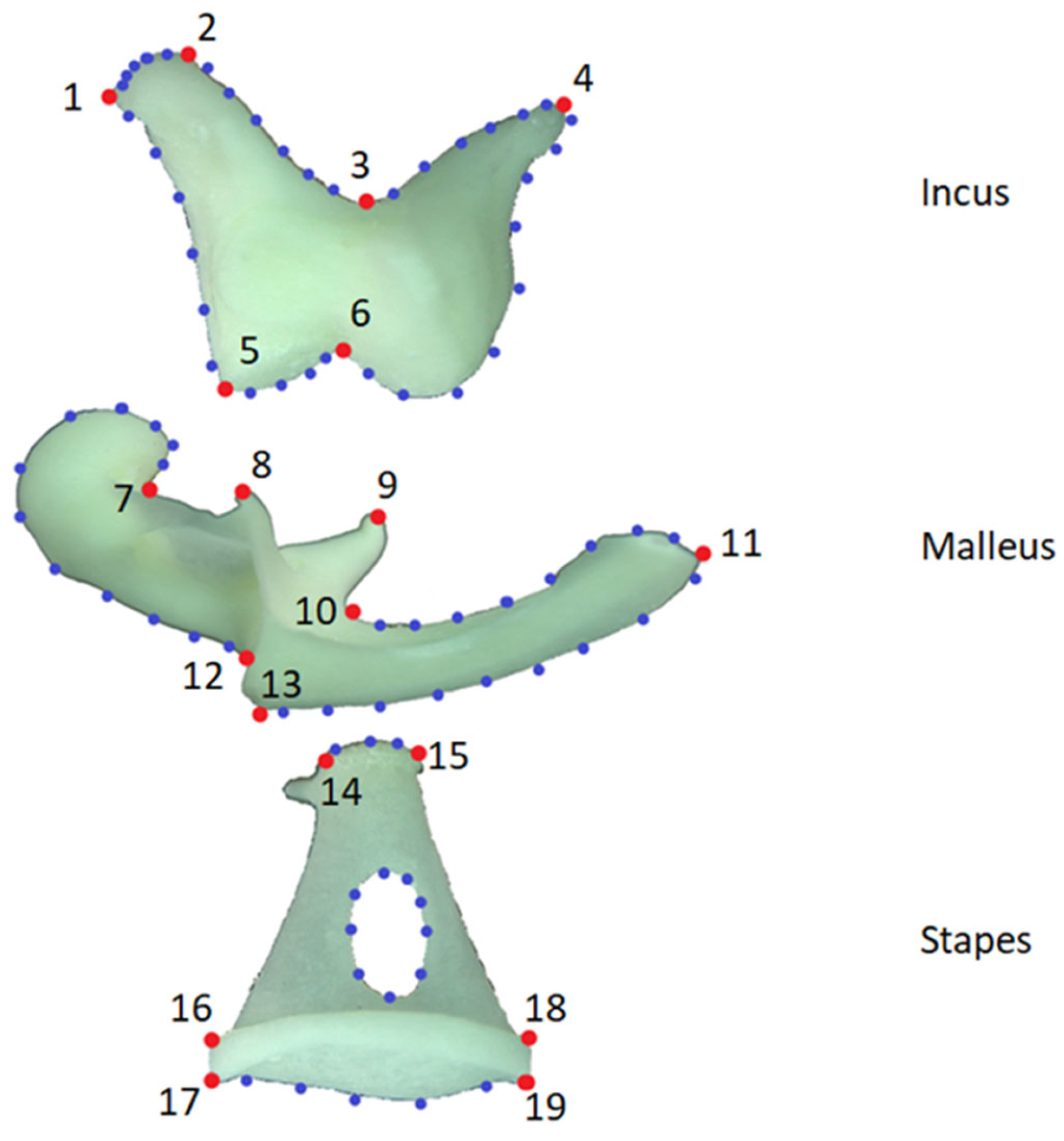
2.2. Morphometric–Analysis
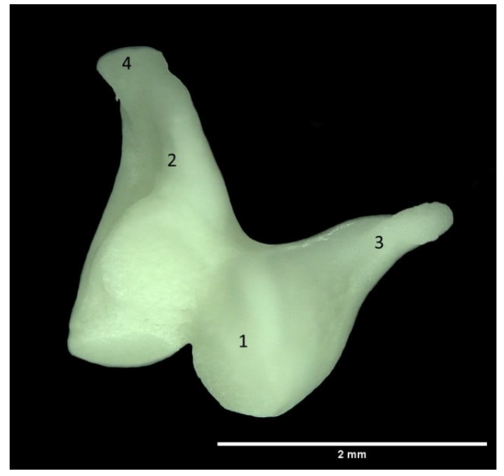
- LM—length of the malleus
- WHM—width of the head of the malleus
- LHM—length of the head of the malleus
- LhM—length of the handle of the malleus
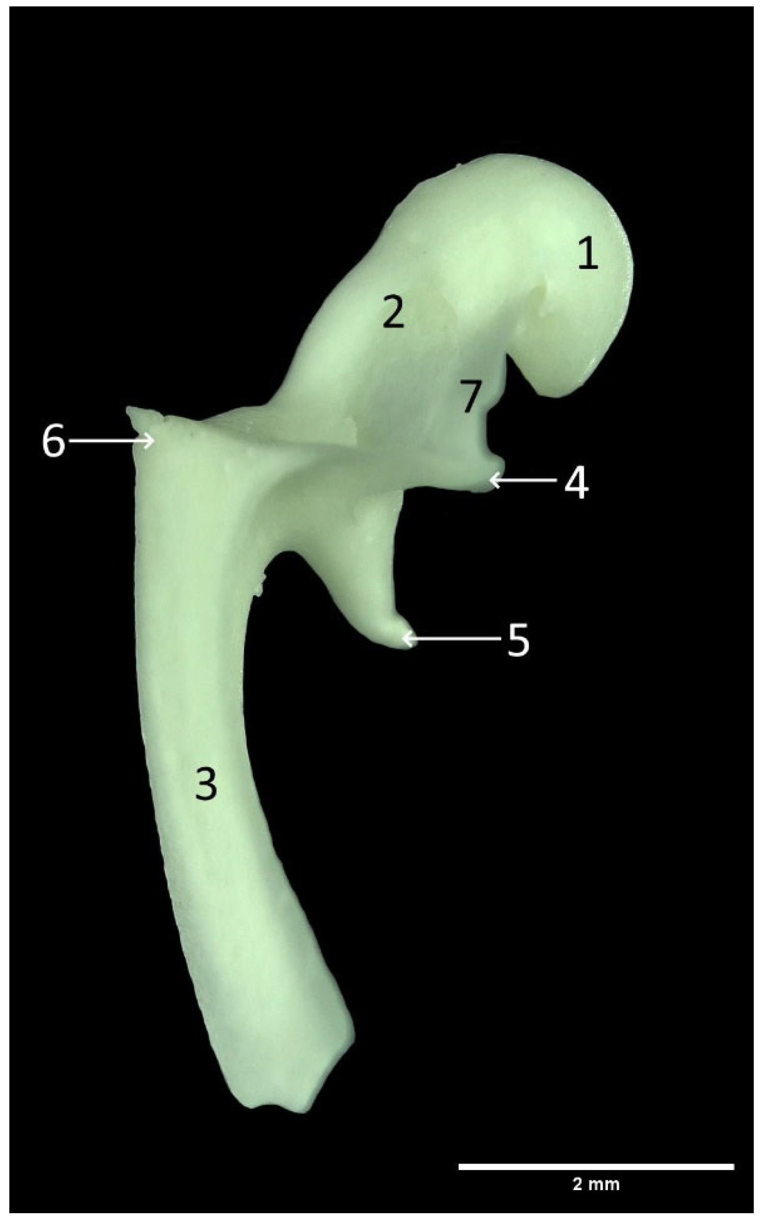
- LI—length of the incus
- LLC—length of long crus of the incus
- LSC—length of short crus of the incus
- HBI—height of the body of the incus
- WBI—width of the body of the incus
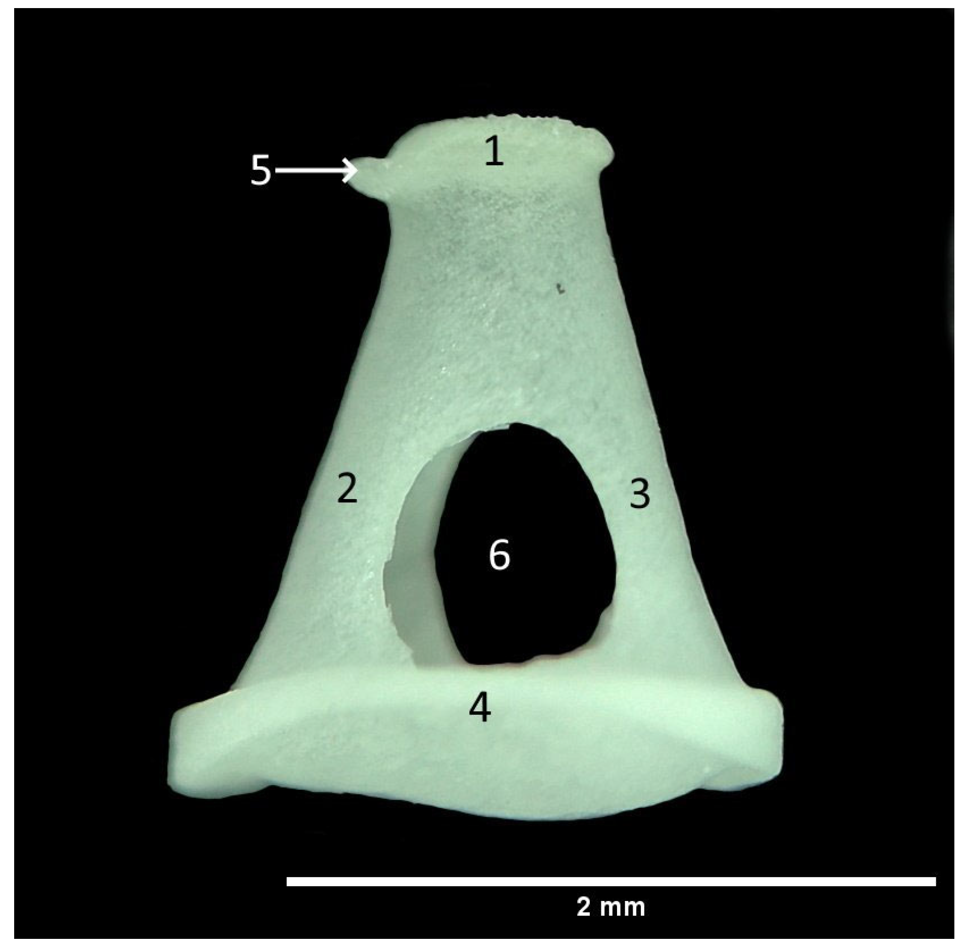
- LS—length of the stapes
- LCC—length of the caudal crus of the stapes
- LRC—length of the rostral crus of the stapes
- WHS—width of the head of the stapes
- WBS—width of the base of the stapes
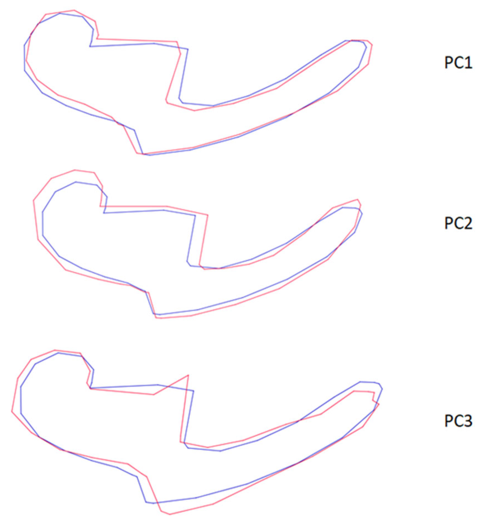
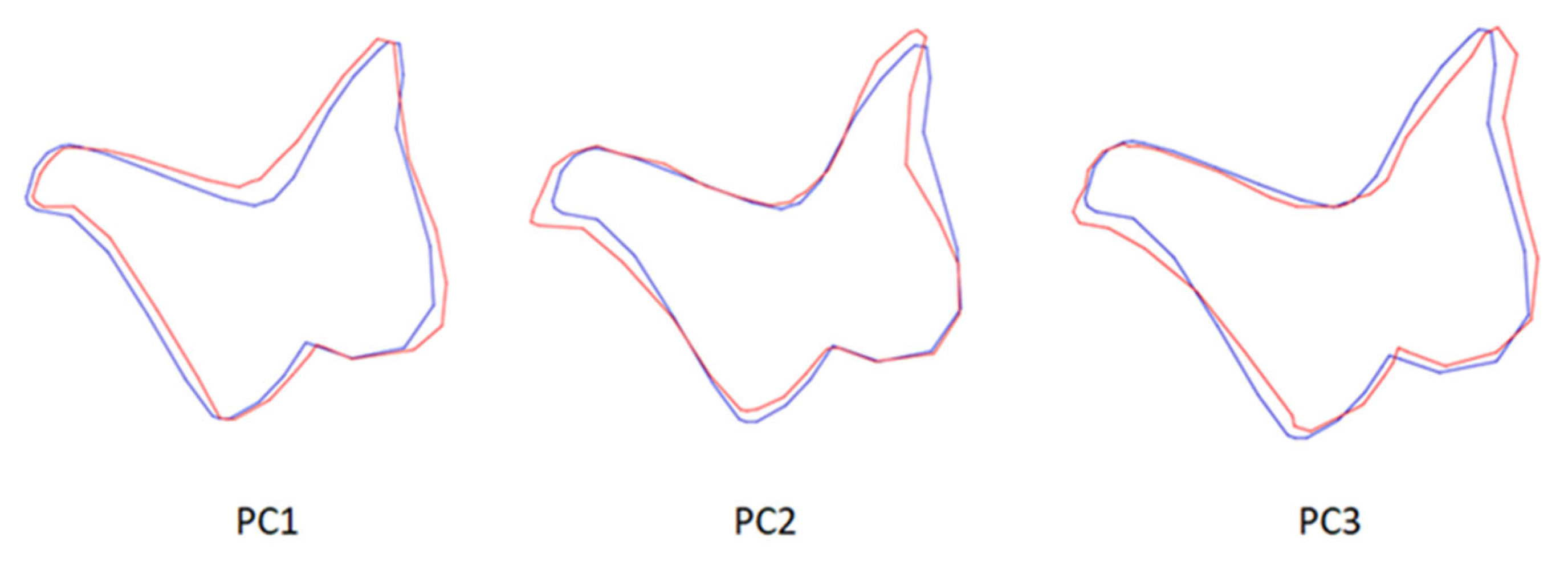
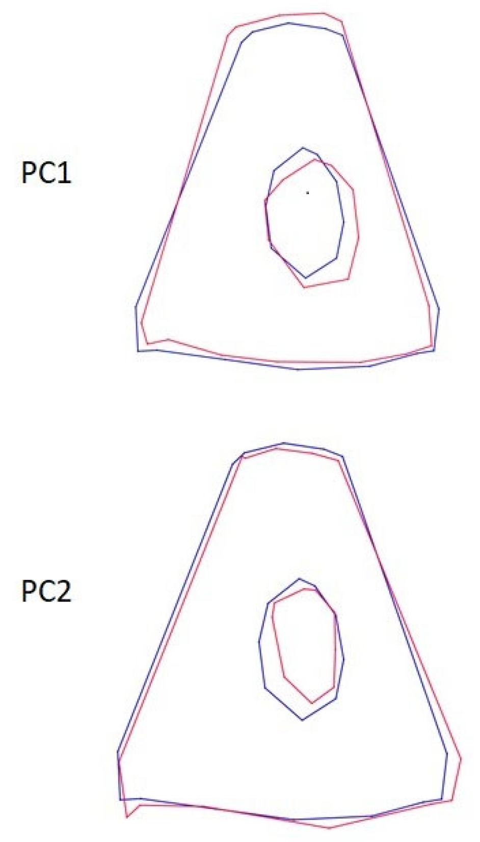
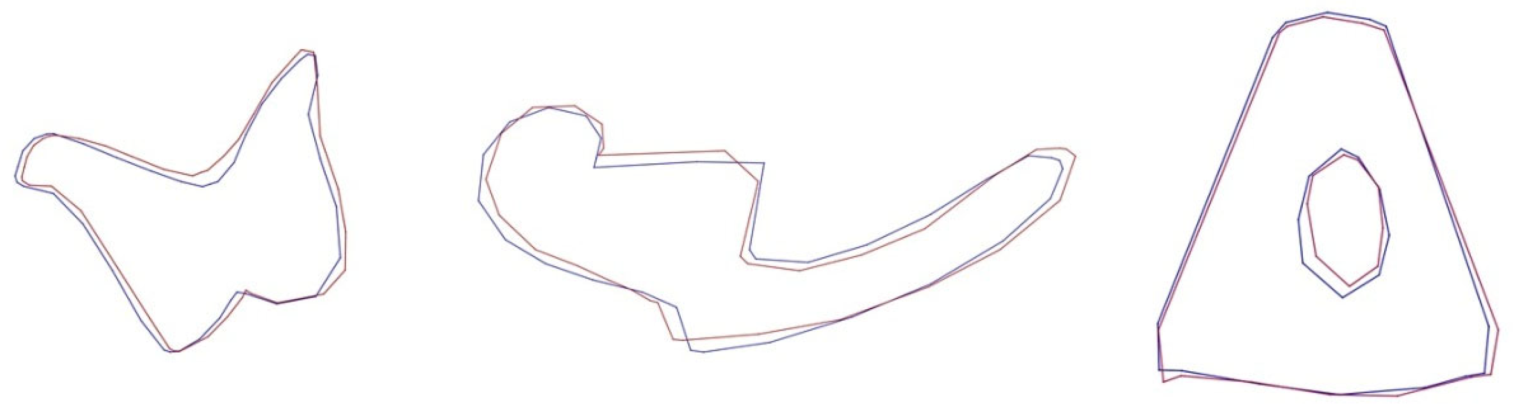
2.3. Geometric Analysis
2.4. Statistical Analysis
3. Results
4. Discussion
5. Conclusions
Author Contributions
Funding
Institutional Review Board Statement
Informed Consent Statement
Data Availability Statement
Acknowledgments
Conflicts of Interest
References
- Porobić, J.M. Geometric-Morphometric Analyses of Golden Jackal (Canis aureus) and Red Fox (Vulpes vulpes) Skulls from the Territory of Serbia: Biogeographical Aspects of Morphological Variability. Ph.D. Thesis, University of Belgrade, Belgrade, Serbia, 2017. [Google Scholar]
- Lloyd, H.G. The Red Fox; Batsford: London, UK, 1980. [Google Scholar]
- Gloor, S.; Bontadina, F.; Hegglin, D.; Deplazes, P.; Breitenmoser, U. The rise of urban fox populations in Switzerland. Mamm. Biol. 2001, 66, 155–164. [Google Scholar]
- Malkemper, E.P.; Mason, M.J.; Burda, H. Functional anatomy of the middle and inner ears of the red fox, in comparison to domestic dogs and cats. J. Anat. 2020, 236, 980. [Google Scholar] [CrossRef] [PubMed] [Green Version]
- Nowak, R.M. Walker’s Mammals of the World, 6th ed.; The Johns Hopkins University Press: Baltimore, MD, USA, 1999. [Google Scholar]
- König, H.E.; Liebich, H.G. Veterinary Anatomy of Domestic Mammals: Textbook and Color Atlas, 3rd ed.; Schattauer Co.: Stuttgart, Germany, 2007; pp. 595–601. [Google Scholar]
- Keen, J.A.; Grobbelaar, C.S. The comparative anatomy of the tympanic bulla and auditory ossicles, with a note suggesting their function. Trans. R. Soc. S. Afr. 1941, 28, 307–329. [Google Scholar] [CrossRef]
- Miller, M.E. Anatomy of the Dog; W.B. and Sounders: Philadelphia, PA, USA, 1964; pp. 853–858. [Google Scholar]
- Kurtul, I.; Cevik, A.; Bozkurt, E.U.; Dursun, N. A detailed subgross morphometric study on the auditory ossicles of the New Zealand rabbit. Anat. Histol. Embryol. 2003, 32, 249–252. [Google Scholar] [CrossRef] [PubMed]
- Gürbüz, I.; Demiraslan, Y.; Dayan, M.O.; Aslan, K. Morphometric and macroanatomic examination of auditory ossicles in male wolves (Canis lupus). Folia Morphol. 2019, 78, 600–605. [Google Scholar] [CrossRef] [Green Version]
- Decraemer, W.F.; Khanna, S.M. Measurement, Visualization and Quantitative Analysis of Complete Three-Dimensional Kinematical Data Sets of Human and Cat Middle Ear. In Middle Ear Mechanics in Research and Otology; World Scientific: Singapore, 2004; pp. 3–10. [Google Scholar]
- Puria, S.; Allen, J.B. Measurements and model of the cat middle ear: Evidence of tympanic membrane acoustic delay. J. Acoust. Soc. Am. 1998, 104, 3463–3481. [Google Scholar] [CrossRef] [Green Version]
- Huang, G.; Rosowski, J.; Peake, W. Relating middle-ear acoustic performance to body size in the cat family: Measurements and models. J. Comp. Physiol. 2000, 186, 447–465. [Google Scholar] [CrossRef]
- Cole, L.K. Anatomy and physiology of the canine ear. Vet. Dermatol. 2009, 20, 412–421. [Google Scholar] [CrossRef]
- Malkemper, E.P. The Sensory Biology of the Red Fox—Hearing, Vision, Magnetoreception. Ph.D. Thesis, University of Duisburg-Essen, Duisburg, Germany, 2014. [Google Scholar]
- Ortug, G.; Ortug, C.; Türkmenoğlu, I. Scanning electron microscopic (SEM) examination of the incudomalleal (IM) joint in dogs. Turk. J. Vet. Anim. Sci. 2005, 29, 345–351. [Google Scholar]
- Berghes, C.; Parvu, M.; Cucoanes, M.; Cuca, D. Anatomic Considerations on the Middle Ear in Dog. Sci. Pap. Anim. Sci. Biotechnol. 2010, 43, 450–452. [Google Scholar]
- Martonos, C.; Gudea, A.; Latiu, C.; Blagojevic, M.; Stan, F. Morphological and Morphometrical Aspects of the Auditory Ossicles in the European Badger (Meles Meles). Vet. Sci. 2022, 9, 483. [Google Scholar] [CrossRef]
- Wible, J.R.; Spaulding, M.A. Reexamination of the Carnivora Malleus (Mammalia, Placentalia). PLoS ONE 2012, 7, e50485. [Google Scholar] [CrossRef] [Green Version]
- Martonos, C.; Gudea, A.; Damian, A.; Lacatus, R.; Purdoiu, R.; Cocan, D.; Stan, F.G. Morphological and morphometrical aspects of the auditory ossicles in goat (Capra hircus). Anat. Histol. Embryol. 2021, 50, 184–191. [Google Scholar] [CrossRef]
- Dalga, S.; Aslan, K. A Macroanatomic and Morphometric Study on Ossicula Auditus in Male Hemshin Sheep. Atatürk Üniv. Vet. Bil. Derg. 2019, 14, 114–118. [Google Scholar] [CrossRef]
- Blanke, A.; Aupperle, H.; Seeger, J.; Kubick, C.; Schusser, G.F. Histological study of the external, middle and inner ear of horses. Anat. Histol. Embryol. 2015, 44, 401–409. [Google Scholar] [CrossRef]
- Demiraslan, Y.; Gurbuz, I.; Aslan, K. A Macroanatomic and morphometric study on Auditory ossicles in Donkey (Equus asinus). Istanbul Üniv. Vet. Fak. Derg. 2015, 41, 151–154. [Google Scholar]
- Nazih, A.M. Anatomical Study on the Middle Ear of Donkey (Equus acinus). Int. J. Adv. Res. Biol. Sci. 2017, 4, 110–121. [Google Scholar] [CrossRef]
- Botti, M.; Secci, F.; Ragionieri, L.; Dessole, A.A.; Acone, F. Auditory ossicles in the ruminants: Comparative morphological analysis with the analogues formations of horse. Ann. Fac. Medic. Vet. Parm. 2006, XXVI, 91–96. [Google Scholar]
- Mohammadpour, A.A. Morphological study of auditory ossicles in the mouse. J. Appl. Anim. Res. 2010, 37, 269–271. [Google Scholar] [CrossRef]
- Mohammadpour, A.A. Morphology and morphometrical study of hamster middle ear bones. Int. J. Virtual Real. 2011, 12, 121–126. [Google Scholar]
- Guan, M.; Zhang, J.; Jia, Y.; Cao, X.; Lou, X.; Li, Y.; Gao, X. Middle ear structure and transcanal approach appropriate for middle ear surgery in rabbits. Exp. Ther. Med. 2019, 17, 1248–1255. [Google Scholar] [CrossRef] [Green Version]
- Martonos, C.; Damian, A.; Gudea, A.I.; Bud, I.; Stan, G.F. Morphological and morphometrical study of the auditory ossicles in chinchilla. Anat. Histol. Embryol. 2019, 48, 340–345. [Google Scholar] [CrossRef] [PubMed]
- Wang, X.; Gan, R.Z. 3D finite element model of the chinchilla ear for characterizing middle ear functions. Biomech. Model. Mechanobiol. 2016, 15, 1263–1277. [Google Scholar] [CrossRef] [PubMed] [Green Version]
- Beșoluk, K.; Dayan, M.O.; Eken, E.; Turgut, N.; Aydogdu, S. Macroanatomic structure and morphometric analysis of middle ear in ostrich (Struthio camelus). Arch. Vet. Sci. Med. 2019, 2, 8–16. [Google Scholar] [CrossRef]
- Claes, R.; Muyshondt, P.G.G.; Van Hoorebeke, L.; Dhaene, J.; Dirckx, J.J.J.; Aerts, P. The effect of craniokinesis on the middle ear of domestic chickens (Gallus gallus domesticus). J. Anat. 2017, 230, 414–423. [Google Scholar] [CrossRef] [Green Version]
- Slice, D.E. Geometric morphometrics. Annu. Rev. Anthropol. 2007, 36, 261–281. [Google Scholar] [CrossRef]
- Gündemir, O.; Hadžiomerović, N.; Pazvan, G.; Erdikmen, D.O. Radiometric and geometric morphometric analysis of the carpal joint area in 2-year-old thoroughbred horses. Veterinaria 2021, 70, 209–217. [Google Scholar] [CrossRef]
- Demircioglu, I.; Demiraslan, Y.; Gurbuz, I.; Dayan, M.O. Geometric Morphometric Analysis of Skull and Mandible in Awassi Ewe and Ram. Kafkas Üniv. Vet. Fakültesi Derg. 2021, 27, 43–49. [Google Scholar]
- International Committee on Veterinary Gross Anatomical Nomenclature. Nomina Anatómica Veterinaria, 6th ed.; World Association of Veterinary Anatomists, Ed.; World Association of Veterinary Anatomists: Hannover, Germany, 2017; ISBN 0-9600444-7-7. [Google Scholar]
- Rohlf, F.J. The tps series of software. Hystrix 2015, 26, 9–12. [Google Scholar]
- Mason, M.J. Structure and function of the mammalian middle ear. I: Large middle ears in small desert mammals. J. Anat. 2015, 228, 284–299. [Google Scholar] [CrossRef] [Green Version]
- Rosowski, J. Hearing in Transitional Mammals: Predictions from the middle-ear anatomy and hearing capabilities of extant mammals. In The Evolutionary Biology of Hearing; Springer: New York, NY, USA, 1992; pp. 615–631. [Google Scholar]
- Heffner, H.E. Hearing in large and small dogs: Absolute thresholds and size of the tympanic membrane. Behav. Neurosci. 1983, 97, 310–318. [Google Scholar] [CrossRef]
- Malkemper, E.P.; Topinka, V.; Burda, H. A behavioral audiogram of the red fox (Vulpes vulpes). Hear. Res. 2015, 320, 30–37. [Google Scholar] [CrossRef]
- Wysocki, J. Topographical anatomy and morphometry of the temporal bone of the macaque. Folia Morphol. 2009, 68, 13–22. [Google Scholar]
- Nourinezhad, J.; Abedini, M.; Shamsi, M.M.; Dabbaghi, A.; Janeczek, M. Evaluation of the middle ear in water buffaloes (Bubalus bubalis) by gross anatomy and cone-beam computed tomography. Folia Morphol. 2021, 80, 177–185. [Google Scholar] [CrossRef] [Green Version]
- Szara, T.; Duro, S.; Gündemir, O.; Demircioglu, I. Sex Determination in Japanese Quails (Coturnix japonica) Using Geometric Morphometrics of the Skull. Animals 2022, 12, 302. [Google Scholar] [CrossRef]
- Gürbüz, I.; Aytek, A.I.; Demiraslan, Y.; Onar, V.; Özgel, Ö. Geometric morphometric analysis of cranium of wolf (Canis lupus) and German shepherd dog (Canis lupus familiaris). Kafkas Üniv. Vet. Fakültesi Derg. 2020, 26, 525–532. [Google Scholar] [CrossRef]
- Hadžiomerović, N.; Gündemir, O.; Kovačević, S. Mandible size and shape of the red fox (Vulpes vulpes) and golden jackal (Canis aureus). Adv. Anim. Vet. Sci. 2022, 10, 364–368. [Google Scholar] [CrossRef]
- Gündemir, O.; Özkan, E.; Dayan, M.O.; Aydogdu, S. Sexual analysis in turkey (Meleagris gallopavo) neurocranium using geometric morphometric methods. Turk. J. Vet. Anim. Sci. 2020, 44, 681–687. [Google Scholar] [CrossRef]
- Gürbüz, I.; Demiraslan, Y. Geometric morphometric investigation of incus in horse (Equus ferus caballus) and donkey (Equus Asinus). BSJ. Agric. 2023, 6, 26–31. [Google Scholar] [CrossRef]


| Measurement | No | Right Side Mean ± SD | Left Side Mean ± SD | Red Fox (Malkemper, 2014) [15] | Badger (Martonos et al., 2022) [18] | Wolf—Rs (Gürbüz et al., 2019) [10] |
|---|---|---|---|---|---|---|
| Malleus | ||||||
| LM | 7 | 7.21 ± 0.53 | 7.18 ± 0.53 | 8.14 ± 0.14 | 9.35 ± 0.14 | |
| WHM | 7 | 1.75 ± 0.07 | 1.73 ± 0.10 | 2.26 ± 0.014 | 2.16 ± 0.14 | |
| LHM | 7 | 1.15 ± 0.10 | 1.19 ± 0.10 | 1.60 ± 0.12 | ||
| LhM | 7 | 5.19 ± 0.53 | 5.27 ± 0.42 | 3.22 ± 0.38 * 7.04 ± 0.31 ** | 4.85 ± 0.15 | 6.73 ± 0.67 |
| Incus | ||||||
| LI | 7 | 2.34 ± 0.38 | 2.25 ± 0.09 | 2.72 ± 0.12 | 3.01 ± 0.32 | |
| LLC | 7 | 2.44 ± 0.47 | 2.33 ± 0.11 | 2.06 ± 0.12 | 2.69 ± 0.21 | 3.09 ± 0.23 |
| LSC | 7 | 2.39 ± 0.54 | 2.23 ± 0.17 | 2.14 ± 0.19 | 1.99 ± 0.1 | 2.73 ± 0.65 |
| HBI | 7 | 1.31 ± 0.27 | 1.22 ± 0.08 | 1.48 | 1.73 ± 0.25 | |
| WBI | 7 | 2.19 ± 0.41 | 2.01 ± 0.10 | 2.07 ± 0.14 | 2.26 ± 0.14 | |
| Stapes | ||||||
| LS | 3 | 2.11 ± 0.10 | 2.11 ± 0.10 | 2.11 ± 0.15 | 2.3 ± 0.08 | 2.57 ± 0.12 |
| LCC | 3 | 2.05 ± 0.14 | 2.07 ± 0.11 | 2.09 ± 0.2 | 2.51 ± 0.03 | |
| LRC | 3 | 2.11 ± 0.10 | 2.11 ± 0.10 | 1.95 ± 0.11 | 2.77 ± 0.09 | |
| WHS | 3 | 0.61 ± 0.05 | 0.61± 0.07 | 0.79 ± 0.13 | 0.49 ± 0.12 | |
| WBS | 3 | 1.88 ± 0.10 | 1.86 ± 0.09 | 2.16 | 2.01 ± 0.21 |
| PC | Incus | Malleus | Stapes | ||||||
|---|---|---|---|---|---|---|---|---|---|
| E | % V | C% | E | % V | C% | E | % V | C% | |
| PC1 | 0.00362179 | 49.946 | 49.946 | 0.00373181 | 49.931 | 49.931 | 0.00198681 | 58.485 | 58.485 |
| PC2 | 0.00204002 | 28.133 | 78.078 | 0.00172400 | 23.067 | 72.998 | 0.00141034 | 41.515 | 100.000 |
| PC3 | 0.00085646 | 11.811 | 89.889 | 0.00100988 | 13.512 | 86.510 | - | - | - |
| PC4 | 0.00050179 | 6.920 | 96.809 | 0.00049064 | 6.565 | 93.075 | - | - | - |
| PC5 | 0.00023140 | 3.191 | 100.000 | 0.00033112 | 4.430 | 97.505 | - | - | - |
| PC6 | - | - | - | 0.00018645 | 2.495 | 100.000 | - | - | - |
Disclaimer/Publisher’s Note: The statements, opinions and data contained in all publications are solely those of the individual author(s) and contributor(s) and not of MDPI and/or the editor(s). MDPI and/or the editor(s) disclaim responsibility for any injury to people or property resulting from any ideas, methods, instructions or products referred to in the content. |
© 2023 by the authors. Licensee MDPI, Basel, Switzerland. This article is an open access article distributed under the terms and conditions of the Creative Commons Attribution (CC BY) license (https://creativecommons.org/licenses/by/4.0/).
Share and Cite
Hadžiomerović, N.; Gundemir, O.; Tandir, F.; Avdić, R.; Katica, M. Geometric and Morphometric Analysis of the Auditory Ossicles in the Red Fox (Vulpes vulpes). Animals 2023, 13, 1230. https://doi.org/10.3390/ani13071230
Hadžiomerović N, Gundemir O, Tandir F, Avdić R, Katica M. Geometric and Morphometric Analysis of the Auditory Ossicles in the Red Fox (Vulpes vulpes). Animals. 2023; 13(7):1230. https://doi.org/10.3390/ani13071230
Chicago/Turabian StyleHadžiomerović, Nedžad, Ozan Gundemir, Faruk Tandir, Rizah Avdić, and Muhamed Katica. 2023. "Geometric and Morphometric Analysis of the Auditory Ossicles in the Red Fox (Vulpes vulpes)" Animals 13, no. 7: 1230. https://doi.org/10.3390/ani13071230