
Relationships between the Content of Micro- and Macroelements in Animal Samples and Diseases of Different Etiologies


Abstract
:Simple Summary
Abstract
1. Introduction
1.1. MMEs in Animal Tissues and Organs
1.2. The Moscow Zoo
1.3. The Ivanovo Zoo
1.4. The Yaroslavl Zoo
1.5. The Uglich Zoo Station
2. Materials and Methods
2.1. Living Creatures as Objects
2.2. Methods
3. Results and Discussion
3.1. The Moscow Zoo
3.2. The Ivanovo Zoo
3.3. The Yaroslavl Zoo
3.4. The Uglich Zoo Station
3.5. A General Description of Animal Diseases
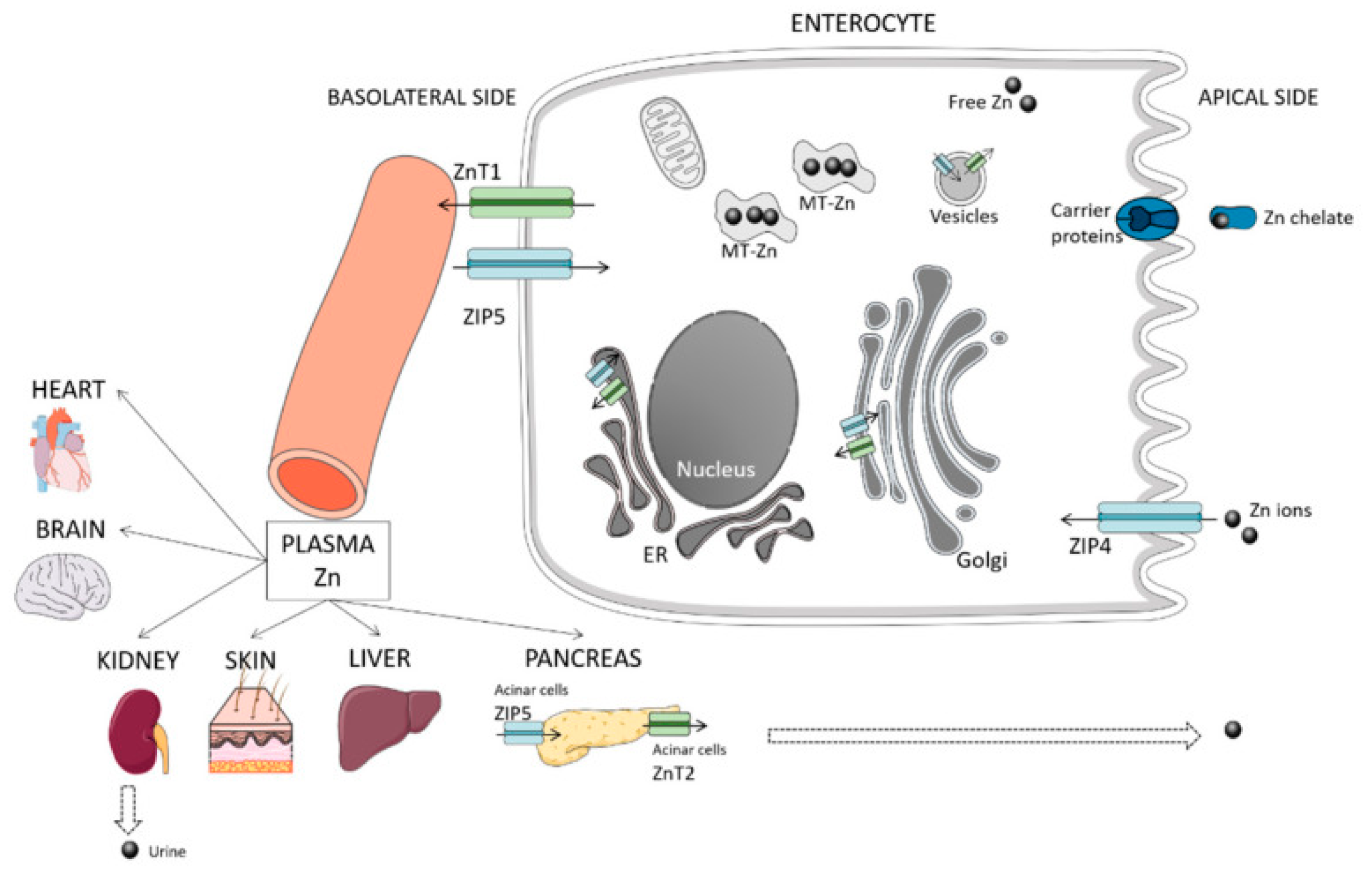
3.6. Zinc
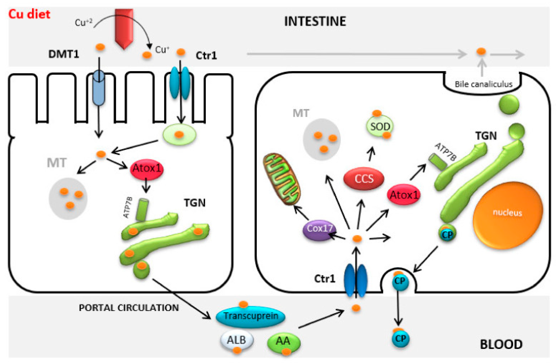
3.7. Copper
3.8. Iron
3.9. Lead (Plumbum)
3.10. Cadmium
3.11. Arsenic
4. Conclusions
Author Contributions
Funding
Institutional Review Board Statement
Informed Consent Statement
Data Availability Statement
Conflicts of Interest
References
- Avtsyn, P.A.; Zhavoronkov, A.A.; Rish, M.A.; Strochkova, L.S. Human Microelementoses: Etiopathology, Classification, Organopathology; Medicine: Moscow, Russia, 1991; p. 496. [Google Scholar]
- Antipov, A.A. Physiological and biochemical features and effects of interactions in the assimilation and metabolism of nutrients in poultry (review). Probl. Biol. Product. Anim. 2010, 2, 5–43. (In Russian) [Google Scholar]
- Bondarev, A.Y. Toxicants in the organisms of the wolf and some other mammals of the Altai Territory. Bull. Altai State Agrar. Univ. 2012, 5, 44–49. (In Russian) [Google Scholar]
- Bykova, E.A.; Gashev, S.N. Features of the accumulation of microelements in the body of small mammals under conditions of urbanization. Proc. Samara Sci. Cent. Russ. Acad. Sci. 2014, 16, 1144–1148. (In Russian) [Google Scholar]
- Drozdova, E.A.; Aleshina, E.S. Influence of iron nanoparticles on blood biochemistry and indicators of nonspecific immunity of poultry. Russ. J. Immunol. 2017, 11, 303–305. (In Russian) [Google Scholar]
- Ivanov, S.D. Iron as a carcinogenic ecotoxicant. Toxicol. Bull. 2011, 2, 34–41. (In Russian) [Google Scholar]
- Voronina, O.A.; Bogolyubova, N.V.; Zaitsev, S.Y. Mineral composition of cow milk—A mini review. Sel’skokhozyaistvennaya Biol. Agric. Biol. 2022, 57, 681–693. (In Russian) [Google Scholar] [CrossRef]
- Fieten, H.; Leegwater, P.A.; Watson, A.L.; Rothuizen, J. Canine models of copper toxicosis for understanding mammalian copper metabolism. Mamm. Genome 2012, 23, 62–75. [Google Scholar] [CrossRef] [Green Version]
- Kozłowska, B.; Sochanowicz, B.; Kraj, L.; Palusinska, M.; Kołsut, P.; Szymanski, Ł.; Lewicki, S.; Kruszewski, M.; Załeska-Kociecka, M.; Leszek, P. Clinical and Molecular Aspects of Iron Metabolism in Failing Myocytes. Life 2022, 12, 1203. [Google Scholar] [CrossRef]
- Pederiva, S.; Crescio, M.I.; Ingravalle, F.; Abete, M.C.; Marchis, D.; Squadrone, S. Processed animal proteins (PAPs) in animal nutrition: Assessment of the chemical risk of essential and non-essential elements. Journal of Trace Elements in Medicine and Biology 2022, 71, 126959. [Google Scholar] [CrossRef]
- Pereira, A.M.; Guedes, M.; Matos, E.; Pinto, E.; Almeida, A.A.; Segundo, M.A.; Correia, A.; Vilanova, M.; Fonseca, A.J.M.; Cabrita, A.R.J. Effect of Zinc Source and Exogenous Enzymes Supplementation on Zinc Status in Dogs Fed High Phytate Diets. Animals 2020, 10, 400. [Google Scholar] [CrossRef] [Green Version]
- Pedrinelli, V.; Zafalon, R.V.A.; Rodrigues, R.B.A.; Perini, M.P.; Conti, R.M.C.; Vendramini, T.H.A.; de Carvalho Balieiro, J.C.; Brunetto, M.A. Concentrations of macronutrients, minerals and heavy metals in home-prepared diets for adult dogs and cats. Sci. Rep. 2019, 9, 13058. [Google Scholar] [CrossRef] [Green Version]
- López-Alonso, M.; Miranda, M.; García-Partida, P.; Mendez, A.; Castillo, C.; Benedito, J.L. Toxic and trace metal concentrations in liver and kidney of dogs. Biol. Trace Elem. Res. 2007, 116, 185–202. [Google Scholar] [CrossRef]
- Dodd, S.A.S.; Shoveller, A.K.; Fascetti, A.J.; Yu, Z.Z.; Ma, D.W.L.; Verbrugghe, A. A Comparison of Key Essential Nutrients in Commercial Plant-Based Pet Foods Sold in Canada to American and European Canine and Feline Dietary Recommendations. Animals 2021, 11, 2348. [Google Scholar] [CrossRef]
- Nekrasov, R.V.; Bogolyubova, N.V.; Semenova, A.A.; Nasonova, V.V.; Polishchuk, E.K. Dihydroquercetin influence on clinical and biochemical blood parameters of pigs under conditions of stress load. Vopr. Pitan. Probl. Nutr. 2021, 90, 74–84. (In Russian) [Google Scholar] [CrossRef]
- Nekrasov, R.V.; Ivanov, G.A.; Chabaev, M.G.; Zelenchenkova, A.A.; Bogolyubova, N.V.; Nikanova, D.A.; Sermyagin, A.A.; Bibikov, S.O.; Shapovalov, S.O. Effect of Black Soldier Fly (Hermetia illucens L.) Fat on Health and Productivity Performance of Dairy Cows. Animals 2022, 12, 2118. [Google Scholar] [CrossRef]
- Zaitsev, S.Y.; Kolesnik, N.S.; Bogolyubova, N.V. Correlations between the Major Amino Acids and Biochemical Blood Parameters of Pigs at Controlled Fattening Duration. Molecules 2022, 27, 2278. [Google Scholar] [CrossRef]
- Zaitsev, S.Y.; Belous, A.A.; Voronina, O.A.; Savina, A.A.; Rykov, R.A.; Bogolyubova, N.V. Correlations Between Antioxidant and Biochemical Parameters of Blood Serum of Duroc Breed Pigs. Animals 2021, 11, 2400. [Google Scholar] [CrossRef]
- Tajchman, K.; Ukalska-Jaruga, A.; Bogdaszewski, M. Comparison of the accumulation of macro- and microelements in the bone marrow and bone of wild and farmed red deer (Cervus elaphus). BMC Vet. Res. 2021, 17, 324. [Google Scholar] [CrossRef]
- González-Montaña, J.R.; Escalera-Valente, F.; Alonso, A.J.; Lomillos, J.M.; Robles, R.; Alonso, M.E. Relationship between vitamin B12 and cobalt metabolism in domestic ruminant: An update. Animals. 2020, 10, 1855. [Google Scholar] [CrossRef]
- Clarkson, A.H.; Paine, S.; Martín-Tereso, J.; Kendall, N.R. Copper physiology in ruminants: Trafficking of systematic copper, adaptations to variation in nutritional supply and thiomolybdate challenge. Nut. Res. Revs. 2020, 33, 43–49. [Google Scholar] [CrossRef]
- Upadhaya, S.D.; Kim, I.H. Importance of micronutrients in bone health of monogastric animals and techniques to improve the bioavailability of micronutrient supplements—A review. Asian-Australas. J. Anim. Sci. 2020, 33, 1885–1895. [Google Scholar] [CrossRef] [Green Version]
- Linder, M.C. Copper Homeostasis in Mammals, with Emphasis on Secretion and Excretion. A Review. Int. J. Mol. Sci. 2020, 21, 4932. [Google Scholar] [CrossRef]
- Kalisińska, E. Mammals and Birds as Bioindicators of Trace Element Contaminations in Terrestrial Environments. In Mammals and Birds as Bioindicators of Trace Element Contaminations in Terrestrial Environments; Kalisińska, E., Ed.; Springer: Cham, Switzerland, 2019; pp. 21–53. [Google Scholar] [CrossRef]
- Grech, B.J. Mechanistic insights into the treatment of iron-deficiency anemia and arthritis in humans with dietary molybdenum. Eur. J. Clin. Nutr. 2021, 75, 1170–1175. [Google Scholar] [CrossRef] [PubMed]
- Ward, D.M.; Cloonan, S.M. Mitochondrial Iron in Human Health and Disease. Annu Rev Physiol. 2019, 81, 453–482. [Google Scholar] [CrossRef] [PubMed]
- Skalny, A.V.; Rudakov, I.A. Bioelements in Medicine; Publishing House “ONIX 21st Century”, Mir: Moscow, Russia, 2004; p. 272. (In Russian) [Google Scholar]
- Shchedrina, A.V. Elementary medicine: Deficiency is dangerous for health. Bus. Environ. J. 2003, 2, 34–37. (In Russian) [Google Scholar]
- Navidshad, B.; Mohammadrezaei, M.; Zarei, M.; Valizadeh, R.; Karamati, S.; Rezaei, F.; Jabbari, S.; Kachoei, R.; Esmaeilinasab, P. The new progresses in trace mineral requirements of broilers, a review. Iranian J. Appl. Anim. Sci. 2019, 9, 9–16. [Google Scholar]
- Faghih-Mohammadi, F.; Seidavi, A.; Bouyeh, M. The effects of chelated micro-elements feeding in broiler breeder hens and their progeny: A review. Trop. Anim. Health Prod. 2022, 54, 1–14. [Google Scholar] [CrossRef]
- Binkowski, Ł.J. Arsenic, As. In Mammals and Birds as Bioindicators of Trace Element Contaminations in Terrestrial Environments; Kalisińska, E., Ed.; Springer: Cham, Switzerland, 2019; pp. 463–481. [Google Scholar] [CrossRef]
- Ramos, H.D.; Medellín, R.A.; Morton-Bermea, O. Insectivorous bats as biomonitor of metal exposure in the megalopolis of Mexico and rural environments in central Mexico. Environ. Res. 2020, 185, 109–293. [Google Scholar] [CrossRef]
- Gamberg, M.; Pratte, I.; Brammer, J.; Cuyler, C.; Elkin, B.; Gurney, K.; Kutz, S.; Larter, N.C.; Muir, D.; Wang, X.; et al. Renal trace elements in barren-ground caribou subpopulations: Temporal trends and differing effects of sex, age and season. Sci. Total Environ. 2020, 1, 138305. [Google Scholar] [CrossRef]
- Korbecki, J.; Gutowska, I.; Chlubek, D.; Baranowska-Bosiacka, I. Lead (Pb) in the tissues of Anatidae, Ardeidae, Sternidae and Laridae of the Northern Hemisphere: A review of environmental studies. Environ. Sci. Pollut. Res. 2019, 26, 12631–12647. [Google Scholar] [CrossRef] [Green Version]
- Gruz, A.; Mackle, O.; Bartha, A.; Szabo, R.; Deri, J.; Budai, P.; Lehel, J. Biomonitoring of toxic metals in feathers of predatory birds from eastern regions of Hungary. Environ. Sci. Pollut. Res. 2019, 26, 26324–26331. [Google Scholar] [CrossRef] [Green Version]
- Wang, J.; Pantopoulos, K. Regulation of cellular iron metabolism. Biochem J. 2011, 434, 365–381. [Google Scholar] [CrossRef] [PubMed] [Green Version]
- Durkalec, M.; Nawrocka, A.; Krzysiak, M.; Larska, M.; Kmiecik, M.; Posyniak, A. Trace elements in the liver of captive and free-ranging European bison (Bison bonasus L.). Chemosphere 2017, 193, 454–463. [Google Scholar] [CrossRef] [PubMed]
- Eskov, E.K.; Gorbunova, E.V.; Lavrinovich, V.V. Pollutants and essential elements in different parts of the body of the red fox and their habitat. Proc. Orenbg. State Agrar. Univ. 2009, 22, 291–294. (In Russian) [Google Scholar]
- Kharlamov, A.V.; Frolov, A.N.; Zavyalov, O.A.; Miroshnikov, A.M. Informativity of biosubstrates in assessing the elemental status of farm animals (review). Bull. Beef Cattle Breed. 2014, 4, 53–58. (In Russian) [Google Scholar]
- Sidorov, P.I.; Novikova, I.A. Screening method for assessing health factors. Hyg. Sanit. 2010, 2, 85–89. (In Russian) [Google Scholar]
- Stepanova, M.V.; Timakov, A.V.; Yarlykov, N.G. Nosological profile of non-contagious diseases of wild animals in a zoo. Vet. Dr. 2019, 5, 45–53. (In Russian) [Google Scholar]
- Stepanova, M.V.; Ostapenko, V.A. Neoplasms in animals of zoological institutions. Vet. Anim. Husb. Biotechnol. 2019, 4, 55–62. (In Russian) [Google Scholar]
- Stepanova, M.V.; Timakov, A.V.; Yarlykov, N.G. Analysis of diseases of the digestive system of wild animals in a zoo. Int. Bull. Vet. Med. 2019, 3, 92–99. (In Russian) [Google Scholar]
- Cosselman, K.E.; Navas-Acien, A.; Kaufman, J.D. Environmental factors in cardiovascular disease. Nat. Rev. Cardiol. 2015, 12, 627. [Google Scholar] [CrossRef]
- Hackett, T.L.; Stefanowicz, D.; Aminuddin, F.; Sin, D.D.; Connett, J.E.; Anthonisen, N.R.; Sandford, A.J. Effect of gene environment interactions on lung function and cardiovascular disease in COPD. Int. J. Chronic Obstr. Pulm. Dis. 2011, 6, 277. [Google Scholar] [CrossRef] [Green Version]
- Hegazy, A.A.; Zaher, M.M.; Abd el-Hafez, M.A.; Morsy, A.A.; Saleh, R.A. Relation between anemia and blood levels of lead, copper, zinc and iron among children. BMC Res. Notes 2010, 3, 133. [Google Scholar] [CrossRef] [Green Version]
- Kim, J.; Lee, Y.; Yang, M. Environmental exposure to lead (Pb) and variations in its susceptibility. J. Environ. Sci. Health Part C 2014, 32, 159–185. [Google Scholar] [CrossRef]
- Marras, C.; Goldman, S.M. Genetics meets environment: Evaluating gene–environment interactions in neurologic diseases. Semin. Neurol. Thieme Med. Publ. 2011, 31, 553–562. [Google Scholar] [CrossRef] [PubMed]
- Modgil, S.; Lahiri, D.K.; Sharma, V.L.; Anand, A. Role of early life exposure and environment on neurodegeneration: Implications on brain disorders. Transl. Neurodegener. 2014, 3, 9. [Google Scholar] [CrossRef] [Green Version]
- Nikolić, M.; Nikić, D.; Stanković, A. Effects of air pollution on red blood cells in children. Environ. Stud. 2008, 17, 267. [Google Scholar]
- Sheehan, W.J.; Phipatanakul, W. Difficult to control asthma: Epidemiology and its link with environmental factors. Curr. Opin. Allergy Clin. Immunol. 2015, 15, 397. [Google Scholar] [CrossRef] [Green Version]
- Ashford, N.A.; Bauman, P.; Brown, H.S.; Clapp, R.W.; Finkel, A.M.; Gee, D.; Sass, J.B. Cancer risk: Role of environment. Science 2015, 347, 727. [Google Scholar] [CrossRef] [PubMed]
- Skal’nyy, A.V. Mikroelementy: Bodrost’, Zdorov’e, Dolgoletie. [Trace Elements: Vitality, Health, Longevity]; MIR: Moscow, Russia, 2019. (In Russian) [Google Scholar]
- Ugarte, M.; Osborne, N.N. Recent advances in the understanding of the role of zinc in ocular tissues. Metallomics 2014, 6, 189–200. [Google Scholar] [CrossRef]
- Marquez, A.; Urbina, M.; Quintal, M.; Obregon, F.; Salazar, V.; Lim, L. Extracellular zinc chelator in vivo on system of taurine in retina: Transport, concentrations and localization of transporter. J. Clin. Exp. Ophthalmol. 2017, 8, 137–148. [Google Scholar] [CrossRef]
- Madl, J.E.; McIlnay, T.R.; Powell, C.C.; Gionfriddo, J.R. Depletion of taurine and glutamate from damaged photoreceptors in the retinas of dogs with primary glaucoma. Am. J. Vet. Res. 2005, 66, 791–799. [Google Scholar] [CrossRef] [PubMed]
- Rosolen, S.G.; Neveux, N.; Sahel, J.A.; Picaud, S.; Froger, N. Evaluation of the taurine concentrations in dog plasma and aqueous humour: A pilot study. Adv. Exp. Med. Biol. 2013, 775, 145–154. [Google Scholar] [PubMed]
- Gammoh, N.Z.; Rink, L. Zinc in infection and inflammation. Nutrients 2017, 9, 624. [Google Scholar] [CrossRef] [Green Version]
- Heidarpour, M.; Soltani, S.; Mohri, M.; Khoshnegah, J. Canine visceral leishmaniasis: Relationships between oxidative stress, liver and kidney variables, trace elements, and clinical status. Parasitol. Res. 2012, 111, 1491–1496. [Google Scholar] [CrossRef]
- Bilandžić, N.; Dežđek, D.; Sedak, M.; Dokić, M.; Simić, B.; Rudan, N.; Brstilo, M.; Lisicin, T. Trace elements in tissues of wild carnivores and omnivores in Croatia. Bull. Environ. Contam. Toxicol. 2012, 88, 94–99. [Google Scholar] [CrossRef]
- Ogawa, Y.; Kinoshita, M.; Shimada, S.; Kawamura, T. Zinc and skin disorders. Nutrients 2018, 10, 199. [Google Scholar] [CrossRef] [Green Version]
- Bin, B.H.; Bhin, J.; Takaishi, M.; Toyoshima, K.-E.; Kawamata, S.; Ito, K.; Hara, T.; Watanabe, T.; Irie, T.; Takagishi, T. Requirement of zinc transporter ZIP10 for epidermal development: Implication of the ZIP10-p63 axis in epithelial homeostasis. Proc. Natl. Acad. Sci. USA 2017, 114, 12243–12248. [Google Scholar] [CrossRef] [Green Version]
- Pereira, A.M.; Maia, M.R.G.; Fonseca, A.J.M.; Cabrita, A.R.J. Zinc in Dog Nutrition, Health and Disease: A Review. Animals 2021, 11, 978. [Google Scholar] [CrossRef]
- Hoffmann, G.; Jones, P.G.; Biourge, V.; van den Ingh, T.S.; Mesu, S.J.; Bode, P.; Rothuizen, J. Dietary management of hepatic copper accumulations in Labrador Retrievers. Vet. Intern. Med. 2009, 23, 957–963. [Google Scholar] [CrossRef]
- Skal’nyy, A.V. (Ed.) Mikroelementy [Microelements], 4th ed.; Fabrika Bloknotov: Moscow, Russia, 2018; p. 295. (In Russian) [Google Scholar]
- Center, S.A. Metabolic, antioxidant, nutraceutical, probiotic, and herbal therapies relating to the management of hepatobiliary disorders. Vet. Clin. North Am. Small Anim. Pract. 2004, 34, 67–172. [Google Scholar] [CrossRef]
- Fallah, A.; Mohammad-Hasani, A.; Colagar, A.H. Zinc is an Essential Element for Male Fertility: A Review of Zn Roles in Men’s Health, Germination, Sperm Quality, and Fertilization. J. Reprod. Infertil. 2018, 19, 69–81. [Google Scholar] [PubMed]
- Mogielnicka-Brzozowska, M.; Kowalska, N.; Fraser, L.; Kordan, W. Proteomic characterization of zinc-binding proteins of canine seminal plasma. Reprod. Domest. Anim. 2015, 50, 1017–1021. [Google Scholar] [CrossRef] [PubMed]
- Kuhlman, G.; Rompala, R.E. The influence of dietary sources of zinc, copper and manganese on canine reproductive performance and hair mineral content. Nutrition 1998, 128, 2603S–2605S. [Google Scholar] [CrossRef] [Green Version]
- Wilson, R.L.; Leemaqz, S.Y.; Goh, Z.; McAninch, D.; Jankovic-Karasoulos, T.; Leghi, G.E.; Phillips, J.A.; Colafella, K.M.; Tran, C.; O’Leary, S. Zinc is a critical regulator of placental morphogenesis and maternal hemodynamics during pregnancy in mice. Sci. Rep. 2017, 7, 15137. [Google Scholar] [CrossRef] [Green Version]
- Brodzki, A. Copper and zinc concentration in skin neoplastic tissues in dogs. Bull.–Vet. Inst. Pulawy 2007, 51, 271–273. [Google Scholar]
- Harro, C.C.; Smedley, R.C.; Buchweitz, J.P.; Langlois, D.K. Hepatic copper and other trace mineral concentrations in dogs with hepatocellular carcinoma. Vet. Intern. Med. 2019, 33, 2193–2199. [Google Scholar] [CrossRef] [Green Version]
- Enginler, S.O.; Toydemir, T.S.F.; Ates, A.; Ozturk, B.; Erdogan, O.; Ozdemir, S.; Kirsan, I.; Or, M.E.; Arun, S.S.; Barutcu, U.B. Examination of Oxidative/Antioxidative Status and Trace Element Levels in Dogs with Mammary Tumors. Bulg. J. Agric. Sci. 2015, 5, 1086–1091. [Google Scholar]
- Wang, F.; Jiao, P.; Qi, M.; Frezza, M.; Dou, Q.P.; Yan, B. Turning tumor-promoting copper into an anti-cancer weapon via high-throughput chemistry. Curr. Med. Chem. 2010, 17, 2685–2698. [Google Scholar] [CrossRef] [Green Version]
- Lazarchick, J. Update on anemia and neutropenia in copper deficiency. Curr. Opin. Hematol. 2012, 19, 58–60. [Google Scholar] [CrossRef]
- López-Alonso, M.; Miranda, M. Copper Supplementation, A Challenge in Cattle. Animals 2020, 10, 1890. [Google Scholar] [CrossRef]
- Gürgöze, M.K.; Ölçücü, A.; Aygün, A.D.; Taşkin, E.; Kiliç, M. Serum and hair levels of zinc, selenium, iron, and copper in children with iron-deficiency anemia. Biol. Trace Elem. Res. 2006, 111, 23–29. [Google Scholar] [CrossRef] [PubMed]
- Wittung-Stafshede, P.A. Copper Story: From Protein Folding and Metal Transport to Cancer. Isr. J. Chem. 2016, 56, 671–681. [Google Scholar] [CrossRef]
- Denoyer, D.; Masaldan, S.; La Fontaine, S.; Cater, M.A. Targeting copper in cancer therapy: “Copper That Cancer”. Metallomics 2015, 7, 1459–1476. [Google Scholar] [CrossRef]
- Lener, M.R.; Scott, R.J.; Wiechowska-Kozłowska, A.; Serrano-Fernández, P.; Baszuk, P.; Jaworska-Bieniek, K.; Gromowski, T. Serum concentrations of selenium and copper in patients diagnosed with pancreatic cancer. Cancer research and treatment. Off. J. Korean Cancer Assoc. 2016, 48, 1056. [Google Scholar]
- Karimi, G.; Shahar, S.; Homayouni, N.; Rajikan, R.; Bakar, N.F.A.; Othman, M.S. Association between trace element and heavy metal levels in hair and nail with prostate cancer. Asian Pac. J. Cancer Prev. 2012, 13, 4249–4253. [Google Scholar] [CrossRef] [Green Version]
- Donma, M.M.; Donma, O.; Tas, M.A. Hair zinc and copper concentrations and zinc: Copper ratios in pediatric malignancies and healthy children from southeastern Turkey. Biol. Trace Elem. Res. 1993, 36, 51–63. [Google Scholar] [CrossRef]
- Bost, M.; Houdart, S.; Oberli, M.; Kalonji, E.; Huneau, J.F.; Margaritis, I. Dietary copper and human health: Current evidence and unresolved issues. J. Trace Elem. Med. Biol. 2016, 35, 107–115. [Google Scholar] [CrossRef] [PubMed]
- Weiss, G.; Goodnough, L.T. Anemia of chronic disease. New Engl. J. Med. 2005, 352, 1011–1023. [Google Scholar] [CrossRef] [PubMed] [Green Version]
- Roy, C.N.; Andrews, N.C. Anemia of inflammation: The hepcidin link. Curr. Opin. Hematol. 2005, 2, 107–111. [Google Scholar] [CrossRef]
- Pantopoulos, K.; Porwal, S.K.; Tartakoff, A.; Devireddy, L. Mechanisms of mammalian iron homeostasis. Biochemistry 2012, 51, 5705–5724. [Google Scholar] [CrossRef]
- Flora, G.; Gupta, D.; Tiwari, A. Toxicity of lead: A review with recent updates. Interdiscip. Toxicol. 2012, 5, 47–58. [Google Scholar] [CrossRef] [PubMed]
- Salnikova, E.V. Ecological and Geochemical Monitoring of the Influence of Copper, Cadmium and Lead on the Zinc Status of the Population of the Orenburg Region FGBOU VO; Orenburg State University: Orenburg, Russia, 2018; p. 274. (In Russian) [Google Scholar]
- Liu, Z.; Yu, Y.; Li, X.; Wu, A.; Mu, M.; Li, N.; Chen, X. Maternal lead exposure and risk of congenital heart defects occurrence in offspring. Reprod. Toxicol. 2015, 51, 16. [Google Scholar] [CrossRef] [PubMed]
- Özel, Ş.; Ozyer, S.; Aykut, O.; Çinar, M.; Yılmaz, O.H.; Caglar, A.; EnginUstun, Y. Maternal second trimester blood levels of selected heavy metals in pregnancies complicated with neural tube defects. J. Matern.—Fetal Neonatal Med. 2018, 32, 2547–2553. [Google Scholar] [CrossRef] [PubMed]
- Montrose, L.; Faulk, C.; Francis, J.; Dolinoy, D.C. Perinatal lead (Pb) exposure results in sex and tissue-dependent adult DNA methylation alterations in murine IAP transposons. Environ. Mol. Mutagen. 2017, 58, 540–550. [Google Scholar] [CrossRef] [Green Version]
- Sanchez, O.F.; Lee, J.; Hing, N.Y.K.; Kim, S.E.; Freeman, J.L.; Yuan, C. Lead (Pb) exposure reduces global DNA methylation level by noncompetitive inhibition and alteration of dnmt expression. Metallomics 2017, 9, 149–160. [Google Scholar] [CrossRef]
- Li, H.; Li, X.; Liu, J.; Jin, L.; Yang, F.; Wang, J.; Gao, Y. Correlation between serum lead and thyroid diseases: Papillary thyroid carcinoma, nodular goiter, and thyroid adenoma. Int. J. Environ. Health Res. 2017, 27, 409–419. [Google Scholar] [CrossRef]
- Tyrrell, J.B.; Hafida, S.; Stemmer, P.; Adhami, A.; Leff, T. Lead (Pb) exposure promotes diabetes in obese rodents. J. Trace Elem. Med. Biol. 2017, 39, 221–226. [Google Scholar] [CrossRef]
- Chen, X.; Zhou, H.; Li, X.; Wang, Z.; Zhu, G.; Jin, T. Effects of lead and cadmium co-exposure on hemoglobin in a Chinese population. Environ. Toxicol. Pharmacol. 2015, 39, 758–763. [Google Scholar] [CrossRef]
- Shah, F.; Kazi, T.G.; Afridi, H.I.; Kazi, N.; Baig, J.A.; Shah, A.Q.; Wadhwa, S.K. Evaluation of status of trace and toxic metals in biological samples (scalp hair, blood, and urine) of normal and anemic children of two age groups. Biol. Trace Elem. Res. 2011, 141, 131–149. [Google Scholar] [CrossRef]
- Suh, Y.J.; Lee, J.E.; Lee, D.H.; Yi, H.G.; Lee, M.H.; Kim, C.S.; Kim, S.K. Prevalence and relationships of iron deficiency anemia with blood cadmium and vitamin D levels in Korean women. J. Korean Med. Sci. 2016, 31, 25–32. [Google Scholar] [CrossRef]
- Zhang, T.; Liu, J.; Zhang, J.; Zhang, N.; Yang, X.; Qu, H.; Xi, L.; Han, J. Effects of dietary zinc levels on the growth performance, organ zinc content, and zinc retention in broiler chickens. Rev. Bras. Cienc. Avic. 2018, 20, 127–132. [Google Scholar] [CrossRef] [Green Version]
- Semenova, A.V.; Chernova, O.F.; Gorbacheva, M.V.; Perfilova, T.V. Species diagnostic features of wool fibers of representatives of the camelid family, as criteria for the identification of textile material. Des. Technol. 2020, 76, 49–57. (In Russian) [Google Scholar]
- Horiguchi, H.; Oguma, E.; Kayama, F. Cadmium induces anemia through interdependent progress of hemolysis, body iron accumulation, and insufficient erythropoietin production in rats. Toxicol. Sci. 2011, 122, 198–210. [Google Scholar]
- Jomova, K.; Jenisova, Z.; Feszterova, M.; Baros, S.; Liska, J.; Hudecova, D.; Rhodes, C.J.; Valko, M. Arsenic: Toxicity, oxidative stress and human disease. J. Appl. Toxicol. 2011, 31, 95–107. [Google Scholar] [CrossRef] [PubMed]


| Group of Nosologies | MME, mg/kg | |||||
|---|---|---|---|---|---|---|
| Zn | Cu | Fe | Pb | Cd | As | |
| Skin diseases | 0.43 * | 0.19 | 0.41 | 0.72 * | 0.08 | −0.04 |
| Diseases of the hearing organs | −0.14 | 0.24 | 0.34 | −0.11 | 0.17 | 0.01 |
| Diseases of the musculoskeletal system | 0.43 | 0.24 * | 0.51 | 0.41 | 0.12 | 0.05 |
| Diseases of the respiratory system | −0.26 | 0.14 | 0.11 | 0.01 | 0.34 | 0.29 |
| Diseases of the digestive system | 0.38 | 0.12 | 0.10 | 0.62 | 0.23 | 0.11 |
| Metabolic diseases | 0.03 | −0.05 | 0.12 | 0.27 * | 0.18 | 0.11 |
| Diseases of the cardiovascular system | −0.01 | 0.37 * | 0.24 | 0.63 | 0.18 * | 0.16 |
| Diseases of the organs of vision | 0.42 | 0.22 | 0.41 | 0.22 | −0.21 | 0.08 |
| Diseases of the nervous system | 0.02 | 0.11 | 0.09 | 0.11 * | −0.24 | 0.23 |
| Diseases of the reproductive system | 0.21 | 0.17 | 0.13 | −0.08 | −0.01 | 0.41 |
| Diseases of the excretory system | 0.11 | 0.04 | 0.04 | 0.34 * | −0.01 | 0.01 |
| Oncological diseases | 0.28 * | 0.19 | 0.11 * | 0.03 | 0.17 | 0.15 |
| Group of Nosologies | ME, mg/kg | |||||
|---|---|---|---|---|---|---|
| Zn | Cu | Fe | Pb | Cd | As | |
| Skin diseases | 56.9 ± 8.4 * ↓ | 15.9 ± 2.45 | 543 ± 113 | 6.99 ± 2.48 | 1.01 ± 0.48 | 0.110 ± 0.102 |
| Diseases of the hearing organs | 129 ± 11.2 | 11.7 ± 14.5 | 428 ± 48.6 | 8.01 ± 4.24 | 1.52 ± 0.93 | 0.211 ± 0.071 |
| Diseases of the musculoskeletal system | 143 ± 2.42 | 3.95 ± 4.57 * ↓ | 395 ± 38.5 | 7.11 ± 3.24 | 3.15 ± 1.01 | 0.342 ± 0.241 |
| Diseases of the respiratory system | 166 ± 3.84 | 13.8 ± 9.42 | 288 ± 69.0 | 6.99 ± 2.97 | 1.13 ± 0.24 | 0.533 ± 0.042 |
| Diseases of the digestive system | 62.1 ± 6.51 * ↓ | 12.5 ± 7.15 | 343 ± 57.5 | 6.97 ± 2.11 | 1.56 ± 0.64 | 0.241 ± 0.112 |
| Metabolic diseases | 74.5 ± 19.5 ↓ | 14.6 ± 4.52 | 354 ± 15.8 | 8.01 ± 3.54 | 1.85 ± 1.03 | 0.653 ± 0.431 |
| Diseases of the cardiovascular system | 142 ± 54.9 | 18.7 ± 3.54 ↑ | 128 ± 11.8 * ↓ | 6.88 ± 1.02 | 5.01 ± 0.68 * ↑ | 0.342 ± 0.123 |
| Diseases of the organs of vision | 49.7 ± 33.8 * ↓ | 14.7 ± 8.41 | 302 ± 65.8 | 7.55 ± 1.24 | 2.03 ± 1.51 | 0.429 ± 0.221 |
| Diseases of the nervous system | 169 ± 64.5 | 11.6 ± 5.64 | 342 ± 54.2 | 14.6 ± 2.94 * ↑ | 3.48 ± 0.86 * ↑ | 0.538 ± 0.341 |
| Diseases of the reproductive system | 98.8 ± 29.6 ↓ | 11.9 ± 8.31 | 298 ± 51.4 | 11.68 ± 1.06 ↑ | 1.56 ± 1.02 | 0.668 ± 0.339 |
| Diseases of the excretory system | 146 ± 25.4 | 11.7 ± 5.42 | 270 ± 185 | 12.97 ± 2.84 ↑ | 2.11 ± 0.63 | 0.951 ± 0.022 * ↑ |
| Oncological diseases | 155 ± 49.8 | 21.9 ± 2.48 * ↑ | 298 ± 67.8 | 9.01 ± 2.48 | 2.46 ± 1.39 | 0.638 ± 0.109 |
| Average level | 130 ± 14.3 | 17.7 ± 4.89 | 388 ± 48.8 | 5.73 ± 0.93 | 1.25 ± 0.17 | 0.768 ± 0.181 |
Disclaimer/Publisher’s Note: The statements, opinions and data contained in all publications are solely those of the individual author(s) and contributor(s) and not of MDPI and/or the editor(s). MDPI and/or the editor(s) disclaim responsibility for any injury to people or property resulting from any ideas, methods, instructions or products referred to in the content. |
© 2023 by the authors. Licensee MDPI, Basel, Switzerland. This article is an open access article distributed under the terms and conditions of the Creative Commons Attribution (CC BY) license (https://creativecommons.org/licenses/by/4.0/).
Share and Cite
Stepanova, M.V.; Sotnikova, L.F.; Zaitsev, S.Y. Relationships between the Content of Micro- and Macroelements in Animal Samples and Diseases of Different Etiologies. Animals 2023, 13, 852. https://doi.org/10.3390/ani13050852
Stepanova MV, Sotnikova LF, Zaitsev SY. Relationships between the Content of Micro- and Macroelements in Animal Samples and Diseases of Different Etiologies. Animals. 2023; 13(5):852. https://doi.org/10.3390/ani13050852
Chicago/Turabian StyleStepanova, Marina V., Larisa F. Sotnikova, and Sergei Yu. Zaitsev. 2023. "Relationships between the Content of Micro- and Macroelements in Animal Samples and Diseases of Different Etiologies" Animals 13, no. 5: 852. https://doi.org/10.3390/ani13050852