
One Health: Animal Models of Heritable Human Bleeding Diseases


Hemopet, 938 Stanford Street, Santa Monica, CA 90403, USA
Animals 2023, 13(1), 87; https://doi.org/10.3390/ani13010087
Submission received: 15 November 2022
/
Revised: 21 December 2022
/
Accepted: 25 December 2022
/
Published: 26 December 2022
(This article belongs to the Section Veterinary Clinical Studies)
Abstract
:Simple Summary
Animal models of human and animal diseases have been studied for decades in both experimental and clinical research with the findings applied to their management and therapy. Today, molecular and genomic research has led to the gene editing and gene therapies of an increasing number of these disorders. This review summarizes current knowledge about the molecular genetics and therapeutic approaches applied to the heritable human and animal bleeding diseases.
Abstract
Animal models of human and animal diseases have long been used as the lynchpin of experimental and clinical research. With the discovery and implementation of novel molecular and nano-technologies, cellular research now has advanced to assessing signal transduction pathways, gene editing, and gene therapies. The contribution of heritable animal models to human and animal health as related to hemostasis is reviewed and updated with the advent of gene editing, recombinant and gene therapies.
1. Introduction
The identification of a group of mammalian genomes and their sequencing has led to the current genomic revolution (Table 1) [1]. In 2021, the Alliance for Regenerative Medicine published a review of the rare human diseases currently undergoing 61 different clinical trials which included examples of applied gene therapy [2]. A plethora of articles and opinions also has appeared in the recent global scientific literature [3,4,5,6,7,8,9,10,11]. These include hematological, ophthalmological and metabolic conditions. Most are based upon the CRISPR-Cas9 technology of Doudna and Charpentier and colleagues, along with the parallel work of Zhang and colleagues at Harvard and MIT’s Broad Institute [2,3,4,5,6,7,8,9,10,11,12].
As recently described and summarized by author Walter Issacson, the 2020 Nobel Prize winning genomic research of Jennifer Doudna and Emmanuelle Charpentier helped catapult molecular research into the CRISPR gene-editing era [12]. Similar findings were published by Virginijus Šikšnys and associates in Lithuania [12,13].
CRISPR is a genetic engineering technique that allows the genomes of living organisms to be modified primarily with a simplified bacterial CRISPR-Cas9 antiviral defense system that delivers the Cas9 nuclease complexed with a synthetic guide RNA (gRNA) into a cell. The genome of the cell can be cut at any desired location, such as Cas9 or Cas12, thereby permitting existing genes to be removed and/or new ones added in vivo [12,13,14,15].
Meanwhile, recombinant AAV vectors, based on nonpathogenic parvoviruses, have been used or are currently in use in 264 Phase I/II/III human clinical trials for these diseases [9,10,11,16,17,18,19,20,21,22,23]. While unexpected, remarkable clinical efficacy has also been achieved, the use of such high doses has been shown to provoke host immune responses culminating in serious adverse events including the deaths of four patients [9,10,16,17,18,19,20,21,22,23].
To address these limitations, scientists have developed capsid-modified next-generation (NextGen) AAV serotype vectors [9,16,17,18,19,20,21,22,23]. These new recombinant AAV vectors are up to 80-fold more efficacious at reduced doses. Regulatory approval using AAV5 gene therapy with valoctocogene roxaparvovec for hemophilia A was granted in the European Union in August 2022 and is pending in the United States, Meanwhile, the US FDA in November 2022 approved the AAV vector use of etranacogene dezaparvovec for treatment of adults with hemophilia B [16,24,25,26,27,28,29,30,31,32,33,34,35].
Additional data have derived from animal models to evaluate efficacy and safety, including the mouse, ‘humanized’ mouse, dog, monkey, and other non-human primates (Table 1). Viral vector characterization is also an important application in gene therapy [9,10,11,16,17,18,19,20,21,22,23,24].
Regardless, therapeutic gene and cell therapy research and development no doubt will progress when advanced technologies and services for viral vector design and manufacturing are adopted. Such technologies, in combination with technologies for CRISPR-based gene editing, RNA interference, base editing, and prime editing, will move innovative therapeutics forward [12,13,14,15,16,17,18,19,20,21,22,23,24,25,26,27,28,29,30,31,32,33,34,35].
This review addresses the topic with respect to the heritable mammalian bleeding disorders, the history of which is summarized below (Table 2). Please note that the acquired human bleeding disorders also are seen in animals (e.g., thrombocytopenia and thrombopathia, liver disease, rodenticide exposure and thrombosis with disseminated intravascular coagulation) [36,37].
2. Inherited Hemostatic Disorders in People and Animals
For decades, studies of the role of blood cells, plasma, lymph, and the vascular endothelium of humans have relied upon in vivo and in vitro animal models for their scientific advancements and understanding [19,22,25,29,31,36,37,38,39,40,41,42,43,44,45,46,47,48,49,50,51,52,53,54,55,56,57,58,59,60,61,62,63,64,65,66,67,68,69,70,71,72,73,74,75,76,77,78,79,80,81,82,83,84,85,86,87,88,89,90,91,92,93,94,95,96,97,98,99,100,101,102,103,104,105,106,107,108,109,110,111,112,113,114,115,116,117,118,119,120,121,122,123,124]. In that regard, hemostatic disorders that parallel those seen in people also have been recognized in companion animals for decades, and recent studies have focused on their management with recombinant and gene therapy [50,52,57] (Table 2 and Table 3). These heritable bleeding diseases occur most often as a consequence of inbreeding and line breeding—in rare breeds of dogs and cats, which by necessity are inbred, and in breeds in which particular animals are popular competition show winners and are used extensively for breeding [36,37,38,39,40,43,44,45,46,47,48,49] (Figure 1). Interestingly, the most common of all canine heritable disorders are essentially the same or similar in purebred and mixed breed dogs, as documented in a recent review that included more than 152 genetic disease variants in more than 100,000 dogs [84].
Diagnosis of these hemostatic disorders is more accurate when age-and sex-matched controls are used for coagulation studies [85,86,88,124], whereas for platelet function assessment. Mucosal bleeding time, whole blood, platelet-rich plasma and washed platelets have been the samples of choice [124] (Table 3).
The cloning of the factor VIII and factor IX genes occurred more than three decades ago [15,16,17,18,19,20,21,22,23,24,25,26,27,28,29,30,31,32,33,34,35,39,53,57,59,61,81] (Table 3). Since then, major advances in the application of molecular genetics and gene therapy to the diagnostics and clinical management of hemophilia have led to the generation of novel bioengineered recombinant clotting factor concentrates and the recent successes with AAV gene therapy for Factor VIII using AAV5, AAV6, AAV8, AAV-LK03, and AAVhum37 and factor IX with AAV-2, AAVS3L, AAV5, AAV6, AAV*, AAVSPK-100, and AAVrh10 ([16,17,18,19,20,21,22,23,24,25,26,31] (Table 1)).
Given these innovations and efforts, the clinical benefit of gene therapy in hemophilia has been relatively recent following on the outcome of Phase 3 clinical trials [25,30,31,34]. Despite the very high cost of development ($ 2.5 million for hemophilia A and $3.5 million for hemophilia B [34], these advances in gene therapy have clearly enhanced the safety and efficacy of hemophilia clinical care, reduced the described ‘societal burden’ from their care, and have vastly improved the quality of life of these patients [34].
The use of gene therapy has been and still is a major goal in hemostasis research ([16,17,18,19,20,21,22,23,24,25,26,27,28,29,30,31,32,33,34,35,39,59,61,62,124] (Table 2 and Table 3)). The first hemophilia gene therapy products approved for clinical use, as stated above, have used AAV gene therapy with the nonpathogenic parvoviruses, valoctocogene roxaparvovec for hemophilia A and etranacogene dezaparvovec for treatment of adults with hemophilia B [24,25,26,27,28,30,34]
A review by Kerri Wachter in the AABB News of February 2022, reviewed the gene biotherapy trials for patients with blood disorders [35]. Specifically mentioned was progress with sickle cell disease, and hemophilia A (Factor VIII deficiency) and B (Factor IX deficiency), which are caused by single-gene mutations [34,35]. These patients no longer should need prophylactic or therapeutic infusions of plasma or recombinant Factor VIII and IX, respectively. These gene therapies use adenovirus-associated viral vectors to deliver the missing DNAs to the patient’s liver for synthesis. This process of gene editing essentially deletes the mutant section of their DNA and inserts the normal section of DNA. In addition to gene editing. other genome approaches use gene addition, gene silencing and gene correction [5,6,7,8,9,10,11,12,35].
A severely affected young hemophiliac born since the mid-1990s with access to recombinant factor VIII and IX replacement therapy, can anticipate a normal life expectancy with little to no permanent complications from excessive bleeding [24,25,26,27,34,52]. This therapy when given by necessity every other day is exceedingly expensive, and there are still serious treatment concerns [30,34]. Firstly, some patients will develop neutralizing antibodies during the first 50 infusions of therapeutic factor VIII [30]. Secondly, placement of a central venous access device is typically needed which has the life-threatening risks of infection and thrombosis. Prolonging the biological efficacy of infused recombinant factor VIII has been a goal in this field [50,52,57].
Hemophilic boys receiving plasma-derived transfusion therapies in the early 1980s also had a 75–95% risk of acquiring human immuno-deficiency virus (HIV) and /or hepatitis C infection, respectively [34,50]. Since 1990, however, improved screening of plasma donors and commercial plasma fractionation and production protocols have shown no transmission of HIV, hepatitis C, or other virus associated with any of these modern plasma-derived factor VIII preparations [27,28,34,35,50].
The availability of several commercial recombinant factor VIII, factor IX, and von Willebrand factor products since the mid-1990s has largely supplanted the plasma-derived products [30,34,50,52,57]. When recombinant von Willebrand protein is infused along with recombinant factor VIII, the risk of developing a clotting factor inhibitor is reduced. Similarly, the amount of human albumin that the cell lines need for stability in producing the recombinant factor VIII has been reduced in each step, making the product safer [50,52,57].
Currently, a dozen or more gene transfer, gene editing and genetically modified cell therapy trials for hemophilia have been performed and are ongoing [16,17,18,19,20,21,22,23,24,25,26,27,28,29,30,31,32,33,34,35].
2.1. Hemophilia A (Factor VIII: C Deficiency)
Hemophilia A is an X-chromosome-linked recessive disease carried by the female and manifested in the male. In animals, female hemophiliacs can be produced, however, by the mating of hemophilic males to carrier females [36,37]. This situation has occurred with mild forms of the disease in inbred families of purebred dogs and cats where affected males survive to sexual maturity and can reproduce. Hemophilia is the most commonly reported, severe inherited coagulation defect of animals, and has been recognized in most breeds of dogs, in mongrel dogs, in many breeds of cats and mixed breed cats, in horses and cattle [36,37,39,40,41,42,43,44,45,46] (Table 3).
Recombinant human factor VIII produced by Genetech, Inc., was first infused in early 1970 into a Boxer dog with hemophilia A in this author’s clinic in rural Albany, NY [28]. The small volume of concentrated factor VIII took about five minutes and the toenail bleeding time checked beforehand at over ten minutes was dramatically reduced to just 3 drops of serous-tinged fluid. Thus, this breakthrough study in a hemophilic dogs showed clinical success of a commercial recombinant human factor VIII [27,36,37,38,50,51,52]. Parallel infusion and gene therapy studies in hemophilic dogs included not only plasma-derived and recombinant factor VIII but also recombinant factor VIIa to induce its sustained expression, as therapy to help generate factor Xa and bypass the need for factor VIII [29,57,59,61,62,66].
2.2. Hemophilia B (Factor IX Deficiency; Christmas Disease)
An X-chromosomal-linked recessive disease like hemophilia A, hemophilia B has been reported in at least 26 breeds of dogs and 3 breeds of cats [36,37,38,39,40,41,42,43,44,45,46,47,48,49,51] (Table 3). It was first recognized in families of the Cairn Terrier and British Shorthair cat [36,37,40,43,44,45]. Results of diagnostic screening tests are basically the same as those as described for hemophilia A although specific tests are required to identify the defect factor IX deficiency rather than factor VIII:C deficiency. Affected animals have very low circulating levels of factor IX and carrier females have levels reduced to about half normal (40–60%) [36,37,45].
Treatment and management considerations for pets with hemophilia B are the same as those for hemophilia A except that canine factor IX-rich plasma fractions are given [36,37]. In general, cats with either form of hemophilia are more easily managed as house pets than are dogs with the same diseases. Cats tend to more agile and are lighter and can often lead reasonably healthy, long lives maintained as house pets [40]. Recombinant human factor VIIa and IX infusions and gene therapy have also have been successful with canine hemophilia B [37,51,57,59,61,62,67].
2.3. von Willebrand Disease (vWD)
The multifaceted syndrome known as vWD was first described in humans in 1926 [38]. The first animal model of this disease was described in 1959 in a colony of Poland-China swine, and in 1970, the canine form was discovered in members of a German Shepherd Dog family imported from Germany [39,64,65](Table 3). Their disorder, usually milder than the hemophilias, causes bleeding from mucous membranes and skin, as well as epistaxis, and gastrointestinal and urogenital bleeding. Affected dogs have a prolonged bleeding time with resulting abnormal hemorrhage from surgery. While vWD is the most common inherited bleeding disorder of humans, it also occurs in about five dozen canine breed types, several families of cats, a quarter horse, and an inbred line of Flemish Giant/Chinchilla laboratory rabbits [47,64,65,66,67,68,69,70,71,72,73,74,75,76,77,78,79,80,81,82,83,84]. A high prevalence of the gene for vWD is found in the Doberman Pinscher (~80% prevalence), German Shepherd Dog, Miniature Schnauzer, Golden Retriever, Shetland Sheepdog, Basset Hound, Standard Poodle, Keeshond, Rottweiler, Dachshund, Scottish Terrier, Manchester Terrier, and Pembroke Welsh Corgi. The disorder is either less prevalent or the true prevalence is unknown in other breed types likely because too few animals have been studied [64,65,66,67,68,69,70,71,72,73,74,75,76,77,78,79,80,81,82,83,84].
Type 1 vWD is by far the most common form in canines, and is inherited as an autosomal, incompletely dominant trait with variable clinical and laboratory expression depending upon the degree of penetrance of the mutant gene [36,37,76,77,78,79,80,81,82,83,84]. vWD is analogous to the autosomal recessive Type 3 vWD of humans in four dog breeds: Scottish terriers, Chesapeake Bay retrievers, Shetland sheepdogs, and German wirehaired pointers, in Himalayan cats, and Poland-China swine [35,68,71,73,76]. Homozygous affected individuals cannot produce measurable von Willebrand factor (vWF) and have a moderate to severe bleeding tendency. vWD heterozygotes can be detected by laboratory tests as they have reduced vWF antigen or activity, but are otherwise asymptomatic.
Many additional, less common variants of vWD exist and are classified as Type 2 vWD. These include families of German shorthair pointer dogs, and quarter horses [36,70,82]. Concurrent hypothyroidism can exacerbate bleeding in canine vWD, resulting in the situation where asymptomatic carriers of vWD may exhibit a bleeding tendency if they develop autoimmune thyroiditis and become hypothyroid. This is a common situation that is especially prevalent in Doberman pinschers [36,72]. Hypothyroid dogs also may exhibit thrombocytopenia and mucosal surface bleeding (Table 3). As thyroid supplement non-specifically shortens the bleeding time in animals with mild inherited or acquired vWD and other platelet dysfunctions, clinical experience with its use supports the efficacy, safety and low cost of this approach [36,37,72].
A reliable genetic screening test for identifying Scottish terriers with type 3 vWD is available for this and several other breeds [76]. Strong associations were detected between plasma von Willebrand factor concentration and von Willebrand factor marker genotype. All were homozygous for a 157-base pair intragenic marker allele and homozygous or compound heterozygous for 1 of 4 extragenic marker alleles. These marker genotypes were exclusively detected in dogs with low plasma von Willebrand factor concentration, although some dogs with these genotypes did not have abnormal bleeding [76].
2.4. Inherited Platelet Function Defects
Inherited disorders of platelet function were originally characterized in humans as either being of the Glanzmann’s thrombasthenia or Bernard-Soulier syndrome types. Since then, a wide variety of heritable and acquired platelet disorders called thrombopathias have been identified in people and animals [36,37,39,101,102,103,104,105,106,107,108,109,110,111,112,113,114,115,116,117,118,119,120,121,122,123,124,125] (Table 3). Thrombasthenia is an autosomal disorder where both sexes are affected and both sexes can carry the gene for this disease. In animals, it was first recognized in a family of Otterhound dogs bred by a veterinarian in upstate New York [36,37,101,108,114]. A similar disease has been recognized in other hound breeds and in an occasional cat. The biochemical defect on the membrane or surface of affected animals′ platelets is similar to that of human Glanzmann’s disease, and is caused by a deficiency of platelet GPIIb/IIIa which results in reduced platelet aggregation [113,114]. A deficiency of GPIB-IX-V in Bernard-Soulier Syndrome cases a platelet adhesion defect [106,123]. In the otterhound disorder, the platelets are large as well as dysfunctional; a similar disorder with large “Swiss-cheese”- like platelets was identified in a human patient [101,102].
Basically, the clinical signs of these disorders are similar to those of vWD because patients have long bleeding times. Platelet numbers are usually normal in these diseases but the function of the platelets is impaired. In the thrombopathic disorders, affected animals are born with defective platelet function. These are of several biochemical types and have been recognized in Basset Hounds, American Foxhounds, Spitz, Greater Swiss Mountain Dogs, German Shepherd Dogs, Simmental cattle, several breeds of cats and in a family of Fawn Hooded rats (strain named FH/Wjd) ([103,111,112,113,115,116,117,118,119,120,121,122,123,124] (Table 3)). The clinical signs, again, are similar to those of vWD because the animals have long bleeding times. The disease in Basset Hounds has been quite widespread among North American breeding stock and is caused by an unique, platelet activation signaling defect problem following injury to a blood vessel [106,108,109]. More recently, other platelet disorders have been described in animals, including ADP storage pool deficiency, ADP receptor defect, signaling pathway defects (CalDAG-DEFI and Kindlin-3) for GPIIb/IIIa activation, and a platelet procoagulant defect (Scott Syndrome) resulting in an in vivo coagulopathy [104,117]. A recent in-depth review summarized these findings [6,24] (Table 3).
2.5. Other Inherited Disorders
2.5.1. Factor VII Deficiency
Factor VII deficiency is a mild to moderate bleeding disorder in people characterized by bruising, and soft tissue bleeding from gums, bowel and urinary tract [37,47,89]. It was described in the 1960s in colonies of Beagles bred for biomedical research [37]. Since then, dogs affected by this autosomal recessive trait have been useful for studies that require monitoring liver function, as factor VII is synthesized in the liver and has a very short half-life (~4 h). A novel missense mutation has been identified as causing the relatively high prevalence of this defect in the breed [89].
2.5.2. Factor X Deficiency
Stuart–Prower factor (factor X) deficiency, an uncommon human coagulation disorder, was first described in the 1970s in a family of American Cocker Spaniels [90]. This condition has since been diagnosed in mongrel dogs and the Jack Russell terrier. Very low levels of factor X (<6% to 35%) are present in homozygotes and some heterozygotes, and they have a clinically expressed bleeding disease, whereas most heterozygotes (40–70% factor X) are asymptomatic. When factor X activity is below 20%, the affected dogs usually do not survive neonatal life. Severely affected pups are stillborn or fade and die in the first week or two of life, thereby mimicking the ”fading puppy syndrome”. Necropsy of these pups reveals massive internal bleeding. Signs in adults are mild and bleeding is seen from mucosal surfaces [37,90,91].
2.5.3. Factor XI (PTA) Deficiency
Another rare disorder of humans, factor XI deficiency mostly affects individuals of Jewish background [37,38]. Spontaneous bleeding episodes are mild (hematuria, bruising, epistaxis, menorrhagia) except when patient undergoes surgical procedures. In this case, bleeding usually starts 12–24 h after surgery and can be severe and protracted. Even after minor procedures such as biopsies and tonsillectomy, lethal bleeding has been reported. In animals, this disorder is clinically like the human equivalent and was first described in English Springer Spaniels. It also has been reported in Holstein cattle, Great Pyrenees, and Kerry Blue Terrier dogs [37,92,93,94,95].
2.5.4. Prekallikrein (Fletcher Factor) Deficiency
Prekallikrein is involved in the early surface contact phases of blood clotting. It is the precursor of plasma kallikrein that activates small peptide kinins. In addition to humans, deficiency of prekallikrein has been reported in a family of Belgian horses, and two dog breeds [98,99,100]. In one affected dog, a point mutation was identified in Exon 8 leading to an amino acid substitution in the fourth apple domain of the protein [99].
2.5.5. Factor XII Deficiency (Hageman Trait)
An asymptomatic coagulation deficiency recognized in humans, Hageman Trait (factor XII deficiency) occurs in dogs, and is quite often found in cats [37,96,97,100]. The absence of detectable biological or immunological factor XII is a normal phenomenon of a variety of other species, such as whales, birds (including the common domestic fowl and waterfowl), reptiles, and possibly fish [37].
3. Discussion and Conclusions
Research on animal models has been pivotal essential to our understanding of basic and applied sciences and has led to significant improvements in the management of both human and animal diseases [36,37,38,39,40] (Table 2 and Table 3). Veterinarians and animal scientists have been at the forefront of biomedical research in comparative medicine over the last 50 years [38,39]. The study of naturally occurring or induced animal models of human disease has led to tremendous growth of knowledge in many disciplines, including hematology, immunology, vaccinology, virology and genetics and contributed significantly to new areas of research, such as transplantation and gene therapy [34,36,37,38,39,40,55].
This era began with in vitro manual diagnosis using tilt tube timed assays with test tubes and a 37 °C water bath along with skin and mucosal surface bleeding times [39]. Today, sophisticated genetic, genomic and molecular diagnostics plus the use of safe, blood type compatible blood transfusion products, including blood concentrates, recombinant and stem cell technology are available for humans and companion animals [36,37]. As described above, the first early study with a recombinant human clotting Factor VIII product was infused into a hemophilic boxer in our laboratory at the Griffin Laboratory, NYS Department of Health in Albany, NY. His bleeding time normalized for 48 h and this success help lead to human clinical trials with this technology [27,28,29,30,31,32,33,34,35].
These research animal models also benefitted other animals [37,39,48,49,88,110,116,124]. While information generated from animal-based research experiments has been used primarily to benefit human health and well-being, parallel benefits have been accorded to animals. A classical example is the inherited bleeding disorders discussed here. In fact, this author was surprised how relatively easy it was over the last decades to search for and find parallel animal models of the human diseases of interest [36,37]. The net effect of those basic and comparative medical advances has been to improve diagnostic and treatment modalities in clinical veterinary medicine [36,37,39,48,49].
4. Conclusions
Current molecular markers and gene editing research has yielded practical and innovative clinical applications. For decades, veterinary and comparative geneticists have developed and relied upon biochemical markers of specific genetic traits to identify carrier and affected animals that are used as models of human disease [39,85,86,87,88,124]. More recently, molecular approaches have been developed that can be used to study gene therapeutic approaches for advancing human and animal health and well-being [1,2,3,4,5,6,7,8,9,10,11,12,13,14,15,16,17,18,19,20,21,22,23,24,25,26,27,28,29,30,31,32,33,34,35,50,51,52,53,88,124], Future technological developments, particularly in the areas of gene delivery and cell transplantation, will be critical for the successful clinical implementation of this gene therapy.
Funding
This author and her colleagues have been privileged to have received federal research funding to study the inherited and acquired bleeding disorders of humans and animals for more than 5 decades, NIH NHLBI Grant HL09902. This information was shared globally in person and in print with other human and comparative medicine colleagues.
Institutional Review Board Statement
IRB approval for these studies was not required.
Informed Consent Statement
Not applicable.
Data Availability Statement
Data for these studies can be found in the cited literature.
Conflicts of Interest
The author declares no conflict of interest.
References
- Hotaling, S.; Kelley, J.L.; Frandsen, P.B. Toward a genome sequence for every animal: Where are we now? Proc. Natl. Acad. Sci. USA 2021, 118, e2109019118. [Google Scholar] [CrossRef]
- Alliance for Regenerative Medicine. A new era in the therapeutic journey—Gene therapy as the beacon for rare diseases. In White Paper; Perkin-Elmer Inc.: Waltham, MA, USA, 2021. [Google Scholar]
- Knutsen, A. CRISPR-Based Therapeutics Blaze an In Vivo Path to the Clinic. Genet. Eng. Biotechnol. News 2021, 41, S12, S14–S15. [Google Scholar] [CrossRef]
- Zhang, F. Development of CRISPR-Cas systems for genome editing and beyond. Q. Rev. Biophys. 2019, 52, 31. [Google Scholar] [CrossRef] [Green Version]
- Xu, X.; Chemparathy, A.; Zeng, L.; Kempton, H.R.; Shang, S.; Nakamura, M.; Qi, L.S. Engineered miniature CRISPR-Cas system for mammalian genome regulation and editing. Mol. Cell 2021, 81, 4333–4345. [Google Scholar] [CrossRef] [PubMed]
- Malech, H.L. Treatment by CRISPR-Cas9 gene editing—A proof of principle. N. Engl. J. Med. 2021, 384, 286–287. [Google Scholar] [CrossRef]
- Musunuru, K. CRISPR hits home in a first-in-human study. CRISPR J. 2021, 4, 460. [Google Scholar] [CrossRef]
- Miccio, A. CRISPR’s path to the clinic. CRISPR J. 2022, 5, 2–3. [Google Scholar] [CrossRef] [PubMed]
- Livshits, G. CRISPR genome editing: Into the second decade. GEN Biotechnol. 2022, 1, 37–40. [Google Scholar] [CrossRef]
- Labant, M. The Point of Base Editors: Correcting Point Mutations. Genet. Eng. Biotechnol. News 2021, 41, S17–S19. [Google Scholar] [CrossRef]
- Ledford, H. Beyond CRISPR: A guide to the many other ways to edit a genome. Nature 2016, 536, 137. [Google Scholar] [CrossRef] [PubMed]
- Isaacson, W. The Code Breaker: Jennifer Doudna, Gene Editing, and the Future of the Human Race; Simon & Schuster: New York, NY, USA, 2021. [Google Scholar]
- Lapinaite, A.; Knott, G.J.; Palumbo, C.M.; Lin-Shiao, E.; Richter, M.F.; Zhao, K.T.; Beal, P.A.; Liu, D.R.; Doudna, J.A. DNA capture by a CRISPR-Cas9–guided adenine base editor. Science 2020, 369, 566–571. [Google Scholar] [CrossRef] [PubMed]
- Anzalone, A.V.; Randolph, P.B.; Davis, J.R.; Sousa, A.A.; Koblan, L.W.; Levy, J.M.; Chen, P.J.; Wilson, C.; Newby, G.A.; Raguram, A.; et al. Search-and-replace genome editing without double-strand breaks or donor DNA. Nature 2019, 576, 149–157. [Google Scholar] [CrossRef] [PubMed]
- Uddin, F.; Rudin, C.M.; Sen, T. CRISPR Gene Therapy: Applications, Limitations, and Implications for the Future. Front. Oncol. 2020, 10, 1387. [Google Scholar] [CrossRef] [PubMed]
- Kumar, S.R.; Xie, J.; Hu, S.; Ko, J.; Huang, Q.; Brown, H.C.; Srivastava, A.; Markusic, D.M.; Doering, C.B.; Spencer, H.T.; et al. Coagulation factor IX gene transfer to non-human primates using engineered AAV3 capsid and hepatic optimized expression cassette. Mol. Ther. Methods Clin. Dev. 2021, 23, 98–107. [Google Scholar] [CrossRef]
- Mendell, J.R.; Al-Zaidy, S.A.; Rodino-Klapac, L.R.; Goodspeed, K.; Gray, S.J.; Kay, C.N.; Boye, S.L.; Boye, S.E.; George, L.A. Current clinical applications of in vivo gene therapy with AAVs. Mol. Ther. 2021, 29, 464–488. [Google Scholar] [CrossRef]
- Khimani, A.H.; Thirion, C.; Srivastava, A. AAV Vectors Advance the Frontiers of Gene Therapy. Genet. Eng. Biotechnol. News 2022, 42, 38–40. [Google Scholar] [CrossRef]
- Nguyen, G.N.; Everett, J.K.; Kafle, S.; Roche, A.M.; Raymond, H.E.; Leiby, J.; Wood, C.; Assenmacher, C.-A.; Merricks, E.P.; Long, C.T.; et al. A long-term study of AAV gene therapy in dogs with hemophilia A identifies clonal expansions of transduced liver cells. Nat. Biotechnol. 2021, 39, 47–55. [Google Scholar] [CrossRef]
- Nathwani, A.C.; Tuddenham, E.G.D.; Rangarajan, S.; Rosales, C.; McIntosh, J.; Linch, D.C.; Chowdary, P.; Riddell, A.; Pie, A.J.; Harrington, C.; et al. Adenovirus-associated virus vector-mediated gene transfer in hemophilia B. N. Engl. J. Med. 2011, 365, 2357–2365. [Google Scholar] [CrossRef]
- Rangarajan, S.; Walsh, L.; Lester, W.; Perry, D.; Madan, B.; Laffan, M.; Yu, H.; Vettermann, C.; Pierce, G.F.; Wong, W.Y.; et al. AAV5-Factor VIII gene transfer in severe hemophilia A. N. Engl. J. Med. 2017, 377, 2519–2530. [Google Scholar] [CrossRef]
- Markusic, D.M.; Herzog, R.W.; Aslanidi, G.V.; Hoffman, B.E.; Li, B.; Li, M.; Jayandharan, G.R.; Ling, C.; Zolotukhin, I.; Ma, W.; et al. High-efficiency transduction and correction of murine hemophilia B using AAV2 vectors devoid of multiple surface-exposed tyrosines. Mol. Ther. 2010, 18, 2048–2056. [Google Scholar] [CrossRef]
- Brown, H.C.; Doering, C.B.; Herzog, R.W.; Ling, C.; Markusic, D.M.; Spencer, H.T.; Srivastava, A.; Srivastava, A. Development of a Clinical Candidate AAV3 Vector for Gene Therapy of Hemophilia B. Hum. Gene Ther. 2020, 31, 1114–1123. [Google Scholar] [CrossRef]
- Nathwani, A.C.; Reiss, U.; Tuddenham, E.; Chowdary, P.; McIntosh, J.; Riddell, A.; Pie, J.; Mahlangu, J.N.; Recht, M.; Shen, Y.-M.; et al. Adeno-Associated Mediated Gene Transfer for Hemophilia B:8 Year Follow up and Impact of Removing “Empty Viral Particles” on Safety and Efficacy of Gene Transfer. Blood 2019, 132, 491. [Google Scholar] [CrossRef]
- Nathwani, A.C. Gene therapy for hemophilia. Hematology 2019, 2019, 1–8. [Google Scholar] [CrossRef]
- Lacroix-Desmazes, S.; Voorberg, J.; Lillicrap, D.; Scott, D.W.; Pratt, K.P. Tolerating factor VIII: Recent progress. Front. Immunol. 2020, 10, 2991. [Google Scholar] [CrossRef]
- Batty, P.; Lillicrap, D. Hemophilia gene therapy: Approaching the first licensed product. HemaSphere 2021, 5, e540. [Google Scholar] [CrossRef]
- HEMLIBRA® [emicizumab-kxwh] Manufactured. Genentech, Inc. A Member of the Roche Group 1 DNA Way South San Francisco, CA 94080-499. Available online: https://www.hemlibra.com/ (accessed on 5 March 2021).
- Connelly, S.; Mount, J.; Mauser, A.; Gardner, J.; Kaleko, M.; McClelland, A.; Lothrop, C.J. Complete short-term correction of canine hemophilia A by in vivo gene therapy. Blood 1996, 88, 3846–3853. [Google Scholar] [CrossRef] [Green Version]
- U.S. Food and Drug Administration. FDA Approves Emicizumab·Kxwh for Prevention and Reduction of Bleeding in Patients with Hemophilia A with Factor VIII Inhibitors. Hemophilia·Factor viii. Available online: www.fda.gov/drresources-information-approved-drugs/fda-approves-emicizumckxwh-prevention-and-reduction-bleeding-patlents (accessed on 2 October 2022).
- Chowdary, P.; Shapiro, S.; Makris, M.; Evans, G.; Boyce, S.; Talks, K.; Dolan, G.; Reiss, U.; Phillips, M.; Riddell, A.; et al. Phase 1–2 trial of AAVS3 gene therapy in patients with hemophilia B. N. Engl. J. Med. 2022, 387, 237–247. [Google Scholar] [CrossRef]
- Murphy, S.L.; High, K.A. Gene therapy for haemophilia. Br. J. Haematol. 2008, 140, 479–487. [Google Scholar] [CrossRef] [Green Version]
- George, L.A.; Monahan, P.E.; Eyster, M.E.; Sullivan, S.K.; Ragni, M.V.; Croteau, S.E.; Rasko, J.E.; Recht, M.; Samelson-Jones, B.J.; MacDougall, A.; et al. Multiyear Factor VIII Expression after AAV Gene Transfer for Hemophilia A. N. Engl. J. Med. 2021, 385, 1961–1973. [Google Scholar] [CrossRef]
- Lawrence, L. Gene Therapy and the Future of Hemophilia Treatment. AABB News. 6–10 September 2022. Available online: www.aabb.org (accessed on 2 October 2022).
- Wachter, K. Biotherapy trials transform care for patients with blood disorders. AABB News 2022, 24, 16–20. [Google Scholar]
- Dodds, W.J. Estimating disease prevalence by health surveys and genetic screening. Adv. Vet. Sci. Comp. Med. 1995, 39, 29–96. [Google Scholar] [PubMed]
- Dodds, W.J. Hemostasis. In Clinical Biochemistry of Domestic Animals, 5th ed.; Kaneko, J.J., Bruss, D., Eds.; Academic Press: San Diego, CA, USA, 1997; pp. 1241–1283. [Google Scholar]
- Owen, C.A., Jr. A History of Blood Coagulation; Nichols, W.L., Walter Bowie, E.J., Eds.; Mayo Foundation for Medical Education and Research; Mayo Clinic: Rochester, MN, USA, 2001. [Google Scholar]
- Dodds, W.J. Keynote address: Extending frontiers in the art of medicine. In Proceedings of the Milestones in AHVMA, 30th anniversary, AHVMA Meeting, Birmingham, AL, USA, 8–11 September 2012. [Google Scholar]
- Peterson, J.L.; Couto, C.G.; Wellman, M.L. Hemostatic disorders in cats: A retrospective study and review of the literature. J. Vet. Int. Med. 1995, 9, 298–303. [Google Scholar] [CrossRef] [PubMed]
- Stokol, T.; Parry, B.W.; Mansell, P.D.; Richardson, J.L. Hematorrhachis associated with hemophilia A in three German shepherd dogs. J. Am. Anim. Hosp. Assoc. 1994, 30, 239–243. [Google Scholar]
- Joseph, S.A.; Brooks, M.B.; Coccari, P.J.; Riback, S.C. Hemophilia A in a German shorthaired pointer: Clinical presentations and diagnosis. J. Am. Anim. Hosp. Assoc. 1995, 32, 25–28. [Google Scholar] [CrossRef] [PubMed]
- Brinkhous, K.M.; Davis, P.D.; Graham, J.B.; Dodds, W.J. Expression and linkage of genes for X-linked hemophilias A and B in the dog. Blood 1973, 41, 577–585. [Google Scholar] [CrossRef] [PubMed] [Green Version]
- Feldman, D.G.; Brooks, M.B.; Dodds, W.J. Hemophilia B (factor IX deficiency) in a family of German shepherd dogs. J. Am. Vet. Med. Assoc. 1995, 206, 1901–1905. [Google Scholar]
- Maggio-Price, L.; Dodds, W.J. Factor IX deficiency (hemophilia B) in a family of British shorthair cats. J. Am. Vet. Med. Assoc. 1993, 203, 1702–1704. [Google Scholar]
- Dodds, W.J.; Moynihan, A.C.; Fisher, T.M.; Trauner, D.B. The frequencies of inherited blood and eye diseases as determined by genetic screening programs. J. Am. Anim. Hosp. Assoc. 1981, 17, 697–704. [Google Scholar]
- Dodds, W.J. Contributions and future directions of hemostasis research. J. Am. Vet. Med. Assoc. 1988, 193, 1157–1160. [Google Scholar] [PubMed]
- Stefanon, G.; Stefanon, B.; Stefanon, G.G.; Dodds, W.J. Inherited and acquired canine bleeding disorders in northeastern Italy. Canine Pract. 1993, 18, 15–23. [Google Scholar]
- Brooks, M. Hereditary bleeding disorders in dogs and cats. Vet. Med. 1999, 94, 555–564. [Google Scholar]
- Powell, J.S. Recombinant factor VIII in the management of hemophilia A: Current use and future promise. Ther. Clin. Risk. Manag. 2009, 5, 391–402. [Google Scholar] [CrossRef] [PubMed] [Green Version]
- Lozier, J.N.; Nichols, T.C. Animal models of hemophilia and related bleeding disorders. Semin. Hematol. 2013, 50, 175–184. [Google Scholar] [CrossRef] [PubMed] [Green Version]
- Young, G.; Mahlangu, J.; Kulkarni, R.; Nolan, B.; Liesner, R.; Pasi, J.; Barnes, C.; Neelakantan, S.; Gambino, G.; Cristiano, L.M.; et al. Recombinant factor VIII Fc fusion protein for the prevention and treatment of bleeding in children with severe hemophilia A. Thromb. Haemost. 2015, 13, 967–977. [Google Scholar] [CrossRef] [PubMed]
- Brinkhous, K.M. Gene transfer in the hemophilias: Retrospect and prospect. Thromb. Res. 1992, 67, 329–338. [Google Scholar] [CrossRef] [PubMed]
- Bouma, B.N.; Dodds, W.J.; van Mourik, J.A.; Sixma, J.J.; Webster, W.P. Infusion of human and canine factor VIII in dogs with von Willebrand’s disease: Studies of the von Willebrand and factor VIII synthesis stimulating factors. Scand. J. Haematol. 1976, 17, 263–275. [Google Scholar] [CrossRef] [PubMed]
- Giles, A.R.; Tinlin, S.; Hoogendoorn, H.; Fournel, M.A.; Ng, P.; Pancham, N. In vivo characterization of recombinant factor VIII in a canine model of hemophilia A (factor VIII deficiency). Blood 1988, 72, 335–339. [Google Scholar] [CrossRef]
- Christopherson, P.W.; Bacek, L.M.; King, K.B.; Boudreaux, M.K. Two novel missense mutations associated with hemophilia A in a family of Boxers, and a German Shepherd dog. Vet. Clin. Pathol. 2014, 43, 312–316. [Google Scholar] [CrossRef]
- Brinkhous, K.; Sigman, J.; Read, M.; Stewart, P.; McCarthy, K.; Timony, G.; Leppanen, S.; Rup, B.; Keith, J.J.; Garzone, P.; et al. Recombinant human factor IX: Replacement therapy, prophylaxis, and pharmacokinetics in canine hemophilia B. Blood 1986, 88, 2603–2610. [Google Scholar] [CrossRef] [Green Version]
- Evans, J.P.; Brinkhous, K.M.; Brayer, G.D.; Reisner, H.M.; High, K.A. Canine hemophilia B resulting from a point mutation with unusual consequences. Proc. Natl. Acad. Sci. USA 1989, 86, 10095–10099. [Google Scholar] [CrossRef] [Green Version]
- Mount, J.D.; Herzog, R.W.; Tillson, D.M.; Goodman, S.A.; Robinson, N.; McCleland, M.L.; Bellinger, D.; Nichols, T.C.; Arruda, V.R.; Lothrop, C.D.; et al. Sustained phenotypic correction of hemophilia B dogs with a factor IX null mutation by liver-directed gene therapy. Blood 2002, 99, 2670–2676. [Google Scholar] [CrossRef] [PubMed] [Green Version]
- Brooks, M.B.; Gu, W.; Barnas, J.L.; Ray, J.; Ray, K. A Line 1 insertion in the factor IX gene segregates with mild hemophilia B in dogs. Mamm. Genome 2003, 14, 788–795. [Google Scholar] [CrossRef] [PubMed]
- Margaritis, P.; Roy, E.; Aljamali, M.N.; Downey, H.D.; Giger, U.; Zhou, S.; Merricks, E.; Dillow, A.; Ezban, M.; Nichols, T.C.; et al. Successful treatment of canine hemophilia by continuous expression of canine FVIIa. Blood 2009, 113, 3682–3689. [Google Scholar] [CrossRef] [PubMed]
- Margaritis, P. Long-term expression of canine FVIIa in hemophilic dogs. Thromb. Res. 2010, 125 (Suppl. 1), S60–S62. [Google Scholar] [CrossRef] [Green Version]
- Brenig, B.; Steingräber, L.; Shan, S.; Xu, F.; Hirschfeld, M.; Andag, R.; Spengeler, M.; Dietschi, E.; Mischke, R.; Leeb, T. Christmas disease in a Hovawart family resembling human hemophilia B Leyden is caused by a single nucleotide deletion in a highly conserved transcription factor binding site of the F9 gene promoter. Haematologica 2019, 104, 2307–2313. [Google Scholar] [CrossRef] [Green Version]
- Dodds, W.J. Canine von Willebrand’s disease. J. Lab. Clin. Med. 1970, 76, 713–721. [Google Scholar]
- Dodds, W.J. Further studies of canine von Willebrand’s disease. Blood 1975, 45, 221–230. [Google Scholar] [CrossRef] [Green Version]
- Brinkhous, K.M.; Sandberg, H.; Garris, J.B.; Mattsson, C.; Palm, M.; Griggs, T.; Read, M.S. Purified human factor VIII procoagulant protein: Comparative hemostatic response after infusions into hemophilic and von Willebrand disease dogs. Proc. Natl. Acad. Sci. USA 1985, 82, 8752–8756. [Google Scholar] [CrossRef]
- Brinkhous, K.M.; Hedner, U.; Garris, J.B.; Diness, V.; Read, M.S. Effect of recombinant factor VIIa on the hemostatic defect in dogs with hemophilia A, hemophilia B, and von Willebrand disease. Proc. Natl. Acad. Sci. USA 1989, 86, 1382–1386. [Google Scholar] [CrossRef] [Green Version]
- Raymond, S.L.; Jones, D.W.; Brooks, M.B.; Dodds, W.J. Clinical and laboratory features of a severe form of von Willebrand disease in Shetland sheepdogs. J. Am. Vet. Med. Assoc. 1990, 197, 1342–1346. [Google Scholar]
- Meyers, K.; Wardrop, K.; Dodds, W.; Brassard, J. Effect of exercise, DDAVP, and epinephrine on the factor VIII:C-von Willebrand factor complex in normal dogs and von Willebrand factor deficient Doberman pinscher dogs. Thromb. Res. 1990, 57, 97–108. [Google Scholar] [CrossRef] [PubMed]
- Brooks, M.; Leith, G.S.; Allen, A.K.; Woods, P.R.; Benson, R.E.; Dodds, W.J. Bleeding disorder (von Willebrand disease) in a quarter horse. J. Am. Vet. Med. Assoc. 1991, 198, 114–116. [Google Scholar] [PubMed]
- Brooks, M.; Dodds, W.J.; Raymond, S.L. Epidemiologic features of von Willebrand’s disease in Doberman Pinchers, Scottish Terriers, and Shetland Sheepdogs: 260 cases (1984-1988). J. Am. Vet. Med. Assoc. 1992, 200, 1123–1127. [Google Scholar] [PubMed]
- Dodds, W.J. Hypothyroidism and von Willebrand factor. J. Am. Vet. Med. Assoc. 1995, 206, 594–595. [Google Scholar]
- Brooks, M.B.; Raymond, S.L.; Catalfamo, J.L. A severe recessive form of von Willebrand’s disease in German Wirehaired Pointers. J, Am. Vet. Med. Assoc. 1996, 209, 926–929. [Google Scholar]
- Brooks, M.; Raymond, S.; Catalfamo, J. Plasma von Willebrand factor antigen concentration as a predictor of von Willebrand’s disease status in German Wirehaired Pointers. J. Am. Vet. Med. Assoc. 1996, 209, 930–933. [Google Scholar]
- Turecek, P.L.; Gritsch, H.; Pichler, L.; Auer, W.; Fischer, B.; Mitterer, A.; Mundt, W.; Schlokat, U.; Dorner, F.; Brinkman, H.J.M.; et al. In vivo characterization of recombinant von Willebrand factor in dogs with von Willebrand disease. Blood 1997, 90, 3555–3567. [Google Scholar] [CrossRef]
- Venta, P.J.; Li, J.; Yuzbasiyan-Gurkan, V.; Brewer, G.J.; Schall, W.D. Mutation causing von Willebrand’s disease in Scottish Terriers. J. Vet. Intern. Med. 2000, 14, 10–19. [Google Scholar] [CrossRef]
- Riehl, J.; Okura, M.; Mignot, E.; Nishino, S. Inheritance of von Willebrand’s disease in a colony of Doberman Pinchers. Am. J. Vet. Res. 2000, 61, 115–120. [Google Scholar] [CrossRef] [Green Version]
- Brooks, M.B.; Erb, H.N.; Foureman, P.A.; Ray, K. von Willebrand disease phenotype and von Willebrand marker genotype in Doberman Pinchers. Am. J. Vet. Res. 2001, 62, 364–369. [Google Scholar] [CrossRef]
- van Dongen, A.M.; van Leeuwen, M.; Slappendel, R.J. Canine von Willebrand’s disease type 2 in German wirehair pointers in the Netherlands. Vet. Record 2001, 148, 80–82. [Google Scholar] [CrossRef] [PubMed]
- Brooks, M.; Castillo-Juárez, H.; Oltenacu, P. Heritability of plasma von Willebrand factor antigen concentration in German Wirehaired pointers. Vet. Q. 2001, 23, 126–128. [Google Scholar] [CrossRef] [PubMed]
- Gadisseur, A.; Berneman, Z.; Schroyens, W.; Michiels, J.J. Laboratory diagnosis of von Willebrand disease type 1/2E (2A subtype IIE), type 1 Vicenza and mild type 1 caused by mutations in the D3, D4, B1-B3 and C1-C2 domains of the von Willebrand factor gene. Role of von Willebrand factor multimers and the von Willebrand factor propeptide/antigen ratio. Acta Haematol. 2009, 121, 128–138. [Google Scholar] [CrossRef] [PubMed]
- Vos-Loohuis, M.; van Oost, B.A.; Dangel, C.; Langbein-Detsch, I.; Leegwater, P.A. A novel VWF variant associated with type 2 von Willebrand disease in German Wirehaired Pointers and German Shorthaired Pointers. Anim. Genet. 2017, 48, 493–496. [Google Scholar] [CrossRef]
- Segert, J.H.; Seidel, J.-M.; Wurzer, W.J.; Geretschlaeger, A.M. vWDI is inherited in an autosomal dominant manner with incomplete penetrance, in the Kromfohrländer breed. Canine Genet. Epidemiol. 2019, 6, 3. [Google Scholar] [CrossRef]
- Donner, J.; Anderson, H.; Davison, S.; Hughes, A.M.; Bouirmane, J.; Lindqvist, J.; Lytle, K.M.; Ganesan, B.; Ottka, C.; Ruotanen, P.; et al. Frequency and distribution of 152 genetic disease variants in over 100,000 mixed breed and purebred dogs. PLoS Genet. 2018, 14, e1007361. [Google Scholar] [CrossRef]
- Hall, C.A.; London, A.R.; Moynihan, A.C.; Dodds, W.J. Hereditary factors VII and IX deficiency in a large kindred. Brit. J. Haematol. 1975, 29, 319–328. [Google Scholar] [CrossRef]
- Dodds, W.J.; Moynihan, A.C.; Benson, R.E.; Hall, C.A. The value of age and sex-matched controls for coagulation studies. Brit. J. Haematol. 1975, 29, 305–317. [Google Scholar] [CrossRef]
- Benson, R.E.; Dodds, W.J. lmmunological characterization of canine factor VIII. Blood 1976, 48, 521–529. [Google Scholar] [CrossRef] [Green Version]
- Nichols, T.C.; Hough, C.; Agersø, H.; Ezban, M.; Lillicrap, D. Canine models of inherited bleeding disorders in the development of coagulation assays, novel protein replacement and gene therapies. J. Thromb. Haemost. 2016, 14, 894–905. [Google Scholar] [CrossRef] [Green Version]
- Callan, M.B.; Aljamali, M.N.; Margaritis, P.; Griot-Wenk, M.E.; Pollak, E.S.; Werner, P.; Giger, U.; High, K.A. A novel missense mutation responsible for factor VII deficiency in research Beagle colonies. J. Thromb. Haemost. 2006, 4, 2616–2622. [Google Scholar] [CrossRef]
- Dodds, W.J. Canine factor X (Stuart-Prower factor) deficiency. J. Lab. Clin. Med. 1973, 82, 560–566. [Google Scholar]
- Heuss, J.; Weatherton, L. A case of factor X deficiency in a Chihuahua dog. Can. Vet. J. 2016, 57, 865–868. [Google Scholar] [PubMed]
- Dodds, W.J.; Kull, J.E. Canine factor XI (PTA) deficiency. J. Lab. Clin. Med. 1971, 78, 746–752. [Google Scholar] [PubMed]
- Gentry, P.A.; Ross, M.L. Coagulation factor XI deficiency in Holstein cattle: Expression and distribution of factor XI activity. Can. J. Vet. Res. 1993, 57, 242–247. [Google Scholar]
- Knowler, C.; Giger, U.; Dodds, W.J.; Brooks, M. Factor XI deficiency in Kerry Blue Terriers. J. Am. Vet. Med. Assoc. 1994, 205, 1557–1561. [Google Scholar] [PubMed]
- Tcherneva, E.; Giger, U. Molecular base of coagulation factor XI deficiency in Kerry Blue Terrier. Bulg. J. Vet. Med. 2007, 10, 247–255. [Google Scholar]
- Bender, D.E.; Kloos, M.T.; Pontius, J.U.; Hinsdale, M.E.; Bellinger, D.A. Molecular characterization of cat factor XII gene and identification of a mutation causing factor XII deficiency in a domestic shorthair cat colony. Vet. Pathol. 2015, 52, 312–320. [Google Scholar] [CrossRef] [Green Version]
- Maruyama, H.; Hosoe, H.; Nagamatsu, K.; Kano, R.; Kamata, H. A novel missense mutation in the factor XII gene in a litter of cats with factor XII deficiency. J. Vet. Med. Sci. 2017, 79, 822–826. [Google Scholar] [CrossRef] [Green Version]
- Geor, R.; Jackson, M.L.; Lewis, K.D.; Fretz, P.B. Prekallikrein deficiency in a family of Belgian horses. J. Am. Vet. Med. Assoc. 1990, 197, 741–745. [Google Scholar]
- Okawa, T.; Yanase, T.; Miyama, T.S.; Hiraoka, H.; Baba, K.; Tani, K.; Okuda, M.; Mizuno, T. Prekallikrein deficiency in a dog. J. Vet. Med. Sci. 2011, 73, 107–111. [Google Scholar] [CrossRef] [PubMed] [Green Version]
- Otto, C.M.; Dodds, W.J.; Greene, C.E. Factor XII and partial prekallikrein deficiencies in a dog with recurrent gastrointestinal hemorrhage. J. Am. Vet. Med. Assoc. 1991, 198, 129–131. [Google Scholar] [PubMed]
- Dodds, W.J. Familial canine thrombocytopathy. Thromb. Diath. Haemorrh. 1967, 26, 241–248. [Google Scholar]
- Smith, T.P.; Dodds, W.J.; Tartaglia, A.P. Thrombasthenic-thrombopathic thrombocytopenia with giant “Swiss-cheese” platelets: A case report. Ann. Intern. Med. 1973, 79, 828–834. [Google Scholar] [CrossRef]
- Raymond, S.L.; Dodds, W.J. Characterization of the fawn-hooded rat as a model for hemostatic studies. Thromb. Diath. Haemorrh. 1975, 33, 361–369. [Google Scholar] [CrossRef]
- Callan, M.B.; Bennett, J.S.; Phillips, D.K.; E Haskins, M.; E Hayden, J.; Anderson, J.G.; Giger, U. Inherited platelet delta-storage pool disease in dogs causing severe bleeding—An animal model for a specific ADP deficiency. Thromb. Haemost. 1995, 74, 949–953. [Google Scholar] [CrossRef]
- Raymond, S.L.; Dodds, W.J. Platelet membrane glycoproteins in normal dogs and dogs with hemostatic defects. J. Lab. Clin. Med. 1979, 93, 607–613. [Google Scholar]
- Catalfamo, J.; Raymond, S.L.; White, J.G.; Dodds, W.J. Defective platelet-fibrinogen interaction in hereditary canine thrombopathia. Blood 1986, 67, 1568–1577. [Google Scholar] [CrossRef] [PubMed]
- Boudreaux, M.K.; Dodds, W.J.; Slauson, D.O.; Catalfamo, J.L. Evidence for regulatory control of canine platelet phosphodiesterase. Biochem. Blophys.Res. Comm. 1986, 140, 589–594. [Google Scholar] [CrossRef]
- Catalfamo, J.L.; Dodds, W.J. Hereditary and acquired thrombopathias. Vet. Clin. North Am. Small An. Pract. 1988, 18, 185–193. [Google Scholar] [CrossRef]
- Patterson, W.R.; Estry, D.W.; Schwartz, K.A.; Borchert, R.D.; Bell, T.G. Absent platelet aggregation with normal fibrinogen binding in Basset hound hereditary thrombopathy. Thromb. Haemost. 1989, 62, 1011–1015. [Google Scholar] [CrossRef] [PubMed]
- Brooks, M.; Catalfamo, J. Buccal mucosa bleeding time is prolonged in canine models of primary hemostatic disorders. Thromb. Haemost. 1993, 70, 777–780. [Google Scholar] [CrossRef] [PubMed]
- Boudreaux, M.K.; Crager, C.; Dillon, A.; Stanz, K.; Toivio-Kinnucan, M. Identification of an intrinsic platelet function defect in Spitz dogs. J. Vet. Intern. Med. 1994, 8, 93–98. [Google Scholar] [CrossRef] [PubMed]
- Searcy, G.P.; Frojmovic, M.M.; McNicol, A.; Robertson, C.; Wong, T.; Gerrard, J.M. Platelets from bleeding Simmental cattle mobilize calcium, phosphorylate myosin light chain and bind normal numbers of fibrinogen molecules but have abnormal cytoskeletal assembly and aggregation in response to ADP. Thromb. Haemost. 1994, 71, 240–246. [Google Scholar] [PubMed]
- Lipscomb, D.L.; Bourne, C.; Boudreaux, M.K. Two genetic defects in αIIb are associated with type I Glanzmann’s thrombasthenia in a Great Pyrenees dog: A 14-base insertion in Exon 13 and a splicing defect of intron 13. Vet. Pathol. 2000, 37, 581–588. [Google Scholar] [CrossRef] [PubMed] [Green Version]
- Boudreaux, M.K.; Catalfamo, J.L. Molecular and genetic basis for thrombasthenic thrombopathia in Otterhounds. Am. J. Vet. Res. 2001, 62, 1797–1804. [Google Scholar] [CrossRef]
- Boudreaux, M.K.; Catalfamo, J.L.; Klok, M. Calcium-diacylglycerol guanine nucleotide exchange factor I gene mutations associated with loss of function in canine platelets. Transl. Res. 2007, 150, 81–92. [Google Scholar] [CrossRef] [Green Version]
- Boudreaux, M.K. Characteristics, diagnosis, and treatment of inherited platelet disorders in mammals. J. Am. Vet. Med. Assoc. 2008, 233, 1251–1259. [Google Scholar] [CrossRef]
- Brooks, M.; Etter, K.; Catalfamo, J.; Brisbin, A.; Bustamante, C.; Merzey, J. A genome-wide linkage scan in German shepherd dogs localizes platelet procoagulant efficiency (scott syndrome) to canine chromosome 27. Gene 2010, 450, 70–75. [Google Scholar] [CrossRef] [Green Version]
- Boudreaux, M.K.; Martin, M. P2Y12 receptor gene mutation associated with postoperative hemorrhage in a Greater Swiss Mountain dog. Vet. Clin. Pathol. 2011, 40, 202–206. [Google Scholar] [CrossRef]
- Dodds, W.J.; Laverdure, D.R. The Canine Thyroid Epidemic; DogWise Publ.: Wenatchee, WA, USA, 2011; 192p. [Google Scholar]
- Marta, G.N.; de Campos, F.P.F. Immune thrombocytopenia and autoimmune thyroid disease: A Controversial Overlap. Autops. Case Rep. 2015, 5, 45–48. [Google Scholar] [CrossRef] [PubMed]
- Lentaigne, C.; Freson, K.; Laffan, M.A.; Turro, E.; Ouwehand, W.H. Inherited platelet disorders: Towards DNA-based diagnosis. Blood 2016, 127, 2814–2823. [Google Scholar] [CrossRef] [PubMed] [Green Version]
- Flores, R.S.; Boudreaux, M.K.; Vasquez, B.; Bristow, P.; Aronson, L.R.; Santoro-Beer, K.; Callan, M.B. Heterozygosity for P2Y12 receptor gene mutation associated with postoperative hemorrhage in a Greater Swiss Mountain dog. Vet. Clin. Pathol. 2017, 46, 569–574. [Google Scholar] [CrossRef] [PubMed]
- Gentilini, F.; Turba, M.E.; Giancola, F.; Chiocchetti, R.; Bernardini, C.; Dajbychova, M.; Jagannathan, V.; Drögemüller, M.; Drögemüller, C. A large deletion in the GP9 gene in Cocker Spaniel dogs with Bernard-Soulier syndrome. PLoS ONE 2019, 14, e0220625. [Google Scholar] [CrossRef] [PubMed]
- Cortese, L.; Christopherson, P.W.; Pelagalli, A. Platelet function and therapeutic applications in dogs: Current status and future prospects. Animals 2020, 10, 201. [Google Scholar] [CrossRef]
- Ero, M.; Ng, C.; Sambar, C.; Kain, J. This, that, or both: Platelet aggregation and platelet genetics. Med. Lab. Observ. 2022, 54, 32–34. [Google Scholar]
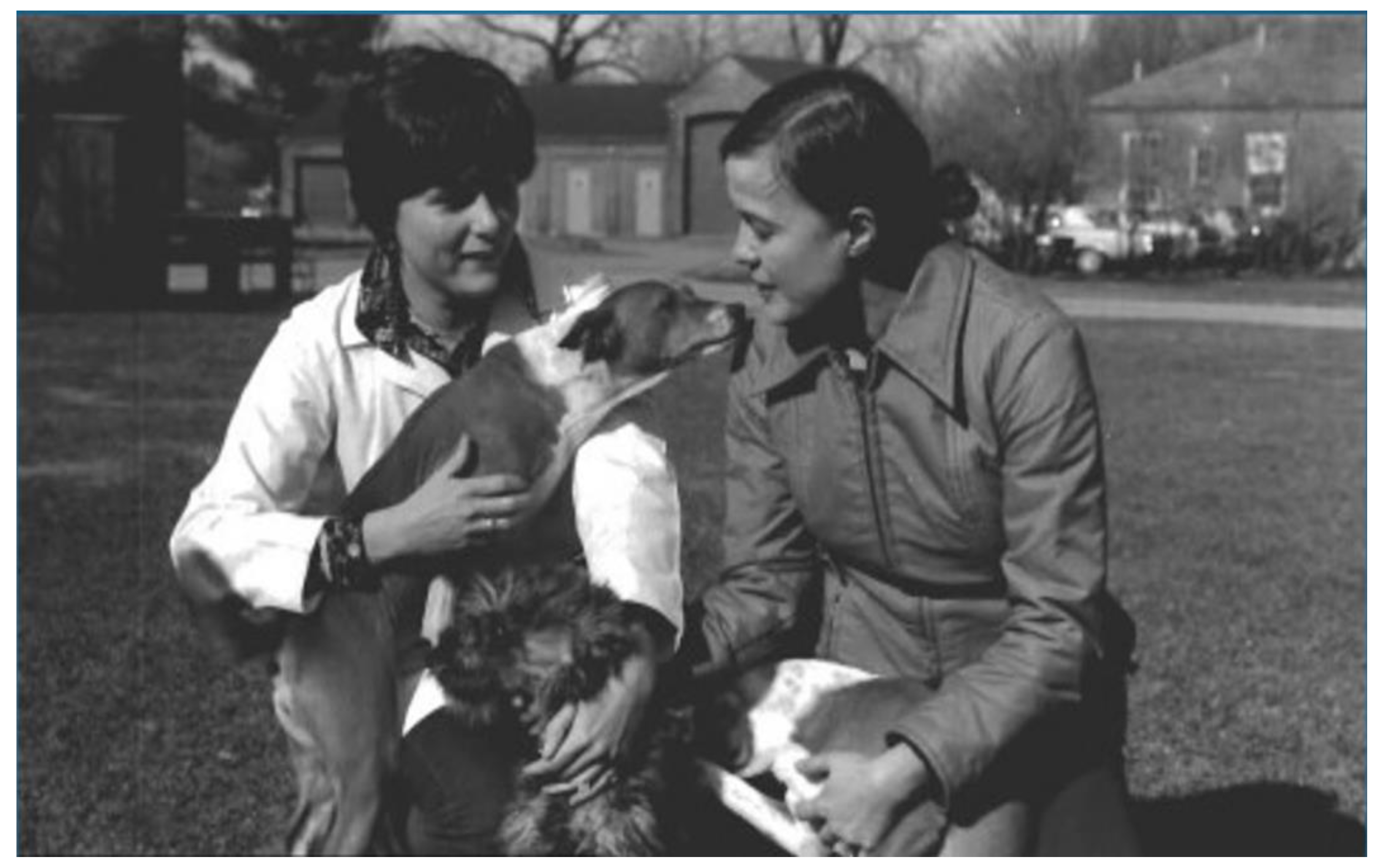
Figure 1.
The Author and technician, the late Joanne Kull, with hemophilic dogs at Griffin Laboratory, New York State Department of Health, 1975 [39].
Figure 1.
The Author and technician, the late Joanne Kull, with hemophilic dogs at Griffin Laboratory, New York State Department of Health, 1975 [39].


Table 1.
Timeline of animal genome sequencing [1].
Table 1.
Timeline of animal genome sequencing [1].
| Species | Date Sequence Published |
|---|---|
| Human | 2001 |
| Mouse | 2002 |
| Rat | 2004 |
| Chicken | 2004 |
| Non-Human Primate | |
| Chimpanzee | 2005 |
| Rhesus macaque | 2007 |
| Orangutan | 2011 |
| Dog | 2005 |
| Cat | 2007 |
| Cow | 2009 |
| Horse | 2009 |
| Turkey | 2010 |
| Pig | 2012 |
| Goat | 2017 |
| Time Period | Discoveries |
|---|---|
| 1700s | Long after Hippocrates, Aristotle, Celsus and Galen found freshly drawn blood to clot, blood clotting became linked to hemostasis (the cessation of bleeding). |
| 1800s | Thrombosis first recognized by Virchow; platelets are discovered by Bizzozero; familial bleeding tendency in males (hemophilia) is first recognized. |
| 1900s | Morawitz described the classical theory of blood coagulation. |
| 1930–1940s | Disputes arose between scientists about factors that form and dissolve clots; more clotting factor disorders are recognized in people (von Willebrand Disease) and dogs (hemophilia). |
| 1950s | von Willebrand disease identified in Poland-China pigs; factor VII identified in dog plasma after coumarin therapy prolonged the blood clotting in vitro. |
| 1970–1980s | von Willebrand Disease described in German Shepherd Dogs imported to North America from Germany, and then in many dog breeds, cats, and rabbits; hemophilia described in cats and horses; factors I, IX, X, XI and XII deficiencies documented in dogs, cats, cattle, goats; and platelet defects described in dogs, rats, and mice. |
| 1990s–today | More of these bleeding disorders found in domestic and companion animals, including the documentation of familial pre-kallikrein and kallikrein deficiencies. |
Table 3.
Genome Wide Associations (GWAS) for heritable canine bleeding disorder traits.
| Bleeding Disorder | GWAS; Genes | Breeds Affected | References |
|---|---|---|---|
| Hemophilia A (Factor VIII Deficiency) | Boxer, single nucleotide change C to G at nucleotide 1412 (1412 C>G)in Exon 10, results in arginine to proline at amino acid 471 (P471R) in A2 domain German Shepherd Dog, single nucleotide change G to A at nucleotide 1643 (1643 G>A)in Exon 11, results in tyrosine to cysteine at amino acid 548 (C548Y) in A2 domain | Many, also mixed breeds, cats, horses | [19,21,29,39,43,56] |
| Hemophilia B (Christmas Disease; Factor IX Deficiency) | Missense mutation G to nucleotide 1477, glycine 379-glutamic acid Insertional mutation in line 1 of canine FIX gene Nucleoside deletion of transcription factor binding site of FIX gene promotor | Cairn Terrier, Hovawart, German Wired-Haired Pointer (Drathaar), 23 other breeds, and cats | [16,20,33,58,59,60,61,62,63] |
| von Willebrand Disease, Types 1, 2, 3 | Type 1, Doberman, homozygous 157-base-pair intragenic marker allele+ heterozygous 1 of 4 extragenic marker alleles Type 2, GSHP nucleotide variant at Exon 28 Type 3, single nucleotide deletion in Exon coding VWF prepeptide (Scottish Terrier), splice site mutation Intron 16 (Dutch Kooiker)VWFc.4937A>G A/A, G/G | Many, prevalent in Doberman Pinscher, Shetland Sheepdog, Scottish Terrier, Golden Retriever, Pembroke Welsh Corgi, Chesapeake Bay Retriever, German Short-Haired Pointer (GSHP), German Wire-Haired Pointer (Drathaar), ~ 50 other breeds, cats, Poland. China swine | [76,77,78,81,82,83,84,88] |
| Factor VII Deficiency | Missense G96E mutation at Exon 5. Glycine 26 to Glutamic acid, 31% frequency in breed | Beagle, more than 14 other breeds | [89] |
| Factor X Deficiency (Stuart–Prower Disease) | Homozygous deletion of factor X gene(s) is lethal | American Cocker Spaniel, Jack Russell Terrier | [91] |
| Factor XI Deficiency | Kerry Blue, mutation of F11 gene, homozygotes affected, 90 bp insertion, Chr16:44477343-44477344, 10 bp duplication (dup GCACAAAGCT) Chr:44477344-44477353 | English Springer Spaniel, Kerry Blue Terrier, and Holstein cattle | [95] |
| Factor XII Deficiency (Hageman Trait) | Cats, novel mutation (c.1631 G >C) at Exon 13 of feline F12 gene, results in amino acid change (p.GS54A) | Miniature Poodle, cats, reptiles, marine mammals, birds | [96,97] |
| Prekallikrein Deficiency | G to A transversion at Exon 8 | Shih Tzu. American Hairless Terrier, others, and Belgian horse | [99] |
| Thrombasthenia (Glanzmann’s Disease); Bernard-Soulier Syndrome | Otterhounds, single nucleotide change at G1193 (1000) at Exon 12 of gene encoding for glycoprotein GPIIb, substitution of histidine for aspartic acid at 398 (367) of calcium -binding domain of GPIIb Single ITGA2B gene mutation on chromosome 9, chr9:19054488-19054488: G>C American Cockers, single glycoprotein 9 (GP9) deletion at Exon coding on chromosome 20 Great Pyrenees, 14-base insertion in Exon 13 and a splicing defect of Intron 13 Deletion of P2Y12 in Greater Swiss Mountain Dog and Bichon Frise | Otterhounds American Cocker Spaniel, Greater Swiss Mountain Dog (GSMD), Great Pyrenees, Bichon Frise | [113,114,121,123,124] |
| Thrombopathia | RASGRP-1; chr18:52417313-52417315: 3 bp deletion (del TCT) Autosomal recessive procoagulant deficiency at canine chromosome 27 | Basset Hound, Spitz, and cats, Simmental cattle, Greater Swiss Mountain Dog, German Shepherd Dog, Fawn-Hooded (FHwjd) rat | [103,106,108,111,112,115,117,121,122,124,125] |
| Thrombocytopenia | Associated with Hashimoto’s lymphocytic thyroiditis (3-5 genes of major histocompatibility complex, MHC, as in humans) | American Cocker Spaniel, Old English Sheepdog, Standard Poodle, Vizsla, Weimaraner, Akita, Samoyed, Shih Tzu, Long -Haired Dachshund, Kerry Blue Terrier, other white/fawn and dilute-color breeds and hybrids | [118,120] |
| Macrothrombocytopenia | Norfolk Terrier, Cairn Terrier, Chihuahua, Danish-Swedish Farm Dog, Kritikos Lagonikos, Wesr Highland White Terrier, Parson Russell Terrier, Marenma and Abruzees Sheepdog | [124] |
Disclaimer/Publisher’s Note: The statements, opinions and data contained in all publications are solely those of the individual author(s) and contributor(s) and not of MDPI and/or the editor(s). MDPI and/or the editor(s) disclaim responsibility for any injury to people or property resulting from any ideas, methods, instructions or products referred to in the content. |
© 2022 by the author. Licensee MDPI, Basel, Switzerland. This article is an open access article distributed under the terms and conditions of the Creative Commons Attribution (CC BY) license (https://creativecommons.org/licenses/by/4.0/).
Share and Cite
MDPI and ACS Style
Dodds, W.J. One Health: Animal Models of Heritable Human Bleeding Diseases. Animals 2023, 13, 87. https://doi.org/10.3390/ani13010087
AMA Style
Dodds WJ. One Health: Animal Models of Heritable Human Bleeding Diseases. Animals. 2023; 13(1):87. https://doi.org/10.3390/ani13010087
Chicago/Turabian StyleDodds, W. Jean. 2023. "One Health: Animal Models of Heritable Human Bleeding Diseases" Animals 13, no. 1: 87. https://doi.org/10.3390/ani13010087
Note that from the first issue of 2016, this journal uses article numbers instead of page numbers. See further details here.