Article

First Description of the Mitogenome Features of Neofoleyellides Genus (Nematoda: Onchocercidae) Isolated from a Wild Bird (Pyrrhocorax pyrrhocorax)

1
School of Public Health, Shantou University, Shantou 515063, China
2
College of Animal Science and Veterinary Medicine, Heilongjiang Bayi Agricultural University, Daqing 163319, China
3
Center for Biological Disaster Prevention and Control, National Forestry and Grassland Administration, Shenyang 110034, China
*
Author to whom correspondence should be addressed.
These authors contributed equally to this work.
Animals 2022, 12(20), 2854; https://doi.org/10.3390/ani12202854
Submission received: 4 October 2022 / Revised: 14 October 2022 / Accepted: 19 October 2022 / Published: 20 October 2022
(This article belongs to the Topic Host–Parasite Interactions)

Abstract

:

Simple Summary

Filarioidea, a superfamily of nematodes, presently includes 42 species divided into six genera, mainly in the family Onchocercidae, which have been reported to infect a wide range of hosts, including reptiles, birds, and mammals. Current limitations in molecular characterization methods and species identification are the main obstacles to a better understanding of the biology of Onchocercidae species, particularly in wildlife. Thus, the objective of the present study was to sequence and analyze the complete mt genome of Neofoleyellides sp. isolated from a wild bird (Pyrrhocorax pyrrhocorax) and to assess its phylogenetic position in the Onchocercidae family. The evaluated Neofoleyellides sp. mt genome was consistent with the molecular pattern of the Onchocercidae family: 36 subunits consisting of 12 PCGs, 2 rRNAs, and 22 tRNAs. Phylogenetic analyses based on the 18S rRNA gene, cox1 gene, and 12 PCGs showed consistent results, which strongly supported monophyly of the genus Neofoleyellides. These findings enriched the gene database and improved our knowledge of the molecular characteristics of the Onchocercidae family, which provide useful genetic markers to study the population genetics, molecular biology, and phylogenetics of these Onchocercidae nematodes.

Abstract

The Onchocercidae family is composed of more than 30 valid nematode species with notable zoonotic potential. Current limitations in molecular characterization methods and species identification are the main obstacles to a better understanding of the biology of Onchocercidae species, particularly in wildlife. This study describes for the first time the complete mitochondrial (mt) genome sequence of Neofoleyellides sp. isolated from a wild bird (Pyrrhocorax pyrrhocorax) and belonging to the Neofoleyellides genus (Nematoda: Onchocercidae). The mt genome of Neofoleyellides sp. (GenBank accession number: ON641583) was a typical circular DNA molecule of 13,628 bp in size with an AT content of 76.69%. The complete mt genome comprised 36 functional subunits, including 12 protein-coding genes (PCGs), 2 ribosomal RNA genes, and 22 transfer RNA genes. The most common start codon was ATT/ATG except for nad2 with TTG, and TAA was the termination codon for all protein-coding genes (PCGs). Phylogenetic analysis of the concatenated and aligned amino acid sequences of the 12 PCGs showed that the trees generated using different methods (Bayesian inference and maximum likelihood) with different partition schemes shared similar topologies. The isolated Neofoleyellides sp. was placed in the Onchocercidae family and formed a sister branch with the genera Onchocerca and Dirofilaria. The entire mt genome of Neofoleyellides sp. presented in this study could provide useful data for studying the population genetics and phylogenetic relationships of Onchocercidae species.

1. Introduction

Filarioidea, a superfamily of nematodes, presently includes 42 species divided into six genera, mainly in the family Onchocercidae, with a worldwide distribution [1]. Onchocercidae nematodes are transmitted by blood-sucking arthropods, and some genera such as Dirofilaria and Onchocerca have been linked to cases of blindness in humans [2]. Blood parasites have been the focus of various studies, and several reports have indicated that the microfilariae of nematodes of the Onchocercidae family can be found in the blood of birds [3,4]. Onchocercidae nematodes have been reported to infect a wide range of hosts, including reptiles, birds, and mammals [5,6]. Although some Adult worms of Onchocercidae have been detected in different tissues of bird hosts, there is limited information on wild birds [7]. Therefore, determining their presence in the final host is still challenging. Most of the previous research on Neofoleyellides has focused on the host of amphibians, and the phylogenetic relationships of amphibian onchocercids has been examined using molecular genetic methods (18S rDNA and cox1 gene sequences), in which all species parasitizing amphibians were recovered as the most distantly related genera of the family tree [5,8]. However, relationships between Foleyellides, Neofoleyellides, and Ochoterenella remained unresolved. In addition, there is limited phylogenetic information on adult Neofoleyellides worms in birds; additionally, the molecular detection of these parasites has not been performed.
Pyrrhocorax pyrrhocorax, a large corvid with a body length of 36–48 cm, mainly feeds on locusts, grasshoppers, mosquitoes and ants, which share a similar diet to amphibians; thus, it carries the same risk of Neofoleyellides infection. Unpredictable changes in ecological, biological, and social conditions caused by globalization have led to alterations in the prevalence, spread, and geographical range of infections. Therefore, the continuous surveillance of emerging infectious diseases is crucial, especially for emerging zoonotic parasites such as Onchocercidae nematodes, whose species diversity remains insufficiently explored [9].
Although the morphological and biological aspects of the parasite species were targeted for a phylogenetic reconstruction, the rapid and accurate identification of vector species is difficult due to the morphological homogeneity between species at certain life stages and the lack of necessary taxonomic expertise [10]. Molecular methods are of great significance for simplifying the detection of filarioids in hosts and species identification [2]. The strict maternal inheritance of the mitochondrial (mt) genome, lack of recombination, and rapid evolutionary rates have been widely used to determine species evolution, especially for classification at high taxonomic levels [11]. Furthermore, phylogenetic analysis based on molecular methods can be used to identify sister species (closest relatives in the phylogenetic tree) that share a common ancestor with the species of interest, and is useful to understand their role in the transmission cycle of pathogens [12].
The limited molecular characterization information of the avian Filarioidea species remains a major obstacle to a better understanding of the biology of these parasites, especially in wild animals. Thus, the objective of the present study was to sequence and analyze the complete mt genome of Neofoleyellides sp. isolated from a wild bird (Pyrrhocorax pyrrhocorax) and to assess its phylogenetic position in the Onchocercidae family. The findings could provide novel and useful genetic markers for further studies on species identification and molecular epidemiology.

2. Materials and Methods

2.1. Sample Collection, Total DNA Extraction and Molecular Detection

A total of three Adult Neofoleyellides worms were isolated from the intestinal tract of the wild bird Pyrrhocorax pyrrhocorax in Xining City, Qinghai Province, China (36°36′ N, 101°77′ E). The collected specimens were identified based on external morphological characteristics using dichotomous keys to represent the Filarioidea superfamily [9]. The collected worms were thoroughly washed with physiological saline three times and fixed in 75% (v/v) ethanol at −80 °C until use.
In order to identify the worm, total genomic DNA was extracted from all three adult worms using the TIANamp Genomic DNA Kit (Tiangen, Beijing, China) according to the manufacturer’s instructions. Molecular identification was conducted by polymerase chain reaction (PCR) targeting the 18S ribosomal RNA (rRNA) and mitochondrial cox1 gene, which has been previously described in several studies [9,13]. The primers are listed in Table S1. During amplification of both gene sequences, PCR was performed in the same reaction system: a 25 μL system including 12.5 μL of 2× Ex Taq Mix PCR buffer (Takara), 0.5 μm of each primer, 1.5 μL of DNA sample, and 10 μL of nuclease-free water.

2.2. Construction of the Genomic Library and Sequencing

For the genomic library construction, Total DNA was extracted from one adult worm, and was standardized at a concentration of 0.3 ng/L. The Illumina NovaSeq sequencing platform was used to adopt the whole-genome shotgun (WGS) strategy based on next-generation sequencing. After sequencing, a DNA library was constructed with an insert size of 400 bp using the paired-end method, which was conducted by Personalbio (Nanjing, China). The sequenced raw data were transferred to a computer workstation, and the analysis and genomic characterization steps were performed. The quality metrics of the obtained data were assessed using FastQC v. 0.11.9 software (available online: https://www.bioinformatics.babraham.ac.uk/projects/fastqc/ (accessed on 1 August 2019)), and Trim Galore v. 0.6.5 was used to remove adapter sequences (available online: https://www.bioinformatics.babraham.ac.uk/projects/trim_galore/ (accessed on 19 November 2019)). Finally, further quality checks and data validation were performed using FastQC.

2.3. Genomic Assembly

The A5 miseq v20150522 and SPAdes v3.9.0 softwares were used to construct contig and scaffold sequences, respectively, which were examined using pre-processed high-quality second-generation sequencing data for each species [14,15]. Furthermore, collinearity analysis was conducted using the Mummer v3.1 software to fill gaps among the contigs [16], and the corrected mt sequence was obtained using the Pilon v1.18 software [17]. Finally, the assembled mt genome sequence was compared with reference Onchocercidae family sequences (Onchocerca lupi, GenBank ID: NC056960; Wuchereria bancrofti, GenBank ID: AP017705) and manually validated.

2.4. Annotation and Bioinformatic Analysis

The complete mt genome sequence was functionally annotated using the MITOS web server (http://mitos.bioinf.uni-leipzig.de/index.py (accessed on 16 June 2017)) [18], and the secondary structures of transfer RNAs (tRNAs) were identified using tRNAscan-SE (http://lowelab.ucsc.edu/tRNAscan-SE (accessed on 4 May 2021)). An online open reading frame finder (https://www.ncbi.nlm.nih.gov/orffinder/ (accessed on 4 May 2021)) was used to analyze and translate protein-coding genes (PCGs).
DNAStar (v. 5.0) was used to calculate the A+T and G+C contents of each gene, and the AT skew and GC skew values were calculated using the formulas AT skew = (A − T)/(A + T) and GC skew = (G − C)/(G + C) for the coding genes [19]. Synonymous codon usage bias in PCGs was analyzed using CodonW [20], and relative synonymous codon usage (RSCU) values were determined. Sliding window analysis and pairwise comparisons of the proportions of non-synonymous (dN) and synonymous (dS) substitutions (dN/dS) among the obtained sequences were performed using DnaSP (v. 6) [21]. Sliding window analysis was used to estimate the nucleotide diversity (π) for every 200 bp (in 25 bp overlapping steps), and nucleotide diversity was plotted according to the midpoint position.

2.5. Phylogenetic Analysis

The concatenated amino acid sequences of the complete Neofoleyellides sp. mt genome were aligned with the corresponding amino acid sequences of 12 Filarioidea species available in GenBank, using Gongylonema pulchrum (NC026687) as outgroup. All 12 PCG sequences obtained in this study were extracted together with those of other taxa available in the GenBank data repository, and the MAFFT software with the option (L-INS-I) was also employed to determine the gene boundaries [22]. Phylogenetic relationships between the analyzed species were determined using two methods: maximum likelihood (ML) and Bayesian inference (BI). MEGA X software was used for the ML method, and bootstrapping was performed with 1000 replicates. MrBayes 3.1 was used to construct the BI tree, and four independent Markov chain runs were performed for 1,000,000 metropolis-coupled MCMC generations, sampling a tree every 100 generations. The first 25% (2500) of the trees were omitted as burn-ins, and the remaining trees were used to calculate Bayesian posterior probabilities. Phylogenetic trees were visualized using FigTree (v. 1.42) [23].

3. Results and Discussion

3.1. Acquisition of 18S rDNA and Mitochondrial cox1 Genes

The specimen was identified as Neofoleyellides sp., confirmed by PCR-based sequencing of 18S rDNA and cox1 genes. The amplified 18S rDNA showed 96.9% of nucleotide identity with that of Neofoleyellides spp. (GenBank accession number: MW599275). Besides, the cox1 gene was identical for 98.67% to the sequence of Neofoleyellides sp. (GenBank accession number: MW774895). Phylogenetic analyses of the 18S rDNA and cox1 gene were placed in the same genus of Neofoleyellides (Figures S1 and S2). Filarial nematodes are widespread parasites that infect all classes of vertebrates except fish. Many of these are of socioeconomic and medical importance [1]. To date, only a few studies on the Neofoleyellides genus have focused on the phylogenetic relationships of amphibian onchocercids based on 18S rDNA and cox1 gene sequences, and there is a lack of information for birds [24]. Birds play an important role in the spread of diseases, and a timely and accurate identification of the pathogens they carry is particularly important for the prevention and control of unknown diseases. In this study, we reported for the first time the mitochondrial genome of Neofoleyellides sp. isolated from a wild bird Pyrrhocorax pyrrhocorax, which suggests that Neofoleyellides are not only parasitic on amphibians, but may have a wider host distribution.

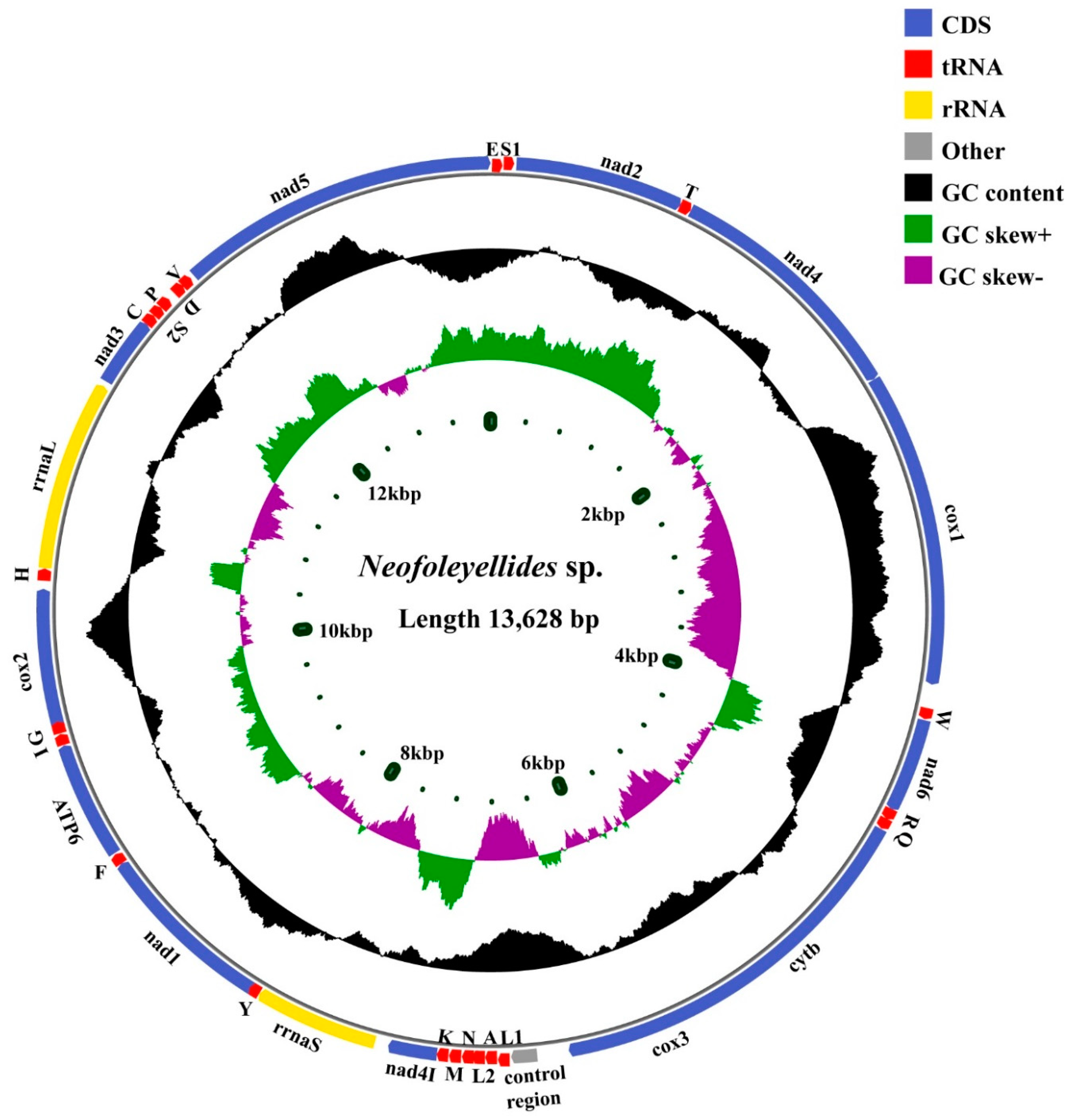
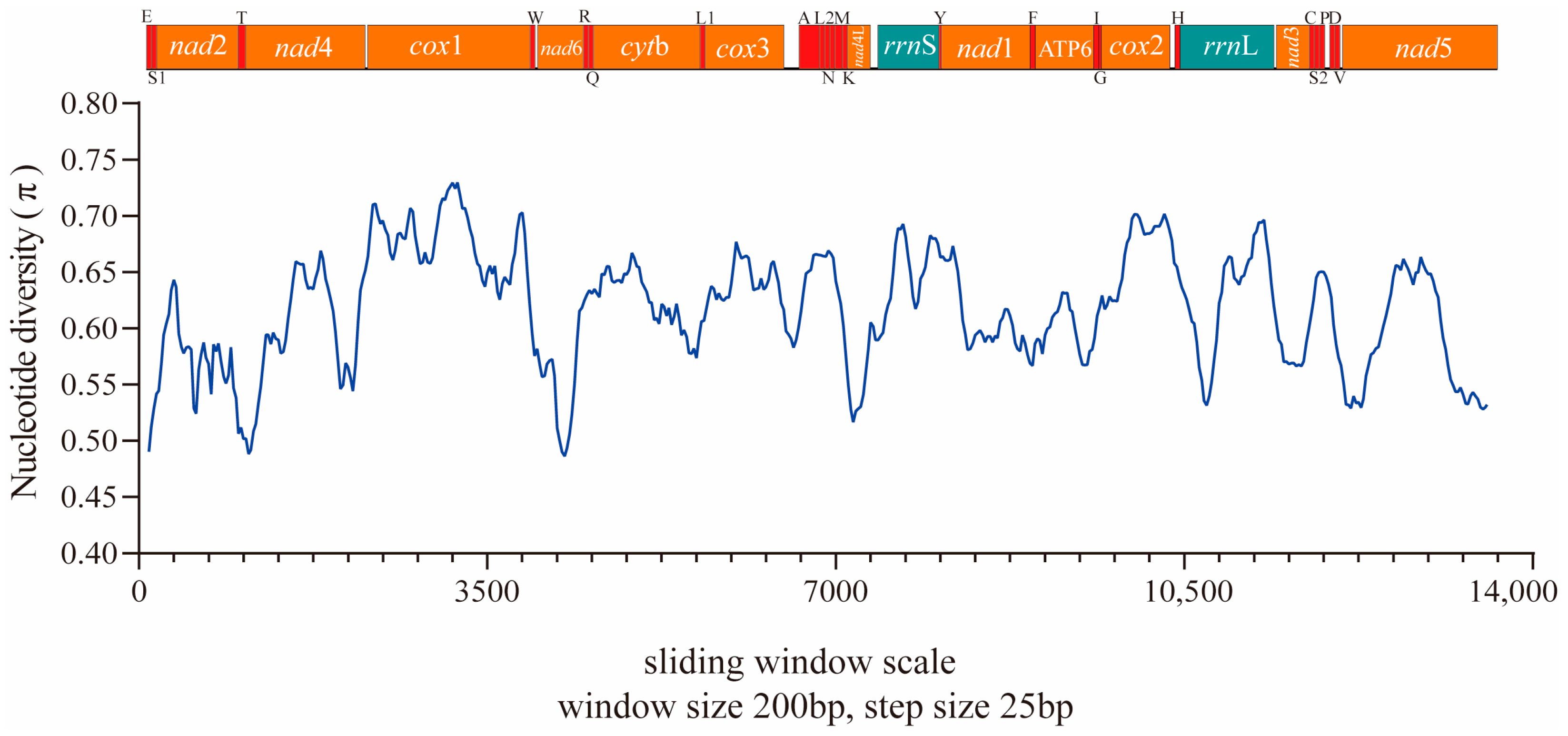
3.2. Mitogenome Organization and Composition

The complete mt genome of Neofoleyellides sp. (GenBank accession number: ON641583) was a typical circular DNA molecule, 13,628 bp in size and contained 36 functional and conserved subunits: 12 PCGs (including cox1-3, nad1-6, nad4L, atp6, and cytb), 22 tRNAs, and 2 rRNAs (rrnL and rrnS) (Figure 1). Similar to the genes of other Onchocercidae nematodes, all genes were organized along the N (forward) strands (Table 1). Taking into account the PCGs, tRNAs, and rRNAs, the mitogenome had an average AT content of 76.69%, which was similar to that of previously reported mt genomes including those of D. repens (76.06%), and B. malayi (75.46%) available in GenBank (NC029975, NC004298).
Among the 12 PCGs, nad3 had the highest A+T content (83.62%), followed by nad6 (83.33%), and atp6 (78.41%). Although cox1 had the lowest A+T content, it was still greater than 69.65%. A high AT content indicates a greater proportion of adenine and cytosine than thymine and guanine on the majority chain [25]. Analysis of the compositional asymmetry of the total mitogenome yielded negative values for AT skews (−0.41) and positive values for GC skews (0.50), which are consistent with the values for nematode parasites [26]. The AT skew values within the mt genome ranged from −0.22 (rrnL) to −0.59 (nad5), and the GC skew values ranged from 0.35 (rrnS) to 0.89 (nad4L) (Figure 2). The control region, which is known to initiate replication in vertebrates and invertebrates, was located between cox3 and trnA and varied in size. The control region (also known as the AT-rich region) is naturally rich in homopolymers and highly variable in terms of the length and rates of mutations, which may constitute an impairment to next-generation sequencing [27,28]. Alternative methods can be used to address this shortcoming; conventional PCR and Sanger sequencing allow target amplification, as demonstrated in similar studies [29].
The locations of the two rRNA genes (rrnS and rrnL) were identical in the mt genome, where rrnS was flanked by nad4L and trnY, and rrnL was flanked by trnH and nad3; the locations of these rRNA genes are similar to those in other filarial parasites [30]. The mt genome is unable to express all of the components required to assemble mt protein complexes. tRNAs play an essential role in amino acid transport, and the remaining proteins and RNAs involved in mt biogenesis are transported from the cytoplasm to the mitochondria [31]. The total length of the 22 tRNAs was 1222 bp, and the individual gene lengths varied from 51 bp to 60 bp (Table 1). The most common nucleotide mismatch was G-U, followed by U-U, which play an important role in maintaining the stability of the tRNA secondary structure [32].

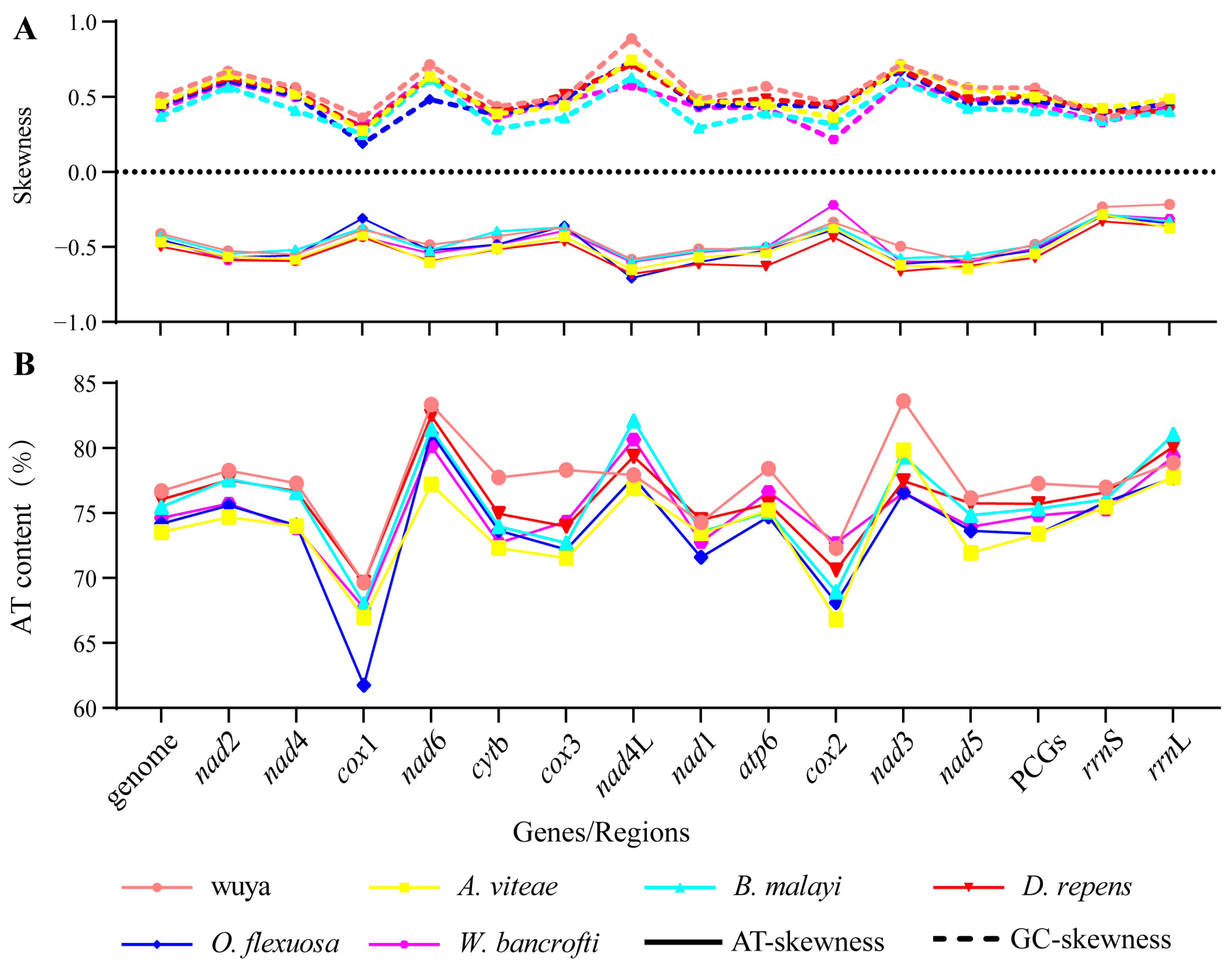
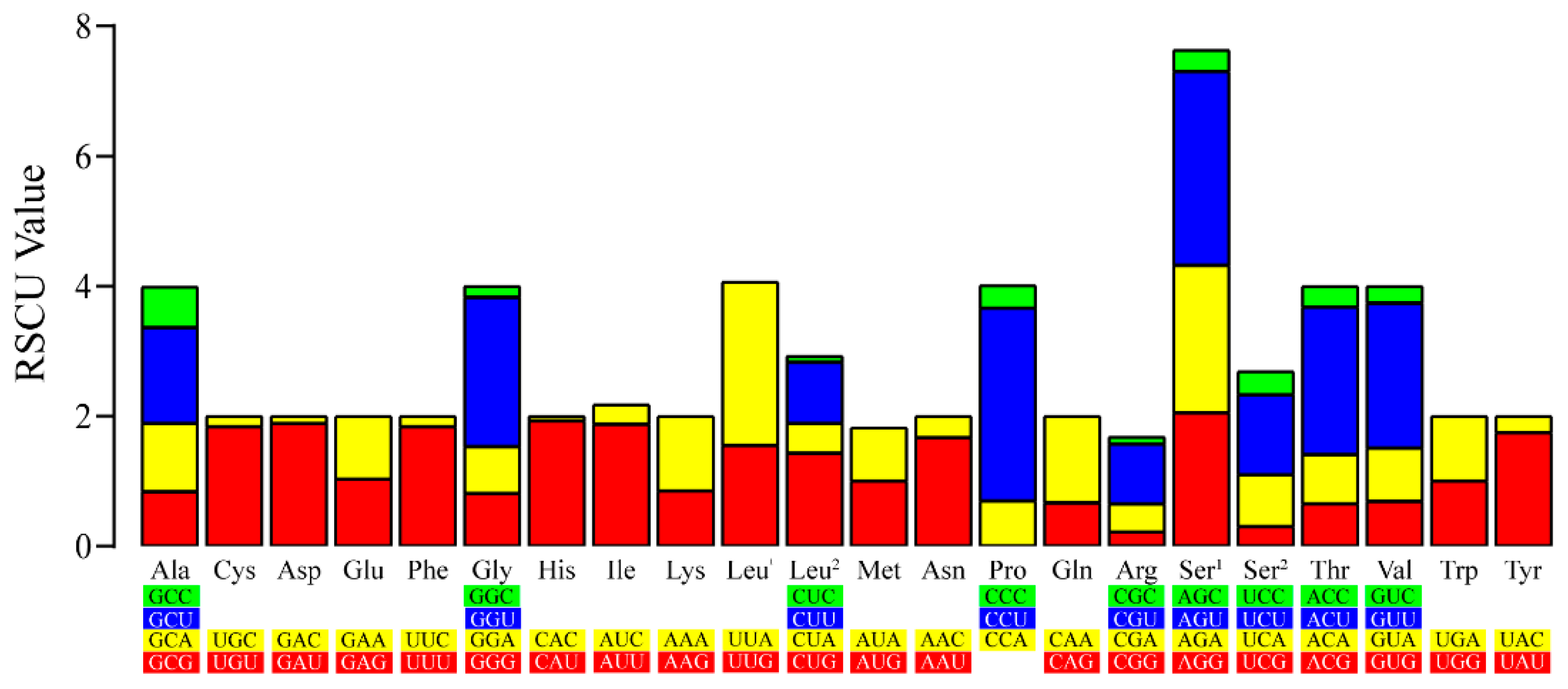
3.3. Characteristics of PCGs

The length of the PCG region in Neofoleyellides sp. was 10,358 bp, and the AT content was 77.27%. The relative lengths of PCGs were in the following order: cox1 > nad5 > nad4 > cytb > nad1 > nad2 > cox3 > cox2 > atp6 > nad6 > nad3 > nad4L. All 12 PCGs (except nad2 with TTG) had ATN as the start codon, with ATT being the most common (nad6, nad4L, cox2, nad4, nad4L, nad3, and nad5), followed by ATG (cox1, cytb, nad1, and nad6), all of which had TAA as the standard termination codon (Table 1). However, in most Onchocercidae family species, nad2 uses TTG as the start codon, which is considered a common feature across various organisms [33]. Termination codons for almost all PCGs are typically complete and incomplete stop codons (T or TA), which have also been observed in all metazoan mitogenomes and can be converted to the complete termination codon TAA through post-transcriptional modification [32].
The PCGs in the mt genome of Neofoleyellides sp. contained 3479 amino acids, including 176 strongly basic (+) amino acids (K, R), 156 strongly acidic (-) amino acids (D, E), 1852 hydrophobic amino acids (A, I, L, F, W, V), and 800 polar amino acids (N, C, Q, S, T, Y). RSCU analysis showed that nearly all codons were used in the mt genome, except for CCG (Pro). CCU (Pro) was the most common codon in the Neofoleyellides sp. mt genome with an RSCU value of 2.52, followed by UUA (Leu1) with an RSCU value of 2.23. Except for CCG (Pro), CAC (His) was the least common codon in the mt genome with an RSCU value of 0.07 (Figure 3). Synonymous codon usage bias may be attributed to various factors including gene function, recombination, mutation bias, GC composition, gene length, codon position, environmental stress, and population size [34]. Codons ending with A or U were more common than those ending with CG or GC, which is a common feature of some species [35].
To investigate the evolutionary pressure acting on different regions of the protein-coding mt genome, the corresponding sequences of each PCG in the studied species were paired based on the non-synonymous and synonymous substitution (dN/dS) ratio. The results indicated that different regions have evolved globally under the effect of negative (or purifying) pressure. The order of influence of evolutionary pressure according to the averages obtained was as follows: cox2 < cox1 < nad1 < cox3 < nad4 < nad4L < nad3 < atp6 < nad2 < nad6 < cytb < nad5 (Figure 4). The dN/dS ratio is a measure of the selective pressures acting on genes, indicating neutral selection (dN/dS = 1), negative or purifying selection (dN/dS < 1), and positive or diversifying selection (dN/dS > 1) [36,37]. Previous studies have reported that the dN/dS ratios of four protein-coding genes (nad2, nad3, nad5, and nad6) are > 1, suggesting that these genes have evolved under positive or diversifying selection [26]. However, the dN/dS ratio range in our study was large because of the lack of complete mitogenome data for the Neofoleyellides genus. In addition to dN/dS analysis, nucleotide diversity was compared between the mt sequences obtained in the present study and those of other members of the Onchocercidae family. Nucleotide diversity was calculated for a 200 bp window, plotted against the midpoint of this window, and moved in 25 bp steps across the alignment. The values of nucleotide diversity (π) of the evaluated sequences were in the range of 0.48–0.73 (Figure 5).

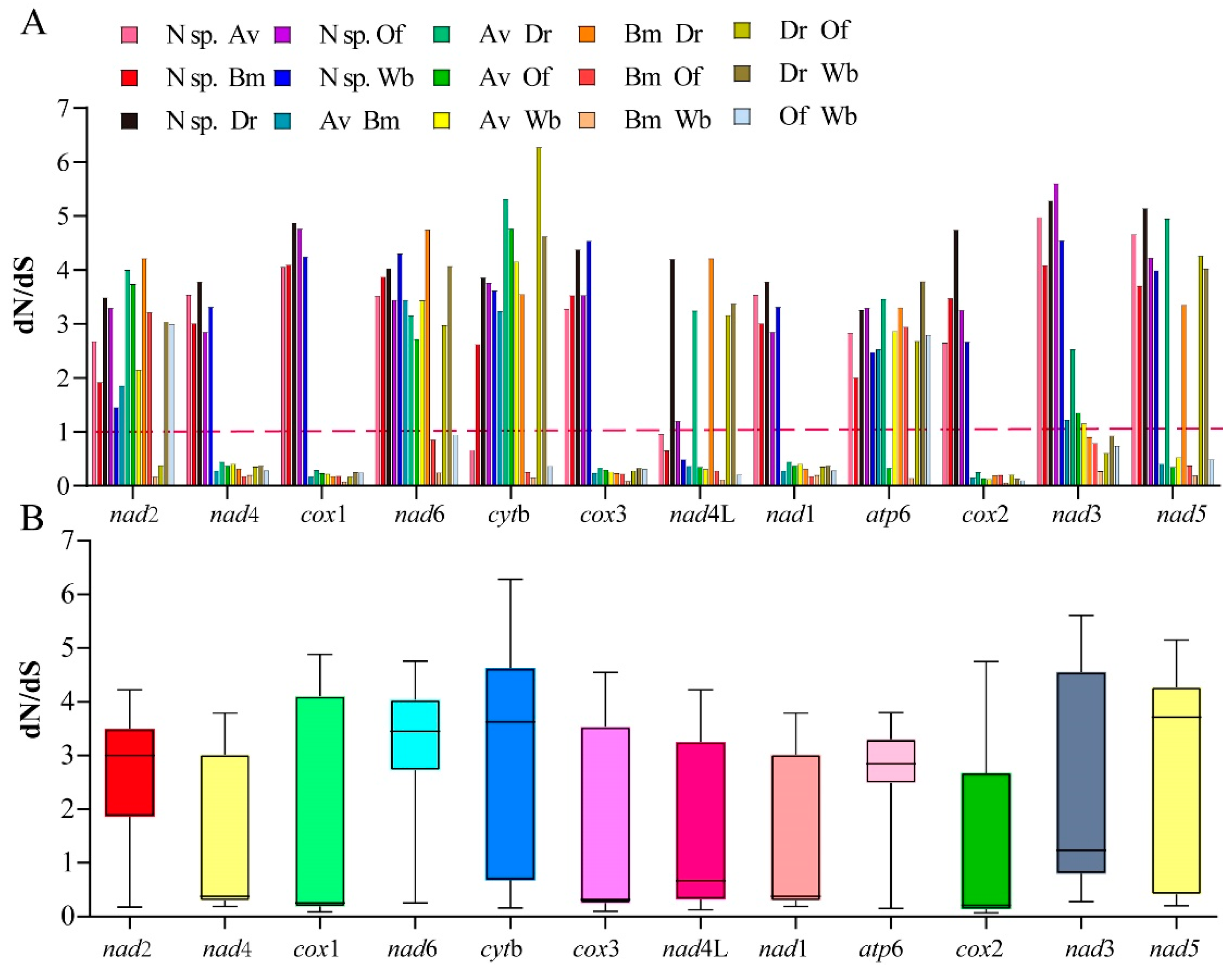
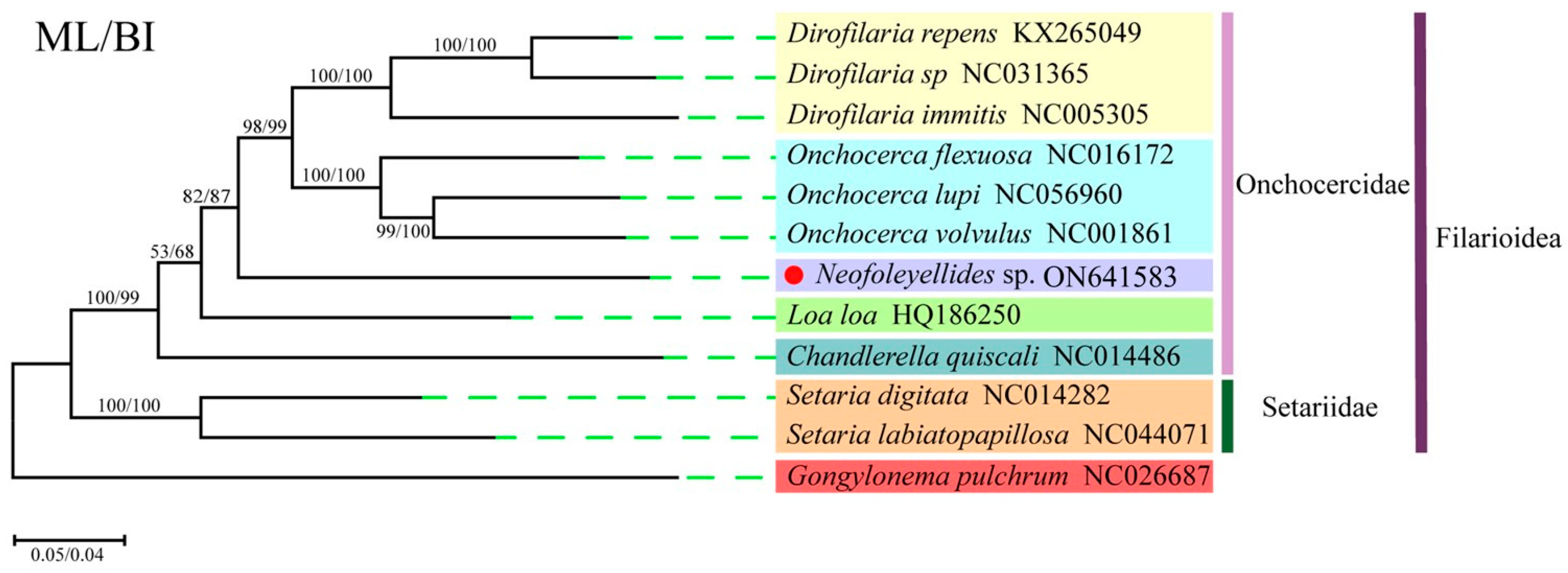
3.4. Phylogenetic Analyses

Phylogenetic analysis of the protein-coding regions was performed with 12 PCGs from 12 taxa (1 from this study and 11 from the GenBank database) using two analytical approaches (BI and ML) with G. pulchrum (Diptera: Spiruroidea) as the outgroup (Figure 6). In general, trees generated using different methods with different partition schemes share similar topologies. The Filarioidea superfamily had a topology with two well-supported clades corresponding to the families Setariidae and Onchocercidae. The Onchocercidae clade, which contained nine species, had five subclades representing the genera Dirofilaria, Onchocerca, Neofoleyellides, Loa, and Chandlerella, which is consistent with the results of previous studies [26,38]. In this study, the isolated Neofoleyellides sp. was placed in the Onchocercidae family, which formed a sister branch with the genera Onchocerca and Dirofilaria (posterior probability = 100). However, due to the lack of mt genome data for the Neofoleyellides genus, the adult worm formed an independent branch in the Onchocercidae family. Nevertheless, combined with 18S and cox1 gene evolution results, we determined that the parasites isolated from Pyrrhocorax pyrrhocorax belonged to the Neofoleyellides genus.
In previous studies, morphological and biological aspects were targeted for phylogenetic reconstruction; however, this method has drawbacks, especially in cases with diverse and ancient taxa [39]. Various studies have demonstrated that mtDNA sequences are valuable genetic markers for phylogenetic studies of members of Nematoda [39,40], and the advent of molecular tools has allowed a deeper investigation of the taxonomic relationships between organisms [41]. To date, only a few species have been confirmed to be infected by Neofoleyellides parasites, and more data are needed to understand their diversity and distribution, especially in wild animals [9]. However, the effect of the species of Neofoleyellides on host health has not been specifically investigated; only some pathological changes have been observed in frogs [24]. In addition, there is limited phylogenetic information on birds infected with Neofoleyellides parasites; thus, whether these parasites contribute to a risk of zoonosis is also worthy of further investigation. Therefore, mt characterization studies and the consequent inclusion of more filarial nematodes in public databases would allow more comprehensive phylogenetic reconstruction analyses and aid the elucidation of taxonomic relationships.

4. Conclusions

The present study is the first to determine the complete mt genome sequences of Neofoleyellides sp. isolated from a wild bird Pyrrhocorax pyrrhocorax. The evaluated Neofoleyellides sp. mt genome was consistent with the molecular pattern of the Onchocercidae family: 36 subunits consisting of 12 PCGs, 2 rRNAs, and 22 tRNAs. Phylogenetic analyses based on the 18S rRNA gene, cox1 gene, and 12 PCGs showed consistent results, which strongly supported monophyly of the genus Neofoleyellides. These findings enriched the gene database and improved our knowledge on the molecular characteristics of the Onchocercidae family, which provides useful genetic markers to study the population genetics, molecular biology, and phylogenetics of these Onchocercidae nematodes.

Supplementary Materials

The following supporting information can be downloaded at: https://www.mdpi.com/article/10.3390/ani12202854/s1. Figure S1. Phylogenetic reconstruction based on the 18S rRNA gene using the maximum likelihood method with Setaria yehi as the outgroup. The sequenced 18S gene of Neofoleyellides sp. was marked with a red circle. Figure S2. Phylogenetic reconstruction based on the cox1 gene using the maximum likelihood method with Setaria yehi as the outgroup. The sequenced cox1 gene of Neofoleyellides sp. was marked with a red circle. Table S1. Polymerase chain reaction primers used to verify the 18S and cox1 genes of Neofoleyellides sp.

Author Contributions

Data curation, X.M., F.W., L.X. and S.Q.; Formal analysis, M.Z.; Methodology, Y.X.; Resources, Q.C.; Software, Q.L.; Writing—original draft, T.W. and X.M.; Writing—review and editing, Q.C. All authors have read and agreed to the published version of the manuscript.

Funding

This work was supported by the National Natural Science Foundation of China (Grant No. 32072885) and the STU Scientific Research Foundation for Talents (Grant No. NTF21043).

Institutional Review Board Statement

Not applicable.

Informed Consent Statement

Not applicable.

Data Availability Statement

All data generated during this study are available as tables and figures included in this published article and its supplementary information files. The genome sequence data that support the findings of this study are available in GenBank of NCBI at (https://www.ncbi.nlm.nih.gov/ accessed on 18 October 2022) under the accession no. ON641583.

Acknowledgments

We are thankful for the financial support provided by the National Natural Science Foundation of China (Grant No. 32072885) and the STU Scientific Research Foundation for Talents (Grant No. NTF21043).

Conflicts of Interest

The authors declare no conflict of interest.

References

  1. Martin, C.; Bain, O.; Jouvenet, N.; Raharimanga, V.; Robert, V.; Rousset, D. First report of Litomosa spp. (Nematoda: Filarioidea) from Malagasy bats; review of the genus and relationships between species. Parasite 2006, 13, 3–10. [Google Scholar] [CrossRef] [PubMed] [Green Version]
  2. Bain, O.; Otranto, D.; Diniz, D.G.; dos Santos, J.N.; de Oliveira, N.P.; Frota de Almeida, I.N.; de Almeida, R.N.F.; de Almeida, L.N.F.; Dantas-Torres, F.; de Almeida Sobrinho, E.F. Human intraocular filariasis caused by Pelecitus sp. nematode, Brazil. Emerg. Infect. Dis. 2011, 17, 867–869. [Google Scholar] [CrossRef] [PubMed]
  3. Bennett, G.F.; Earlé, R.A.; Du Toit, H.; Huchzermeyer, F.W. A host-parasite catalogue of the haematozoa of the sub-Saharan birds. Onderstepoort. J. Vet. Res. 1992, 59, 1–73. [Google Scholar] [PubMed]
  4. Mat Udin, A.S.; Uni, S.; Zainuri, N.A.; Abdullah Halim, M.R.; Belabut, D.A. Morphological characteristics of microfilariae in blood smears of the common treeshrew Tupaia glis (Mammalia: Scandentia) in Gemas, Negeri Sembilan, Malaysia. Trop. Biomed. 2020, 37, 1152–1157. [Google Scholar]
  5. Lefoulon, E.; Bain, O.; Bourret, J.; Junker, K.; Guerrero, R.; Cañizales, I.; Kuzmin, Y.; Satoto, T.B.T.; Cardenas-Callirgos, J.M.; Lima, S.D.S.; et al. Shaking the tree: Multi-locus sequence typing usurps current onchocercid (filarial nematode) phylogeny. PLoS Negl. Trop. Dis. 2015, 9, e0004233. [Google Scholar] [CrossRef]
  6. Roe, C.C.; Yaglom, H.; Howard, A.; Urbanz, J.; Verocai, G.G.; Andrews, L.; Harrison, V.; Barnes, R.; Lyons, T.; Bowers, J.R.; et al. Coyotes as reservoirs for Onchocerca lupi, United States, 2015–2018. Emerg. Infect. Dis. 2020, 26, 2989–2993. [Google Scholar] [CrossRef]
  7. Bain, O.; Casiraghi, M.; Martin, C.; Uni, S. The Nematoda Filarioidea: Critical analysis linking molecular and traditional approaches. Parasite 2008, 15, 342–348. [Google Scholar] [CrossRef] [Green Version]
  8. Kuzmin, Y.; Netherlands, E.C.; du Preez, L.H.; Svitin, R. Two new species of Neofoleyellides (Nematoda: Onchocercidae) parasitising anuran amphibians in South Africa. Int. J. Parasitol. Parasites. Wildl. 2021, 14, 298–307. [Google Scholar] [CrossRef]
  9. Muñoz-García, C.I.; López-Díaz, O.; Osorio-Sarabia, D.; Martínez-Hernández, F.; Villalobos, G.; Isaak-Delgado, A.B.; Rendón-Franco, E.; Carreño-Cervantes, A.; Contreras-Patiño, D.R.; Berriatua, E.; et al. New insights into the clinico-histopathological and molecular features of Pelecitus (Filarioidea: Onchocercidae) from a raptor bird. Parasitol. Res. 2018, 117, 3319–3325. [Google Scholar] [CrossRef]
  10. Murugan, K.; Vadivalagan, C.; Karthika, P.; Panneerselvam, C.; Paulpandi, M.; Subramaniam, J.; Wei, H.; Aziz, A.T.; AlSalhi, M.; Devanesan, S.; et al. DNA barcoding and molecular evolution of mosquito vectors of medical and veterinary importance. Parasitol. Res. 2016, 115, 107–121. [Google Scholar] [CrossRef]
  11. Gao, Y.; Zhang, Y.; Yang, X.; Qiu, J.-H.; Duan, H.; Xu, W.-W.; Chang, Q.-C.; Wang, C.-R. Mitochondrial DNA evidence supports the hypothesis that Triodontophorus species belong to Cyathostominae. Front. Microbiol. 2017, 8, 1444. [Google Scholar] [CrossRef] [PubMed]
  12. Shragai, T.; Tesla, B.; Murdock, C.; Harrington, L.C. Zika and chikungunya: Mosquito-borne viruses in a changing world. Ann. N. Y. Acad. Sci. 2017, 1399, 61–77. [Google Scholar] [CrossRef] [PubMed] [Green Version]
  13. Binkienė, R.; Chagas, C.R.F.; Bernotienė, R.; Valkiūnas, G. Molecular and morphological characterization of three new species of avian Onchocercidae (Nematoda) with emphasis on circulating microfilariae. Parasit. Vectors. 2021, 14, 137. [Google Scholar] [CrossRef] [PubMed]
  14. Bankevich, A.; Nurk, S.; Antipov, D.; Gurevich, A.A.; Dvorkin, M.; Kulikov, A.S.; Lesin, V.M.; Nikolenko, S.I.; Pham, S.; Prjibelski, A.D.; et al. SPAdes: A new genome assembly algorithm and its applications to single-cell sequencing. J. Comput. Biol. 2012, 19, 455–477. [Google Scholar] [CrossRef] [Green Version]
  15. Coil, D.; Jospin, G.; Darling, A.E. A5-miseq: An updated pipeline to assemble microbial genomes from Illumina MiSeq data. Bioinformatics 2015, 31, 587–589. [Google Scholar] [CrossRef] [Green Version]
  16. Kurtz, S.; Phillippy, A.; Delcher, A.L.; Smoot, M.; Shumway, M.; Antonescu, C.; Salzberg, S.L. Versatile and open software for comparing large genomes. Genome. Biol. 2004, 5, R12. [Google Scholar] [CrossRef] [Green Version]
  17. Walker, B.J.; Abeel, T.; Shea, T.; Priest, M.; Abouelliel, A.; Sakthikumar, S.; Cuomo, C.A.; Zeng, Q.; Wortman, J.; Young, S.K.; et al. Pilon: An integrated tool for comprehensive microbial variant detection and genome assembly improvement. PLoS ONE 2014, 9, e112963. [Google Scholar] [CrossRef]
  18. Bernt, M.; Donath, A.; Jühling, F.; Externbrink, F.; Florentz, C.; Fritzsch, G.; Pütz, J.; Middendorf, M.; Stadler, P.F. MITOS: Improved de novo metazoan mitochondrial genome annotation. J. Mol. Phylogenetics Evol. 2013, 69, 313–319. [Google Scholar] [CrossRef]
  19. Burland, T.G. DNASTAR’s Lasergene sequence analysis software. J. Methods Mol. Biol. 2000, 132, 71–91. [Google Scholar]
  20. Do Nascimento, B.L.S.; Da Silva, F.S.; Nunes-Neto, J.P.; De Almeida Medeiros, D.B.; Cruz, A.C.R.; Da Silva, S.P.; da Silva e Silva, L.H.; de Oliveira Monteiro, H.A.; Dias, D.D.; Vieira, D.B.R.; et al. First description of the mitogenome and phylogeny of Culicinae species from the Amazon region. Genes 2021, 12, 1983. [Google Scholar] [CrossRef]
  21. Librado, P.; Rozas, J. DnaSP v5: A software for comprehensive analysis of DNA polymorphism data. Bioinformatics 2009, 25, 1451–1452. [Google Scholar] [CrossRef] [PubMed] [Green Version]
  22. Katoh, K.; Standley, D.M. MAFFT multiple sequence alignment software version 7: Improvements in performance and usability. Mol. Biol. Evol. 2013, 30, 772–780. [Google Scholar] [CrossRef] [PubMed]
  23. Chen, Y.; Chen, S.; Kang, J.; Fang, H.; Dao, H.; Guo, W.; Lai, C.; Lai, M.; Fan, J.; Fu, L.; et al. Evolving molecular epidemiological profile of human immunodeficiency virus 1 in the southwest border of China. PLoS ONE 2014, 9, e10757824. [Google Scholar] [CrossRef] [Green Version]
  24. Netherlands, E.C.; Svitin, R.; Cook, C.A.; Smit, N.J.; Brendonck, L.; Vanhove, M.; Du Preez, L.H. Neofoleyellides boerewors n. gen. n. sp. (Nematoda: Onchocercidae) parasitising common toads and mosquito vectors: Morphology, life history, experimental transmission and host-vector interaction in situ. Int. J. Parasitol. 2020, 50, 177–194. [Google Scholar] [CrossRef]
  25. Da Silva, A.F.; Machado, L.C.; De Paula, M.B.; Vieira, C.J.D.S.P.; Bronzoni, R.V.D.M.; Santos, M.A.V.D.M.; Wallau, G.L. Culicidae evolutionary history focusing on the culicinae subfamily based on mitochondrial phylogenomics. Sci. Rep. 2020, 10, 18823. [Google Scholar] [CrossRef]
  26. Liu, G.-H.; Gasser, R.B.; Otranto, D.; Xu, M.-J.; Shen, J.-L.; Mohandas, N.; Zhou, D.-H.; Zhu, X.-Q. Mitochondrial genome of the eyeworm, Thelazia callipaeda (Nematoda: Spirurida), as the first representative from the family Thelaziidae. PLoS Negl. Trop. Dis. 2013, 7, e2029. [Google Scholar] [CrossRef] [Green Version]
  27. Hardy, C.M.; Court, L.N.; Morgan, M.J.; Webb, C.E. The complete mitochondrial DNA genomes for two lineages of Aedes notoscriptus (Diptera: Culicidae). Mitochondrial. DNA A DNA Mapp. Seq. Anal. 2016, 27, 2024–2025. [Google Scholar]
  28. Matthews, B.J.; Dudchenko, O.; Kingan, S.B.; Koren, S.; Antoshechkin, I.; Crawford, J.E.; Glassford, W.J.; Herre, M.; Redmond, S.N.; Rose, N.H.; et al. Improved reference genome of Aedes aegypti informs arbovirus vector control. Nature 2018, 563, 501–507. [Google Scholar] [CrossRef]
  29. Bang, W.J.; Won, M.H.; Cho, S.T.; Ryu, J.; Choi, K.S. A multiplex PCR assay for six Aedini species, including Aedes albopictus. Parasit. Vectors 2021, 14, 380. [Google Scholar] [CrossRef]
  30. Yatawara, L.; Wickramasinghe, S.; Rajapakse, R.P.; Agatsuma, T. The complete mitochondrial genome of Setaria digitata (Nematoda: Filarioidea): Mitochondrial gene content, arrangement and composition compared with other nematodes. Mol. Biochem. Parasitol. 2010, 173, 32–38. [Google Scholar] [CrossRef]
  31. Luo, Q.-C.; Hao, Y.-J.; Meng, F.; Li, T.-J.; Ding, Y.-R.; Hua, Y.-Q.; Bin Chen, B. The mitochondrial genomes of Culex tritaeniorhynchus and Culex pipiens pallens (Diptera: Culicidae) and comparison analysis with two other Culex species. Parasit. Vectors. 2016, 9, 406. [Google Scholar] [CrossRef] [PubMed] [Green Version]
  32. Roe, C.C.; Urbanz, J.; Andrews, L.; Verocai, G.G.; Engelthaler, D.M.; Hepp, C.M.; Sahl, J.W. Complete mitochondrial genome of Onchocerca lupi (Nematoda, Onchocercidae). Mitochondrial. DNA B Resour. 2021, 6, 2572–2574. [Google Scholar] [CrossRef] [PubMed]
  33. Beckenbach, A.T.; Stewart, J.B. Insect mitochondrial genomics 3: The complete mitochondrial genome sequences of representatives from two neuropteroid orders: A dobsonfly (order Megaloptera) and a giant lacewing and an owlfly (order Neuroptera). Genome 2009, 52, 31–38. [Google Scholar] [CrossRef] [PubMed]
  34. Behura, S.K.; Severson, D.W. Codon usage bias: Causative factors, quantification methods and genome-wide patterns: With emphasis on insect genomes. Biol. Rev. Camb. Philos. Soc. 2013, 88, 49–61. [Google Scholar] [CrossRef] [PubMed]
  35. Hao, Y.-J.; Zou, Y.-L.; Ding, Y.-R.; Xu, W.-Y.; Yan, Z.-T.; Li, X.-D.; Fu, W.-B.; Li, T.-J.; Chen, B. Complete mitochondrial genomes of Anopheles stephensi and An. dirus and comparative evolutionary mitochondriomics of 50 mosquitoes. Sci. Rep. 2017, 7, 7666. [Google Scholar] [CrossRef]
  36. Meganathan, P.R.; Dubey, B.; Batzer, M.A.; Ray, D.A.; Haque, I. Complete mitochondrial genome sequences of three Crocodylus species and their comparison within the Order Crocodylia. Gene 2011, 478, 35–41. [Google Scholar] [CrossRef]
  37. Li, H.; Liu, H.; Shi, A.; Stys, P.; Zhou, X.; Cai, W. The complete mitochondrial genome and novel gene arrangement of the unique-headed bug Stenopirates sp. (Hemiptera: Enicocephalidae). PLoS ONE 2012, 7, e29419. [Google Scholar] [CrossRef] [Green Version]
  38. Traversa, D.; Costanzo, F.; Iorio, R.; Aroch, I.; Lavy, E. Mitochondrial cytochrome c oxidase subunit 1 (cox1) gene sequence of Spirocerca lupi (Nematoda, Spirurida): Avenues for potential implications. Vet. Parasitol. 2007, 146, 263–270. [Google Scholar] [CrossRef]
  39. Weeraratne, T.C.; Surendran, S.N.; Parakrama Karunaratne, S.H.P. DNA barcoding of morphologically characterized mosquitoes belonging to the subfamily Culicinae from Sri Lanka. Parasit. Vectors. 2018, 11, 266. [Google Scholar] [CrossRef] [Green Version]
  40. Small, S.T.; Tisch, D.J.; Zimmerman, P.A. Molecular epidemiology, phylogeny and evolution of the filarial nematode Wuchereria bancrofti. Infect. Genet. Evol. 2014, 28, 33–43. [Google Scholar] [CrossRef] [Green Version]
  41. Ferreira de Freitas, L.; Bartholomay, L.C. The Taxonomic History of Ochlerotatus Lynch Arribálzaga, 1891 (Diptera: Culicidae). Insects 2021, 12, 452. [Google Scholar] [CrossRef] [PubMed]
Figure 1. Structural representation of Neofoleyellides sp. mitogenome. From inside to outside, the first circle represents the length scale, the second circle represents the GC skew, the third circle represents the GC content, and the fourth circle represents the arrangement of PCGs, tRNAs, and rRNAs; each tRNA is identified by a unique letter abbreviation.
Animals 12 02854 g001
Figure 2. AT/GC contents and AT/GC skews of the investigated mitogenomes. (A) AT GC skews. (B) AT contents (%).
Animals 12 02854 g002
Figure 3. Relative synonymous codon usage (RSCU) values of the obtained mitogenomes. RSCU values are represented on the y-axis, and codons for the amino acids of the Onchocercidae family are represented on the x-axis.
Animals 12 02854 g003
Figure 4. Ratio of the rates of non-synonymous (dN) to synonymous (dS) nucleotide substitutions (dN/dS). (A) Bar graph of the pairwise ratios of dN/dS for each of the mitochondrial subunits of the investigated species. (B) Box chart illustrating the average pairwise ratios of dN/dS for each of the mitochondrial subunits of the investigated species. The dN/dS ratios are plotted on the y-axis, and the PCGs are plotted on the x-axis.
Animals 12 02854 g004
Figure 5. Nucleotide diversity (π) of the mitogenomes obtained in this study. The blue line indicate nucleotide diversity values based on the evaluation of sequences from B. malayi, D. repens, O. flexuosa, and A. viteae. The π values (plotted on the y-axis) were calculated in a 200 bp sliding window with 25 bp steps. The lengths of the aligned sequences of the two evaluated groups are plotted on the x-axis. The limits of each gene are indicated above the graph. Vertical red bars represent tRNAs, orange rectangles represent PCGs, and cyan bars represent rrnL and rrnS.
Animals 12 02854 g005
Figure 6. Phylogenetic reconstruction based on the concatenated amino acid sequences of 12 PCGs of 12 mitogenomes analyzed using the maximum likelihood and Bayesian inference methods. The sequenced specie of Neofoleyellides sp. in this study are marked with a red circle.
Animals 12 02854 g006
Table 1. Mitochondrial genome organization of Neofoleyellides sp.
GenesStrandPosition and
Length (bp)
Initiation CodonStop CodonAnticodon
trnEN1–57 (57) TTC
trnS1N58–108 (51) TCT
nad2N108–944 (838)TTGTA(A)
trnTN944–999 (56) TGT
nad4N1001–2224 (1224)ATATAA
cox1N2226–3878 (1653)ATGTAG
trnWN3879–3933 (55) TCA
nad6N3939–4400 (462)ATTTAA
trnRN4399–4452 (54) ACG
trnQN4452–4505 (54) TTG
cytbN4509–5592 (1084)ATGT(AA)
trnL1N5592–5647 (56) TAG
cox3N5644–6426 (783)ATATAA
trnAN6777–6833 (57) TGC
trnL2N6836–6895 (60) TAA
trnNN6890–6946 (57) GTT
trnMN6951–7009 (59) CAT
trnKN7013–7068 (56) CTT
nad4LN7069–7308 (240)ATTTAA
rrnSN7310–7985 (676)ATATAA
trnYN7986–8039 (54) GTA
nad1N8042–8911 (870)TTGTAG
trnFN8911–8964 (54) GAA
atp6N8971–9549 (579)ATGTAA
trnIN9553–9607 (55) GAT
trnGN9612–9669 (58) TCC
cox2N9669–10,358 (690)ATTTAG
trnHN10,358–10,416 (59) GTG
rrnLN10,418–11,386 (969)ATTT(AA)
nad3N11,380–11,721 (342)ATTTAA
trnCN11,720–11,774(55) GCA
trnS2N11,776–11,826(51) TGA
trnPN11,828–11,882 (55) AGG
trnDN11,922–11,976 (55) GTC
trnVN11,976–12,029 (54) TAC
nad5N12,029–13,621 (1593)ATTTAA
Publisher’s Note: MDPI stays neutral with regard to jurisdictional claims in published maps and institutional affiliations.

