Article

Description and Molecular Characterization of Two Species of Avian Blood Parasites, with Remarks on Circadian Rhythms of Avian Haematozoa Infections

by
Carolina Romeiro Fernandes Chagas
*,
Rasa Binkienė
and
Gediminas Valkiūnas
Nature Research Centre, Akademijos 2, 08412 Vilnius, Lithuania
*
Author to whom correspondence should be addressed.
Animals 2021, 11(12), 3490; https://doi.org/10.3390/ani11123490
Submission received: 2 November 2021 / Revised: 2 December 2021 / Accepted: 2 December 2021 / Published: 7 December 2021
(This article belongs to the Special Issue Avian Blood Parasites)

Abstract

:

Simple Summary

The diversity of avian blood parasites is remarkable, and they are often found in co-infections, which is a challenge for wildlife parasitology research. Co-infections not only make parasite morphological and molecular identification difficult, but also might influence the infection dynamics and disease manifestation. This study investigated blood parasites infections in a Eurasian blackbird (Turdus merula) and a Song thrush (Turdus philomelos). A new Lankesterella species was found and described. The molecular characterization of this parasites as well as Splendidofilaria mavis, an avian filarioid nematode, was developed. As several blood infections were present in co-infection, we used this opportunity to investigate the daily changes in the parasite intensity seen in the blood of these birds. A peak of the Plasmodium matutinum parasitemia was seen during the daytime. Leucocytozoon spp. showed some parasitemia peaks close to the evening and night. Trypanosoma spp. and S. mavis parasitemia increased during the night. Data about daily variation of Haemoproteus, Lankesterella, and S. mavis parasites were obtained for the first time. No pattern in parasitemia dynamics was identified in Haemoproteus and Lankesterella infections.

Abstract

Avian blood parasites are remarkably diverse and frequently occur in co-infections, which predominate in wildlife. This makes wildlife pathogen research challenging, particularly if they belong to closely related groups, resulting in diagnostic problems and poor knowledge about such infections as well as the patterns of their co-occurrence and interactions. This is particularly true due to the periodicity (circadian rhythms) of parasitemia, which means that different parasitemia and parasite stages might be found throughout the day. We analysed blood samples from a Eurasian blackbird (Turdus merula) and a Song thrush (Turdus philomelos). This study aimed to describe a new avian Lankesterella species and molecularly characterize and redescribe Splendidofilaria mavis, a common avian filarioid nematode. Additionally, it was possible to investigate the circadian rhythms of the avian blood parasites belonging to Plasmodium, Haemoproteus, Leucocytozoon, and Trypanosoma, which occurred in co-infection in the same avian host individuals. Different circadian rhythms were seen in different parasites, with Plasmodium sp. peaks occurring at midday, Leucocytozoon spp. peaks mainly during the evening and night, and Trypanosoma spp. and microfilariae peaks at midnight. No periodicity was seen in Haemoproteus and Lankesterella species infections. The time of parasitemia peaks most likely coincides with the time of vectors’ activity, and this should be beneficial for transmission. Knowledge about the circadian rhythms is needed for better understanding patterns in host-parasite interactions and disease transmission.

1. Introduction

Avian blood parasites are an ecological group that is composed of protists and helminths which have been markedly unevenly studied [1]. For example, some haemosporidian parasites (Plasmodium and Haemoproteus) are extensively studied due to their close relationships to human malaria agents [2,3], while other avian blood pathogens such as Lankesterella and filarioid nematodes remain poorly investigated and insufficient knowledge is available even about basic stages of their life cycles [4,5]. One of the main challenges that is faced by researchers in wildlife parasitology is the presence of co-infections which are commonly observed in naturally infected wild birds [6,7,8,9] and even predominate in some bird populations [10]. As a result, both molecular diagnostics that use general primers [11,12] and morphological identifications might be remarkably challenging [13,14]. Moreover, blood parasite infections are markedly dynamic, resulting in variation of the parasitemia in the same host individuals not only in a long-term perspective but also during the same day [3,15,16,17].
The daily rhythms of biological activity, also known as circadian rhythms, were reported in numerous prokaryotic and eukaryotic organisms, including parasites. The periodicity of biological activity might trigger immunological responses and hormone levels that allow the organisms to anticipate changes in their external environment, resulting in fitness advantages [15,18,19,20]. This is a particularly sensitive issue for parasites which need not only to avoid the host immunological system, but also to increase their chances of being transmitted to the new hosts. The optimization of transmission is particularly important for vector-transmitted parasites whose survival needs to be adjusted for the accession of specific vectors at a particular stage of the pathogen life cycle when invasive stages are available [15,18,19,20]. For example, a clear circadian rhythm was observed in the Wuchereria bancrofti, a filarioid nematode of humans. This parasite is transmitted mainly by Culex mosquitoes which are active during the night when parasitemia of microfilariae increases as well [21]. Parasites with a direct life cycle of transmission also benefit from circadian rhythms. For example, the invasive stages (oocysts) of avian Isospora species were massively released in the environment only in the late afternoon. This minimized direct exposure to sunlight, reducing desiccation and destruction by the solar ultraviolet radiation of the oocysts and, consequently, contributed to their better survival [22].
Knowledge about parasite diversity and parasitic diseases is essential in a changing world not only regarding human and domestic animal health, but also for a better understanding of wildlife disease epidemiology and evolutionary patterns [23]. This is true concerning poorly investigated parasite circadian rhythms, which a better understanding might show new directions for the development of prophylactic measures for diseases control and prevention. This might be helpful to improve research on parasite biology, gene expression, experimental investigations, pharmacological studies, and finally contribute to the development of measures to prevent new life-threatening infections to humans. However, the knowledge about many groups of wildlife parasites remains sparse and focused mainly on parasite species that are important in human and veterinary medicine or are agents of zoonotic diseases. This study aimed to contribute with new knowledge about common, but neglected, avian blood parasites. The main tasks were (i) to describe a new species of Lankesterella infecting a Eurasian blackbird (Turdus merula), and (ii) molecularly characterize Splendidofilaria mavis, a filarioid nematode infecting the Eurasian blackbird and a Song thrush (Turdus philomelos). Because both studied avian hosts were co-infected with several species of blood parasites, we used this opportunity to investigate the circadian rhythms of these parasites as well as other pathogens that were found in co-infection and belong to Plasmodium, Haemoproteus, Leucocytozoon, and Trypanosoma genera.

2. Materials and Methods

2.1. Selection of Donor Birds and Examination of Blood Films

The study was conducted in Ventės Ragas Ornithological Station, Lithuania (55°20′28.1″ N, 21°11′25.3″ E) during May 2019. To select donors, wild birds were caught using funnel traps, mist nets, and ‘zigzag’ traps. About 50 µL of blood was taken by puncturing the vena ulnaris cutanea using heparinized capillary tubes. A few drops of blood were used to prepare blood films and a few drops of blood were stored in SET buffer (0.05 M tris, 0.15 M NaCl, 0.5 M EDTA, pH 8.0) for molecular analysis. The remaining blood was kept in the capillary tubes and processed using the buffy coat method [5]. Briefly, the blood samples were collected in heparinized capillary tubes, sealed with plasticine at one end, and centrifugated for 5 min. at 10,000 rpm. Next, the capillary tubes were placed above a glass slide and the buffy coat area was examined under low magnification (100×) in a microscope. Then, wet preparations were prepared by breaking the capillary tubes about 1 mm below the buffy coat layer. The buffy coat and plasma were transferred to a glass slide and screened using a microscope under 400× magnification.
The blood films were air-dried using a battery-operated fan, fixed by immersion in absolute methanol for 1 s, dried at room temperature, and stained with a 10% Giemsa solution for 1 h [3]. Microscopic examination of the blood films consisted of screening each slide for 15–20 min at low magnification (400×) and at least 100 fields were examined at high magnification (1000×) using an Olympus CX23 light microscope (Olympus, Tokyo, Japan). A total of two birds were selected for this study. These were: a Eurasian blackbird (Turdus merula) that was infected with Plasmodium, Haemoproteus, Leucocytozoon, Trypanosoma, Lankesterella and microfilaria of filarioid nematode, and a Song thrush (Turdus philomelos) that was infected with Haemoproteus, Leucocytozoon, and microfilaria of filarioid nematode.
Images of the haemosporidians, Trypanosoma, and Lankesterella parasites were collected using an Olympus BX41 light microscope that was equipped with an Olympus DP-12 digital camera and the Olympus DP-Soft v. 3.2 imaging software (Olympus, Tokyo, Japan). These images were used for illustration of the morphology and measurement of the described parasites. Statistical analyses were carried out using ‘R studio’ v.3.4.3. Voucher preparations of Plasmodium matutinum lineage pLINN1 (accession numbers NS49369 and NS49370), Haemoproteus minutus hTURDUS2 (NS49371 and NS49372), Leucocytozoon spp. lNEVE01 and lTURMER15 (NS49373 and NS49374), Lankesterella bivacuolata n. sp. LanTurd1 (NS49367 and NS49368), Trypanosoma avium (NS49375 and NS49376), Trypanosoma everetti group (NS49377 and NS49378), Haemoproteus homominutus unknown lineage (NS49379 and NS49380), and Leucocytozoon sp. lSTUR1 (NS49381 and NS49382) were deposited in Nature Research Centre.

2.2. Circadian Rhythm Investigation

To follow parasitemia changes during a day, the selected birds were kept in cages with water and food ad libitum, at room temperature (~22 °C), and with a natural light:dark photoperiod (about 17:7 h). The Eurasian blackbird was observed for five days and the Song thrush for four days. Blood was taken for microscopic and molecular analysis from both experimental birds every 6 h (6 h, 12 h, 18 h and 24 h) using the same methodology as described in Section 2.1. At the end of the observation period, both birds were euthanized and dissected; adult worms of filarioid nematodes were collected for species identification (see description below).
Parasitemia intensity was determined using different methods depending on the parasite genus, as shown in Table 1. The blood stages were classified according to the certain parasite life cycle during the analysis of the data for determination of circadian rhythms. In Plasmodium parasites these were young and mature meronts, and young and mature gametocytes; in Haemoproteus and Leucocytozoon, young and mature gametocytes were examined separately as well (see Section 3.1). In Trypanosoma (see Section 3.1), Lankesterella (see Section 3.2) and microfilariae (see Section 3.3) infections, only one blood stage was observed and analysed, i.e., trypomastigotes, sporozoites, and microfilariae, respectively.

2.3. Collection of Filarioid Worms

After the investigation of the circadian rhythm, the infected birds were euthanized and immediately dissected. The dissected organs and other tissues were placed in Petri dishes containing 0.9% saline solution and the parasites were recovered using dissecting needles and pipettes under a stereomicroscope (MБС-9). The subcutaneous tissues, joints of the leg and wings, brain, eyes, heart, connective tissues of trachea and oesophagus, lungs, air sacs, and body cavities for adult filarioid nematodes were examined according to Binkienė et al. [4]. Adult alive nematodes were examined under the microscope in 0.9% saline solution and then stored in 70% ethanol. For light-microscope examination, they were cleared with glycerine [28]. Blood from the liver and lungs was collected and stored in absolute ethanol for molecular analysis. Blood films from the liver and lungs were also prepared from these organs; these preparations were fixed, stained, and examined as the blood films (see description above). All blood films and preparations of nematodes were examined using an Olympus BX 51 light microscope equipped with differential interference contrast optics and a digital image analysis system (DeltaOptical DLTCam Viewer 3.7.8301). The identification of the parasite species was done using keys and species descriptions [29,30,31]. Voucher parasite specimens were deposited in the Nature Research Centre, Vilnius, Lithuania (accession numbers 1082–1106).

2.4. DNA Extraction, PCR and Molecular Identification of Parasites

Two methodologies were used for DNA extraction. For peripheral blood samples, the DNA was extracted using an ammonium acetate protocol [32]. For the blood samples that were obtained from the organs and adult nematodes, DNA extraction was done according to Stunžėnas et al. [33] with a slight modification according to Petkevičiūtė et al. [34]; mainly, the samples were well dried for 15 min on a sterile microscope glass slide before the DNA extraction. Polymerase chain reaction (PCR) protocols were applied for the determination of parasite species and/or their genetic lineages using general primers that were specific for each parasite group (Table 1). To control for false amplifications, all PCR reactions were run using one negative (ultra-pure water) and one positive (according to the targeted parasite) control.
The amplification success was determined using electrophoresis in a 2% agarose gel. Positive PCR products were precipitated and sequenced from both strands using the Big Dye Terminator V3.1 Cycle Sequencing Kit (Thermo Fisher Scientific, Vilnius, Lithuania) and ABI PRISM™ 3100 capillary sequencing robot (Applied Biosystems, Foster City, CA, USA). The obtained sequences were edited and aligned using BioEdit software [35] to create a consensus sequence. The presence of double peaks in electropherograms was considered as an indication of a co-infection with the parasite of target group. The obtained consensus sequences were aligned using BLAST (Basic Local Alignment Search Tool) and compared with sequences from MalAvi database (http://mbio-serv2.mbioekol.lu.se/Malavi/blast.html, accessed on 12 October 2021, for haemosporidian parasites) and GenBank database (http//www.ncbi.nlm.nih.gov/BLAST, accessed on 12 October 2021, for other blood parasites). For filarioid nematodes, the sequences that were obtained from the microfilaria and the adult worms were compared with each other to prove that they belonged to the same species. All detected parasite DNA sequences were deposited in GenBank: P. matutinum pLINN1 (OK646331), H. minutus hTURDUS2 (OK646332), H. asymmetricus hTUPHI01 (OK721103), Leucocytozoon sp. lNEVE01 (OK646333), Leucocytozoon sp. lTURMER15 (OK646334), Leucocytozoon sp. lSTUR1 (OK646335), L. bivacuolata n. sp. (OK644714), T. avium (OK605056), S. mavis 28S (OK644715-OK644718), and S. mavis coxI (OK631737-OK391740).

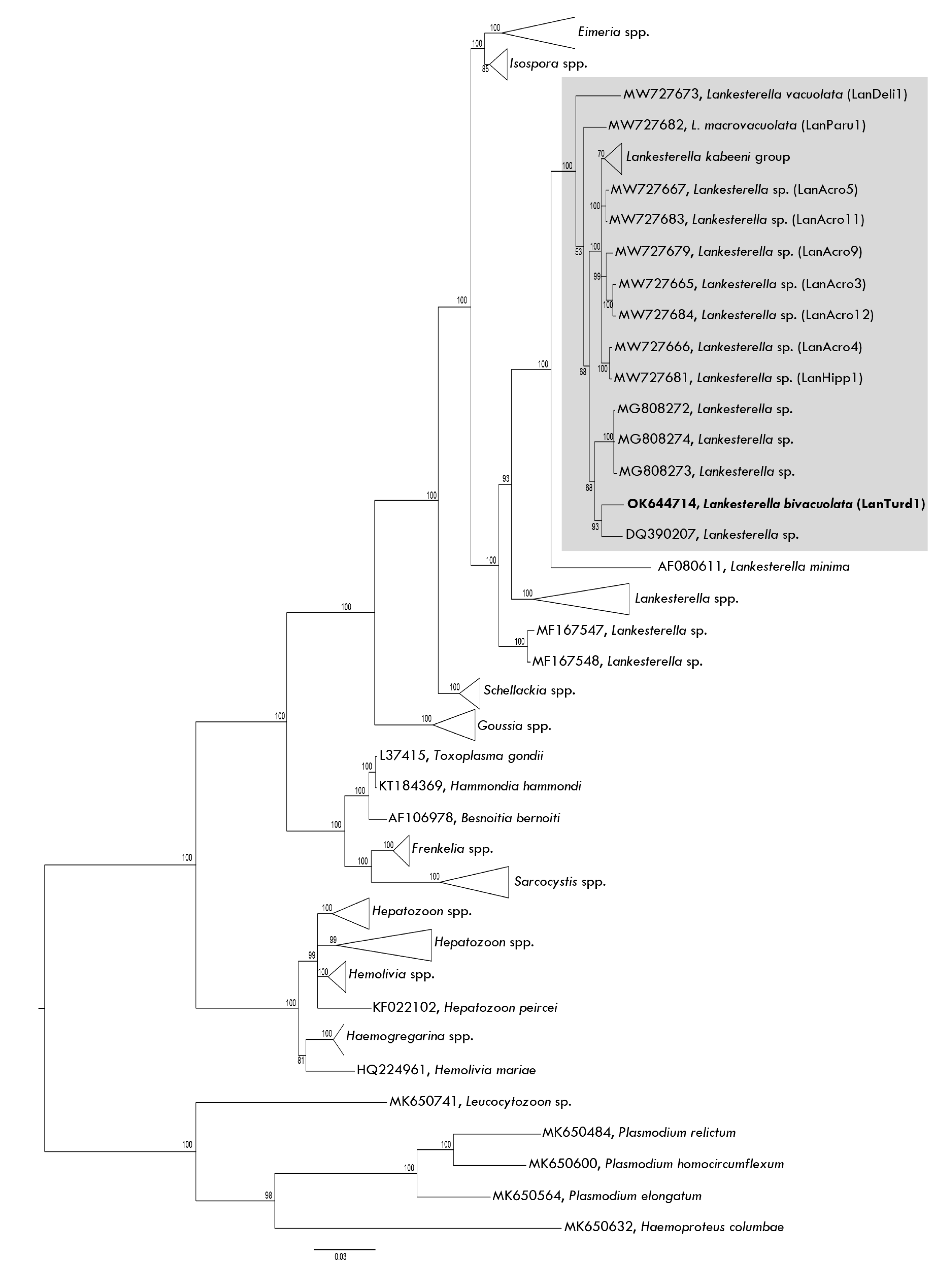
2.5. Plylogenetic Analysis

Phylogenetic analysis was done to determine the relationships of the new Lankesterella species and the newly molecularly characterized Splendidofilaria mavis with other Lankesterella parasites and filarioid nematodes, respectively.
A Bayesian phylogeny was constructed for Lankesterella parasites using 18S rRNA gene partial sequences. The alignment consisted of sequences from 12 closely related parasite genera, including the one that was obtained in this study. We used the same DNA sequences as described by Chagas et al. [26]. Sequences of haemosporidian parasites (Plasmodium, Haemoproteus, and Leucocytozoon species) were used as an outgroup.
A Bayesian phylogeny was constructed using S. mavis nuclear 28S gene partial sequence and software MrBayes version 3.2 [36]. Even though two genes were used for the identification of S. mavis (Table 1), only 28S was used in the phylogenetic inference because it seemed to be the best marker for filarioid nematode species identification [4]. The alignment consisted of 26 DNA sequences, including the ones that were obtained in this study. A sequence of Ascaridia galli was used as an outgroup.
For both Lankesterella sp. and S. mavis phylogenetic inference, the sequences were aligned using MAFFT software [37]. The general time-reversible (GTR) was selected by the software MrModelTest2 software [38] as the best-fit substitution model. The run was conducted with four chains and with a sampling frequency of every 100th generation over 3 million generations. We discarded 25% of the trees as ‘burn-in’. The remaining trees were used to construct a consensus tree. The phylogenetic trees were visualized using FigTree 1.4 software [39].

3. Results

3.1. Parasite Morphological and Molecular Identification

3.1.1. Eurasian Blackbird Infections

The Eurasian blackbird was infected with Plasmodium matutinum (cytb lineage pLINN1) (Figure 1a–e); Haemoproteus minutus (hTURDUS2) (Figure 1f–j); Leucocytozoon spp. (lNEVE01 and lTURMER15) (Figure 1k–o); Lankesterella bivacuolata n. sp. (18S lineage LanTurd1, see description in Section 3.2); Trypanosoma avium (Figure 1p); and Trypanosoma everetti group parasites (Figure 1q,r), and Splendidofilaria mavis (morphological features presented in Section 3.3). Parasite identification was supported by morphological and/or DNA barcoding methods.
The identification of the lineage pLINN1 of P. matutinum was confirmed in blood films due to the combination of the following characteristics: the presence of large vacuoles in the erythrocytic meronts (Figure 1a), which possess between 10 and 24 merozoites (Figure 1b) and the presence of roundish fully grown gametocytes (Figure 1d,e) possessing relatively small pigment granules. A detailed description and molecular characterization of this parasite were developed by Valkiūnas et al. [40].
Haemoproteus minutus (hTURDUS2) was readily distinguished morphologically in blood films due to (i) the presence of tiny fully grown gametocytes which do not fill the erythrocytes up to their poles and possess a few pigment granules (Figure 1i,j); (ii) the availability of growing gametocytes that do not touch erythrocyte envelope (Figure 1h); (iii) the pale staining of macrogametocytes (compare Figure 1u with Figure 1v); (iv) the absence of one readily distinguishable vacuole in the majority of the macrogametocytes, a feature of Haemoproteus homominutus (see description below); and (v) the predominant terminal position of nuclei in macrogametocytes (Figure 1h,i). For a detailed parasite description, see [3].
Leucocytozoon species identification was difficult. The observed mature gametocytes and host-cell nuclei were similar to Leucocytozoon dubreuili (Figure 1k–o), a common parasite of Turdus birds. Mainly, the gametocytes developed in roundish host cells, whose nuclei were more or less dumbbell-shaped with thickening at both ends and with the nuclei extending more than half of the circumference of gametocytes [3]. However, even though we obtained two DNA sequences of Leucocytozoon parasites, it was impossible to morphologically link them to certain species due to co-infection of morphologically similar gametocytes. The circadian rhythms’ analysis was done on the Leucocytozoon parasite genus level.
Morphotypes of the reported Trypanosoma species belonged to Trypanosoma avium and Trypanosoma everetti group (Figure 1p–r). These parasites were readily distinguishable from each other, particularly due to their different size and different position of kinetoplasts in trypomastigotes (compare Figure 1p with Figure 1q,r). However, even though it was possible to obtain T. avium DNA sequence, all the detected electropherograms still showed co-infections. As a result, certain linkage of DNA sequence information and morphotypes was impossible and the DNA sequences of all seen Trypanosoma species infecting this bird could not be determined. Since trypomastigotes of T. avium and T. everetti were readily distinguishable in blood films, we used this opportunity to proceed with the circadian rhythms’ analysis using only morphological criteria. For detailed descriptions of both Trypanosoma parasites, see Baker [41] and Molyneux [42].
In all, a total of 15 specimens of adult worms were recovered in the ankle joints of the Eurasian blackbird. The morphology of these nematodes corresponded to the description of Splendidofilaria mavis [30].

3.1.2. Song Thrush Infections

The Song thrush was infected with Haemoproteus homominutus (unknown lineage) (Figure 1s–w), Leucocytozoon sp. (lSTUR1) (Figure 1x–ab) and Splendidofilaria mavis (morphological features presented in Section 3.3). Parasite identification was supported by morphological and/or DNA barcoding methods.
The Haemoproteus that was infecting this bird was morphologically identical to Haemoproteus homominutus (Figure 1s–w) due to (i) the presence of small fully grown gametocytes which fill the erythrocytes up to their poles (Figure 1v,w) and possess numerous pigment granules (Figure 1u–w), (ii) the availability of growing gametocytes that do not touch erythrocyte envelope (Figure 1s,t), (iii) the pale staining of macrogametocytes (compare Figure 1u with Figure 1v), (iv) the presence of one readily distinguishable vacuole in the majority of the macrogametocytes (Figure 1s), and (v) the subterminal location of nuclei in the majority of macrogametocytes (Figure 1s,u). A detailed description of this pathogen was provided by Valkiūnas et al. [43]. However, the only DNA sequence that was amplified from this bird blood samples was of Haemoproteus asymmetricus (lineage hTUPHI01), but the gametocytes of this parasite were not seen in the examined blood films, indicating light parasitemia or sub-patent infection that was hardly detectable under a microscope.
This bird also was infected by Leucocytozoon sp. (lSTUR1) (Figure 1x–ab), which has not been identified to species level yet. The mature gametocytes and host cells (Figure 1z–ab) of this Leucocytozoon parasite were similar to L. dubreuili (see description above).
Additionally, Splendidofilaria mavis was also present in the Song thrush. As in the Eurasian blackbird case, adult worms (three individuals in total) were recovered from the ankle joints. These worms were morphologically indistinguishable from the worms that were seen in the Eurasian blackbird (see description below).

3.2. Description of New Lankesterella Species (Eimeriorina, Apicomplexa)

Lankesterella bivacuolata n. sp. (Figure 2, Table 2).
Type host: The Eurasian blackbird (Turdus merula) Linnaeus, 1758 (Passeriformes, Turdidae).
Additional hosts: Unknown.
Type locality: Ornithological Station, Ventės Ragas, Lithuania (55°20’28.1” N 21°11’25.3” E).
Site of infection: Mononuclear leukocytes; no other data.
Prevalence: 1 out of 1 examined Eurasian blackbird was infected.
Type specimens: Hapantotype (accession blood film numbers NS49367 and NS49368, adult Turdus merula, parasitemia intensity of 4% or 4 parasites per 100 mononuclear leukocytes, Giemsa-stained blood films, Ornithological Station, Ventės Ragas, collected 9 May 2019 by M. Ilgūnas) were deposited in the Nature Research Centre, Vilnius, Lithuania. Parahapantotype (accession numbers G466230 and G466231, other data as for the hapantotype) was deposited in the Queensland Museum, Brisbane, Australia. Lankesterella parasites were marked with circles in type preparations. A co-infection with P. matutinum (pLINN1), H. minutus (hTURDUS2), Leucocytozoon spp. (lNEVE01 and lTURMER15), T. avian, T. everetti group and microfilaria of S. mavis are present in the type-material.
DNA sequences: 18S ribosomal RNA gene lineage LanTurd1 (1030 bp, GenBank accession number OK644714).
Distribution: This infection has been reported only in the type hosts and type locality as far. There is no sequence with 100% similarity deposited in GenBank.
Vector: Unknown.
ZooBank registration: the life science identifier (LSID) for the new species L. bivacuolata n. sp. is urn:lsid:zoobank.org:pub:15143435-D7D8-4174-ADC7-2A777D8CA9C8.
Etymology: the species name refers to the presence of two small vacuole-like structures in the cytoplasm of sporozoites (Figure 2), which is a characteristic feature of this parasite.
Sporozoites (Figure 2, Table 2) develop in mononuclear leukocytes. The parasites are elongated, with approximately equally rounded ends (Figure 2a,c,h); they were only occasionally seen occupying the entire cytoplasmic space of host cells (Figure 2j,n), and usually, the cytoplasm remnants were well-visible in infected cells (Figure 2c,d,m). The sporozoite cytoplasm stains moderately dark reddish (Figure 2a–p), and it is heterogeneous in appearance. The outline is even. The sporozoites are closely appressed to the infected cell nuclei, which were displaced towards the cell envelope; host-cell nuclei were markedly deformed, frequently assuming an elongated shape (Figure 2a,f,g,l,m), resulting in numerous deformed host-parasite complexes present in blood films. Occasionally, the sporozoites were seen splitting the infected cell nuclei into two portions (Figure 2c,o). Sporozoites length is smaller than the length of host cell nuclei (Figure 2a,d,f,g). The parasite nuclei are elongated, band-like in form; they assume a central or slightly subcentral position and usually occupy the entire width of the sporozoites (Figure 2). Two vacuole-like structures were present in each sporozoite, a characteristic feature of this species. These structures were oval or roundish in shape, relatively small (Figure 2g,i,n; Table 2), and pale-stained; they were frequently seen to be closely appressed to the parasite nuclei and often located on both sides of the nuclei, touching them (Figure 2g,i,p). However, the vacuole-like structures were also seen in other positions in the parasite cytoplasm (Figure 2n). Few free sporozoites were seen in the blood films, and the two vacuole-like structures were also visible (Figure 2p).
Taxonomic remarks: the origin of the host cells (mononuclear leukocytes) can be readily identified in blood films due to the basophilic cytoplasm (Figure 2c,d,m,k), which is not the case in other avian blood cells [44]. The most distinctive morphological feature of Lankesterella bivacuolata n. sp. (LanTurd1) is the presence of two small vacuole-like structures, frequently located closely appressed to the parasite nuclei on opposite sides of the nuclei (Figure 2g,i,p). Additionally, the infected host cells are relatively fragile, resulting in their marked deformation in blood films. The host cell nucleus was occasionally seen divided into two portions by the parasite (Figure 2c,o).
Avian Lankestrella parasites were formerly often attributed to the genus Hepatozoon [26,45,46,47]. Due to that, tt is logical to compare this new parasite species not only with Lankesterella spp., but also with avian Hepatozoon species. The presence of a split nucleus was reported in Hepatozoon pittae, Hepatozoon zosteropis, and Hepatozoon nephrontis [45]. Blood stages of L. bivacuolata n. sp. can be readily distinguished from these parasites due to several features. First, H. pittae [45] is a relatively big parasite, with an average length (10.6 µm), width (3.7 µm) and area (32.1 µm2); that is bigger than in L. bivacuolata n. sp. (Table 2). Second, the vacuole-like structures are not always present in H. zosteropis, and this parasite does not deform infected host cell nuclei [45], but both these features occur in L. bivacuolata n. sp. (Figure 2a,c,f). Third, in H. zosteropis [45], the average width (4.6 µm) and area (36 µm2) are bigger than in L. bivacuolata. Fourth, a “cyst-like” wall surrounding the parasite is present in H. nephrontis; this parasite also possesses round to ovoid, big-size nuclei, which are located in terminal positions in the sporozoites [45]. None of these characteristics are features of L. bivacuolata n. sp. (Figure 2).
Markedly deformed infected cells were observed in Lankesterella vacuolata (LanDeli1) [26]. Lankesterella bivacuolata n. sp. (LanTurd1) can be distinguished from this parasite due to the presence of two vacuole-like structures in the cytoplasm of sporozoites (Figure 2g,i,n,p). Furthermore, infected host cells with elongated nuclei (Figure 2a,f,g,k,m) were present in L. bivacuolata n. sp. (LanTurd1), but not in L. vacuolata (LanDeli1).
In phylogenetic analysis, L. bivacuolata n. sp. (LanTurd1) is closely related to other avian Lankesterella parasites and to Lankesterella minima, a parasite of amphibians (Figure 3). The new species is more closely related to Lankesterella sp. reported in the Eurasian blue tit Cyanistes caeruleus (Figure 3). Phylogenetic analysis supports the conclusion that the avian parasites formerly described as Hepatozoon sp., in fact, belong to Lankesterella.

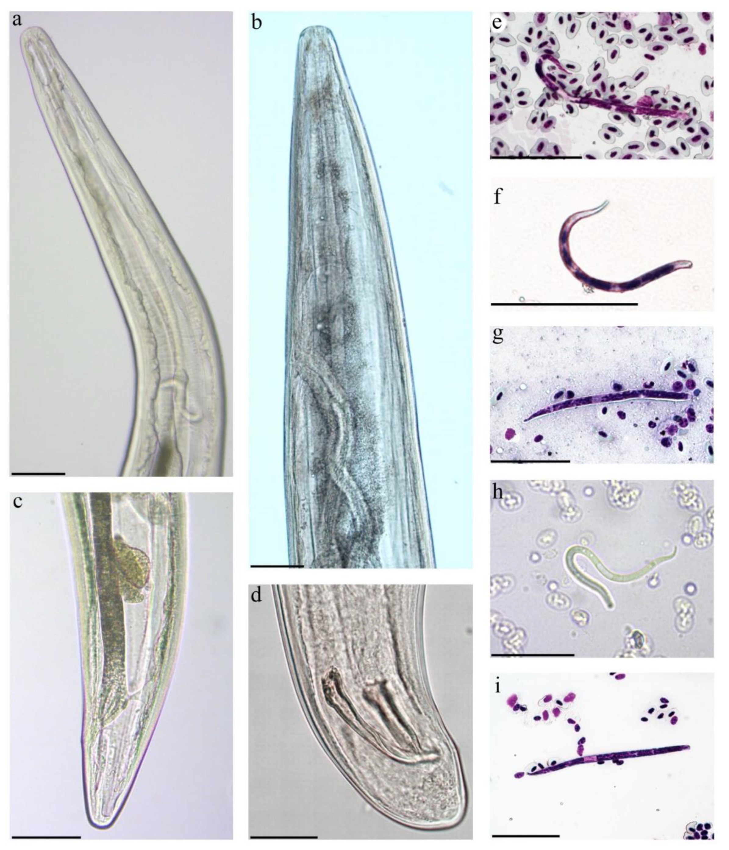
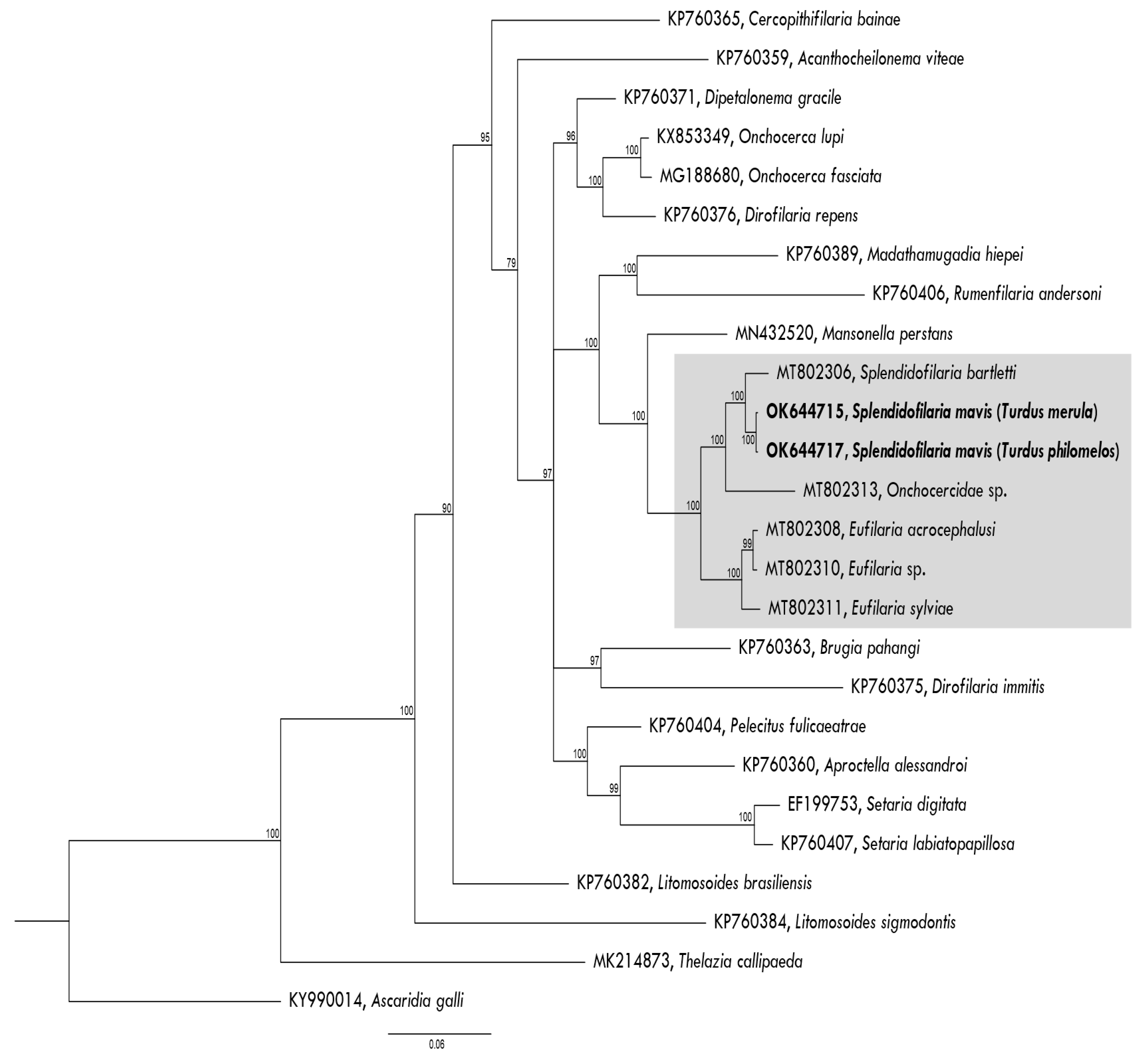
3.3. Description of Splendidofilaria mavis

This study identified microfilariae of S. mavis and developed the molecular characterization of this species. Microfilariae that were detected in the blood films of both examined birds had an anterior rounded extremity and tapering rounded posterior extremity with a smooth cuticle (Figure 4e–i). The morphometric characters of the microfilariae from the Eurasian blackbird and the Song thrush are given in Table 3.
No significant difference was discernible in measurements of the microfilaria that were obtained from the lungs or liver. Some specimens in the blood of the lungs and liver had longer cephalic space than the microfilariae in circulatory blood. The microfilariae from the uterus were shorter (75–95 µm) and had a smaller inner body than microfilariae from the blood and other organs but the body shape and fixed-point values (expressed as percentages of the total body) were similar.
Adult nematodes are slender with slightly attenuated extremities, males are shorter than females, the cuticle is without bosses, the oral opening is small, the oesophagus is thin, it is not externally divided into muscular and glandular part, the vagina is short, directed posteriorly, the spicules are subequal and dissimilar (Figure 4a–d). The morphometric parameters of adult S. mavis in both bird species are similar (Table 4). Although, S. mavis as described from the Song thrush had a shorter oesophagus, a vulva that opens further from the anterior extremity and an ovary that turns up father from posterior extremity in comparison to the Eurasian blackbird, but it corresponds with the descriptions of S. mavis that was found in Germany [48]. Sequences from adult nematodes of the Eurasian blackbird and the Song thrush clustered in one clade with Splendidofilaria bartletti (Figure 5). The sequences of adults and microfilariae coincide.

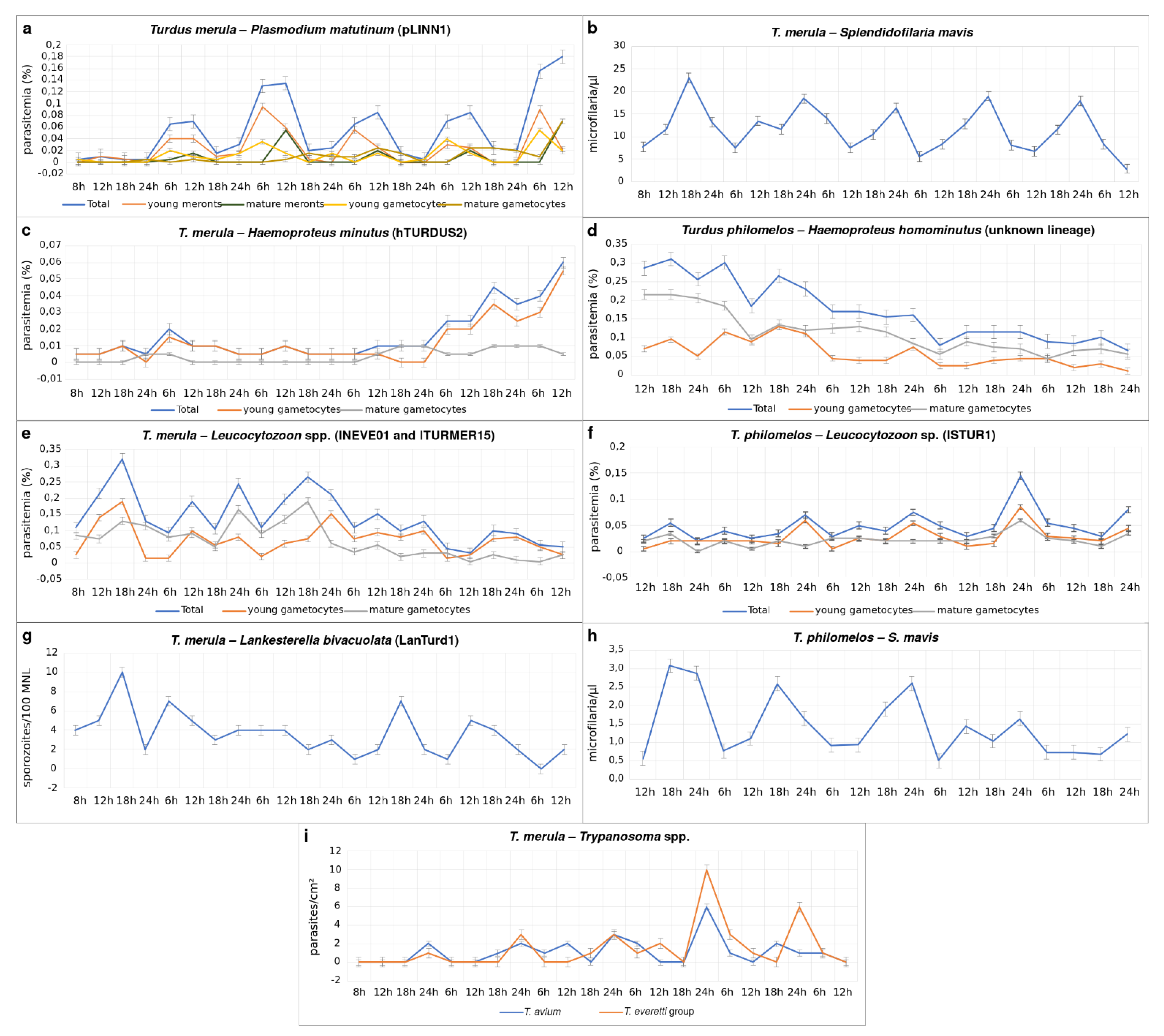
3.4. Circadian Rhythms

3.4.1. Parasites of the Eurasian Blackbird

A 24 h circadian rhythm was observed during P. matutinum (pLINN1) infection, with the parasitemia intensity of young meronts and young gametocytes being higher at 6 h, and mature meronts and mature gametocytes were more often seen at 12 h. During the evening (18 h) and night (24 h), the parasitemia intensity of all P. matutinum stages was usually lower than in the morning (6 h) and midday (12 h) (Figure 6a).
In H. minutus (hTURDUS2) infection, the intensity of parasitemia was low (<0.01%) during the first four days of the observation and increased to 0.06% only on the fifth day, thus being low during the entire period of observation. No circadian rhythm was observed in this infection (Figure 6c).
The Eurasian blackbird was co-infected with two Leucocytozoon lineages (lNEVE01 and lTURMER15). Even though it is possible to speculate that one of these lineages might be L. dubreuili based on the morphology of some of the found gametocytes and their host cells (Figure 1k–o), it is difficult to link the observed gametocytes to a certain lineage because the parasites were similar morphologically. Additionally, many young forms were seen in the blood films (Figure 1e,k). Young Leucocytozoon gametocytes are indistinguishable on a species level. This further complicated the linkage of morphological data and lineage information during the examination of circadian rhythms on Leucocytozoon species. Thus, the parasitemia intensity was not considered separately for these two lineages. We observed an increase of the parasitemia intensity of young gametocytes every 12 hours, mainly at midday (12 h) and midnight (24 h). For mature gametocytes, the parasitemia peaks were observed always as follow: evening (18 h), midday (12 h), and midnight (24 h) (Figure 6e).
No circadian rhythm was observed during the studied period for Lankesterella bivacuolata n. sp. (LanTurd1) infection, with parasitemia intensity slightly increasing irregularly during a day (Figure 6g).
Trypanosoma avium and T. everetti group parasites followed the circadian rhythm (Figure 6i), with their parasitemia increasing mainly at midnight (24 h) (Figure 6i). It is interesting to mention that parasitemia intensity also increased a few times in other times of the day during the observation period, i.e., Trypanosoma avium had one peak at midday (12 h) and one in the evening (18 h), while parasitemia of T. everetti group parasite had one peak at midday (12 h).
Splendidofilaria mavis microfilariae were present in the blood permanently (Figure 6b), however, it was possible to observe its circadian rhythm. Mainly, the lowest parasitemia intensity was in the morning (6 h) and at midday (12 h), while the biggest parasitemia was recorded in the evening (18 h) and night (24 h) (Figure 6b).

3.4.2. Parasites of the Song Thrush

In H. homominutus (unknown lineage) infection, the intensity of parasitemia was around 0.3% at the beginning of the observation, and it decreased along the observation days to 0.1%. As for H. minutus, no circadian rhythm was observed in this infection (Figure 6d).
The song thrush had a single Leucocytozoon (lSTUR1) infection, and in this case, the peaks of parasitemia of young gametocytes were reported at midnight (24 h). Interestingly, the intensity of mature gametocytes was constant throughout the experiment, with small peaks in the morning (6 h) and the evening (18 h) (Figure 6f).
The circadian rhythms of S. mavis in the Song thrush were the same as in the Eurasian blackbird (Figure 6h).

4. Discussion

The key results of this study are (i) the description and molecular characterization of Lankesterella bivacuolata n. sp. (lineage LanTurd1) infecting a Eurasian blackbird; (ii) molecular characterization of the filarioid nematode S. mavis infecting Turdidae birds; (iii) the first description of the circadian rhythm of Plasmodium matutinum (pLINN1) at the lineage level; (iv) the first provided information about the circadian rhythm of Haemoproteus, Lankesterella and S. mavis parasites; and (v) evidence that circadian rhythms of P. matutinum (pLINN1) and S. mavis maintain during co-infection with several different blood parasites. These findings are discussed below.

4.1. Description of New Lankesterella Parasite

A recent study by Chagas et al. [26] reviewed available information about avian Lankesterella blood parasites and pointed out that some avian blood parasites, which have been formerly attributed to the genus Hepatozoon, in fact, belong to Lankesterella genus [46,47,49]. Our phylogenetic analysis supports this conclusion due to the close clustering of all currently available avian Lankesterella lineages, including the lineage LanTurd1 of the new species (Figure 3). We also extend information about the diversity and phylogenetic relationships of these neglected avian parasites by describing L. bivacuolata n. sp. (LanTurd1), which is readily distinguishable from all described Lankesterella species due to the presence of two vacuole-like structures in the sporozoites (Figure 2g,i,n,p).
The available information that is based on the detection of 18S rRNA lineages in avian hosts indicates that Lankesterella parasites likely are specific to birds on their genera levels [26]. Lankesterella bivacuolata n. sp. (LanTurd1) was also found in a Eurasian blackbird in the Labanoras Forest (55°12′25.77″ N, 25°55′26.47″ E), Lithuania (C. Chagas personal communication, data not shown). It is important to mention that the complete life cycle and vectors of avian Lankesterella parasites remain unknown. More studies, particularly those targeting potential vectors, transmission, life cycles, and analyses of other genes should be encouraged to better understand Lankesterella spp. biology and their role in the wild. This study provides information for morphological identification and molecular barcoding of L. bivacuolata n. sp. (LanTurd1), the parasite of the Eurasian blackbird.

4.2. Description and Molecular Characterization of Splendidofilaria mavis

In spite of much former research on morphology and taxonomy of filarioid nematodes [31,50,51,52,53,54], the molecular characterization of only a few avian filarioid nematodes is available [4]. This is unfortunate because molecular barcoding provides opportunities to detect the parasites at the microfilariae stage in circulation avoiding bird dissection and thus might remarkably extend sampling and data collection in wildlife. We contribute to the development of barcoding of Onchocercidae nematodes by providing DNA sequences of S. mavis. This study extends information about the morphology on adult worms of S. mavis and the microfilariae of this parasite, which were found in two different species of thrushes (Turdidae). This provides an opportunity to develop the phylogenetic analysis on relationships of avian wildlife filarioid nematodes (Figure 5). Future research on these parasites using morphological and molecular approaches in parallel should be encouraged. Such approaches open new opportunities for more reliable species identification and would extend screening of birds for Onchocercidae nematodes using analysis of blood samples.
The onchocercidian parasite S. mavis was first found in the redwing (Turdus iliacus, synonym Turdus musicus) in the United Kingdom [30]. Then, this nematode was detected in different species of thrushes in Germany, Austria, and Poland [48,55]. The morphometric parameters of S. mavis that were described in these countries and our measurements are almost identical. Differently from earlier descriptions, our study shows that the vagina can be longer—up to 128 µm instead of 50 µm reported before, and the left spicule can also be longer—up to 116 µm instead of 90 µm. The microfilariae named S. mavis were found in thrushes in Spain, France, and Czech Republic [53,56,57], and also reported in other passerine birds, such as the Hawfinch (Coccothraustes coccothraustes) and the European robin (Erithacus rubecula) [53,58]. These findings indicate that S. mavis might not be strictly specific to the final host. However, there was no convincing evidence that these microfilariae certainly belong to S. mavis. The development of molecular markers for species identification are crucial for future studies on filarioid nematodes that are found in different host species and present at different stages of development because molecular barcoding will minimize misidentifications.
It is worth noting that Splendidofilaria (synonym Ornithofilaria) bohmi was found in the Mistle thrush (Turdus viscivorus) in Europe [59]. According to review by Sonin [54], the morphological differences between this parasite and S. mavis are insignificant; these parasites slightly differ from each other due to the size of spicules. Additional material is needed for a conclusion about validity of the name S. bohmi. Our morphological analysis shows that the length of spicules in S. mavis overlaps with those in S. bohmi (left spicules 103 µm, 116 µm, and 102–125 µm, respectively; right spicules 82 µm, 91 µm, and 86–110 µm, respectively). These two species differ only due to (i) the different distance from anterior extremity to vulva (466–795 µm in S. mavis and 360–380 µm in S. bohmi) and (ii) the shape of microfilariae. Supperer [59] published a good-quality description and pictures of S. bohmi microfilariae, which were over 155 µm in length, and their posterior part was slightly narrowed and broadly rounded. Our study of microfilariae and the microfilariae description given by Gönnert [48] showed that the microfilariae are shorter (73–143 µm). Furthermore, according to our investigation, the posterior extremity of S. mavis microfilariae are tapering rounded, almost pointed, that distinguish them from S. bohmi, indicating possible validity of both names. The shape of the microfilariae that were found in France and Czech Republic [53,56,58] was more similar to S. bohmi microfilariae because their posterior has a broadly rounded end than in S. mavis. Despite the differences in the shape of microfilariae, the morphology of the adult worms was almost identical. To prove that these parasites are different species, further taxonomy studies are needed, ideally by a combination of morphological features and molecular markers.

4.3. Circadian Rhythms of Blood Parasites during Co-Infections

Considering that over 260 avian haemosporidian parasites are described and likely many more exist [60], the available information about their circadian rhythms is certainly insufficient. Most of the studied avian parasite species belong to Plasmodium, but the available information deals mainly with the circadian rhythms of erythrocytic merogony, with rare exceptions when diurnal fluctuation in gametocytes intensity was considered [3,16]. Circadian rhythms have never been investigated in Haemoproteus infections, and information is available only about circadian rhythms of two Leucocytozoon parasites, i.e., Leucocytozoon simondi and L. smithi [3]. Avian Lankesterella are the most poorly investigated parasites, and this study provided the first information on their circadian rhythms. Daily parasitemia cycles in Trypanosoma parasites are available mainly for species that infect humans, those with zoonotic potential and that are economically important [21]. A few studies address this issue in Trypanosoma parasites of amphibians [61]. Filarioid nematodes are relatively well-investigated with regard to circadian rhythms, with many studies conducted, even though they are also more focused on species that infect humans and other mammals [21,62,63].
It is important to note that this study does not propose any conclusions on circadian rhythms in each parasite group because it is limited by a few observations in only two avian hosts. Additionally, co-infection was present in both studied hosts, raising a question about the possible influence of parasites on each other. However, we used this opportunity to describe the available data to get maximum information from these birds which were dissected for research. Data about the circadian rhythms of avian blood parasites during co-infections are absent, thus might provide ideas for further experimental investigations. Below, we discuss the main information that was obtained about circadian rhythms by different parasite groups.

4.3.1. Plasmodium Infections

The periodicity of several avian Plasmodium species has been investigated. Most of them have an erythrocytic merogony cycle of 24 h, with the peak of meront maturation varying in different species along the day. For instance, P. matutinum, P. giovannolaia, and P. elongatum have the biggest intensity of mature meronts in the morning; in P. cathemerium and P. circumflexum infections, a peak was seen in the evening; and in P. gabaldoni, P. nucleophilum, and P. homocircumflexum (lineage pCOLL4), the intensity of meronts was higher at midday [3,16]. In P. gallinaceum and P. relictum, an erythrocytic merogony cycle of 36 h was reported [3]. In this study, we determined the circadian rhythms of the lineage pLINN1 of P. matutinum for the first time; it was close to 24 h, as formerly reported for an unknown lineage of the same parasite species [64], with a bigger intensity of mature meronts seen in the morning (6 h) and a bigger intensity of mature gametocytes at midday (12 h) (Figure 6a).
Previous studies have reported that Plasmodium sp. parasitemia intensity and the parasitemia dynamics might differ in different avian hosts [65,66] and also during co-infections of two Plasmodium species [67]. In the present study, the circadian rhythm of the mature meronts remained the same in P. matutinum (pLINN1), as indicated for an unknown lineage of P. matutinum by Garnham [64] and Valkiūnas [3]. Importantly, our study showed that the circadian rhythms of mature meronts remained even during co-infections of several species and lineages of haemosporidian and other blood parasites, indicating the relatively stable pattern of the periodicity in P. matutinum. This avian malaria parasite is widespread in thrushes [68] and can be recommended for experimental studies on circadian rhythms of avian malaria infections with rapid (24 h) cycles of erythrocytic merogony.

4.3.2. Haemoproteus Infections

Only gametocytes are present in the circulation during Haemoproteus infection. No circadian rhythm was discernible during parasitemia of both studied Haemoproteus species, i.e., H. minutus (hTURDUS2) (Figure 1f–j) and H. homominutus (the parasite was identified only through microscopic examination) (Figure 1s–w). It is interesting to note that H. minutus (hTURDUS2) parasitemia increased throughout the course of this study (Figure 6c), but reverse dynamic of parasitemia was seen during H. homominutus infection. Mainly, the parasitemia intensity decreased during the observation (Figure 6d). It is possible to speculate that stress conditions might influence the increase of H. minutus (hTURDUS2) parasitemia in the Eurasian blackbird. The stress in this host might be particularly severe due to the presence of seven other co-infecting blood parasites. It is known that capturing wild animals leads to an increase in corticosteroid levels, a hormone indicative of physiological stress. This increase can result in an impairment of the immune response of the infected animals and might lead to a higher parasitemia [69,70]. At the same time, the studied birds had access to food ad libitum, which might have increased the availability of resources that they could use to mount an immunological response and reduce parasitemia [71]. That might be the case in the Song thrush. This bird was infected with H. homominutus and co-infected with only two other parasite species, so might be under less parasite-induced stress pressure, resulting in a decrease of parasitemia during this study (Figure 6d). The role of immunological responses on the parasitemia of haemosporidian parasites remains insufficiently studied, particularly in wildlife.
This study further points out difficulties in haemosporidian parasite diagnostics during co-infections. Mainly, even though the intensity of parasitemia of gametocytes of H. homominutus were intense, and the parasite was readily recognizable in blood films of the Song thrush (Figure 1v,w), we did not recover this parasite sequence in the samples. However, the sequence H. asymmetricus (hTUPHI01) was detected. These two genetically closely related parasites often parasitize thrushes; they are sometimes reported in co-infection due to readily distinguishable gametocytes [72]. It is possible to speculate that the studied Song thrush was co-infected with H. homominutus and H. asymmetricus, but the latter infection was latent. A template for H. asymmetricus (hTUPHI01) DNA amplification might come from the remnants of the tissue stages, which are difficult to visualize on blood films, but not from gametocytes. The presence of haemosporidian co-infections has been frequently reported in wild birds [6,7,8,9], and such infections even predominate in some bird populations in Europe [10]. Conventional PCR-based methods are not always able to diagnose these co-infections [11,73] because general primers might have a better match to certain DNA sequences. This study further highlights the importance of both microscopy examination and PCR-based tools in wildlife haemosporidian research [68].

4.3.3. Leucocytozoon Infections

Information about circadian rhythm is available only for two Leucocytozoon species, i.e., Leucocytozoon simondi and Leucocytozoon smithi, the parasites of domestic ducks and turkeys, respectively. Only gametocytes are present in the circulation in Leucocytozoon infections and parasitemia usually increase during the daytime [3]. Information about circadian rhythms of Leucocytozoon species infecting thrushes and other wild birds is absent.
In this study, the Eurasian blackbird was co-infected with two genetic lineages of Leucocytozoon sp., in which gametocytes were hardly distinguishable morphologically and were unidentified to species levels. The identification of Leucocytozoon parasites using morphological characteristics of their blood stages (gametocytes) is challenging due to the limited number of valuable taxonomic morphological characters, which are often similar in many described species. Additionally, gametocytes and host cells of these parasites are fragile and often are deformed in blood films (for example, see Figure 1z,ab). As a result, we were unable to identify Leucocytozoon species during co-infections which were determined by the PCR-based method. In other words, the circadian rhythm was not determined for each Leucocytozoon parasite lineage separately. However, we observed that young gametocytes were present every 12 hours in the blood, while mature gametocytes can be found at different times of the day (Figure 6e).
The Leucocytozoon sp. (lSTUR1) was present in a single infection in the Song thrush. During this infection, young gametocytes were more frequently observed in the evening (18 h) and night (24 h), and mature gametocytes were predominant every 12 hours, in the evening (18 h) and morning (6 h) (Figure 6f).
The available data on parasitemia dynamics of Leucocytozoon species suggest that different parasite species and/or lineages might have different circadian rhythms. This might prevent infection of the same individual vector with several different lineages of related parasites of the same genus, and thus might contribute to minimizing competition during the sporogony process. It is interesting to mention that Leucocytozoon parasites are transmitted by Simuliidae blackflies [3]. These blood-sucking insects are particularly active during the daytime [74]. The presence of mature gametocytes in the circulation during this time would be beneficial for transmission and should be evolutionary preferable.

4.3.4. Lankesterella Infection

Avian Lankesterella parasites remain a neglected group of pathogens with numerous unclear issues in their basic biology, particularly related to their pathogenicity and transmission [26,46,47,49]. This is the first study that has addressed the circadian rhythm of Lankesterella parasites which are probably transmitted by different mosquito species [26].
We did not observe any circadian rhythm in the parasitemia intensity of L. bivacuolata n. sp. (LanTurd1) during the studied period (Figure 6g). Further studies are needed for a better understanding of whether the circadian rhythm occurs in other species of Lankesterella.
Only the final stage of the life cycle (sporozoites) is present in bird blood, and the vectors serve mainly as a reservoir of infection, which does not develop in insects. Birds are likely to get infected mainly by eating infected insects in which the parasite can persist over a month after the initial infection [26]. Circadian rhythms might be of biologically low value for the transmission of such parasites which might be not specific to certain vector species resulting in possible transmission during different times of the day. However, more studies are needed for a better understanding of the vector biology of Lankesterella spp.

4.3.5. Trypanosoma Infections

Circadian rhythms were recognizable in parasitemia of both T. avium and T. everetti group parasites with increases of parasitemia intensity at night close to 24 h (Figure 6i). These parasites might be transmitted by Culicoides biting midges [27,75], which are more active during the night as well. Trypanosoma avium was also reported to be transmitted by Simuliidae insects [76]. These insects are more active during the day, however, only a few peaks of parasitemia were seen during Trypanosoma infections during the daytime in the present study. This might suggest that Simuliidae species may not be the main vectors of the examined parasites. Culicoides biting midges are more active during the sunset and the sunrise in Europe [77]. An increase of parasitemia close to the night period should be beneficial for transmission. It is interesting to note that an increase of parasitemia during the night was reported in some other Trypanosoma species. For example, Trypanosoma lewisi, T. duttoni, and T. congolense, which parasitize rodents [78,79]. However, this is certainly not a general pattern in Trypanosoma species. For example, parasitemia of T. rotatorium, a parasite of frogs, increases during the daytime close to 12 h [61].
Parasitemia of avian trypanosomes is usually low in birds [80]. The obtained information about the daytime peaks of parasitemia of common T. avium and T. everetti group parasites is helpful for the selection of donor hosts for the pathogen strains isolation and experimental research.

4.3.6. Microfilariae Infections

This study shows that S. mavis microfilariae exhibited a nocturnal periodicity in both studied birds. Judging from the daily parasitemia intensity of microfilariae, the best time for the transmission of this pathogen should be close to the night when numerous parasites were seen in the circulation, suggesting a greater probability to infect vectors. Nevertheless, microfilariae were observed in the circulation during the entire day (Figure 6b,h). Hence, vectors can also have access to infection during the entire day. The available knowledge shows that the circadian rhythms of microfilariae depend markedly on the parasite and host species. The parasitemia peaks can be nocturnal, diurnal, or even irregular. An increase of microfilariae parasitemia usually coincides with a period of maximum activity of the local arthropods vector species [81]. This is particularly well-studied in the human parasite Loa loa, where microfilariae exhibit a marked diurnal parasitemia periodicity and this is well-associated with the time of maximum activity of its intermediate hosts, the deer flies Chrysops silacea and Chrysops dimidiate [82]. Meanwhile, the microfilariae of Wuchereria bancrofti, another pathogenic human parasite, exhibit a marked nocturnal periodicity and are transmitted by mosquitoes of different genera, which are active at night (Aedes, Anopheles, Culex, and Monsonia).
Few studies are addressing the circadian parasitemia rhythms of avian filarioid nematodes. These are: Cardiofilaria nilesi, Chandlerella striatospicula, Splendidofilaria picacardina, and S. fallisensis [21,63]. The circadian rhythm of S. picacardina microfilariae in the Black-billed magpie (Pica hudsonia) exhibited nocturnal periodicity, as also was the case with S. mavis in our study. However, the biggest parasitemia intensity of microfilariae was seen during the daytime in S. fallisensis, a parasite of North American ducks [21]. Females of black flies feed on blood during the day [74], therefore it is not surprising that the peak of S. fallisensis microfilaria in the blood also occurred during this period of the day. The vectors of S. picacardina and S. mavis remain unknown. Many Onchocercidae species of mammals exhibit a nocturnal peak of microfilariae parasitemia and their vectors are species of Aedes, Anopheles, Armigeres, Coquillettidia, Culex, Culicoides, Mansonia, and Ochlerotatus mosquitoes [28,83,84,85,86,87,88]. This indicated that ornithophilic mosquitoes from these genera likely can be the vectors of nocturnal Splendidofilaria species as well.

5. Conclusions

This study described one new species of an avian blood parasite, Lankesterella bivacuolata n. sp. (lineage LanTurd1) and provided the molecular characterization and detailed morphological description of the filarioid nematode Splendidofilaria mavis, a common parasite of passerine birds. Importantly, microfilariae of this nematode were not only described and illustrated, but also characterized molecularly. Both the molecular characterization and the microfilariae description open new opportunities for prominent parasite sampling that is aimed at a better understanding of the biology of filarioid nematodes using harmless-for-host methods. The molecular characterization of both these pathogens expand the opportunities for phylogenetic research on blood parasites of related genera.
The information provided about circadian rhythms suggests directions for the planning of future targeting experimental observations on this insufficiently understood subject. It was shown that clear circadian rhythms of parasitemia are maintained in Plasmodium matutinum (pLINN1) and S. mavis infections even during the co-infection of numerous different parasites, thus being relatively insensitive for the influence of other pathogens that are present in same host and indicating possible essential importance of the parasitemia fluctuation in these parasites’ biology and transmission.
This was the first study that addressed the daily variation in parasitemia of avian blood parasites belonging to Haemoproteus, Lankesterella and S. mavis. Circadian rhythms do not occur in Haemoproteus and Lankesterella parasite species, at least during co-infections.

Author Contributions

Conceptualization, C.R.F.C. and R.B.; methodology, C.R.F.C., R.B., and G.V.; formal analysis, C.R.F.C. and R.B.; investigation, C.R.F.C., R.B., and G.V.; resources, G.V., and C.R.F.C.; data curation, C.R.F.C. and R.B.; writing—original draft preparation, C.R.F.C., R.B., and G.V.; writing—review and editing, C.R.F.C., R.B., and G.V.; supervision, G.V.; funding acquisition, G.V. and C.R.F.C. All authors have read and agreed to the published version of the manuscript.

Funding

This research was funded by the Research Council of Lithuania (09.3.3-LMT-K-712-02-0004 and 09.3.3-LMT-K-712-23-0010) and the Open Access to research infrastructure of the Nature Research Centre under Lithuanian open access network initiative.

Institutional Review Board Statement

The study was conducted according to the guidelines of the Declaration of Helsinki and approved by the Environmental Protection Agency, Vilnius, Lithuania (permit number 23 and date of approval 19 April 2019).

Informed Consent Statement

Not applicable.

Data Availability Statement

The data that are presented in this study are in GenBank database (https://www.ncbi.nlm.nih.gov/genbank/, accessed on 27 October 2021) (accession numbers: P. matutinum pLINN1, OK646331; H. minutus hTURDUS2, OK646332; H. asymmetricus hTUPHI01, OK721103, Leucocytozoon sp. lNEVE01, OK646333; Leucocytozoon sp. lTURMER15, OK646334; Leucocytozoon sp. lSTUR1, OK646335; L. bivacuolata n. sp., OK644714; T. avium, OK605056; S. mavis 28S, OK644715-OK644718; and S. mavis coxI, OK631737-OK391740).

Acknowledgments

The authors are grateful to Mikas Ilgūnas for assistance during fieldwork, and Rasa Bernotienė and Tajana Iezhova, for the assistance in the laboratory.

Conflicts of Interest

The authors declare no conflict of interest.

References

  1. Atkinson, C.T.; Thomas, N.; Hunter, B. Parasitic Disease of Wild Birds; John Wiley & Sons: Ames, IA, USA, 2008. [Google Scholar]
  2. Santiago-Alarcon, D.; Marzal, A. Avian Malaria and Related Parasites in the Tropics: Ecology, Evolution and Systematics; Springer: Cham, Switzerland, 2020; ISBN 978-3-030-51632-1. [Google Scholar]
  3. Valkiūnas, G. Avian Malaria Parasites and Other Haemosporidia; CRC Press: Boca Raton, FL, USA, 2005. [Google Scholar]
  4. Binkienė, R.; Chagas, C.R.F.; Bernotienė, R.; Valkiūnas, G. Molecular and Morphological Characterization of Three New Species of Avian Onchocercidae (Nematoda) with Emphasis on Circulating Microfilariae. Parasit. Vectors 2021, 14, 137. [Google Scholar] [CrossRef]
  5. Chagas, C.R.F.; Binkienė, R.; Ilgūnas, M.; Iezhova, T.; Valkiūnas, G. The Buffy Coat Method: A Tool for Detection of Blood Parasites without Staining Procedures. Parasit. Vectors 2020, 13, 104. [Google Scholar] [CrossRef] [Green Version]
  6. Clark, N.J.; Wells, K.; Dimitrov, D.; Clegg, S.M. Co-Infections and Environmental Conditions Drive the Distributions of Blood Parasites in Wild Birds. J. Anim. Ecol. 2016, 85, 1461–1470. [Google Scholar] [CrossRef] [PubMed] [Green Version]
  7. Reis, S.; Melo, M.; Covas, R.; Doutrelant, C.; Pereira, H.; de Lima, R.; Loiseau, C. Influence of Land Use and Host Species on Parasite Richness, Prevalence and Co-Infection Patterns. Int. J. Parasitol. 2021, 51, 83–94. [Google Scholar] [CrossRef] [PubMed]
  8. Van Rooyen, J.; Lalubin, F.; Glaizot, O.; Christe, P. Avian Haemosporidian Persistence and Co-Infection in Great Tits at the Individual Level. Malar. J. 2013, 12, 40. [Google Scholar] [CrossRef] [PubMed] [Green Version]
  9. Soares, L.; Ellis, V.A.; Ricklefs, R.E. Co-Infections of Haemosporidian and Trypanosome Parasites in a North American Songbird. Parasitology 2016, 143, 1930–1938. [Google Scholar] [CrossRef] [PubMed]
  10. Valkiūnas, G.; Iezhova, T.A.; Shapoval, A.P. High Prevalence of Blood Parasites in Hawfinch Coccothraustes Coccothraustes. J. Nat. Hist. 2003, 37, 2647–2652. [Google Scholar] [CrossRef]
  11. Bernotienė, R.; Palinauskas, V.; Iezhova, T.; Murauskaitė, D.; Valkiūnas, G. Avian Haemosporidian Parasites (Haemosporida): A Comparative Analysis of Different Polymerase Chain Reaction Assays in Detection of Mixed Infections. Exp. Parasitol. 2016, 163, 31–37. [Google Scholar] [CrossRef] [PubMed]
  12. Ciloglu, A.; Ellis, V.A.; Bernotienė, R.; Valkiūnas, G.; Bensch, S. A New One-Step Multiplex PCR Assay for Simultaneous Detection and Identification of Avian Haemosporidian Parasites. Parasitol. Res. 2019, 118, 191–201. [Google Scholar] [CrossRef]
  13. Valkiūnas, G.; Iezhova, T.A.; Loiseau, C.; Chasar, A.; Smith, T.B.; Sehgal, R.N.M. New Species of Haemosporidian Parasites (Haemosporida) from African Rainforest Birds, with Remarks on Their Classification. Parasitol. Res. 2008, 103, 1213–1228. [Google Scholar] [CrossRef]
  14. Valkiūnas, G.; Križanauskienė, A.; Iezhova, T.A.; Hellgren, O.; Bensch, S. Molecular Phylegenetic Analysis of Circumnuclear Hemoproteids (Haemosporida: Haemoproteidae) of Sylviid Birds, with a Description of Haemoproteus Parabelopolskyi Sp. No. J. Parasitol. 2007, 93, 680–687. [Google Scholar] [CrossRef] [PubMed]
  15. Hawking, F. Circadian and other Rhythms of Parasites. In Advances in Parasitology; Elsevier: Amsterdam, The Netherlands, 1975; Volume 13, pp. 123–182. ISBN 978-0-12-031713-4. [Google Scholar]
  16. Palinauskas, V.; Žiegytė, R.; Ilgūnas, M.; Iezhova, T.A.; Bernotienė, R.; Bolshakov, C.; Valkiūnas, G. Description of the First Cryptic Avian Malaria Parasite, Plasmodium Homocircumflexum n. Sp., with Experimental Data on Its Virulence and Development in Avian Hosts and Mosquitoes. Int. J. Parasitol. 2015, 45, 51–62. [Google Scholar] [CrossRef] [PubMed]
  17. Pigeault, R.; Caudron, Q.; Nicot, A.; Rivero, A.; Gandon, S. Timing Malaria Transmission with Mosquito Fluctuations. Evol. Lett. 2018, 2, 378–389. [Google Scholar] [CrossRef] [PubMed] [Green Version]
  18. Martinez-Bakker, M.; Helm, B. The Influence of Biological Rhythms on Host–Parasite Interactions. Trends Ecol. Evol. 2015, 30, 314–326. [Google Scholar] [CrossRef] [PubMed] [Green Version]
  19. Rijo-Ferreira, F.; Takahashi, J.S.; Figueiredo, L.M. Circadian Rhythms in Parasites. PLOS Pathog. 2017, 13, e1006590. [Google Scholar] [CrossRef] [PubMed]
  20. Westwood, M.L.; O’Donnell, A.J.; de Bekker, C.; Lively, C.M.; Zuk, M.; Reece, S.E. The Evolutionary Ecology of Circadian Rhythms in Infection. Nat. Ecol. Evol. 2019, 3, 552–560. [Google Scholar] [CrossRef]
  21. Anderson, R.C. Nematode Parasites of Vertebrates: Their Development and Transmission; Cabi: Wallingford, UK, 2000. [Google Scholar]
  22. Dolnik, O.V.; Metzger, B.J.; Loonen, M.J.J.E. Keeping the Clock Set under the Midnight Sun: Diurnal Periodicity and Synchrony of Avian Isospora Parasites Cycle in the High Arctic. Parasitology 2011, 138, 1077–1081. [Google Scholar] [CrossRef] [Green Version]
  23. Calistri, P.; Iannetti, S.; Danzetta, M.L.; Narcisi, V.; Cito, F.; Di Sabatino, D.; Bruno, R.; Sauro, F.; Atzeni, M.; Carvelli, A.; et al. The Components of ‘One World-One Health’ Approach. Transbound. Emerg. Dis. 2013, 60, 4–13. [Google Scholar] [CrossRef] [PubMed] [Green Version]
  24. Godfrey, R.D.; Fedynich, A.M.; Pence, D.B. Quantification of Hematozoan in Blood Smears. J. Wildl. Dis. 1987, 23, 558–565. [Google Scholar] [CrossRef]
  25. Hellgren, O.; Waldenström, J.; Bensch, S. A New PCR Assay for Simultaneous Studies of Leucocytozoon, Plasmodium and Haemoproteus from Avian Blood. J. Parasitol. 2004, 90, 797–802. [Google Scholar] [CrossRef] [PubMed]
  26. Chagas, C.R.F.; Harl, J.; Preikša, V.; Bukauskaitė, D.; Ilgūnas, M.; Weissenböck, H.; Valkiūnas, G. Lankesterella (Apicomplexa, Lankesterellidae) Blood Parasites of Passeriform Birds: Prevalence, Molecular and Morphological Characterization, with Notes on Sporozoite Persistence In Vivo and Development In Vitro. Animals 2021, 11, 1451. [Google Scholar] [CrossRef] [PubMed]
  27. Bernotienė, R.; Iezhova, T.A.; Bukauskaitė, D.; Chagas, C.R.F.; Kazak, M.; Valkiūnas, G. Development of Trypanosoma Everetti in Culicoides Biting Midges. Acta Trop. 2020, 210, 105555. [Google Scholar] [CrossRef] [PubMed]
  28. Singh, S.N. Studies on the Helminth Parasites of Birds in Hyderabad State; Nematoda. J. Helminthol. 1948, 22, 77–92. [Google Scholar] [CrossRef]
  29. Bartlett, C.M.; Anderson, R.C. Lemdana Wernaarti n.Sp. and Other Filarioid Nematodes from Bubo Virginianus and Asio Otus (Strigiformes) in Ontario, Canada, with a Revision of Lemdana and a Key to Avian Filarioid Genera. Can. J. Zool. 1987, 65, 1100–1109. [Google Scholar] [CrossRef]
  30. Leiper, R.Th. Description of Filaria Mavis n. Sp. from the Trush. Zoologist 1909, 13, 337–339. [Google Scholar]
  31. Olsen, O.W.; Braun, C.E. New Species of Splendidofilaria and Chandlerella (Filarioidea: Nematoda), with Keys to the Species, from the Band-Tailed Pigeon (Columba Fasciata Fasciata) in the Rocky Mountain Region. Gt. Basin Nat. 1976, 36, 445–457. [Google Scholar]
  32. Sambrook, J.; Russel, D.W. Molecular Cloning: A Laboratory Manual, 3rd ed.; Cold Spring Harbour Laboratory Press: New York, NY, USA, 2001. [Google Scholar]
  33. Stunžėnas, V.; Petkevičiūtė, R.; Stanevičiūtė, G. Phylogeny of Sphaerium Solidum (Bivalvia) Based on Karyotype and Sequences of 16S and ITS1 RDNA. Open Life Sci. 2011, 6, 105–117. [Google Scholar] [CrossRef] [Green Version]
  34. Petkevičiūtė, R.; Stunžėnas, V.; Stanevičiūtė, G. Differentiation of European Freshwater Bucephalids (Digenea: Bucephalidae) Based on Karyotypes and DNA Sequences. Syst. Parasitol. 2014, 87, 199–212. [Google Scholar] [CrossRef] [PubMed]
  35. Hall, T.A. A User-Friendly Biological Sequence Alignment Editor and Analysis Program for Windows 98/98/NT. Nucleic. Acid Symp. Ser. 1999, 41, 95–98. [Google Scholar]
  36. Ronquist, R.; Huelsenbeck, J.P. MrBayes 3: Bayesian Phylogenetic Inference under Mixed Models. Bioinformatics 2003, 19, 1572–1574. [Google Scholar] [CrossRef] [PubMed] [Green Version]
  37. Katoh, K.; Rozewicki, J.; Yamada, K.D. MAFFT Online Service: Multiple Sequence Alignment, Interactive Sequence Choice and Visualization. Brief. Bioinform. 2019, 20, 1160–1166. [Google Scholar] [CrossRef] [PubMed] [Green Version]
  38. Nylander, J.A.A. MrModeltest v2. Program Distributed by the Author: Evolutionary Biology Centre; Uppsala University: Uppsala, Sweden, 2004. [Google Scholar]
  39. Rambout, A. FigTree: Tree Figure Drawing Tool Version 1.4.0; 2006. Available online: http://tree.bio.ed.ac.uk/software/figtree/ (accessed on 10 March 2021).
  40. Valkiūnas, G.; Ilgūnas, M.; Bukauskaitė, D.; Palinauskas, V.; Bernotienė, R.; Iezhova, T.A. Molecular Characterization and Distribution of Plasmodium Matutinum, a Common Avian Malaria Parasite. Parasitology 2017, 144, 1726–1735. [Google Scholar] [CrossRef] [PubMed] [Green Version]
  41. Baker, J.R. Biology of the trypanosomes of birds. In Biology of the Kinetoplastida; Academic Press: London, UK; New York, NY, USA, , 1976; Volume 1, pp. 131–174. ISBN 978-0-12-460201-4. [Google Scholar]
  42. Molyneux, D.H. Trypanosoma Everetti Sp. Nov. a Trypanosome from the Black-Rumped Waxbill Estrilda t. Troglodytes Lichtenstein. Ann. Trop. Med. Parasitol. 1973, 67, 219–222. [Google Scholar] [CrossRef] [PubMed]
  43. Valkiūnas, G.; Ilgūnas, M.; Bukauskaitė, D.; Chagas, C.R.F.; Bernotienė, R.; Himmel, T.; Harl, J.; Weissenböck, H.; Iezhova, T.A. Molecular Characterization of Six Widespread Avian Haemoproteids, with Description of Three New Haemoproteus Species. Acta Trop. 2019, 197, 105051. [Google Scholar] [CrossRef] [PubMed]
  44. Clark, P.; Boardman, W.S.J.; Raidal, S. Atlas of Clinical Avian Hematology; Wiley-Blackwell: Hoboken, NJ, USA, 2009. [Google Scholar]
  45. Bennett, G.F.; Earlé, R.A.; Peirce, M.A. New Species of Avian Hepatozoon (Apicomplexa: Haemogregarinidae) and a Re-Description of Hepatozoon Neophrontis (Todd & Wohlbach, 1912) Wenyon, 1926. Syst. Parasitol. 1992, 23, 183–193. [Google Scholar] [CrossRef]
  46. Biedrzycka, A.; Kloch, A.; Migalska, M.; Bielański, W. Molecular Characterization of Putative Hepatozoon Sp. from the Sedge Warbler (Acrocephalus Schoenobaenus). Parasitology 2013, 140, 695–698. [Google Scholar] [CrossRef] [Green Version]
  47. Merino, S.; Martínez, J.; Martínez-de la Puente, J.; Criado-Fornelio, Á.; Tomás, G.; Morales, J.; Lobato, E.; García-Fraile, S. Molecular Characterization of the 18S RDNA Gene of an Avian Hepatozoon Reveals That It Is Closely Related to Lankesterella. J. Parasitol. 2006, 92, 1330–1335. [Google Scholar] [CrossRef] [PubMed]
  48. Gönnert, R.V. Zur Frage Der Artzugehörigkeit von Filaria Mavis Leiper, 1909. Festschr. Bernhard Nocht Zum 1937, 80, 159–162. [Google Scholar]
  49. Martínez, J.; Merino, S.; Badás, E.P.; Almazán, L.; Moksnes, A.; Barbosa, A. Hemoparasites and Immunological Parameters in Snow Bunting (Plectrophenax Nivalis) Nestlings. Polar Biol. 2018, 41, 1855–1866. [Google Scholar] [CrossRef]
  50. Bartlett, C.M. New, Known and Unidentified Species of Eulimdana (Nematoda): Additional Information on Biologically Unusual Filarioids of Charadriiform Birds. Syst. Parasitol. 1992, 23, 209–230. [Google Scholar] [CrossRef]
  51. Bartlett, C.M.; Anderson, R.C. Filarioid Nematodes (Filarioidea: Onchocercidae) of Corvus Brachyrhynchos Brachyrhynchos Brehm in Southern Ontario, Canada and a Consideration of the Epizootiology of Avian Filariasis. Syst. Parasitol. 1980, 2, 77–102. [Google Scholar] [CrossRef]
  52. Bartlett, C.M.; Bain, O. New Avian Filarioids (Nematoda: Splendidofilariinae): Dessetfilaria Guianensis Gen. n., Sp. n., Andersonfilaria Africanus Gen. n., Sp. n., and Splendidofilaria Chandenieri Sp. n. Proc. Helminthol. Soc. Wash. 1987, 54, 1–14. [Google Scholar]
  53. Haas, M.; Baruš, V.; Benedikt, V.; Literák, I. Microfilariae in Birds in the Czech Republic, Including a Note on Adult Nematodes Eufilaria Delicata in a Song Thrush Turdus Philomelos. Parasitol. Res. 2011, 109, 645–655. [Google Scholar] [CrossRef] [PubMed]
  54. Sonin, M.D. Filariata of Animals and Man and Diseases Caused by Them. 2: Diplotriaenoidea; Osnovy nematodologii; Israel Program for Scientific Translations: Jerusalem, Israel, 1975; ISBN 978-0-7065-1504-6. [Google Scholar]
  55. Okulewicz, A. Ornithofilaria mavis (Leiper 1909) Gönnert, 1937 in blackbird (Turdus merula L.) in Poland]. Wiad. Parazytol. 1981, 27, 669–671. [Google Scholar] [PubMed]
  56. Chabaud, A.G.; Golvan, Y. Nouvelle Filaire Parasite Des Grives En France. Ann. Parasitol. Hum. Comparée 1956, 31, 405–413. [Google Scholar] [CrossRef] [Green Version]
  57. Lopez Caballero, E.J. Incidencia del parasitismo por formas de la superfamilia Filarioidea (Nematoda: Spirurina) en aves del genero Turdus (Passeriformes: Musacapidae). Boletin Estac. Cent. Ecol. 1978, 7, 67–71. [Google Scholar]
  58. Hauptmanová, K.; Baruš, V.; Literák, I.; Benedikt, V. Haemoproteids and Microfilariae in Hawfinches in the Czech Republic. Helminthologia 2004, 4, 125–133. [Google Scholar]
  59. Supperer, R. Zwei neue Filarien (s. l.), Eufilaria delicata spec. nov. und Ornithofilaria böhmi spec. nov. aus der Misteldrossel, Turdus viscivorus L. Z. Für Parasitenkd. 1958, 18. [Google Scholar] [CrossRef]
  60. Fecchio, A.; Chagas, C.R.F.; Bell, J.A.; Kirchgatter, K. Evolutionary Ecology, Taxonomy, and Systematics of Avian Malaria and Related Parasites. Acta Trop. 2020, 204, 105364. [Google Scholar] [CrossRef]
  61. Southworth, G.C.; Mason, G.; Seed, J.R. Studies on Frog Trypanosomiasis. I. A 24-Hour Cycle in the Parasitemia Level of Trypanosoma Rotatorium in Rana Clamitans from Louisiana. J. Parasitol. 1968, 54, 255. [Google Scholar] [CrossRef]
  62. Di Cesare, A.; Otranto, D.; Di Giulio, E.; Simonato, G.; Latrofa, M.S.; La Torre, F.; Coccia, G.; Traversa, D. Microfilarial Periodicity of Dirofilaria Repens in Naturally Infested Dogs. Parasitol. Res. 2013, 112, 4273–4279. [Google Scholar] [CrossRef] [PubMed]
  63. Gooneratne, B.W.M. On Cardiofilaria Nilesi in Experimentally Infected Chickens with a Note on the Morphology and Periodicity of the Microfilariae. J. Helminthol. 1969, 43, 311–317. [Google Scholar] [CrossRef]
  64. Garnham, P.C.C. Malaria Parasites and Other Haemosporidia; Blackwell Scientific: Oxford, UK, 1966; ISBN 978-0-632-01770-6. [Google Scholar]
  65. Ilgūnas, M.; Bukauskaitė, D.; Palinauskas, V.; Iezhova, T.A.; Dinhopl, N.; Nedorost, N.; Weissenbacher-Lang, C.; Weissenböck, H.; Valkiūnas, G. Mortality and Pathology in Birds Due to Plasmodium (Giovannolaia) Homocircumflexum Infection, with Emphasis on the Exoerythrocytic Development of Avian Malaria Parasites. Malar. J. 2016, 15, 256. [Google Scholar] [CrossRef] [PubMed] [Green Version]
  66. Palinauskas, V.; Valkiūnas, G.; Bolshakov, C.V.; Bensch, S. Plasmodium Relictum (Lineage P-SGS1): Effects on Experimentally Infected Passerine Birds. Exp. Parasitol. 2008, 120, 372–380. [Google Scholar] [CrossRef] [PubMed]
  67. Palinauskas, V.; Žiegytė, R.; Šengaut, J.; Bernotienė, R. Different Paths—The Same Virulence: Experimental Study on Avian Single and Co-Infections with Plasmodium Relictum and Plasmodium Elongatum. Int. J. Parasitol. 2018, 48, 1089–1096. [Google Scholar] [CrossRef] [PubMed]
  68. Harl, J.; Himmel, T.; Valkiūnas, G.; Ilgūnas, M.; Bakonyi, T.; Weissenböck, H. Geographic and Host Distribution of Haemosporidian Parasite Lineages from Birds of the Family Turdidae. Malar. J. 2020, 19, 335. [Google Scholar] [CrossRef] [PubMed]
  69. Davis, A.K.; Maney, D.L.; Maerz, J.C. The Use of Leukocyte Profiles to Measure Stress in Vertebrates: A Review for Ecologists. Funct. Ecol. 2008, 22, 760–772. [Google Scholar] [CrossRef]
  70. Romano, M.C.; Jiménez, P.; Miranda-Brito, C.; Valdez, R.A. Parasites and Steroid Hormones: Corticosteroid and Sex Steroid Synthesis, Their Role in the Parasite Physiology and Development. Front. Neurosci. 2015, 9, 224. [Google Scholar] [CrossRef] [PubMed] [Green Version]
  71. Appleby, B.M.; Anwar, M.A.; Petty, S.J. Short-term and Long-term Effects of Food Supply on Parasite Burdens in Tawny Owls, Strix Aluco. Funct. Ecol. 1999, 13, 315–321. [Google Scholar] [CrossRef]
  72. Valkiūnas, G.; Ilgūnas, M.; Bukauskaitė, D.; Duc, M.; Iezhova, T.A. Description of Haemoproteus Asymmetricus n. Sp. (Haemoproteidae), with Remarks on Predictability of the DNA Haplotype Networks in Haemosporidian Parasite Taxonomy Research. Acta Trop. 2021, 218, 105905. [Google Scholar] [CrossRef] [PubMed]
  73. Chagas, C.R.F.; Valkiūnas, G.; Nery, C.V.C.; Henrique, P.C.; Gonzalez, I.H.L.; Monteiro, E.F.; de Oliveira Guimarães, L.; Romano, C.M.; Kirchgatter, K. Plasmodium (Novyella) Nucleophilum from an Egyptian Goose in São Paulo Zoo, Brazil: Microscopic Confirmation and Molecular Characterization. Int. J. Parasitol. Parasites Wildl. 2013, 2, 286–291. [Google Scholar] [CrossRef] [PubMed] [Green Version]
  74. Addler, P.H.; McCreadie, J.W. Black Flies (Simuliidae). In Medical and Veterinary Entomology; Elsevier: San Diego, CA, USA, 2019; ISBN 978-0-12-814043-7. [Google Scholar]
  75. Svobodová, M.; Dolnik, O.V.; Čepička, I.; Rádrová, J. Biting Midges (Ceratopogonidae) as Vectors of Avian Trypanosomes. Parasit. Vectors 2017, 10, 224. [Google Scholar] [CrossRef] [PubMed]
  76. Votýpka, J.; Svobodová, M. Trypanosoma Avium: Experimental Transmission from Black Flies to Canaries. Parasitol. Res. 2004, 92, 147–151. [Google Scholar] [CrossRef] [PubMed]
  77. Mullen, G.R.; Murphree, C.S. Biting Midges (Ceratopogonidae). In Medical and Veterinary Entomology; Elsevier: San Diego, CA, USA, 2019; pp. 213–236. ISBN 978-0-12-814043-7. [Google Scholar]
  78. Cornford, E.M.; Freeman, B.J.; MacInnis, A.J. Physiological Relationships and Circadian Periodicities in Rodent Trypanosomes. Trans. R. Soc. Trop. Med. Hyg. 1976, 70, 238–243. [Google Scholar] [CrossRef]
  79. Hawking, F. Circadian Rhythms of Trypanosoma Congolense in Laboratory Rodents. Trans. R. Soc. Trop. Med. Hyg. 1978, 72, 592–595. [Google Scholar] [CrossRef]
  80. Zídková, L.; Cepicka, I.; Szabová, J.; Svobodová, M. Biodiversity of Avian Trypanosomes. Infect. Genet. Evol. 2012, 12, 102–112. [Google Scholar] [CrossRef] [PubMed]
  81. Hawking, F. The 24-Hour Periodicity of Microfilariae: Biological Mechanisms Responsible for Its Production and Control. Proc. R. Soc. Lond. B Biol. Sci. 1967, 169, 59–76. [Google Scholar] [CrossRef]
  82. Kelly-Hope, L.; Paulo, R.; Thomas, B.; Brito, M.; Unnasch, T.R.; Molyneux, D. Loa Loa Vectors Chrysops Spp.: Perspectives on Research, Distribution, Bionomics, and Implications for Elimination of Lymphatic Filariasis and Onchocerciasis. Parasit. Vectors 2017, 10, 172. [Google Scholar] [CrossRef] [PubMed] [Green Version]
  83. Bain, O. Recherches Sur La Morphogénèse Des Filaires Chez l’hôte Intermédiaire. Ann. Parasitol. Hum. Comparée 1972, 47, 251–303. [Google Scholar] [CrossRef]
  84. Campbell, J.R.; Soekartono; Purnomo; Atmosoedjono, S.; Marwoto, H. Experimental Wuchereria Kalimantani Infection in the Leaf Monkey, Presbytis Cristata. Ann. Trop. Med. Parasitol. 1986, 80, 141–142. [Google Scholar] [CrossRef]
  85. Hibler, C.P. Onchocercidae (Nematoda: Filarioidea) of the American Magpie, Pica Pica Hudsonia (Sabine), in Northern Colorado; Colorado State University: Fort Collins, CO, USA, 1964. [Google Scholar]
  86. Kobayasi, H. On the development of Microfilaria bancrofli in the body of the mosquito (Culex fatigans). Acta Jpn. Med. Trop. 1940, 2, 63–88. [Google Scholar]
  87. Manguin, S.; Bangs, M.J.; Pothikasikorn, J.; Chareonviriyaphap, T. Review on Global Co-Transmission of Human Plasmodium Species and Wuchereria Bancrofti by Anopheles Mosquitoes. Infect. Genet. Evol. 2010, 10, 159–177. [Google Scholar] [CrossRef] [PubMed]
  88. Webber, W.A.F. Dirofilaria Aethiops Webber, 1955, a Filarial Parasite of Monkeys III. The Larval Development in Mosquitoes. Parasitology 1955, 45, 388–400. [Google Scholar] [CrossRef] [PubMed]
Figure 1. Blood parasites that were found in a Eurasian blackbird (Turdus merula) (ar) and a Song thrush (Turdus philomelos) (sab). Plasmodium matutinum (cytochrome b lineage pLINN1): growing (a) and mature (b) meronts, growing (c) and mature (d) macrogametocytes, and microgametocyte (e). Haemoproteus minutus (hTURDUS2): young gametocytes (f,g), growing macrogametocyte (h), mature macrogametocyte (i) and microgametocyte (j). Leucocytozoon sp. (unknown lineage): young (k) and growing (l,m) gametocytes, mature macrogametocyte (n) and microgametocyte (o). Trypanosoma avium (p) and Trypanosoma everetti (q,r) group parasites. Haemoproteus homominutus (unknown lineage): growing gametocytes (s,t), and mature macrogametocyte (u) and microgametocytes (v,w). Leucocytozoon sp. (lSTUR1): young (x) and advanced growing (y) gametocytes, mature macrogametocyte (z) and microgametocytes (aa,ab). Triangle arrowhead (►), merozoite. Short-barbed arrow ( Animals 11 03490 i001
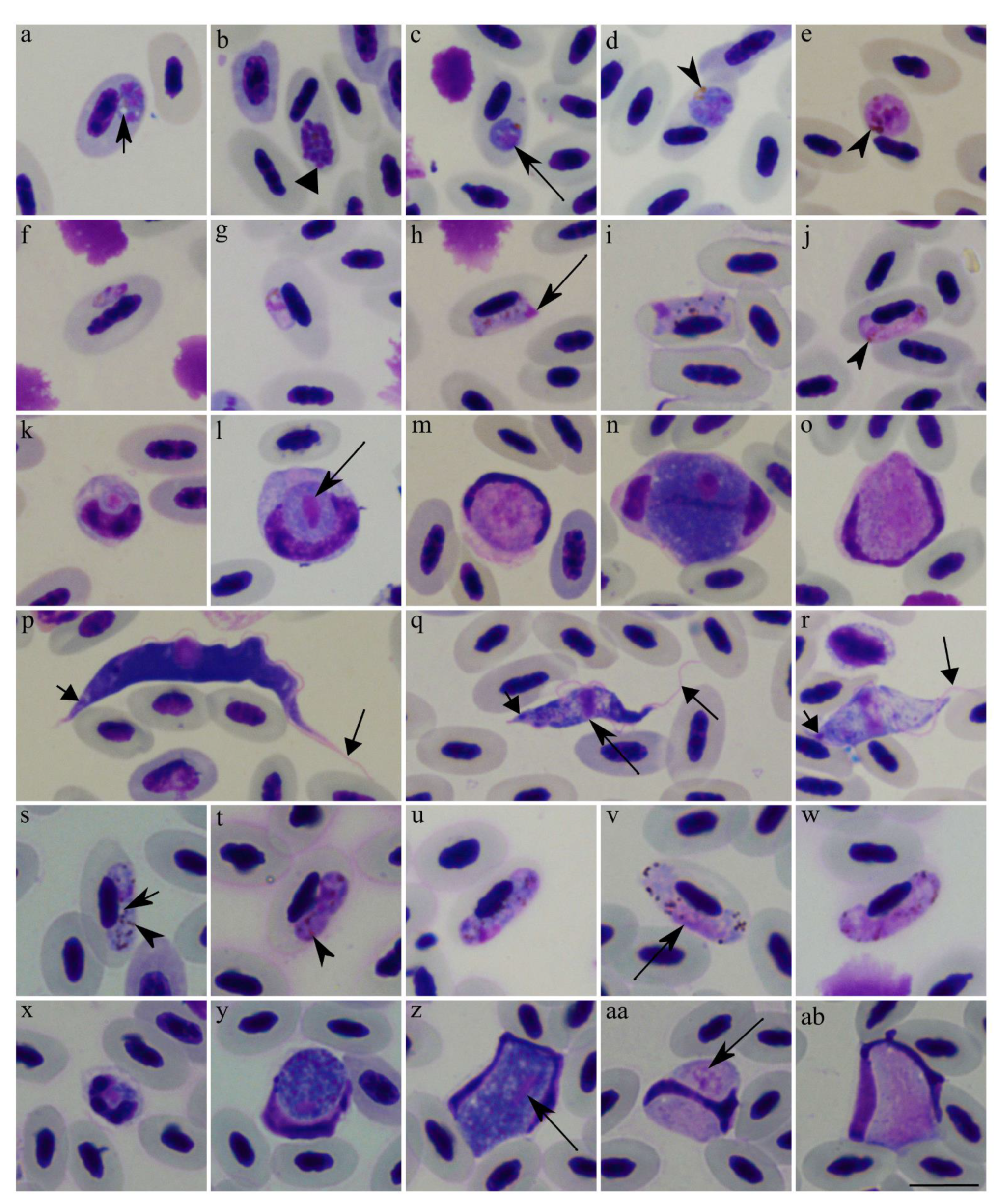
), vacuole. Arrowhead ( Animals 11 03490 i002), pigment granules. Long arrow ( Animals 11 03490 i003), parasite nucleus. Short triangle arrow ( Animals 11 03490 i004), kinetoplast. Long triangle arrow ( Animals 11 03490 i005), flagellum. Scale bar: 10 µm. Methanol fixed and Giemsa-stained blood films.
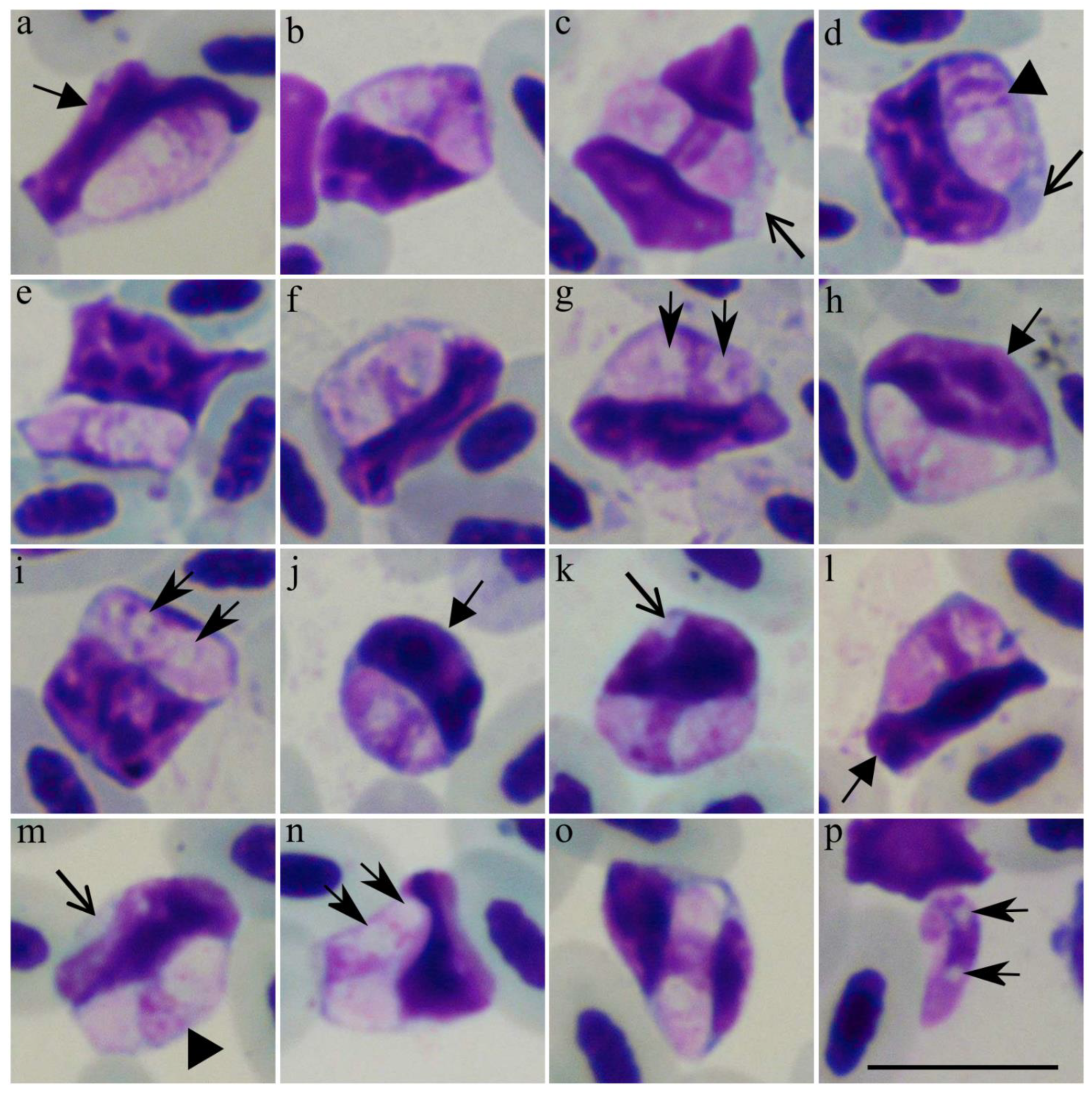
Animals 11 03490 g001
Figure 2. Intracellular (ao) and a free (p) sporozoites of Lankesterella bivacuolata n. sp. (18S rRNA lineage LanTurd1) from a Eurasian blackbird (Turdus merula). Parasites develop in mononuclear leukocytes, which can be readily distinguished from other leukocytes due to basophilic staining of the cell cytoplasm (c,d,l,m). Note that sporozoites markedly deform the host cell nuclei, which were frequently distorted and assumed elongated shapes (a,f,g,k,m). Two vacuole-like structures were closely appressed to the parasite nuclei (g,i,n,p), which are elongate and occupy the entire width of the sporozoites (d,m). Triangle arrowhead (►), parasite nucleus. Short-barbed arrow ( Animals 11 03490 i001), vacuole-like structures. Short triangle arrow ( Animals 11 03490 i004), host cell nuclei. Simple wide arrow ( Animals 11 03490 i006), host cell cytoplasm. Scale bar: 10 µm. Methanol fixed and Giemsa-stained blood films.
Animals 11 03490 g002
Figure 3. Bayesian phylogenetic inference tree based on the partial 18S rRNA gene sequences of Lankesterella parasites and other closely related Apicomplexa species. The tree was rooted with haemosporidian parasite DNA sequences. Lankesterella bivacuolata n. sp. (LanTurd1) found in a Eurasian blackbird (Turdus merula) and other avian Lankesterella parasites clustered together in one well supported clade (grey box). The name of the new species is given in bold font. GenBank accession numbers are provided for all the sequences. Parasite 18S rRNA lineage codes of Lankesterella parasites are given in parentheses. Nodal support values indicate Bayesian posterior probability.
Animals 11 03490 g003
Figure 4. Splendidofilaria mavis from a Eurasian blackbird (Turdus merula) (ag) and a Song thrush (Turdus philomelos) (h,i). Male anterior extremity (alive worm) (a). Female anterior extremity (alive worm) (b). Female posterior extremity (alive worm) (c). Male posterior extremity (ethanol-fixed worm) (d). Microfilariae from peripheral blood, Giemsa-stained (e,i). Microfilariae obtained from female uterus, Giemsa-stained (f). Microfilariae from liver blood films, Giemsa-stained (g). Alive microfilaria in blood (h). Scale bars: 100 µm (a,b); 50 µm (ci).
Animals 11 03490 g004
Figure 5. Bayesian phylogenetic inference tree based on partial 28S nuclear gene sequences of Splendidofilaria mavis and other closely related filarioid nematodes. The tree was rooted with Ascaria galli DNA sequence. Splendidofilaria mavis found in a Eurasian blackbird (Turdus merula) and a Song thrush (Turdus philomelos) and Splendidofilaria bartletti clustered together in a well-supported clade (grey box). Sequences that were obtained in this study are given in bold. The GenBank accession numbers are provided. Nodal support values indicate Bayesian posterior probability.
Animals 11 03490 g005
Figure 6. Circadian rhythms of parasites infecting a Eurasian blackbird (Turdus merula) (ac,e,g,i) and a Song thrush (Turdus philomelos) (d,f,h). Plasmodium matutinum (cytochrome b lineage pLINN1) (a), Haemoproteus minutus (hTURDUS2) (c), Haemoproteus homominutus (unknown lineage) (d), Leucocytozoon sp. (co-infection of lNEVE01 and lTURMER15) (e), Leucocytozoon sp. (lSTUR1) (f), Lankesterella bivacuolata n. sp. (18S rRNA lineage LanTurd1) (g), Trypanosoma avium and Trypanosoma everetti group (i), and Splendidofilaria mavis (b,h). Intensity of parasitemia and day hours of the blood sampling were indicated on the ordinate and abscissa, respectively. Vertical bars indicate standard errors.
Animals 11 03490 g006
Table 1. Methodology used to determine parasitemia intensity and PCR-protocol applied for DNA amplification of blood parasites found in the Eurasian blackbird (Turdus merula) and the Song thrush (Turdus philomelos).
Parasite GenusParasitemia Intensity DeterminationPCR-ProtocolGene Amplified
PlasmodiumInfected cells on 20,000 erythrocytes [24] [25]Cytochrome b (cytb)
HaemoproteusInfected cells on 20,000 erythrocytes [24][25]cytb
LeucocytozoonInfected cells on 20,000 erythrocytes [24][25]cytb
LankesterellaInfected cells on 100 mononuclear leukocytes [26][26]18S ribosomal RNA (18S)
TrypanosomaTrypomastigotes seen in 1 cm2 of blood film[27]18S
SplendidofilariaMicrofilariae seen 1 µL of blood [4]28S nuclear (28S) and cytochrome oxidase I (coxI)
Table 2. Morphometry of sporozoites of Lankesterella bivacuolata n. sp. (18S rRNA lineage LanTurd1). All the measurements are given in micrometres.
FeatureMeasurements (n = 21) a
Sporozoite
Length8.2–9.9 (9.1 ± 0.4) b
Width2.6–4.0 (3.4 ± 0.4)
Area21.9–29.3 (25.7 ± 2.2)
Sporozoite nucleus
Length1.0–2.5 (2.0 ± 0.4)
Width1.5–3.6 (2.7 ± 0.6)
Area2.7–5.4 (3.9 ± 0.7)
Sporozoite vacuole-like structure
Length1.0–2.0 (1.5 ± 0.2)
Width1.4–2.9 (2.2 ± 0.4)
Area1.2–3.8 (2.8 ± 0.7)
a Number of measured parasites. b Minimum and maximum values are provided, followed in parentheses by the arithmetic mean and standard deviation.
Table 3. Measurements of Splendidofilaria mavis microfilariae from the blood, lungs, and uterus form the Eurasian blackbird (Turdus merula) and the Song thrush (Turdus philomelos).
Turdus merulaTurdus philomelos
ParameterBlood (n = 25)Lungs (n = 20)Uterus (n = 7)Blood (n = 10)Lungs (n = 17)
MC%MC%MC%MC%MC%
Length 83–120-86–143-75–95-96–124-73–113-
Maximum width4–6-5–6-3–5-4–6-4–6-
Inner body length6–14-5–15-2–5-6–12-4–13-
Cephalic space1–4-1–7-2–6-1–3-1–7-
Distance from AE to:
nerve ring 16–2618–2417–3419–2818–2522–2618–2717–2416–2718–25
excretory pore24–4728–3526–4930–3524–3130–3631–4029–3522–3926–36
inner body anterior end44–7047–5644–8146–5741–5352–5653–6951–5639–6248–55
inner body posterior end50–8557–6551–9055–6544–5855–6160–7958–6443–7358–67
anal pore67–10881–8770–11980–8560–7879–8282–10583–8759–9680–88
MC, morphometric characteristics are given in micrometres; %, values of fixed points expressed in percentage; (-), not evaluated; AE, anterior extremity.
Table 4. Measurements of adult Splendidofilaria mavis from the Eurasian blackbird (Turdus merula) and the Song thrush (Turdus philomelos) that were examined in this study. All measurements are given in micrometres, except for body length, which is given in millimetres.
FeatureTurdus merulaTurdus philomelos
Male (n = 1)Female (n = 4)Male (n = 1)Female (n = 1)
Body length811–17815
Maximum width345320–403253380
Oesophagus length8901010–1026729791
Vagina length-108–128-82
Left spicule length116-103-
Right spicule length82-91-
Nerve ring from AE118128–182118126
Vulva from AE-466–646-795
Testes from AE547-546-
Ovary from PE-86–239-447
Cloaca/Anus from PE5373–11070153
AE, anterior extremity; PE, posterior extremity; (-) not evaluated.
Publisher’s Note: MDPI stays neutral with regard to jurisdictional claims in published maps and institutional affiliations.

