Zoonotic Diseases: Etiology, Impact, and Control

 ,
,  ,
,  ,
,  ,
,
Abstract
:1. Introduction
2. Classification of Zoonoses
3. Zoonoses of Domestic Animals
4. Zoonoses of Pets, Companion Animals, and Birds
5. Zoonoses of Fish and Aquatic Environments
6. Zoonoses Associated with Food-Borne Pathogens
7. Potential Zoonoses Transmitted by Edible Insects
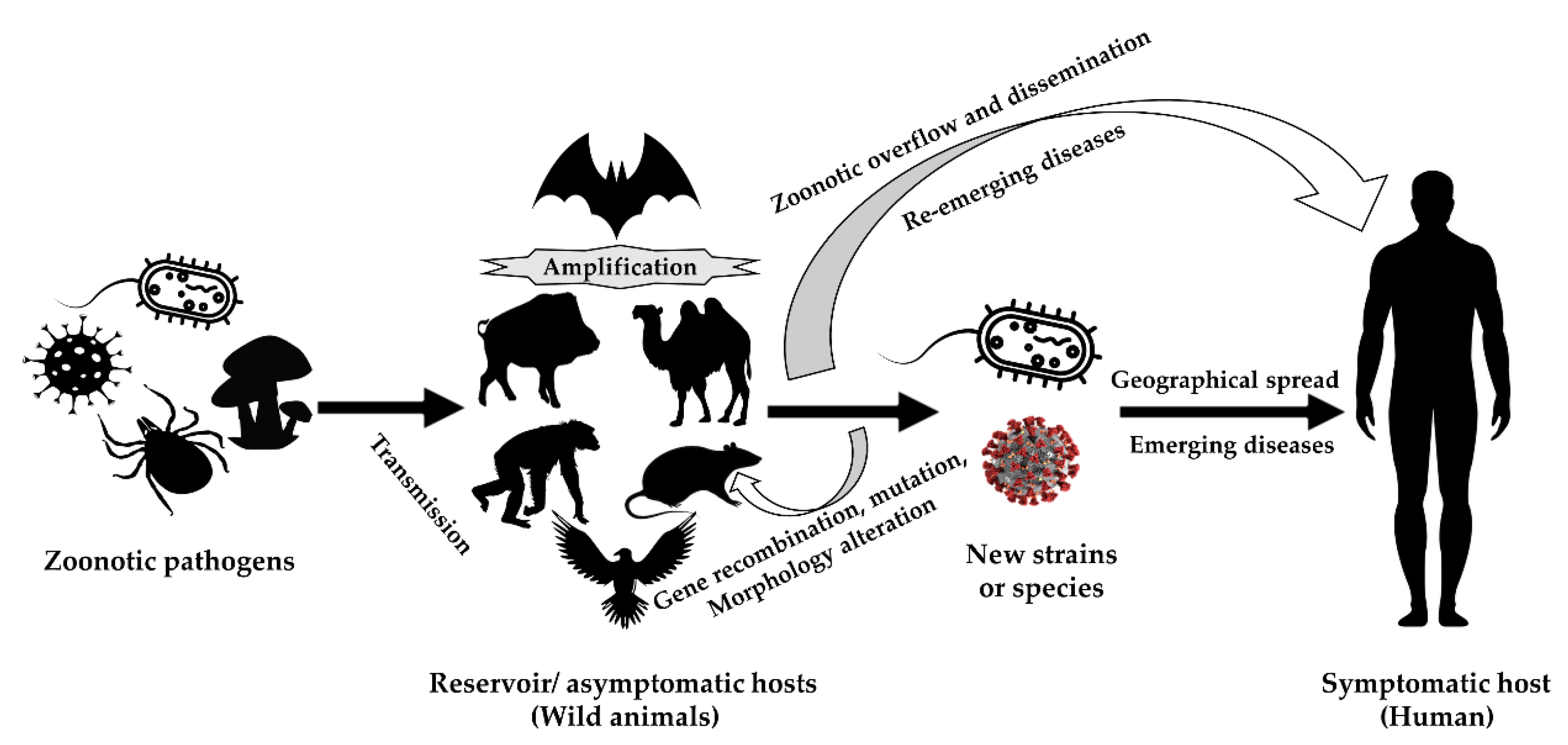
8. Emerging and Re-Emerging Zoonoses
8.1. Wild Animals and Re-Emerging Zoonoses
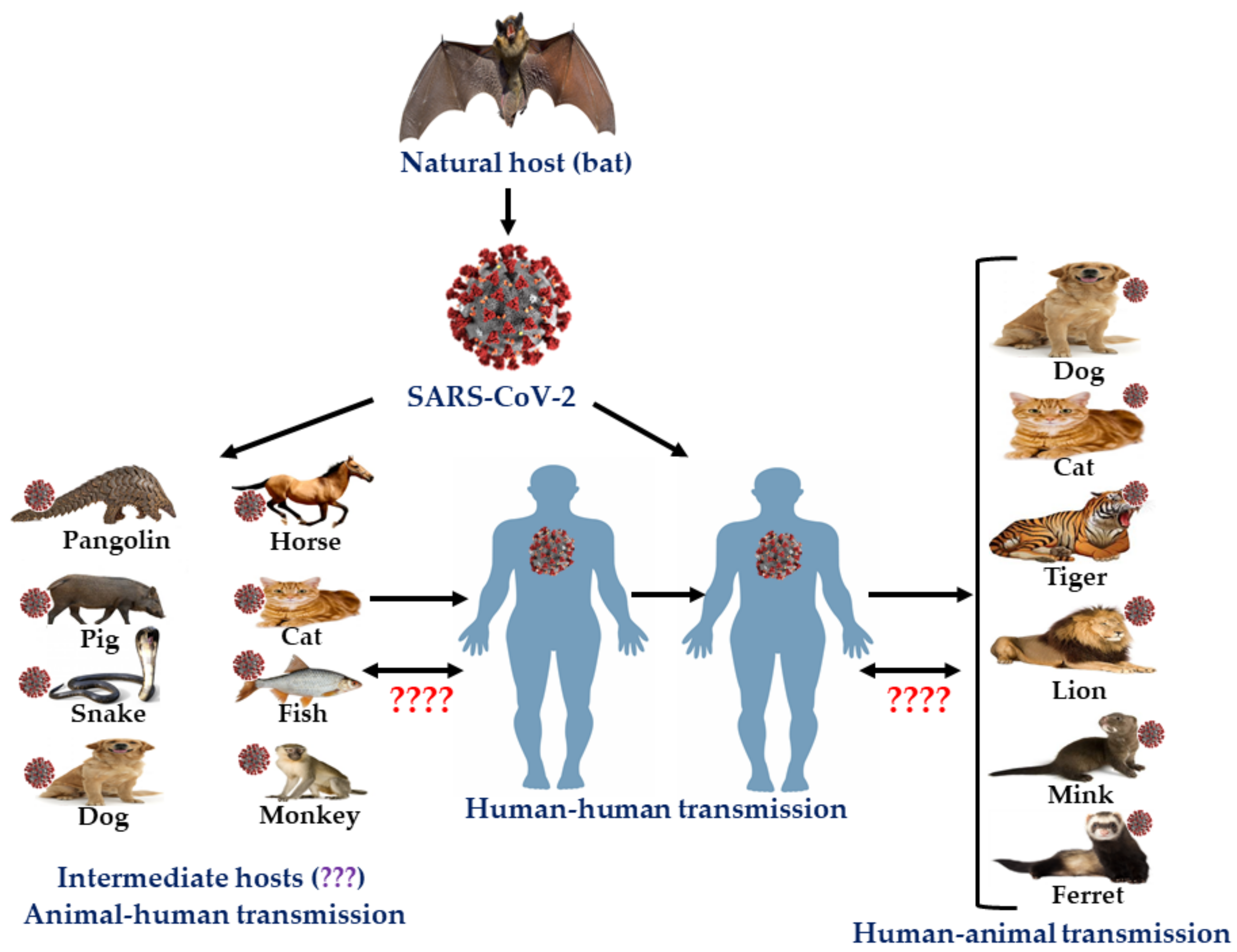
8.2. Zoonotic Coronaviruses
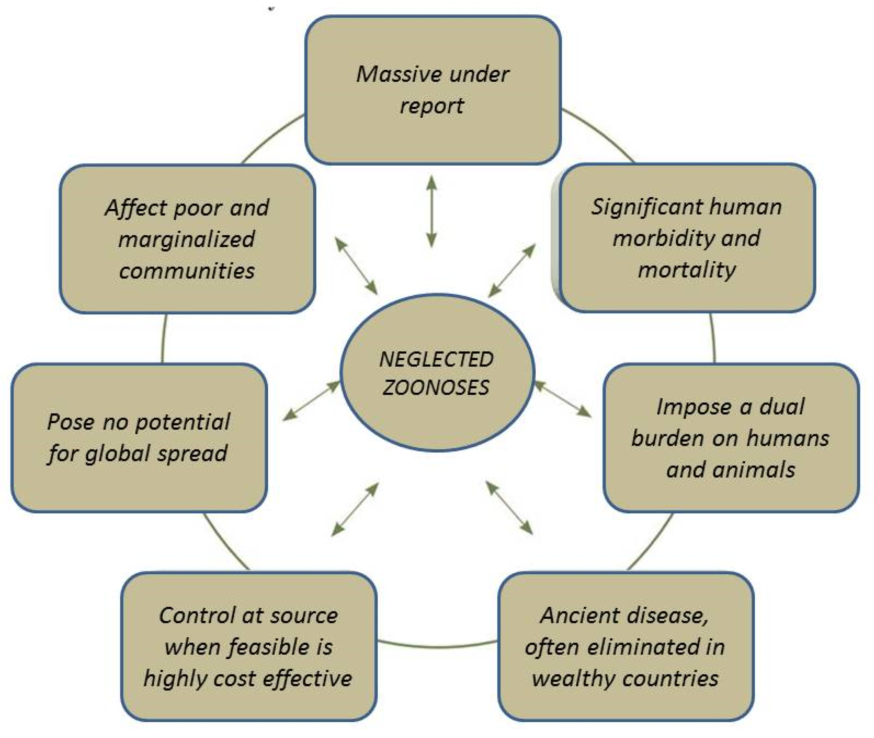
9. Neglected Zoonoses
10. Impact of Zoonoses
11. Control of Zoonoses
- Pathogen surveillance to detect and identify pathogens.
- Serological surveillance to detect the presence of pathogens in the blood of humans or animals through monitoring immune responses.
- Syndrome surveillance to determine the propensity of diseases through data analysis based on symptoms. This analysis-based surveillance cannot be used identify the presence of pathogens.
- Risk surveillance to detect risk factors responsible for the transmission of disease. This control strategy cannot be used to determine the clinical features of multifarious diseases along with their prevalence.
Zoonoses and One Health
12. Recommendations
- Active and wider zoonoses surveillance and monitoring with advanced tools like satellite-based remote sensing system and molecular epidemiological tools.
- Disease reporting and notification service.
- Giving priority to zoonoses and action team formation.
- Available diagnostic facilities and skilled manpower.
- Cooperation at regional, national, subnational, and international levels.
- One health-based approach comprising both veterinarians and medical doctors in addition to environmental experts and other professionals.
- Ensuring adequate regular and emergency funding.
- Mass campaigning on public awareness on zoonoses.
- More research on disease epidemiology, risk factors, pathogen virulence, host biology, and vector biology.
- Wildlife monitoring and wildlife protection.
- Ensure safe food production of animal origin.
- Ensure safety of infectious laboratories to avoid the accidental spread of zoonotic infections and bioterrorism.
- Protection of environment.
- National and international educational programs to make people aware of zoonoses and hygiene.
13. Conclusions
Author Contributions
Funding
Acknowledgments
Conflicts of Interest
References
- Thompson, A.; Kutz, S. Introduction to the Special Issue on ‘Emerging Zoonoses and Wildlife’. Int. J. Parasitol. Parasites Wildl. 2019, 9, 322. [Google Scholar] [CrossRef]
- World Health Organization. Asia Pacific Strategy for Emerging Diseases: 2010. Manila: WHO Regional Office for the Western Pacific. Available online: https://iris.wpro.who.int/bitstream/handle/10665.1/7819/9789290615040_eng.pdf (accessed on 20 July 2020).
- Slingenbergh, J. World Livestock 2013: Changing Disease Landscapes; Food and Agriculture Organization of the United Nations (FAO): Rome, Italy, 2013; p. 2. [Google Scholar]
- World Health Organization. WHO Health Topic Page: Zoonoses. Available online: https://www.who.int/topics/zoonoses/en/ (accessed on 20 July 2020).
- Taylor, L.H.; Latham, S.M.; Woolhouse, M.E. Risk factors for human disease emergence. Philos. Trans. R. Soc. Lond. B Biol. Sci. 2001, 356, 983–989. [Google Scholar] [CrossRef] [PubMed]
- Grace, D.; Mutua, F.; Ochungo, P.; Kruska, R.; Jones, K.; Brierley, L.; Lapar, L.; Said, M.; Herrero, M.; Phuc, P.M.; et al. Mapping of poverty and likely zoonoses hotspots. In Zoonoses Project 4. Report to the UK Department for International Development; International Livestock Research Institute: Nairobi, Kenya, 2012. [Google Scholar]
- Chomel, B.B. Zoonoses. In Encyclopedia of Microbiology, 3rd ed.; Elsevier Inc., University of California: Davis, CA, USA, 2009; pp. 820–829. [Google Scholar]
- Hubálek, Z. Emerging human infectious diseases: Anthroponoses, zoonoses, and sapronoses. Emerg. Infect. Dis. 2003, 9, 403–404. [Google Scholar] [CrossRef]
- McDaniel, C.J.; Cardwell, D.M.; Moeller, R.B.; Gray, G.C. Humans and cattle: A review of bovine zoonoses. Vector Borne Zoonotic Dis. 2014, 14, 1–19. [Google Scholar] [CrossRef] [PubMed]
- Bae, S.E.; Son, H.S. Classification of viral zoonosis through receptor pattern analysis. BMC Bioinform. 2011, 12, 96. [Google Scholar] [CrossRef] [PubMed] [Green Version]
- Mortimer, P.P. Influenza: The centennial of a zoonosis. Rev. Med. Virol. 2019, 29, e2030. [Google Scholar] [CrossRef] [Green Version]
- Huang, Y.J.S.; Higgs, S.; Vanlandingham, D.L. Arbovirus-mosquito vector-host interactions and the impact on transmission and disease pathogenesis of arboviruses. Front. Microbiol. 2019, 10, 22. [Google Scholar] [CrossRef]
- Pavlovsky, E.N. Natural Nidality of Transmissible Diseases with Special Reference to the Landscape Epidemiology of Zooanthroponoses; University of Illinois Press: Champaign, IL, USA, 1966. [Google Scholar]
- Beaty, B.J.; Marquardt, W.C. The Biology of Disease Vector; University Press of Colorado: Niwot, CO, USA, 1996. [Google Scholar]
- Somov, G.P.; Litvin, V.J. Saprophytism and Parasitism of Pathogenic Bacteria—Ecological Aspects; Nauka: Novosibirsk, Russia, 1988. (In Russian) [Google Scholar]
- Schwabe, C.W. Veterinary medicine and human health. In Veterinary Medicine and Human Health; The Williams & Wilkins Company, 428 E. Preston St.: Baltimore, MD, USA, 1964. [Google Scholar]
- Olayemi, A.; Adesina, A.S.; Strecker, T.; Magassouba, N.F.; Fichet-Calvet, E. Determining Ancestry between Rodent-and Human-Derived Virus Sequences in Endemic Foci: Towards a More Integral Molecular Epidemiology of Lassa Fever within West Africa. Biology 2020, 9, 26. [Google Scholar] [CrossRef] [Green Version]
- Cerdà-Cuéllar, M.; Moré, E.; Ayats, T.; Aguilera, M.; Muñoz-González, S.; Antilles, N.; Ryan, P.G.; González-Solís, J. Do humans spread zoonotic enteric bacteria in Antarctica? Sci. Total Environ. 2019, 654, 190–196. [Google Scholar] [CrossRef]
- Adesokan, H.K.; Akinseye, V.O.; Streicher, E.M.; Van Helden, P.; Warren, R.M.; Cadmus, S.I. Reverse zoonotic tuberculosis transmission from an emerging Uganda I strain between pastoralists and cattle in South-Eastern Nigeria. BMC Vet. Res. 2019, 15, 1–7. [Google Scholar] [CrossRef] [Green Version]
- Messenger, A.M.; Barnes, A.N.; Gray, G.C. Reverse zoonotic disease transmission (zooanthroponosis): A systematic review of seldom-documented human biological threats to animals. PLoS ONE 2014, 9, e89055. [Google Scholar] [CrossRef] [PubMed] [Green Version]
- County of Los Angeles Public Health. Overview of Zoonoses. Available online: http://www.lapublichealth.org/vet/guides/vetzooman.htm (accessed on 20 July 2020).
- Morand, S.; McIntyre, K.M.; Baylis, M. Domesticated animals and human infectious diseases of zoonotic origins: Domestication time matters. Infect. Genet. Evol. 2014, 24, 76–81. [Google Scholar] [CrossRef] [PubMed] [Green Version]
- McNeill, W.H. Plagues and People; Anchor Press: New York, NY, USA, 1976. [Google Scholar]
- Klous, G.; Huss, A.; Heederik, D.J.; Coutinho, R.A. Human–livestock contacts and their relationship to transmission of zoonotic pathogens, a systematic review of literature. One Health 2016, 2, 65–76. [Google Scholar] [CrossRef] [PubMed] [Green Version]
- Pearce-Duvet, J.M. The origin of human pathogens: Evaluating the role of agriculture and domestic animals in the evolution of human disease. Biol. Rev. 2006, 81, 369–382. [Google Scholar] [CrossRef]
- Samad, M.A. Public health threat caused by zoonotic diseases in Bangladesh. Bangladesh J. Vet. Med. 2011, 9, 95–120. [Google Scholar] [CrossRef] [Green Version]
- Ghasemzadeh, I.; Namazi, S.H. Review of bacterial and viral zoonotic infections transmitted by dogs. J. Med. Life 2015, 8, 1–5. [Google Scholar]
- Goel, A.K. Anthrax: A disease of biowarfare and public health importance. World J. Clin. Cases 2015, 3, 20–33. [Google Scholar] [CrossRef]
- Kamal, S.M.; Rashid, A.K.; Bakar, M.A.; Ahad, M.A. Anthrax: An update. Asian Pac. J. Trop. Biomed. 2011, 1, 496–501. [Google Scholar] [CrossRef] [Green Version]
- Torgerson, P.R.; Torgerson, D.J. Public health and bovine tuberculosis: What’s all the fuss about? Trends Microbiol. 2010, 18, 67–72. [Google Scholar] [CrossRef] [Green Version]
- Bayraktar, B.; Bulut, E.; Barış, A.B.; Toksoy, B.; Dalgıc, N.; Celikkan, C.; Sevgi, D. Species distribution of the Mycobacterium tuberculosis complex in clinical isolates from 2007 to 2010 in Turkey: A prospective study. J. Clin. Microbiol. 2011, 49, 3837–3841. [Google Scholar] [CrossRef] [Green Version]
- Bayraktar, B.; Togay, A.; Gencer, H.; Kockaya, T.; Dalgic, N.; Bulut, E. Mycobacterium caprae causing lymphadenitis in a child. Pediatr. Infect. Dis. J. 2011, 30, 1012–1013. [Google Scholar] [PubMed]
- Moda, G.; Daborn, C.J.; Grange, J.M.; Cosivi, O. The zoonotic importance of Mycobacterium bovis. Tuber. Lung Dis. 1996, 77, 103–108. [Google Scholar] [CrossRef]
- Ocepek, M.; Pate, M.; Žolnir-Dovč, M.; Poljak, M. Transmission of Mycobacterium tuberculosis from human to cattle. J. Clin. Microbiol. 2005, 43, 3555–3557. [Google Scholar] [CrossRef] [Green Version]
- Hull, N.C.; Schumaker, B.A. Comparisons of brucellosis between human and veterinary medicine. Infect. Ecol. Epidemiol. 2018, 8, 1500846. [Google Scholar] [CrossRef]
- WHO. The Control of Neglected Zoonotic Diseases: From Advocacy to Action: Report of the Fourth International Meeting Held at WHO Headquarters, Geneva, Switzerland, 19–20 November 2014; World Health Organization: Geneva, Switzerland, 2015; p. 44. [Google Scholar]
- Corbel, M.J.; Alton, G.G.; Banai, M.; Díaz, R.; Dranovskaia, B.A.; Elberg, S.S.; Garin-Bastuji, B.; Kolar, J.; Mantovani, A.; Mousa, A.M.; et al. Brucellosis in Humans and Animals; WHO Press: Geneva, Switzerland, 2006. [Google Scholar]
- Rahman, M.S.; Han, J.C.; Park, J.; Lee, J.H.; Eo, S.K.; Chae, J.S. Prevalence of brucellosis and its association with reproductive problems in cows in Bangladesh. Vet. Rec. 2006, 159, 180–182. [Google Scholar] [CrossRef] [PubMed]
- Krebs, J.W.; Mandel, E.J.; Swerdlow, D.L.; Rupprecht, C.E. Rabies surveillance in the United States during 2003. J. Am. Vet. Med. Assoc. 2004, 225, 1837–1849. [Google Scholar] [CrossRef] [Green Version]
- Tang, X.; Luo, M.; Zhang, S.; Fooks, A.R.; Hu, R.; Tu, C. Pivotal role of dogs in rabies transmission, China. Emerg. Infect. Dis. 2005, 11, 1970–1972. [Google Scholar] [CrossRef]
- Liu, Q.; Wang, X.; Liu, B.; Gong, Y.; Mkandawire, N.; Li, W.; Fu, W.; Li, L.; Gan, Y.; Shi, J.; et al. Improper wound treatment and delay of rabies post-exposure prophylaxis of animal bite victims in China: Prevalence and determinants. PLoS Neglect. Trop. Dis. 2017, 11, e0005663. [Google Scholar] [CrossRef] [Green Version]
- World Health Organization. Rabies vaccines: WHO position paper. Wkly. Epidemiol. Rec. 2018, 93, 201–220. [Google Scholar]
- Ghosh, S.; Rana, M.S.; Islam, M.K.; Chowdhury, S.; Haider, N.; Kafi, M.A.H.; Ullah, S.M.; Shah, M.R.A.; Jahan, A.A.; Mursalin, H.S.; et al. Trends and clinico-epidemiological features of human rabies cases in Bangladesh 2006–2018. Sci. Rep. 2020, 10, 1–11. [Google Scholar] [CrossRef] [Green Version]
- Plotkin, S.; Orenstein, W.; Offit, P.; Edwards, M.K. Vaccine. In Plotkin’s Vaccines, 7th ed.; Elsevier: Philadelphia, PA, USA, 2017; pp. 918–942. [Google Scholar]
- Jackson, A.C. Human rabies: A 2016 update. Curr. Infect. Dis. Rep. 2016, 18, 38. [Google Scholar] [CrossRef] [PubMed]
- Mitrabhakdi, E.; Shuangshoti, S.; Wannakrairot, P.; Lewis, R.A.; Susuki, K.; Laothamatas, J.; Hemachudha, T. Difference in neuropathogenetic mechanisms in human furious and paralytic rabies. J. Neurol. Sci. 2005, 238, 3–10. [Google Scholar] [CrossRef] [PubMed]
- Hemachudha, T.; Laothamatas, J.; Rupprecht, C.E. Human rabies: A disease of complex neuropathogenetic mechanisms and diagnostic challenges. Lancet Neurol. 2002, 1, 101–109. [Google Scholar] [CrossRef]
- Dimaano, E.M.; Scholand, S.J.; Alera, M.T.P.; Belandres, D.B. Clinical and epidemiological features of human rabies cases in the Philippines: A review from 1987 to 2006. Int. J. Infect. Dis. 2011, 15, e495–e499. [Google Scholar] [CrossRef] [PubMed] [Green Version]
- Chomel, B.B.; Sun, B. Zoonoses in the bedroom. Emerg. Infect. Dis. 2011, 17, 167–172. [Google Scholar] [CrossRef]
- Halsby, K.D.; Walsh, A.L.; Campbell, C.; Hewitt, K.; Morgan, D. Healthy animals, healthy people: Zoonosis risk from animal contact in pet shops, a systematic review of the literature. PLoS ONE 2014, 9, e89309. [Google Scholar] [CrossRef] [PubMed]
- Day, M.J. Pet-Related Infections. Am. Fam. Physician. 2016, 94, 794–802. [Google Scholar]
- Jacob, J.; Lorber, B. Diseases transmitted by man’s best friend: The dog. Infect. Leis. 2015, 3, 111–131. [Google Scholar] [CrossRef] [Green Version]
- Boseret, G.; Losson, B.; Mainil, J.G.; Thiry, E.; Saegerman, C. Zoonoses in pet birds: Review and perspectives. Vet. Res. 2013, 44, 36. [Google Scholar] [CrossRef] [Green Version]
- Moro, C.V.; Chauve, C.; Zenner, L. Vectorial role of some dermanyssoid mites (Acari, Mesostigmata, Dermanyssoidea). Parasite 2005, 12, 99–109. [Google Scholar] [CrossRef]
- Dorrestein, G.M. Bacterial and parasitic diseases of passerines. Vet. Clin. North Am. Exot. Anim. Pract. 2009, 12, 433–451. [Google Scholar] [CrossRef] [PubMed]
- Jorn, K.S.; Thompson, K.M.; Larson, J.M.; Blair, J.E. Polly can make you sick: Pet bird-associated diseases. Cleve. Clin. J. Med. 2009, 76, 235–243. [Google Scholar] [CrossRef] [PubMed] [Green Version]
- Zaman, S.B.; Sobur, M.A.; Hossain, M.J.; Pondit, A.; Khatun, M.M.; Choudhury, M.A.; Tawyabur, M.; Rahman, M.T. Molecular detection of methicillin-resistant Staphylococcus aureus (MRSA) in ornamental birds having public health significance. J. Bangladesh Agril. Univ. 2020, 18, 415–420. [Google Scholar] [CrossRef]
- Kauffman, M.D.; LeJeune, J. European starlings (Sturnus vulgaris) challenged with Escherichia coli O157 can carry and transmit the human pathogen to cattle. Lett. Appl. Microbiol. 2011, 53, 596–601. [Google Scholar] [CrossRef] [PubMed]
- Belchior, E.; Barataud, D.; Ollivier, R.; Capek, I.; Laroucau, K.; De Barbeyrac, B.; Hubert, B. Psittacosis outbreak after participation in a bird fair, Western France, December 2008. Epidemiol. Infect. 2011, 139, 1637–1641. [Google Scholar] [CrossRef] [Green Version]
- Vanrompay, D.; Harkinezhad, T.; Van de Walle, M.; Beeckman, D.; Van Droogenbroeck, C.; Verminnen, K.; Leten, R.; Martel, A.; Cauwerts, K. Chlamydophila psittaci transmission from pet birds to humans. Emerg. Infect. Dis. 2007, 13, 1108–1110. [Google Scholar] [CrossRef]
- Chomel, B.B. Emerging and Re-Emerging Zoonoses of Dogs and Cats. Animals 2014, 4, 434–445. [Google Scholar] [CrossRef] [Green Version]
- Burgos-Cáceres, S. Canine rabies: A looming threat to public health. Animals 2011, 1, 326–342. [Google Scholar] [CrossRef] [Green Version]
- Faires, M.C.; Tater, K.C.; Weese, J.S. An investigation of methicillin-resistant Staphylococcus aureus colonization in people and pets in the same household with an infected person or infected pet. J. Am. Vet. Med. Assoc. 2009, 235, 540–543. [Google Scholar] [CrossRef] [Green Version]
- Klotz, S.A.; Ianas, V.; Elliott, S.P. Cat-scratch disease. Am. Fam. Physician 2011, 83, 152–155. [Google Scholar]
- Boylan, S. Zoonoses associated with fish. Vet. Clin. Exot. Anim. Pract. 2011, 14, 427–438. [Google Scholar] [CrossRef] [PubMed]
- Alworth, L.C.; Harvey, S.B. IACUC issues associated with amphibian research. ILAR J. 2007, 48, 278–289. [Google Scholar] [CrossRef] [PubMed]
- Haenan, O.L.M.; Evans, J.J.; Berthe, F. Bacterial infections from aquatic species: Potential for and prevention of contact zoonoses. Rev. Sci. Tech. 2013, 32, 497–507. [Google Scholar] [CrossRef] [PubMed]
- Abbot, S.L.; Janda, J.M.; Johnson, J.A.; Farmer, J.J. Vibrio and related organisms. In Manual of Clinical Microbiology, 9th ed.; Murray, P.R., Barron, E.J., Jorgensen, J.H., Landry, M.L., Pfaller, M.A., Eds.; ASM Press: Washington, DC, USA, 2007; pp. 723–733. [Google Scholar]
- Austin, B. Vibrios as causal agents of zoonoses. Vet. Microbiol. 2010, 140, 310–317. [Google Scholar] [CrossRef] [PubMed] [Green Version]
- Zhang, Q.; Dong, X.; Chen, B.; Zhang, Y.; Zu, Y.; Li, W. Zebrafish as a useful model for zoonotic Vibrio parahaemolyticus pathogenicity in fish and human. Dev. Comp. Immunol. 2016, 55, 159–168. [Google Scholar] [CrossRef] [PubMed]
- Zereen, F.; Akter, S.; Sobur, M.A.; Hossain, M.T.; Rahman, M.T. Molecular detection of Vibrio cholerae from human stool collected from SK Hospital, Mymensingh, and their antibiogram. J. Adv. Vet. Anim. Res. 2019, 6, 451–455. [Google Scholar] [CrossRef]
- Chen, B.Y.; Wang, C.Y.; Wang, C.L.; Fan, Y.C.; Weng, I.T.; Chou, C.H. Prevalence and persistence of Listeria monocytogenes in ready-to-eat tilapia sashimi processing plants. J. Food Prot. 2016, 79, 1898–1903. [Google Scholar] [CrossRef]
- Kušar, D.; Zajc, U.; Jenčič, V.; Ocepek, M.; Higgins, J.; Žolnir-Dovč, M.; Pate, M. Mycobacteria in aquarium fish: Results of a 3-year survey indicate caution required in handling pet-shop fish. J. Fish Dis. 2017, 40, 773–784. [Google Scholar] [CrossRef]
- Gauthier, D.T.; Rhodes, M.W. Mycobacteriosis in fishes: A review. Vet. J. 2009, 180, 33–47. [Google Scholar] [CrossRef]
- Vega-López, F. Mycobacterium marinum Infection: Fish Tank Granuloma. In Hunter’s Tropical Medicine and Emerging Infectious Diseases, 10th ed.; Elsevier Inc.: Amsterdam, The Netherlands, 2020; pp. 569–570. [Google Scholar]
- Sunil, V.; Harris, A.W.; Sine, B.; Holt, A.M.; Noseworthy, A.L.; Sider, D.; Jamieson, F.B.; White, S.; Johnston, C.; Spohn, O. Investigation of a community cluster of cutaneous Mycobacterium marinum infection, an emerging zoonotic pathogen in aquaculture industry, Haliburton, Kawartha, Pine Ridge District Health Unit, Ontario, Canada, July–August 2015. Zoonoses Public Health 2019, 66, 164–168. [Google Scholar] [CrossRef]
- Christopher, T.; Cassetty, M.D.; Miguel Sanchez, M.D. Mycobacterium marinum infection. Dermatol. Online J. 2004, 10, 21. [Google Scholar]
- Reidarson, T. Cetacea. In Zoo and Wild Animal Medicine, 5th ed.; Fowler, M., Ed.; Saunders: St. Louis, MO, USA, 2003; pp. 442–459. [Google Scholar]
- Dunn, L. Bacterial and mycotic diseases of cetaceans and pinnipeds. In CRC Handbook of Marine Mammal Medicine: Health, Disease, and Rehabilitation; Dierauf, L., Ed.; CRC Press: Boca Raton, FL, USA, 1990; pp. 73–87. [Google Scholar]
- Gauthier, D.T. Bacterial zoonoses of fishes: A review and appraisal of evidence for linkages between fish and human infections. Vet. J. 2015, 203, 27–35. [Google Scholar] [CrossRef] [PubMed]
- Reboli, A.C.; Farrar, W.E. Erysipelothrix rhusiopathiae: An occupational pathogen. Clin. Microbiol. Rev. 1989, 2, 354–359. [Google Scholar] [CrossRef] [PubMed]
- Wang, Q.; Chang, B.J.; Riley, T.V. Erysipelothrix rhusiopathiae. Vet. Microbiol. 2010, 140, 405–417. [Google Scholar] [CrossRef] [PubMed]
- Gorby, G.; Peacock, J. Erysipelothrix rhusiopathiae endocarditis: Microbiologic, epidemiologic, and clinical features of an occupational disease. Rev. Infect. Dis. 1988, 10, 317–325. [Google Scholar] [CrossRef]
- Klauder, J.; Kramer, D.; Nicholas, L.; Kast, C.; Groskin, L.O.R.A.I.N.E. Erysipelothrix rhusiopathiae septicemia: Diagnosis and treatment: Report of fatal case of erysipeloid. JAMA 1943, 122, 938–943. [Google Scholar] [CrossRef]
- Principe, L.; Bracco, S.; Mauri, C.; Tonolo, S.; Pini, B.; Luzzaro, F. Erysipelothrix rhusiopathiae bacteremia without endocarditis: Rapid identification from positive blood culture by MALDI-TOF mass spectrometry. A case report and literature review. Infect. Dis. Rep. 2016, 8, 6368. [Google Scholar] [CrossRef] [Green Version]
- Gibello, A.; Galán-Sánchez, F.; Blanco, M.M.; Rodríguez-Iglesias, M.; Domínguez, L.; Fernández-Garayzábal, J.F. The zoonotic potential of Lactococcus garvieae: An overview on microbiology, epidemiology, virulence factors and relationship with its presence in foods. Res. Vet. Sci. 2016, 109, 59–70. [Google Scholar] [CrossRef]
- Meyburgh, C.M.; Bragg, R.R.; Boucher, C.E. Lactococcus garvieae: An emerging bacterial pathogen of fish. Dis. Aquat. Org. 2017, 123, 67–79. [Google Scholar] [CrossRef]
- Vendrell, D.; Balcázar, J.L.; Ruiz-Zarzuela, I.; De Blas, I.; Gironés, O.; Múzquiz, J.L. Lactococcus garvieae in fish: A review. Comp. Immunol. Microbiol. Infect. Dis. 2006, 29, 177–198. [Google Scholar] [CrossRef]
- Malek, A.; De la Hoz, A.; Gomez-Villegas, S.I.; Nowbakht, C.; Arias, C.A. Lactococcus garvieae, an unusual pathogen in infective endocarditis: Case report and review of the literature. BMC Infect. Dis. 2019, 19, 301. [Google Scholar] [CrossRef] [PubMed] [Green Version]
- Chan, J.F.W.; Woo, P.C.Y.; Teng, J.L.L.; Lau, S.K.P.; Leung, S.S.M.; Tam, F.C.C.; Yuen, K.Y. Primary infective spondylodiscitis caused by Lactococcus garvieae and a review of human L. garvieae infections. Infection 2011, 39, 259–264. [Google Scholar] [CrossRef] [PubMed] [Green Version]
- Kim, J.H.; Go, J.; Cho, C.R.; Kim, J.I.; Lee, M.S.; Park, S.C. First report of human acute acalculous cholecystitis caused by the fish pathogen Lactococcus garvieae. J. Clin. Microbiol. 2013, 51, 712–714. [Google Scholar] [CrossRef] [PubMed] [Green Version]
- Maekawa, S.; Yoshida, T.; Wang, P.C.; Chen, S.C. Current knowledge of nocardiosis in teleost fish. J. Fish Dis. 2018, 41, 413–419. [Google Scholar] [CrossRef]
- Austin, B.; Austin, D. Bacterial Fish Pathogens: Diseases in Farmed and Wild Fish, 3rd ed.; Springer-Praxis: Chichester, UK, 1999. [Google Scholar]
- Orchard, V.A. Nocardial infections of animals in New Zealand, 1976–1978. N. Z. Vet. J. 1979, 27, 159–165. [Google Scholar] [CrossRef]
- Walton, A.M.; Libke, K.G. Nocardiosis in animals. Vet. Med. Small. Anim. Clin. 1974, 69, 1105–1107. [Google Scholar] [PubMed]
- Lederman, E.R.; Crum, N.F. A case series and focused review of nocardiosis: Clinical and microbiologic aspects. Medicine 2004, 83, 300–313. [Google Scholar] [CrossRef]
- Newell, D.G.; Koopmans, M.; Verhoef, L.; Duizer, E.; Aidara-Kane, A.; Sprong, H.; Opsteegh, M.; Langelaar, M.; Threfall, J.; Scheutz, F.; et al. Food-borne diseases—The challenges of 20 years ago still persist while new ones continue to emerge. Int. J. Food Microbiol. 2010, 139, S3–S15. [Google Scholar] [CrossRef]
- World Health Organization. WHO Estimates of the Global Burden of Foodborne Diseases: Foodborne Disease Burden Epidemiology Reference Group 2007–2015; World Health Organization: Geneva, Switzerland, 2015; p. 225. [Google Scholar]
- Thorns, C.J. Bacterial food-borne zoonoses. Rev. Sci. Tech. 2000, 19, 226–239. [Google Scholar] [CrossRef]
- Ievy, S.; Islam, M.S.; Sobur, M.A.; Talukder, M.; Rahman, M.B.; Khan, M.F.R.; Rahman, M.T. Molecular Detection of Avian Pathogenic Escherichia coli (APEC) for the First Time in Layer Farms in Bangladesh and Their Antibiotic Resistance Patterns. Microorganisms 2020, 8, 1021. [Google Scholar] [CrossRef]
- Alam, S.B.; Mahmud, M.; Akter, R.; Hasan, M.; Sobur, A.; Nazir, K.H.M.; Noreddin, A.; Rahman, T.; El Zowalaty, M.E.; Rahman, M. Molecular detection of multidrug resistant Salmonella species isolated from broiler farm in Bangladesh. Pathogens 2020, 9, 201. [Google Scholar] [CrossRef] [PubMed] [Green Version]
- Sobur, M.A.; Sabuj, A.A.M.; Sarker, R.; Rahman, A.M.M.T.; Kabir, S.M.L.; Rahman, M.T. Antibiotic-resistant Escherichia coli and Salmonella spp. associated with dairy cattle and farm environment having public health significance. Vet. World 2019, 12, 984–993. [Google Scholar] [CrossRef] [PubMed] [Green Version]
- Treacy, J.; Jenkins, C.; Paranthaman, K.; Jorgensen, F.; Mueller-Doblies, D.; Anjum, M.; Kaindama, L.; Hartman, H.; Kirchner, M.; Carson, T.; et al. Outbreak of Shiga toxin-producing Escherichia coli O157: H7 linked to raw drinking milk resolved by rapid application of advanced pathogen characterisation methods, England, August to October 2017. Eurosurveill. 2019, 24, 1800191. [Google Scholar] [CrossRef]
- Yara, D.A.; Greig, D.R.; Gally, D.L.; Dallman, T.J.; Jenkins, C. Comparison of Shiga toxin-encoding bacteriophages in highly pathogenic strains of Shiga toxin-producing Escherichia coli O157: H7 in the UK. Microb. Genom. 2020, 6, e000334. [Google Scholar] [CrossRef] [PubMed]
- Mir, R.A.; Kudva, I.T. Antibiotic-resistant Shiga toxin-producing Escherichia coli: An overview of prevalence and intervention strategies. Zoonoses Public Health 2019, 66, 1–13. [Google Scholar] [CrossRef] [PubMed] [Green Version]
- Hanboonsong, Y.; Jamjanya, T.; Durst, P.B. Six-Legged Livestock: Edible Insect Farming, Collection and Marketing in Thailand; RAP Publication No. 2013/03; Food and Agriculture Organization (FAO), Regional Office for Asia and the Pacific Bangog: Bangkok, Thailand, 2013. [Google Scholar]
- Gałęcki, R.; Sokół, R. A parasitological evaluation of edible insects and their role in the transmission of parasitic diseases to humans and animals. PLoS ONE 2019, 14, e0219303. [Google Scholar] [CrossRef] [PubMed] [Green Version]
- Van Huis, A.; Van Itterbeeck, J.; Klunder, H.; Mertens, E.; Halloran, A.; Muir, G.; Vantomme, P. Edible Insects: Future Prospects for Food and Food; Food and Agriculture Organisation of the United Nations: Rome, Italy, 2013. [Google Scholar]
- Belluco, S.; Losasso, C.; Ricci, A.; Maggioletti, M.; Alonzi, C.; Paoletti, M.G. Edible insects: A food security solution or a food safety concern? Anim. Front. 2015, 5, 25–30. [Google Scholar]
- Belluco, S.; Losasso, C.; Maggioletti, M.; Alonzi, C.C.; Paoletti, M.G.; Ricci, A. Edible insects in a food safety and nutritional perspective: A critical review. Compr. Rev. Food Sci. Food Saf. 2013, 12, 296–313. [Google Scholar] [CrossRef]
- Blum, M.S. The limits of entomophagy: A discretionary gourmand in a world of toxic insects. Food Insects Newsl. 1994, 7, 1–6. [Google Scholar]
- Klunder, H.C.; Wolkers-Rooijackers, J.C.M.; Korpela, J.M.; Nout, M.J.R. Microbiological aspects of processing and storage of edible insects. Food Control 2012, 26, 628–631. [Google Scholar] [CrossRef]
- Nelson, W.; Harris, B. Flies, fingers, fomites, and food. Campylo-bacteriosis in New Zealand food-associated rather than food-borne. N. Z. Med. J. 2006, 119, U2128. [Google Scholar] [PubMed]
- Ahmad, A.; Nagaraja, T.G.; Zurek, L. Transmission of Escherichia coli O157:H7 to cattle by house flies. Prev. Vet. Med. 2007, 80, 74–81. [Google Scholar] [CrossRef] [PubMed]
- Sobur, A.; Haque, Z.F.; Sabuj, A.A.; Ievy, S.; Rahman, A.T.; El Zowalaty, M.E.; Rahman, T. Molecular detection of multidrug and colistin-resistant Escherichia coli isolated from house flies in various environmental settings. Future Microbiol. 2019, 14, 847–858. [Google Scholar] [CrossRef] [PubMed]
- Sobur, A.; Hasan, M.; Haque, E.; Mridul, A.I.; Noreddin, A.; El Zowalaty, M.E.; Rahman, T. Molecular Detection and Antibiotyping of Multidrug-Resistant Salmonella Isolated from Houseflies in a Fish Market. Pathogens 2019, 8, 191. [Google Scholar] [CrossRef] [PubMed] [Green Version]
- Wilson, M.E.; Lorente, C.A.; Allen, J.E.; Eberhard, M.L. Gongylonema infection of the mouth in a resident of Cambridge, MA. Clin. Infect. Dis. 2001, 32, 1378–1380. [Google Scholar] [CrossRef] [Green Version]
- Graczyk, T.K.; Knight, R.; Tamang, L. Mechanical transmission of human protozoan parasites by insects. Clin. Microbiol. Rev. 2005, 18, 128–132. [Google Scholar] [CrossRef] [Green Version]
- World Health Organization. Emerging Zoonoses. Available online: https://www.who.int/zoonoses/emerging_zoonoses/en/ (accessed on 18 July 2020).
- Woolhouse, M.E.; Gowtage-Sequeria, S. Host range and emerging and reemerging pathogens. Emerg. Infect. Dis. 2005, 11, 1842–1847. [Google Scholar] [CrossRef]
- Lindahl, J.F.; Grace, D. The consequences of human actions on risks for infectious diseases: A review. Infect. Ecol. Epidemiol. 2015, 5, 30048. [Google Scholar] [CrossRef] [Green Version]
- Kruse, H.; Kirkemo, A.M.; Handeland, K. Wildlife as source of zoonotic infections. Emerg. Infect. Dis. 2004, 10, 2067–2072. [Google Scholar] [CrossRef]
- Cutler, S.J.; Fooks, A.R.; Van der Poel, W.H. Public health threat of new, reemerging, and neglected zoonoses in the industrialized world. Emerg. Infect. Dis. 2010, 16, 1–7. [Google Scholar] [CrossRef]
- Liu, Q.; Cao, L.; Zhu, X.Q. Major emerging and re-emerging zoonoses in China: A matter of global health and socioeconomic development for 1.3 billion. Int. J. Infect. Dis. 2014, 25, 65–72. [Google Scholar] [CrossRef] [PubMed] [Green Version]
- Bao, M.; Pierce, G.J.; Pascual, S.; González-Muñoz, M.; Mattiucci, S.; Mladineo, I.; Cipriani, P.; Bušelić, I.; Strachan, N.J. Assessing the risk of an emerging zoonosis of worldwide concern: Anisakiasis. Sci. Rep. 2017, 7, 43699. [Google Scholar] [CrossRef] [PubMed] [Green Version]
- Naicker, P.R. The impact of climate change and other factors on zoonotic diseases. Arch. Clin. Microbiol. 2011, 2, 2–7. [Google Scholar]
- Jones, K.E.; Patel, N.G.; Levy, M.A.; Storeygard, A.; Balk, D.; Gittleman, J.L.; Daszak, P. Global trends in emerging infectious diseases. Nature 2008, 451, 990–993. [Google Scholar] [CrossRef] [PubMed]
- Wang, Y.; Wang, Y.; Chen, Y.; Qin, Q. Unique epidemiological and clinical features of the emerging 2019 novel coronavirus pneumonia (COVID-19) implicate special control measures. J. Med. Virol. 2020, 92, 568–576. [Google Scholar] [CrossRef] [Green Version]
- Yu, X.J.; Liang, M.F.; Zhang, S.Y.; Liu, Y.; Li, J.D.; Sun, Y.L.; Zhang, L.; Zhang, Q.F.; Popov, V.L.; Li, C.; et al. Fever with thrombocytopenia associated with a novel bunyavirus in China. N. Engl. J. Med. 2011, 364, 1523–1532. [Google Scholar] [CrossRef] [Green Version]
- Tran, X.C.; Yun, Y.; Le Van An, S.H.K.; Thao, N.T.P.; Man, P.K.C.; Yoo, J.R.; Heo, S.T.; Cho, N.H.; Lee, K.H. Endemic severe fever with thrombocytopenia syndrome, Vietnam. Emerg. Infect. Dis. 2019, 25, 1029–1031. [Google Scholar] [CrossRef]
- Li, H.; Lu, Q.B.; Xing, B.; Zhang, S.F.; Liu, K.; Du, J.; Li, X.K.; Cui, N.; Yang, Z.D.; Wang, L.Y.; et al. Epidemiological and clinical features of laboratory-diagnosed severe fever with thrombocytopenia syndrome in China, 2011–2017: A prospective observational study. Lancet Infect. Dis. 2018, 18, 1127–1137. [Google Scholar] [CrossRef]
- Bao, C.J.; Guo, X.L.; Qi, X.; Hu, J.L.; Zhou, M.H.; Varma, J.K.; Cui, L.B.; Yang, H.T.; Jiao, Y.J.; Klena, J.D.; et al. A family cluster of infections by a newly recognized bunyavirus in eastern China, 2007: Further evidence of person-to-person transmission. Clin. Infect. Dis. 2011, 53, 1208–1214. [Google Scholar] [CrossRef]
- Park, S.J.; Kim, Y.I.; Park, A.; Kwon, H.I.; Kim, E.H.; Si, Y.J.; Song, M.S.; Lee, C.H.; Jung, K.; Shin, W.J.; et al. Ferret animal model of severe fever with thrombocytopenia syndrome phlebovirus for human lethal infection and pathogenesis. Nature Microbiol. 2019, 4, 438–446. [Google Scholar] [CrossRef]
- Cui, F.; Cao, H.X.; Wang, L.; Zhang, S.F.; Ding, S.J.; Yu, X.J.; Yu, H. Clinical and epidemiological study on severe fever with thrombocytopenia syndrome in Yiyuan County, Shandong Province, China. Am. J. Trop. Med. Hyg. 2013, 88, 510–512. [Google Scholar] [CrossRef] [Green Version]
- Takahashi, T.; Maeda, K.; Suzuki, T.; Ishido, A.; Shigeoka, T.; Tominaga, T.; Kamei, T.; Honda, M.; Ninomiya, D.; Sakai, T.; et al. The first identification and retrospective study of severe fever with thrombocytopenia syndrome in Japan. J. Infect. Dis. 2014, 209, 816–827. [Google Scholar] [CrossRef] [PubMed]
- Kim, K.H.; Yi, J.; Kim, G.; Choi, S.J.; Jun, K.I.; Kim, N.H.; Choe, P.G.; Kim, N.J.; Lee, J.K.; Oh, M.D. Severe fever with thrombocytopenia syndrome, South Korea, 2012. Emerg. Infect. Dis. 2013, 19, 1892–1894. [Google Scholar] [CrossRef] [PubMed]
- McMullan, L.K.; Folk, S.M.; Kelly, A.J.; MacNeil, A.; Goldsmith, C.S.; Metcalfe, M.G.; Batten, B.C.; Albariño, C.G.; Zaki, S.R.; Rollin, P.E.; et al. A new phlebovirus associated with severe febrile illness in Missouri. N. Engl. J. Med. 2012, 367, 834–841. [Google Scholar] [CrossRef] [PubMed]
- Lee, J.; Chowell, G.; Jung, E. A dynamic compartmental model for the Middle East respiratory syndrome outbreak in the Republic of Korea: A retrospective analysis on control interventions and superspreading events. J. Theor. Biol. 2016, 408, 118–126. [Google Scholar] [CrossRef] [PubMed] [Green Version]
- Hui, D.S. Epidemic and emerging coronaviruses (severe acute respiratory syndrome and Middle East respiratory syndrome). Clin. Chest Med. 2017, 38, 71–86. [Google Scholar] [CrossRef] [PubMed]
- Perlman, S. Another decade, another coronavirus. N. Engl. J. Med. 2020, 382, 760–762. [Google Scholar] [CrossRef]
- de Wit, E.; Feldmann, F.; Cronin, J.; Jordan, R.; Okumura, A.; Thomas, T.; Scott, D.; Cihlar, T.; Feldmann, H. Prophylactic and therapeutic remdesivir (GS-5734) treatment in the rhesus macaque model of MERS-CoV infection. Proc. Natl. Acad. Sci. USA 2020, 117, 6771–6776. [Google Scholar] [CrossRef] [Green Version]
- Wernery, U.; Lau, S.K.; Woo, P.C. Middle East respiratory syndrome (MERS) coronavirus and dromedaries. Vet. J. 2017, 220, 75–79. [Google Scholar] [CrossRef]
- Schwartz, D.A.; Graham, A.L. Potential maternal and infant outcomes from (Wuhan) coronavirus 2019-nCoV infecting pregnant women: Lessons from SARS, MERS, and other human coronavirus infections. Viruses 2020, 12, 194. [Google Scholar] [CrossRef] [Green Version]
- Thompson, R.A.; Polley, L. Parasitology and one health. Int. J. Parasitol. Parasites Wildl. 2014, 3, A1–A2. [Google Scholar] [CrossRef] [PubMed] [Green Version]
- Thompson, R.A. Parasite zoonoses and wildlife: One health, spillover and human activity. Int. J. Parasitol. 2013, 43, 1079–1088. [Google Scholar] [CrossRef] [PubMed]
- Aguirre, A.A. Changing patterns of emerging zoonotic diseases in wildlife, domestic animals, and humans linked to biodiversity loss and globalization. ILAR J. 2017, 58, 315–318. [Google Scholar] [CrossRef] [PubMed]
- Akter, M.; Islam, M.S.; Islam, M.A.; Sobur, M.A.; Jahan, M.S.; Rahman, S.; Nazir, K.N.H.; Rahman, M.T. Migratory birds as the potential source for the transmission of Aspergillus and other fungus to Bangladesh. J. Adv. Vet. Anim. Res. 2020, 7, 338–344. [Google Scholar] [CrossRef] [PubMed]
- Bengis, R.G.; Leighton, F.A.; Fischer, J.R.; Artois, M.; Morner, T.; Tate, C.M. The role of wildlife in emerging and re-emerging zoonoses. Rev. Sci. Tech. OIE. 2004, 23, 497–512. [Google Scholar]
- Williams, E.S.; Yuill, T.; Artois, M.; Fischer, J.; Haigh, S.A. Emerging infectious diseases in wildlife. Rev. Sci. Tech. OIE. 2002, 21, 139–158. [Google Scholar] [CrossRef]
- Cupertino, M.C.; Resende, M.B.; Mayer, N.A.; Carvalho, L.M.; Siqueira-Batista, R. Emerging and re-emerging human infectious diseases: A systematic review of the role of wild animals with a focus on public health impact. Asian Pac. J. Trop. Med. 2020, 13, 99–106. [Google Scholar] [CrossRef]
- Cunningham, A.A.; Daszak, P.; Wood, J.L.N. One health, emerging infectious diseases and wildlife: Two decades of progress? Philos. Trans. R. Soc. Lond. B. Biol. Sci. 2018, 372, 20160167. [Google Scholar] [CrossRef] [Green Version]
- Reperant, L.A.; Mackenzie, J.; Osterhaus, A.D.M.E. Periodic global one health threats update. One Health 2016, 2, 1–7. [Google Scholar] [CrossRef] [Green Version]
- Daszak, P.; Cunningham, A.A.; Hyatt, A.D. Anthropogenic environmental change and the emergence of infectious diseases in wildlife. Acta. Trop. 2001, 78, 103–116. [Google Scholar] [CrossRef]
- Martinez, E.; Cesário, C.; Silva, I.O.; Boere, V. Domestic dogs in rural area of fragmented atlantic forest: Potential threats to wild animals. Cienc. Rural 2013, 43, 111–222. [Google Scholar] [CrossRef] [Green Version]
- Fehr, A.R.; Perlman, S. Coronaviruses: An overview of their replication and pathogenesis. In Coronaviruses; Humana Press: Totowa, NJ, USA, 2015; Volume 1282, pp. 1–23. [Google Scholar]
- Li, G.; Fan, Y.; Lai, Y.; Han, T.; Li, Z.; Zhou, P.; Pan, P.; Wang, W.; Hu, D.; Liu, X.; et al. Coronavirus infections and immune responses. J. Med. Virol. 2020, 92, 424–432. [Google Scholar] [CrossRef] [PubMed]
- Lam, T.T.Y.; Jia, N.; Zhang, Y.W.; Shum, M.H.H.; Jiang, J.F.; Zhu, H.C.; Tong, Y.G.; Shi, Y.X.; Ni, X.B.; Liao, Y.S.; et al. Identifying SARS-CoV-2-related coronaviruses in Malayan pangolins. Nature 2020, 583, 282–285. [Google Scholar] [CrossRef] [PubMed] [Green Version]
- Channappanavar, R.; Perlman, S. Pathogenic human coronavirus infections: Causes and consequences of cytokine storm and immunopathology. Semin. Immunopathol. 2017, 39, 529–539. [Google Scholar] [CrossRef] [PubMed]
- Rahman, T.; Sobur, A.; Islam, S.; Toniolo, A.; Nazir, K.N.H. Is the COVID-19 pandemic masking dengue epidemic in Bangladesh? J. Adv. Vet. Anim. Res. 2020, 7, 218–219. [Google Scholar] [CrossRef]
- World Health Organization. Coronavirus disease (COVID-19) Weekly Epidemiological Update and Weekly Operational Update. Available online: https://www.who.int/docs/default-source/coronaviruse/situation-reports/20200831-weekly-epi-update-3.pdf?sfvrsn=d7032a2a_4 (accessed on 31 August 2020).
- Corman, V.M.; Baldwin, H.J.; Tateno, A.F.; Zerbinati, R.M.; Annan, A.; Owusu, M.; Nkrumah, E.E.; Maganga, G.D.; Oppong, S.; Adu-Sarkodie, Y.; et al. Evidence for an ancestral association of human coronavirus 229E with bats. J. Virol. 2015, 89, 11858–11870. [Google Scholar] [CrossRef] [Green Version]
- Tao, Y.; Shi, M.; Chommanard, C.; Queen, K.; Zhang, J.; Markotter, W.; Kuzmin, I.V.; Holmes, E.C.; Tong, S. Surveillance of bat coronaviruses in Kenya identifies relatives of human coronaviruses NL63 and 229E and their recombination history. J. Virol. 2017, 91, e01953-16. [Google Scholar] [CrossRef] [Green Version]
- Corman, V.M.; Eckerle, I.; Memish, Z.A.; Liljander, A.M.; Dijkman, R.; Jonsdottir, H.; Ngeiywa, K.J.J.; Kamau, E.; Younan, M.; Al Masri, M.; et al. Link of a ubiquitous human coronavirus to dromedary camels. Proc. Natl. Acad. Sci. USA 2016, 113, 9864–9869. [Google Scholar] [CrossRef] [Green Version]
- Hamre, D.; Procknow, J.J. A new virus isolated from the human respiratory tract. Proc. Soc. Exp. Biol. Med. 1966, 121, 190–193. [Google Scholar] [CrossRef]
- McIntosh, K.; Dees, J.H.; Becker, W.B.; Kapikian, A.Z.; Chanock, R.M. Recovery in tracheal organ cultures of novel viruses from patients with respiratory disease. Proc. Natl. Acad. Sci. USA 1967, 57, 933–940. [Google Scholar] [CrossRef] [Green Version]
- Tyrrell, D.A.J.; Cohen, S.; Schilarb, J.E. Signs and symptoms in common colds. Epidemiol. Infect. 1993, 111, 143–156. [Google Scholar] [CrossRef] [Green Version]
- Bradburne, A.F.; Bynoe, M.L.; Tyrrell, D.A. Effects of a “new” human respiratory virus in volunteers. Br. Med. J. 1967, 3, 767–769. [Google Scholar] [CrossRef] [PubMed] [Green Version]
- Donaldson, E.F.; Haskew, A.N.; Gates, J.E.; Huynh, J.; Moore, C.J.; Frieman, M.B. Metagenomic analysis of the viromes of three North American bat species: Viral diversity among different bat species that share a common habitat. J. Virol. 2010, 84, 13004–13018. [Google Scholar] [CrossRef] [Green Version]
- Huynh, J.; Li, S.; Yount, B.; Smith, A.; Sturges, L.; Olsen, J.C.; Nagel, J.; Johnson, J.B.; Agnihothram, S.; Gates, J.E.; et al. Evidence supporting a zoonotic origin of human coronavirus strain NL63. J. Virol. 2012, 86, 12816–12825. [Google Scholar] [CrossRef] [PubMed] [Green Version]
- Van Der Hoek, L.; Pyrc, K.; Jebbink, M.F.; Vermeulen-Oost, W.; Berkhout, R.J.; Wolthers, K.C.; Wertheim-van Dillen, P.M.; Kaandorp, J.; Spaargaren, J.; Berkhout, B. Identification of a new human coronavirus. Nat. Med. 2004, 10, 368–373. [Google Scholar] [CrossRef] [PubMed]
- Abdul-Rasool, S.; Fielding, B.C. Understanding human coronavirus HCoV-NL63. Open Virol. J. 2010, 4, 76–84. [Google Scholar] [CrossRef] [PubMed]
- Fouchier, R.A.; Hartwig, N.G.; Bestebroer, T.M.; Niemeyer, B.; De Jong, J.C.; Simon, J.H.; Osterhaus, A.D. A previously undescribed coronavirus associated with respiratory disease in humans. Proc. Natl. Acad. Sci. USA 2004, 101, 6212–6216. [Google Scholar] [CrossRef] [Green Version]
- Van Der Hoek, L.; Sure, K.; Ihorst, G.; Stang, A.; Pyrc, K.; Jebbink, M.F.; Petersen, G.; Forster, J.; Berkhout, B.; Überla, K. Croup is associated with the novel coronavirus NL63. PLoS Med. 2005, 2, e240. [Google Scholar] [CrossRef]
- Cui, J.; Li, F.; Shi, Z.L. Origin and evolution of pathogenic coronaviruses. Nat. Rev. Microbiol. 2019, 17, 181–192. [Google Scholar] [CrossRef] [Green Version]
- Woo, P.C.; Lau, S.K.; Chu, C.M.; Chan, K.H.; Tsoi, H.W.; Huang, Y.; Wong, B.H.; Poon, R.W.; Cai, J.J.; Luk, W.K.; et al. Characterization and complete genome sequence of a novel coronavirus, coronavirus HKU1, from patients with pneumonia. J. Virol. 2005, 79, 884–895. [Google Scholar] [CrossRef] [Green Version]
- Lau, S.K.; Woo, P.C.; Yip, C.C.; Tse, H.; Tsoi, H.W.; Cheng, V.C.; Lee, P.; Tang, B.S.; Cheung, C.H.; Lee, R.A.; et al. Coronavirus HKU1 and other coronavirus infections in Hong Kong. J. Clin. Microbiol. 2006, 44, 2063–2071. [Google Scholar] [CrossRef] [PubMed] [Green Version]
- Centers for Disease Control and Prevention (CDC). Prevalence of IgG antibody to SARS-associated coronavirus in animal traders—Guangdong Province, China, 2003. MMWR. Morb. Mortal. Wkly. Rep. 2003, 52, 986–987. [Google Scholar]
- Guan, Y.; Zheng, B.J.; He, Y.Q.; Liu, X.L.; Zhuang, Z.X.; Cheung, C.L.; Luo, S.W.; Li, P.H.; Zhang, L.J.; Guan, Y.J.; et al. Isolation and characterization of viruses related to the SARS coronavirus from animals in southern China. Science 2003, 302, 276–278. [Google Scholar] [CrossRef] [PubMed] [Green Version]
- Poon, L.L.; Chu, D.K.; Chan, K.H.; Wong, O.K.; Ellis, T.M.; Leung, Y.H.C.; Lau, S.K.; Woo, P.C.Y.; Suen, K.Y.; Yuen, K.Y.; et al. Identification of a novel coronavirus in bats. J. Virol. 2005, 79, 2001–2009. [Google Scholar] [CrossRef] [PubMed] [Green Version]
- Tu, C.; Crameri, G.; Kong, X.; Chen, J.; Sun, Y.; Yu, M.; Xiang, H.; Xia, X.; Liu, S.; Ren, T.; et al. Antibodies to SARS coronavirus in civets. Emerg. Infect. Dis. 2004, 10, 2244–2248. [Google Scholar] [CrossRef]
- Lau, S.K.; Woo, P.C.; Li, K.S.; Huang, Y.; Tsoi, H.W.; Wong, B.H.; Wong, S.S.; Leung, S.Y.; Chan, K.H.; Yuen, K.Y. Severe acute respiratory syndrome coronavirus-like virus in Chinese horseshoe bats. Proc. Natl. Acad. Sci. USA 2005, 102, 14040–14045. [Google Scholar] [CrossRef] [Green Version]
- Li, W.; Shi, Z.; Yu, M.; Ren, W.; Smith, C.; Epstein, J.H.; Wang, H.; Crameri, G.; Hu, Z.; Zhang, H.; et al. Bats are natural reservoirs of SARS-like coronaviruses. Science 2005, 310, 676–679. [Google Scholar] [CrossRef]
- Ge, X.Y.; Li, J.L.; Yang, X.L.; Chmura, A.A.; Zhu, G.; Epstein, J.H.; Mazet, J.K.; Hu, B.; Zhang, W.; Peng, C.; et al. Isolation and characterization of a bat SARS-like coronavirus that uses the ACE2 receptor. Nature 2013, 503, 535–538. [Google Scholar] [CrossRef]
- van Boheemen, S.; de Graaf, M.; Lauber, C.; Bestebroer, T.M.; Raj, V.S.; Zaki, A.M.; Osterhaus, A.D.; Haagmans, B.L.; Gorbalenya, A.E.; Snijder, E.J.; et al. Genomic characterization of a newly discovered coronavirus associated with acute respiratory distress syndrome in humans. MBio 2012, 3, e00473-12. [Google Scholar] [CrossRef] [PubMed] [Green Version]
- Cotten, M.; Lam, T.T.; Watson, S.J.; Palser, A.L.; Petrova, V.; Grant, P.; Pybus, O.G.; Rambaut, A.; Guan, Y.; Pillay, D.; et al. Full-genome deep sequencing and phylogenetic analysis of novel human betacoronavirus. Emerg. Infect. Dis. 2013, 19, 736–742. [Google Scholar] [CrossRef] [PubMed] [Green Version]
- Annan, A.; Baldwin, H.J.; Corman, V.M.; Klose, S.M.; Owusu, M.; Nkrumah, E.E.; Badu, E.K.; Anti, P.; Agbenyega, O.; Meyer, B.; et al. Human betacoronavirus 2c EMC/2012–related viruses in bats, Ghana and Europe. Emerg. Infect. Dis. 2013, 19, 456–459. [Google Scholar] [CrossRef] [PubMed]
- Lau, S.K.; Zhang, L.; Luk, H.K.; Xiong, L.; Peng, X.; Li, K.S.; He, X.; Zhao, P.S.H.; Fan, R.Y.; Wong, A.C.; et al. Receptor usage of a novel bat lineage C betacoronavirus reveals evolution of Middle East respiratory syndrome-related coronavirus spike proteins for human dipeptidyl peptidase 4 binding. J. Infect. Dis. 2018, 218, 197–207. [Google Scholar] [CrossRef] [PubMed] [Green Version]
- Luo, C.M.; Wang, N.; Yang, X.L.; Liu, H.Z.; Zhang, W.; Li, B.; Hu, B.; Peng, C.; Geng, Q.B.; Zhu, G.J.; et al. Discovery of novel bat coronaviruses in South China that use the same receptor as Middle East respiratory syndrome coronavirus. J. Virol. 2018, 92, e00116-18. [Google Scholar] [CrossRef] [PubMed] [Green Version]
- Reusken, C.B.; Haagmans, B.L.; Müller, M.A.; Gutierrez, C.; Godeke, G.J.; Meyer, B.; Muth, D.; Raj, V.S.; Smits-De Vries, L.; Corman, V.M.; et al. Middle East respiratory syndrome coronavirus neutralising serum antibodies in dromedary camels: A comparative serological study. Lancet Infect. Dis. 2013, 13, 859–866. [Google Scholar] [CrossRef] [Green Version]
- Chan, J.F.; Lau, S.K.; To, K.K.; Cheng, V.C.; Woo, P.C.; Yuen, K.Y. Middle East respiratory syndrome coronavirus: Another zoonotic betacoronavirus causing SARS-like disease. Clin. Microbiol. Rev. 2015, 28, 465–522. [Google Scholar] [CrossRef] [Green Version]
- Raj, V.S.; Farag, E.A.; Reusken, C.B.; Lamers, M.M.; Pas, S.D.; Voermans, J.; Smits, S.L.; Osterhaus, A.D.; Al-Mawlawi, N.; Al-Romaihi, H.E.; et al. Isolation of MERS coronavirus from a dromedary camel, Qatar, 2014. Emerg. Infect. Dis. 2014, 20, 1339–1342. [Google Scholar] [CrossRef]
- Adney, D.R.; Letko, M.; Ragan, I.K.; Scott, D.; van Doremalen, N.; Bowen, R.A.; Munster, V.J. Bactrian camels shed large quantities of Middle East respiratory syndrome coronavirus (MERS-CoV) after experimental infection. Emerg. Microbes Infect. 2019, 8, 717–723. [Google Scholar] [CrossRef]
- Samara, E.M.; Abdoun, K.A. Concerns about misinterpretation of recent scientific data implicating dromedary camels in epidemiology of Middle East respiratory syndrome (MERS). MBio 2014, 5, e01430-14. [Google Scholar] [CrossRef] [Green Version]
- Hilgenfeld, R.; Peiris, M. From SARS to MERS: 10 years of research on highly pathogenic human coronaviruses. Antivir. Res. 2013, 100, 286–295. [Google Scholar] [CrossRef]
- Gao, H.; Yao, H.; Yang, S.; Li, L. From SARS to MERS: Evidence and speculation. Front. Med. 2016, 10, 377–382. [Google Scholar] [CrossRef]
- Coleman, C.M.; Frieman, M.B. Emergence of the Middle East respiratory syndrome coronavirus. PLoS Pathog. 2013, 9, e1003595. [Google Scholar] [CrossRef] [PubMed] [Green Version]
- Zhou, P.; Yang, X.L.; Wang, X.G.; Hu, B.; Zhang, L.; Zhang, W.; Si, H.R.; Zhu, Y.; Li, B.; Huang, C.L.; et al. A pneumonia outbreak associated with a new coronavirus of probable bat origin. Nature 2020, 579, 270–273. [Google Scholar] [CrossRef] [PubMed] [Green Version]
- Huang, C.; Wang, Y.; Li, X.; Ren, L.; Zhao, J.; Hu, Y.; Zhang, L.; Fan, G.; Xu, J.; Gu, X.; et al. Clinical features of patients infected with 2019 novel coronavirus in Wuhan, China. Lancet 2020, 395, 497–506. [Google Scholar] [CrossRef] [Green Version]
- Islam, M.S.; Sobur, M.A.; Akter, M.; Nazir, K.N.H.; Toniolo, A.; Rahman, M.T. Coronavirus Disease 2019 (COVID-19) pandemic, lessons to be learned! J. Adv. Vet. Anim. Res. 2020, 7, 260–280. [Google Scholar] [CrossRef]
- Hossain, M.G.; Akter, S.; Saha, S. SARS-CoV-2 host diversity: An update of natural infections and experimental evidences. J. Microbiol. Immunol. Infect. 2020. [Google Scholar] [CrossRef] [PubMed]
- Maudlin, I.; Eisler, M.C.; Welburn, S.C. Neglected and endemic zoonoses. Philos. Trans. R. SocLond. B. Biol. Sci. 2009, 364, 2777–2787. [Google Scholar] [CrossRef] [Green Version]
- World Health Organization. ICONZ—Integrated Control of Neglected Zoonotic Diseases & United Kingdom. Dept for International Development Research in Use. The Control of Neglected Zoonotic Diseases: Community Based Interventions for NZDs Prevention and Control: Report of the Third Conference Organized with ICONZ, DFID-RiU, SOS, EU, TDR and FAO with the Participation of ILRI and OIE: 23–24 November 2010; World Health Organization; WHO Heaquarters: Geneva, Switzerland, 2011. [Google Scholar]
- World Health Organization. World Health Assembly Adopts Resolution on Neglected Tropical Diseases. Available online: https://www.who.int/neglected_diseases/WHA_66_seventh_day_resolution_adopted/en/ (accessed on 18 July 2020).
- Mrzljak, A.; Novak, R.; Pandak, N.; Tabain, I.; Franusic, L.; Barbic, L.; Bogdanic, M.; Savic, V.; Mikulic, D.; Pavicic-Saric, J.; et al. Emerging and neglected zoonoses in transplant population. World J. Transplant. 2020, 10, 47–63. [Google Scholar] [CrossRef]
- Elelu, N.; Aiyedun, J.O.; Mohammed, I.G.; Oludairo, O.O.; Odetokun, I.A.; Mohammed, K.M.; Bale, J.O.; Nuru, S. Neglected zoonotic diseases in Nigeria: Role of the public health veterinarian. Pan Afr. Med. J. 2019, 32, 36. [Google Scholar] [CrossRef]
- World Health Organization. Neglected Tropical Diseases. Available online: https://www.who.int/neglected_diseases/zoonoses/en/ (accessed on 18 July 2020).
- Meslin, F.X. Impact of zoonoses on human health. Vet. Ital. 2006, 42, 369–379. [Google Scholar]
- ARÁMBULO III, P.V.; Thakur, A.S. Impact of zoonoses in tropical America. Ann. N. Y. Acad. Sci. 1992, 653, 6–18. [Google Scholar] [CrossRef]
- Cascio, A.; Bosilkovski, M.; Rodriguez-Morales, A.J.; Pappas, G. The socio-ecology of zoonotic infections. Clin. Microbiol. Infect. 2011, 17, 336–342. [Google Scholar] [CrossRef] [PubMed] [Green Version]
- Bennett, R.; IJpelaar, J. Updated estimates of the costs associated with thirty four endemic livestock diseases in Great Britain: A note. J. Agric. Econ. 2005, 56, 135–144. [Google Scholar] [CrossRef]
- Martins, S.; Häsler, B.; Rushton, J. Economic Aspects of Zoonoses: Impact of Zoonoses on the Food Industry. In Zoonoses—Infections Affecting Humans and Animals; Sing, A., Ed.; Springer: Dordrecht, The Netherlands, 2015; pp. 1107–1126. [Google Scholar]
- World Bank. People, Pathogens and Our Planet: The Economics of One Health; World Bank: Washington, DC, USA, 2012; Available online: http://documents.worldbank.org/curated/en/2012/06/16360943/peoplepathogens-planet-economics-one-health (accessed on 20 July 2020).
- MARSH Report. The Economic and Social Impact of Emerging Infectious Disease. Available online: http://www.healthcare.philips.com/main/shared/assets/documents/bioshield/ecoandsocialimpactofemerginginfectiousdisease_111208.pdf (accessed on 24 August 2020).
- Keusch, G.T.; Pappaioanou, M.; Gonzalez, M.C.; Scott, K.A.; Tsai, P. National Research Council. Sustaining Global Surveillance and response to emerging zoonotic diseases. In Committee on Achieving Sustainable Global Capacity for Surveillance and Response to Emerging Diseases of Zoonotic Origin; The National Academies Press: Washington, DC, USA, 2009. [Google Scholar]
- Rassy, D.; Smith, R.D. The economic impact of H1N1 on Mexico’s tourist and pork sectors. Health Econ. 2013, 22, 824–834. [Google Scholar] [CrossRef] [PubMed]
- Wright, C. A good example of successful crisis management: The avian influenza outbreak in Chile-What actually happened and what can be learned from their experience. Poult. Int. 2004, 43, 34–39. [Google Scholar]
- Burroughs, T.; Knobler, S.; Lederberg, J. The emergence of zoonotic diseases: Understanding the impact on animal and human health: Workshop summary. In the Emergence of Zoonotic Diseases: Understanding the Impact on Animal and Human Health: Workshop Summary; Burroughs, T., Knobler, S., Lederberg, J., Eds.; National Academy Press: Washington, DC, USA, 2002. [Google Scholar]
- Mitura, V.; Di Piétro, L. Canada’s beef cattle sector and the impact of BSE on farm family income 2000–2003. In Agriculture and Rural Working Paper Series Working Paper No. 69; Canada Statistics, Agricultural Division: Ottawa, ON, Canada, 2004. [Google Scholar]
- Coffey, B.; Mintert, J.; Fox, S.; Schroeder, T.; Valentin, L. Economic Impact of BSE on the US Beef Industry: Product Value Losses, Regulatory Costs and Consumer Reactions; Kansas State University: Kansas, MO, USA, 2005. [Google Scholar]
- Food and Agriculture Organization of the United Nations (FAO). The Monetary Impact of Zoonotic Diseases on Society, Evidence from Three Zoonoses in Kenya. Available online: http://www.fao.org/3/i8968en/I8968EN.pdf (accessed on 24 August 2020).
- Samartino, L.E. Brucellosis in Argentina. Vet. Microbiol. 2002, 90, 71–80. [Google Scholar] [CrossRef]
- Ajogi, I. Settling the nomads in Wase and Wawa-Zange grazing reserves in the Sudan savannah zone of Nigeria IV: Strategies for the control of bovine brucellosis. Niger. Vet. J. 1998, 19, 40–48. [Google Scholar]
- Ozili, P.K.; Arun, T. Spillover of COVID-19: Impact on the Global Economy. Available at SSRN 3562570. 2020. Available online: http://dx.doi.org/10.2139/ssrn.3562570 (accessed on 24 August 2020).
- The World Bank. The Global Economic Outlook during the COVID-19 Pandemic: A Changed World. Available online: https://www.worldbank.org/en/news/feature/2020/06/08/the-global-economic-outlook-during-the-covid-19-pandemic-a-changed-world (accessed on 24 August 2020).
- Bidaisee, S.; Macpherson, C.N. Zoonoses and one health: A review of the literature. J. Parasitol. Res. 2014, 2014, 874345. [Google Scholar] [CrossRef]
- Al-Tayib, O.A. An overview of the most significant zoonotic viral pathogens transmitted from animal to human in Saudi Arabia. Pathogens 2019, 8, 25. [Google Scholar] [CrossRef] [Green Version]
- Ng, V.; Sargeant, J.M. A quantitative approach to the prioritization of zoonotic diseases in North America: A health professionals’ perspective. PLoS ONE 2013, 8, e72172. [Google Scholar] [CrossRef]
- Aenishaenslin, C.; Hongoh, V.; Cissé, H.D.; Hoen, A.G.; Samoura, K.; Michel, P.; Waaub, J.P.; Bélanger, D. Multi-criteria decision analysis as an innovative approach to managing zoonoses: Results from a study on Lyme disease in Canada. BMC Public Health 2013, 13, 897. [Google Scholar] [CrossRef] [Green Version]
- Van der Giessen, J.W.B.; van De Giessen, A.W.; Braks, M.A.H. Emerging Zoonoses: Early Warning and Surveillance in the Netherlands; RIVM: Utrecht, The Netherlands, 2010. [Google Scholar]
- Chomel, B.B. Control and prevention of emerging parasitic zoonoses. Int. J. Parasitol. 2008, 38, 1211–1217. [Google Scholar] [CrossRef] [PubMed]
- Hassell, J.M.; Begon, M.; Ward, M.J.; Fèvre, E.M. Urbanization and disease emergence: Dynamics at the wildlife–livestock–human interface. Trends Ecol. Evol. 2017, 32, 55–67. [Google Scholar] [CrossRef] [PubMed] [Green Version]
- Rahman, M.T. Chikungunya virus infection in developing countries-What should we do? J. Adv. Vet. Anim. Res. 2017, 4, 125–131. [Google Scholar] [CrossRef]
- Murphy, S.C.; Negron, M.E.; Pieracci, E.G.; Deressa, A.; Bekele, W.; Regassa, F.; Wassie, B.A.; Afera, B.; Hajito, K.W.; Walelign, E.; et al. One Health collaborations for zoonotic disease control in Ethiopia. Rev. Sci. Tech. 2019, 38, 51–60. [Google Scholar] [CrossRef]
- Pal, M.; Gebrezabiher, W.; Rahman, M.T. The roles of veterinary, medical and environmental professionals to achieve One Health. J. Adv. Vet. Anim. Res. 2014, 1, 148–155. [Google Scholar] [CrossRef]
- Gibbs, E.P.J. The evolution of One Health: A decade of progress and challenges for the future. Vet. Rec. 2014, 174, 85–91. [Google Scholar] [CrossRef] [Green Version]
- Dahal, R.; Kahn, L. Zoonotic diseases and one health approach. Epidemiology 2014, 10. [Google Scholar] [CrossRef]
- One Health. One Health Commission. Available online: http://www.onehealthcommission.org/ (accessed on 19 July 2020).
- Okello, A.L.; Gibbs, E.P.J.; Vandersmissen, A.; Welburn, S.C. One Health and the neglected zoonoses: Turning rhetoric into reality. Vet. Rec. 2011, 169, 281–285. [Google Scholar] [CrossRef]
- CDC-One health. One Health Zoonotic Disease Prioritization (OHZDP). Available online: https://www.cdc.gov/onehealth/what-we-do/zoonotic-disease-prioritization/index.html (accessed on 19 July 2020).
- Pulliam, J.R.; Epstein, J.H.; Dushoff, J.; Rahman, S.A.; Bunning, M.; Jamaluddin, A.A.; Hyatt, A.D.; Field, H.E.; Dobson, A.P.; Daszak, P. Agricultural intensification, priming for persistence and the emergence of Nipah virus: A lethal bat-borne zoonosis. J. R. Soc. Interface 2012, 9, 89–101. [Google Scholar] [CrossRef] [Green Version]
- Cardiff, R.D.; Ward, J.M.; Barthold, S.W. ‘One medicine—One pathology’: Are veterinary and human pathology prepared? Lab. Invest. 2008, 88, 18–26. [Google Scholar] [CrossRef] [Green Version]
- Pieracci, E.G.; Hall, A.J.; Gharpure, R.; Haile, A.; Walelign, E.; Deressa, A.; Bahiru, G.; Kibebe, M.; Walke, H.; Belay, E. Prioritizing zoonotic diseases in Ethiopia using a one health approach. One Health 2016, 2, 131–135. [Google Scholar] [CrossRef] [PubMed] [Green Version]


| Disease | Etiology | Animal Host | Major Symptoms, System or Organs Involved |
|---|---|---|---|
| Bacterial zoonoses | |||
| Anthrax | Bacillus anthracis | Cattle, horses, sheep, pigs, dogs, bison, elks, white-tailed deer, goats, and mink | Skin, respiratory organs, or GI tract |
| Tuberculosis | Mycobacterium bovis, Mycobacterium caprae, Mycobacterium microti | Cattle, sheep, swine, deer, wild boars, camels, and bison | Respiratory organs bone marrow |
| Brucellosis | Brucella abortus Brucella melitensis, Brucella suis, Brucella canis, | Cattle, goats, sheep, pigs, and dogs | Fever, usually high in the afternoon, back pain, joint pain, poor appetite, and weight loss |
| Bubonic plague | Yersinia pestis | Rock squirrels, wood rats, ground squirrels, prairie dogs, mice, voles, chipmunks, and rabbits | Fever, chills, abdominal pain, diarrhea, vomiting, and bleeding from natural opening |
| Glanders | Burkholderia mallei | Horses, donkeys, and mules | Fever, sweating, muscle aches, chest pain, muscle tightness, and headache |
| Leprosy | Mycobacterium leprae | Monkeys, rats, mice, and cats | Skin lesions |
| Leptospirosis | Leptospira interrogans | Wild and domestic animals including pet dogs | Fever, abdominal pain, jaundice, and red eye |
| Tularemia | Francisella tularensis | Rabbits, squirrels, muskrats, deer, sheep, bull snakes, wild rodents, beavers, cats, and dogs | Joint pain, diarrhea, and dry cough |
| Arcobacter infections | Arcobacter butzleri, Arcobacter cryaerophilus, Arcobacter skirrowii | Cattle, sheep, pigs, and chickens | Abdominal pain, fever, and vomiting |
| Actinomycosis | Actinomyces bovis | Cattle, sheep, horses, pigs, dogs, and other mammals | Swelling of lymph nodes, soft tissues, skin, and abscess |
| Bordetellosis | Bordetella bronchiseptica | Cats and dogs | Respiratory problem |
| Lyme disease | Borrelia burgdorferi | Cats, dogs, and horses | Fever, headache, skin rash, and erythema migrans |
| Campylobacter enteritis | Campylobacter jejuni, Campylobacter coli | Cattle, sheep, chickens, turkeys, dogs, cats, mink, ferrets, and pigs | Enteric disorder |
| Campylobacter fetus infection | Campylobacter fetus subsp. fetus, Campylobacter fetus subsp. testudinum | Cattle, sheep, and goats | Enteric disorder |
| Clostridioides difficile infection | Clostridioides difficile | Cattle, horses, and birds | Pseudomembranous colitis, and diarrhea |
| Corynebacterium ulcerans and Corynebacterium pseudotuberculosis infections | Corynobacterium ulcerans, Corynobacterium pseudotuberculosis | Cattle, dogs, and cats | Diphtheria |
| Enterohemorrhagic Escherichia coli infections | E coli O157:H7 | Cattle, sheep, pigs, deer, dogs, and poultry | Enteritis and Hemolytic–uremic syndrome (HUS) |
| Helicobacter infection | Helicobacter pullorum, Helicobacter suis | Poultry and pigs | Peptic ulcer |
| Vibriosis | Vibrio parahaemolyticus | Farm animals | Enteritis |
| Salmonellosis | Salmonella enterica, Salmonella bongor | Domestic animals, birds, and dogs | Enteritis |
| Ehrlichiosis | Anaplasma phagocytophilum, Ehrlichia ewingii, Ehrlichia chaffeensis, Ehrlichia canis, Neorickettsia sennetsu | Sheep, cattle, deer, dogs, and cats | Fever, headache, fatigue, muscle aches, and occasionally rash |
| Pasteurellosis | Pasteurella multocida | Poultry, pigs, cattle, buffaloes, sheep, goats, deer, cats, dogs, and antelope | Fever, vomiting, diarrhea, and gangrene |
| Viral zoonoses | |||
| Rabies | Rabies virus, Genus—Lyssavirus Family—Rhabdoviridae | Cattle, horses, cats, dogs, bats, monkeys, wolves, skunks, rabbits, and coyotes | Nervous system |
| Newcastle disease | Paramyxovirus, Genus—Avulavirus Family—Paramyxoviridae | Poultry and wild birds | Conjunctivitis |
| Avian influenza | Influenza A virus Genus—Alphainfluenzavirus Family—Orthomyxoviridae | Ducks, chickens, turkeys, dogs, cats, pigs, whales, horses, seals, and wild birds | Flu like symptoms, diarrhea, and pneumonia |
| Rift Valley fever | Rift Valley fever virus Genus—Phlebovirus Family—Bunyaviridae | Buffaloes, camels, cattle, goats, and sheep | Influenza- like fever, muscle pain, joint pain, and headache |
| Ebola virus disease (Ebola Hemorrhagic Fever) | Ebola virus Genus—Ebolavirus Family—Flaviviridae | Monkeys, gorillas, chimpanzees, apes, and wild antelopes | Fever, intense weakness, muscle pain, headache, sore throat, hemorrhage, vomiting, diarrhea, kidney, and liver failure |
| Marburg viral hemorrhagic fever | Marburg virus Genus—Marburgvirus Family—Flaviviridae | Fruit bats and monkeys | Hemorrhage, fever, muscle pains, watery diarrhea, abdominal pain, and non-itchy rash |
| Chikungunya fever | Chikungunya virus Genus—Alphavirus Family—Togaviridae | Monkeys, birds, and rodents | High fever, severe joint pain, muscle pain, and skin rash |
| Dengue fever | Dengue virus Genus—Flavivirus Family—Flaviviridae | Monkeys and dogs | High fever, skin rash, skin hemorrhage, and shock |
| Hantavirus infection (Hantavirus Pulmonary Syndrome) | Hantavirus Genus—Orthohantavirus Family—Hantaviridae | Deer mice, cotton rats, rice rats, white-footed mice, shrews, and moles | Respiratory problem, high fever, dizziness, chills, and abdominal problems |
| Zika fever | Zika virus Genus—Flavivirus Family—Flaviviridae | Apes and monkeys | Fever, pain, and conjunctivitis |
| West Nile fever | West Nile virus Genus—Flavivirus Family—Flaviviridae | Horses, birds, and reptiles | Headache, skin rash, swollen lymph nodes, stiff neck, disorientation, coma, tremors, convulsions, and paralysis |
| AIDS | HIV Genus—Lentivirus Family—Retroviridae | Monkeys and chimpanzees | Immunosuppression, influenza-like symptoms, fever, chills, rash, night sweats, muscle aches, fatigue, swollen lymph nodes |
| Severe acute respiratory syndrome (SARS) | SARS coronavirus (SARS-CoV) Genus—Coronavirus Family—Coronaviridae | Bats, dogs, cats, ferrets, minks, tigers, and lions | influenza-like symptoms, fever, muscle pain, severe cases progress to a respiratory disease and pneumonia |
| Monkey pox | Monkeypox virus Genus—Orthopoxvirus Family—Poxviridae | Squirrels, Gambian poached rats, dormice, different species of monkeys, and others. | Fever, pox lesions on skin |
| Parasitic zoonoses | |||
| Trichinellosis | Trichinella spp. | Pigs, dogs, cats, rats, and other wild species | Gastrointestinal, e.g., nausea, vomiting, diarrhea, and abdominal pain |
| Visceral larva migrans | Baylisascaris procyonis, Toxocara canis, Toxocara cati, and Ascaris suum | Birds, emus, cats, chinchillas, porcupines, prairie dogs, rabbits, weasels, woodchucks, and woodrats | Gastrointestinal, e.g., coughing, shortness of breath, fever, and abdominal pain |
| Cutaneous larval migrans | Ancylostoma braziliense | Dogs and cats | Subcutaneous tissue |
| Hydatidosis | Echinococcus granulosus | Buffaloes, sheep, goats and adult stray or shepherd dogs | Hydatid cysts in liver, lungs, bones, kidneys, spleen, abdominal pain, and respiratory problem |
| Cryptococcosis | Cryptococcus neoformans | Dogs, cattle, horses, sheep, goats, birds, and wild animals | Respiratory problems, fever, nausea, and vomiting |
| Cryptosporidiosis | Cryptosporidium parvum | Cattle, sheep, pigs, goats, horses, and deer | Diarrhea lasting 3–14 days. Abdominal pain, nausea and malaise are frequent. Some patients have a slight fever |
| Fascioliasis | Fasciola hepatica, Fasciola gigantica | Cattle, sheep, goats, and other ruminants | Intense internal bleeding, fever, nausea, swollen liver, skin rashes, and extreme abdominal pain |
| Mycotic/Fungal zoonoses | |||
| Tinea/ringworm infection | Microsporum spp., Trichophyton spp. | All animals like cattle, sheep, goats, cats, and dogs | Skin lesions |
| Aspergillosis | Aspergillus spp. | All domestic animals and birds | Respiratory problems |
| Blastomycosis | Blastomyces dermatitidis | Mostly dogs, cats, and less common in horses, ferrets, deer, wolves, African lions, bottle-nosed dolphins, and sea lions | Fever, malaise, pneumonia, verrucous skin lesions, subacute meningitis, gait abnormalities, and seizures |
| Coccidioidomycosis | Coccidioides immitis, Coccidioides posadasii | Dogs, horses, pigs, and ruminants | Hypersensitivity reaction, fever, erythema nodosum, erythema multiform, arthralgia, pleuritic chest pain, and dry cough |
| Cryptococcosis | Cryptococcus neoformis | Cats, dogs, cattle, horses, sheep, goats, birds, and wild animals | Meningitis, fever, malaise, headache, neck stiffness, photophobia, cough, nausea, and vomiting |
| Sporotrichosis | Sporothrix schenckii | Dogs, cats, horses, cows, camels, dolphins, goats, mules, birds, pigs, rats, and armadillos | Erythematous papulonodular lesions, cough, low-grade fever, weight loss, pulmonary dysfunction, and lung abscess |
| Malassezia infection | Malassezia spp. | Dogs and cats | Pityriasis versicolor, seborrheic dermatitis, atopic eczema, folliculitis, and dandruff |
| Histoplasmosis | Histoplasma capsulatum var. capsulatum | Cats, dogs, rabbits, and rats | Often asymptomatic, fever, productive cough, chest pain, weight loss, hepatosplenomegaly, and hematologic disturbances |
| Rickettsial zoonoses | |||
| Q-Fever | Coxiella burnetti | Cattle, sheep, goats, dogs, cats, chickens, and wild animals | Fever, and skin rash |
| Epidemic typhus | Rickettsia prowazekii | Dogs, lambs, goat kids, calves, donkeys, and young camels | High fever, headache, malaise, myalgia, arthralgias, rashes, CNS manifestations, petechiae, and cough |
| Rocky mountain spotted fever | Rickettsia rickettsii | Rodents and dogs | Fever, headache, rash, malaise, myalgia, anorexia, nausea, vomiting, abdominal pain, and photophobia |
| Queensland tick typhus | Rickettsia australis | Bandicoots, rodents, cattle, wombats, and companion animals | Mild fever, macular, papular, or maculo-papular rash, rigors, myalgia, arthralgia, acute renal failure, headache, and lymphadenopathy |
| Scrub typhus | Orientia tsutsugamushi | Rodents | Fever, rash, myalgia, diffuses lymphadenopathy, necrotic eschar, cough, and headache, diarrhea |
| Chlamydial zoonoses | |||
| Enzootic abortion | Chlamydia abortus | Cattle, horses, sheep, pigs, cats, and rabbits | Abortion |
| Psittacosis | Chlamydia psittaci | Parrots, parakeets, lories, cockatoos, cattle, sheep, and goats | Cough, dyspnea, pleuritic chest pain, epistaxis, sore throat, hemoptysis, fever, malaise, anorexia, chills, nausea, vomiting, myalgias, arthralgias, headache, and abdominal pain |
| Chlamydiosis | Chlamydia felis, Chlamydia trachomatis | Cats and mice | Conjunctivitis, urethritis, cervicitis, pelvic inflammatory disease, ectopic pregnancy, tubal factor infertility, epididymitis, proctitis, and reactive arthritis (sequelae) |
| Protozoal zoonoses | |||
| Trypanosomiasis | Trypanosoma brucei | Antelopes, cattle, camels, and horses | chronic and intermittent fever, headache, pruritus, lymphadenopathy, hepatosplenomegaly, and sleep disturbance |
| Leishmaniasis | Leishmania infantum | Cats, dogs, horses, and bats | Skin lesions, hepatosplenomegaly, and wasting |
| African sleeping sickness | Trypanosoma brucei | Antelopes, cattle, camels, and horses | High fever, headache, nausea, vomiting, and erythematous plaque formation |
| Chagas disease | Trypanosoma cruzi | Domestic pigs and cats, wildlife reservoirs include opossums, armadillos, raccoons, and woodrats | severe myocarditis, meningoencephalitis, swelling or redness of skin, fever, swollen lymph nodes, head or body aches, fatigue, nausea, vomiting, and diarrhea |
| Giardiasis | Giardia lamblia | Dogs, cats, ruminants, and pigs | Diarrhea, abdominal cramping, bloating, flatulence, malaise, nausea, and anorexia |
| Toxocariasis | Toxocara canis, Toxocara cati | Dogs and cats | Fever, anorexia, hepatosplenomegaly, rash, pneumonitis, asthma, and visual impairment |
| Toxoplasmosis | Toxoplasma gondii | Pigs, sheep, goats, poultry, and rabbits | Lymphadenopathy, fever, malaise, night sweats, myalgia, sore throat, and maculopapular rash |
| Balantidiasis | Balantidium coli | Ruminants, pigs, guinea pigs and rats | Chronic diarrhea, occasional dysentery, nausea, foul breath, colitis, abdominal pain, weight loss, and deep intestinal ulcerations |
| Disease caused by acellular non-viral pathogenic agents | |||
| Mad Cow Disease, also known as BSE (Bovine spongiform encephalopathy). In human known as Creutzfeldt–Jakob disease (CJD) | Prion protein | Cattle, sheep, goats, mink, deer, and elks | Ataxia, jerky movements, seizures, dementia, memory loss, and personality changes |
| Agent | Human Disease | Animal Disease | Animal Affected |
|---|---|---|---|
| Mumps virus | Mumps | Parotiditis | Dogs |
| Infectious hepatitis | Hepatitis | Hepatitis | Nonhuman primates |
| Corynebacterium diphtheriae | Diphtheria | Ulcers on teats, mastitis | Cattle |
| Staphylococcus aureus | Furunculosis | Furunculosis, mastitis | Cattle |
| Streptococcus pyogenes | Pharyngitis, scarlet fever | Mastitis | Cattle |
| Giardia lamblia | Nausea, flatulence diarrhea | None known | Beavers |
| Mycobacterium tuberculosis | Tuberculosis | Tuberculosis | Deer, dogs, elephants |
| Virus | HCoV-229E | HCoV-NL63 | HCoV-OC43 | HCoV-HKU1 | SARS-CoV | MERS-CoV | SARS-CoV-2 |
|---|---|---|---|---|---|---|---|
| Genus | Alphacoronavirus | Betacoronavirus | |||||
| Disease | Mild respiratory tract infections | Mild respiratory tract infections and pneumonia | Severe acute respiratory syndrome | Middle East respiratory syndrome | Coronavirus disease 2019 | ||
| Natural host | Bats | Rodents | Bats | ||||
| Intermediate host | Camelids? | Unidentified | Bovines | Unidentified | Palm civets | Dromedary camels | Unidentified |
| Transmission | Respiratory droplets, aerosols, and fomites | Respiratory droplets, aerosols, fomites and fecal–oral | Respiratory droplets and fomites | Respiratory droplets, aerosols, fomites, and fecal–oral (?) | |||
| Incubation period (days) | 2–5 | 2–4 | 2–5 | 2–4 | 2–11 | 2–13 | 1–14 |
| Signs and symptoms | Fever, nasal discharge, sneezing, cough, malaise headache, and sore throat | Fever, dry cough, headache, myalgia, malaise, dyspnea, respiratory distress, and diarrhea | Fever, nasal discharge, sneezing, cough, malaise headache, and sore throat | Fever, cough, running nose, and dyspnea | Fever, headache, dry cough, dyspnea, respiratory distress, malaise, myalgia, and diarrhea | Fever, cough, myalgia, chills, sore throat, dyspnea, pneumonia, arthralgia, diarrhea, vomiting, and acute renal impairment | Fever, dry cough, headache, dyspnea, myalgia, respiratory distress, renal impairment, diarrhea, multiple organ failure |
| Case fatality | N/A | N/A | N/A | N/A | 9.6% | 34.4% | 3.5% |
| Epidemiology | Peak in winter globally | 2002–03 in China, then Globally to 29 countries | 2012 in Middle East, 2015 in South Korea, and Endemic in Middle East | 2019–2020 in China, then progressed to a global pandemic. | |||
| References | [161,162,163,164,165,166,167] | [168,169,170,171,172,173] | [165,174] | [174,175] | [176,177,178,179,180,181,182,183] | [184,185,186,187,188,189,190,191,192,193,194,195,196] | [155,197,198,199] |
| Poverty Interface | Emerging Market Interface | Zoonoses Interface | ||||
|---|---|---|---|---|---|---|
| Poor livestock Keepers | Protein Energy Malnutrition | Monogastrics (TLU) 2010 | Rapid Change Monogastrics 2010–2030 | Zoonoses Burden (GBD) | Endemic Zoonoses Prevalence | Emerging Zoonoses Events |
| Bangladesh | Bangladesh | Bangladesh | Myanmar | India | Nigeria | USA |
| India | India | India | Burkina Faso | Nigeria | Ethiopia | UK |
| Pakistan | Pakistan | Pakistan | India | Congo DR | Tanzania | Australia |
| China | China | Myanmar | Pakistan | China | Togo | France |
| Kenya | Ethiopia | China | Ghana | Ethiopia | India | Brazil |
| Nigeria | Nigeria | Thailand | Afghanistan | Bangladesh | Mali | Canada |
| Sudan | Indonesia | Indonesia | Bangladesh | Pakistan | Vietnam | Germany |
| Congo DR | Congo DR | Vietnam | Liberia | Afghanistan | Sudan | Japan |
| Ethiopia | Angola | Iran | Central African Republic | Angola | Bangladesh | China |
| Chad | ||||||
| Cambodia | ||||||
| Tanzania | Afghanistan | Philippines | Benin | Brazil | Burkina Faso | Sweden |
| Turkey | Sudan | Brazil | Laos | Indonesia | Cameroon | Italy |
| Indonesia | Philippines | Nigeria | Thailand | Niger | Chad | Malaysia |
| Niger | Brazil | Peru | Zimbabwe | Tanzania | Rwanda | Switzerland |
| Uganda | Uganda | South Africa | Kenya | Ghana | Congo DR | |
| Madagascar | Mali | Morocco | Ethiopia | Côte d′Ivoire | Mozambique | Sudan |
| Guinea | Uganda | |||||
| Philippines | South Africa | Ecuador | Guinea-Bissau | Sudan | South Africa | Argentina |
| Afghanistan | Vietnam | Colombia | China | Burkina Faso | Congo DR | India |
| Egypt | Tanzania | Ukraine | Mali | Mali | Egypt | Israel |
| Mozambique | Mozambique | Bolivia | Iraq | Gambia | Peru | |
| Burkina Faso | Malawi | Egypt | Ivory Coast | Trinidad and Tobago | ||
| Uganda | ||||||
| Pakistan | Vietnam | |||||
| Zimbabwe | ||||||
© 2020 by the authors. Licensee MDPI, Basel, Switzerland. This article is an open access article distributed under the terms and conditions of the Creative Commons Attribution (CC BY) license (http://creativecommons.org/licenses/by/4.0/).
Share and Cite
Rahman, M.T.; Sobur, M.A.; Islam, M.S.; Ievy, S.; Hossain, M.J.; El Zowalaty, M.E.; Rahman, A.T.; Ashour, H.M. Zoonotic Diseases: Etiology, Impact, and Control. Microorganisms 2020, 8, 1405. https://doi.org/10.3390/microorganisms8091405
Rahman MT, Sobur MA, Islam MS, Ievy S, Hossain MJ, El Zowalaty ME, Rahman AT, Ashour HM. Zoonotic Diseases: Etiology, Impact, and Control. Microorganisms. 2020; 8(9):1405. https://doi.org/10.3390/microorganisms8091405
Chicago/Turabian StyleRahman, Md. Tanvir, Md. Abdus Sobur, Md. Saiful Islam, Samina Ievy, Md. Jannat Hossain, Mohamed E. El Zowalaty, AMM Taufiquer Rahman, and Hossam M. Ashour. 2020. "Zoonotic Diseases: Etiology, Impact, and Control" Microorganisms 8, no. 9: 1405. https://doi.org/10.3390/microorganisms8091405