Article

Ectothiorhodospira lacustris sp. nov., a New Purple Sulfur Bacterium from Low-Mineralized Soda Lakes That Contains a Unique Pathway for Nitric Oxide Reduction

by
Irina A. Bryantseva
1,*,
John A. Kyndt
2,
Vladimir M. Gorlenko
1 and
Johannes F. Imhoff
3
1
Winogradsky Institute of Microbiology, Research Center of Biotechnology, Russian Academy of Sciences, 33, Bld. 2 Leninsky Ave., 119071 Moscow, Russia
2
College of Science and Technology, Bellevue University, Bellevue, NE 68005, USA
3
GEOMAR Helmholtz Centre for Ocean Research Kiel, Düsternbrooker Weg 20, D-24105 Kiel, Germany
*
Author to whom correspondence should be addressed.
Microorganisms 2023, 11(5), 1336; https://doi.org/10.3390/microorganisms11051336
Submission received: 20 April 2023 / Revised: 12 May 2023 / Accepted: 17 May 2023 / Published: 19 May 2023
(This article belongs to the Special Issue Phototrophic Bacteria 2.0)

Abstract

:
Several strains of a Gram-negative, anaerobic photoautotrophic, motile, rod-shaped bacterium, designated as B14B, A-7R, and A-7Y were isolated from biofilms of low-mineralized soda lakes in central Mongolia and Russia (southeast Siberia). They had lamellar stacks as photosynthetic structures and bacteriochlorophyll a as the major photosynthetic pigment. The strains were found to grow at 25–35 °C, pH 7.5–10.2 (optimum, pH 9.0), and with 0–8% (w/v) NaCl (optimum, 0%). In the presence of sulfide and bicarbonate, acetate, butyrate, yeast extract, lactate, malate, pyruvate, succinate, and fumarate promoted growth. The DNA G + C content was 62.9–63.0 mol%. While the 16S rRNA gene sequences confirmed that the new strains belonged to the genus Ectothiorhodospira of the Ectothiorhodospiraceae, comparison of the genome nucleotide sequences of strains B14B, A-7R, and A-7Y revealed that the new isolates were remote from all described Ectothiorhodospira species both in dDDH (19.7–38.8%) and in ANI (75.0–89.4%). The new strains are also genetically differentiated by the presence of a nitric oxide reduction pathway that is lacking from all other Ectiothiorhodospiraceae. We propose to assign the isolates to the new species, Ectothiorhodospira lacustris sp. nov., with the type strain B14BT (=DSM 116064T = KCTC 25542T = UQM 41491T).

1. Introduction

Purple sulfur bacteria (PSB) are anoxygenic phototrophic Gammaproteobacteria. They are represented by the families Chromatiaceae, Ectothiorhodospiraceae, and Halorhodospiraceae [1]. Members of the family Chromatiaceae form microscope-visible sulfur globules inside the cell, whereas members of Ectothiorhodospiraceae and Halorhodospiraceae deposit sulfur globules extracellularly [2,3,4,5]. Ectothiorhodospiraceae include three genera: Ectothiorhodospira, Thiorhodospira, and Ectothiorhodosinus. The genera Thiorhodospira and Ectothiorhodosinus contain one species each (Trs. sibirica and Ets. mongolicus) [6,7,8]. The genus Ectothiorhodospira contains nine species with validly published names (Ect. mobilis, Ect. haloalkaliphila, Ect. marina, Ect. marismortui, Ect. shaposhnikovii, Ect. vacuolata, Ect. variabilis, Ect. magna, Ect. salini) and a tenth species whose name has not yet been validated (“Ect. imhoffii”) [1].
A large number of genome sequences of different species and strains of Ectothiorhodospiraceae isolated from different habitats at different times are now available [1,9]. The genome sequences of strains B14B, A-7R, and A-7Y, isolated in the late 20th century from low-salinity soda lakes in central Mongolia and southeastern Siberia (Russia), were recently analyzed to establish their taxonomic position [1,10]. Based on a comparison of genome length and G + C content (mol%) in DNA, they belong to a group of species related to Ect. shaposhnikovii and Ect. vacuolata. On the phylogenetic tree of total genomes, strains B14B, A-7R, and A-7Y form a separate branch and have ANI values <90% (75.0–89.4%) relative to any of the other strains of the genus Ectothiorhodospira, but high ANI values between themselves (99.0–99.9%). Therefore, they are considered to represent a new species [1].
Although these strains were isolated over two decades ago, their specific characteristics had not yet been described. Given their unique genomic placement, a more detailed characterization of these strains was warranted. This paper presents morphological and physiological characterization of strains B14B, A-7R, and A-7Y and describes the taxonomic position of the new species. Strain B14B is designated as the type of strain.

2. Materials and Methods

2.1. Bacterial Isolation

The strains studied in this work (B14B, A-7R, and A-7Y) were isolated from natural biofilm samples (thin 1–2 mm microbial mats developing on the surface of sediments) of steppe shallow-water slightly mineralized soda lakes in 1999. The color of the mats was dark green due to the development of unicellular cyanobacteria. Strain B14B was isolated from lake Bezymyannoye (salinity 1–2.0 g/L; pH 9.0) located in central Mongolia. Strains A-7R and A-7Y were isolated from lake Ostozhe (salinity 2.2 g/L; pH 9.2) located in southeastern Siberia (Russia). In these soda lakes, sodium bicarbonate makes up 80% of the total mineralization of water, while NaCl makes up the other 20%. The medium used for isolation and cultivation contained the following (per liter distilled water): NH4Cl 0.5 g, KH2PO4 0.5 g, MgCl2∙6H2O 0.2 g, CaCl2∙2H2O 0.1 g, NaCl 0–5 g, KCl 0.3 g, NaHCO3 3.0 g, yeast extract 0.1 g, CH3COONa 0.5 g, Na2S2O3·6H2O 0.5 g, Na2S·9H2O 0.5 g, vitamin solution 1 mL, trace elements solution according to Pfennig and Lippert [11] 1 mL. The vitamin solution consisted of the following components per 100 mL: thiamin 0.005 g, calcium pantothenate 0.005 g in acidic solution (0.1 N HCl, pH 3); biotin 0.002 g, 4-aminobenzoic acid 0.005 g, nicotinic acid 0.005 g, pyridoxine 0.010 g in alkaline solution (0.1 N NaOH); folic acid 0.002 g, riboflavin 0.005 g, cobalamin 0.500 g in neutral solution (deionized water). Solutions of Na2S·9H2O (10%), Na2S2O3·6H2O (10%, w/v), NaHCO3 (10%, w/v), yeast extract (5%, w/v), and CH3COONa (10%, w/v) were prepared and sterilized separately and added to the medium prior to inoculation. The final pH of the medium was 9.0. The cultures were grown at 25–35 °C under illumination (2000 lx).
Pure cultures were obtained by serial dilutions after repeated transfers of well-isolated colonies grown in 0.5–0.7% (w/v) agar medium. In liquid media, the cultures were grown anaerobically in screw-capped glass bottles at 25–35 °C and illumination of 2000 lx.

2.2. Morphological, Physiological, and Biochemical Characteristics

Cell morphology was studied using an Olympus BX 41 phase contrast microscope (Olympus, Tokyo, Japan). Whole cells and ultrathin sections were examined under a JEOL JEM-100C electron microscope (JEOL, Tokyo, Japan) at 80 kV. Whole cells were contrasted with 1% phosphotungstic acid (PTA). For ultrathin sectioning, the material was treated according to Ryter and Kellenberger [12], dehydrated, and embedded in Epon. The sections were placed on formvar-coated copper grids and contrasted according to Reynolds [13].
Absorption spectra of the pigments were determined within the 350–1100-nm range using an SF-56A spectrophotometer (LOMO, St. Petersburg, Russia) for both whole cells (resuspended in 50% glycerol) and in acetone extracts.
Growth at different temperature, pH, and salinity values was studied using the basic medium with variations in the tested parameters and components. The required pH was adjusted by adding HCl or Na2CO3 (18.25 or 53 g/L solutions, respectively). The concentration of organic substrates was 0.5 g/L. Biomass accumulation was determined at the stationary growth phase as OD650 using a KFK-3 spectrophotometer (ZOMZ, Sergiyev Posad, Russia).
Response to oxygen was determined by growth in agar stabs (0.7%, w/v) in cotton-plugged test tubes. The distance of noticeable bacterial growth from the surface of the medium was recorded after two weeks.

2.3. Genome Sequencing, 16S rRNA Gene Sequencing, Phylogenetic Analyses

The genome sequencing of Ect. strains B14B, A-7R and A-7Y was described in Imhoff et al., 2022 [1]. Average percentage nucleotide identity (ANIb) between the whole genomes was calculated using JSpecies [14]. Digital DNA-DNA hybridization (dDDH) data were obtained using the Type (Strain) Genome Server (TYGS) web server (https://tygs.dsmz.de accessed on 5 May 2023) [15]. The program used the distance formula d4 to calculate a similarity based on sequence identity.
A whole genome-based phylogenetic tree was generated using the CodonTree method within BV-BRC [16], which used PGFams as homology groups. The following genomes were used in the analysis for comparison: Hlr. halophila DSM 244T (=SL1) (CP000544), Mch. gracile DSM 203T (SMDC00000000), Ect. haloalkaliphila ATCC 51935T (AJUE00000000), Ect. haloalkaliphila 9902 (JAJNQR000000000), Ect. variabilis DSM 21381T (=WN22) (JAJNAO000000000), Ect. marina DSM 241T (FOAA00000000), Ect. mobilis DSM 237T (NRSK01000000), Ect. marismortui DSM 4180T (FOUO00000000), Ect. marismortui DG9 (JAJOZD000000000), Ect. vacuolata DSM 2111T (JAJMLZ000000000), Ect. sp. PHS-1 (AGBG00000000) Ect. shaposhnikovii DSM 243T (NRSM00000000), Ect. magna DSM 22250T (FOFO00000000), Ect. sp. BSL-9 (CP011994), Ect. sp. 9100 (JAJNAN000000000), Ect. sp. 9905 (JAJNAM000000000), and Ets. mongolicus DSM 15479T (FTPK00000000). A total of 230 PGFams were found among these selected genomes using the CodonTree analysis, and the aligned proteins and coding DNA from single-copy genes were used for RAxML analysis [17,18]. iTOL version number 6.7.4 was used for tree visualization [19].
For synteny analysis, comparative genome regions were generated in BV-BRC using global PATRIC PGFam families to determine a set of genes that matched a focus gene [16]. All Ectothiorhodospira genomes were used in the search and compared to the B14B genome. The gene set was compared to the focus gene using BLAST and sorted by BLAST scores within BV-BRC [16]. The ‘periplasmic sensor signal transduction histidine kinase PGF_03758479’ was used as the focus gene to analyze synteny of the gene cluster.
The multiple sequence alignment for the 16S rRNA comparisons was performed using MUSCLE [20]. The evolutionary history was inferred by using the maximum likelihood method and Tamura–Nei model [21]. The bootstrap consensus tree inferred from 1000 replicates was taken to represent the evolutionary history of the taxa analyzed. Initial tree(s) for the heuristic search were obtained automatically by applying the maximum parsimony method. A discrete gamma distribution was used to model evolutionary rate differences among sites (5 categories (+G, parameter = 0.6304)). The rate variation model allowed for some sites to be evolutionarily invariable ([+I], 57.63% sites). The branch lengths were measured in the number of substitutions per site. This analysis used 16S rRNA sequences that were obtained from the genome sequences or downloaded from Genbank when genomes were not available, with the following accession numbers: AM902494 (“Ect. imhoffii”) and X93480 (Ect. sp. DSM 239). All positions with less than 95% site coverage were eliminated, i.e., fewer than 5% alignment gaps, missing data, and ambiguous bases were allowed at any position (partial deletion option). There were a total of 1464 positions in the final dataset. Evolutionary analyses were conducted in MEGA X [22], and iTOL was used for tree visualization [19]. Pairwise 16S rRNA comparisons were performed using LALIGN (https://www.ebi.ac.uk/Tools/psa/lalign/ accessed on 6 May 2023) using the genome-derived 16S rRNA sequences, and standard parameters were used.
The nucleotides of the complete DNA sequences of the genomes of the strains B14B, A-7R, and A-7Y were deposited in DDBJ/ENA/GenBank under accession no. JAJNQM000000000, JAJNQO000000000, and JAJNQN000000000, respectively [1].
The 16S rRNA gene sequences of the strains B14B, A-7R, and A-7Y were deposited with GenBank under accession numbers OQ618219, OQ618217, and OQ618218, respectively.

3. Results and Discussion

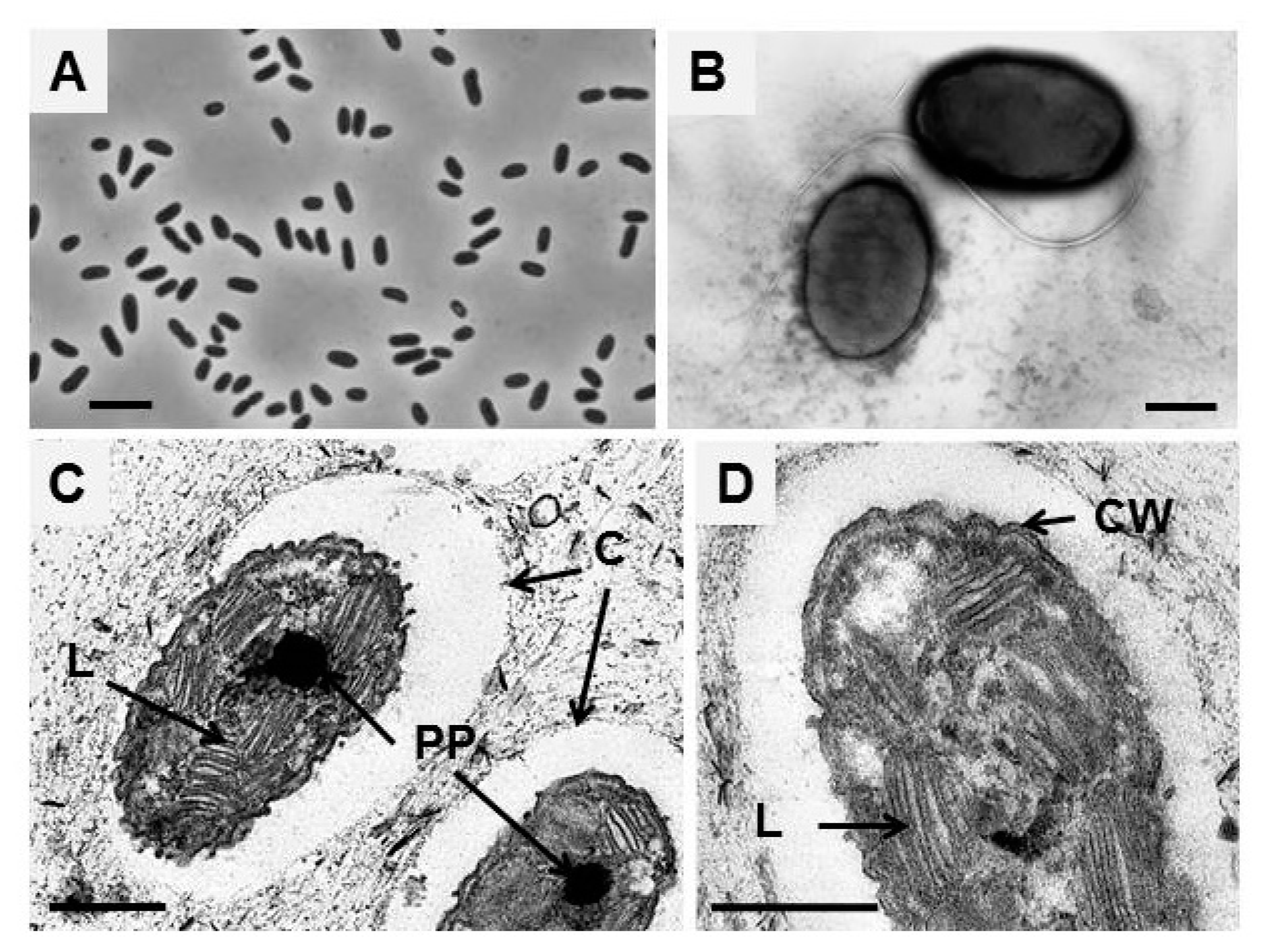
3.1. Morphology and Ultrastructure
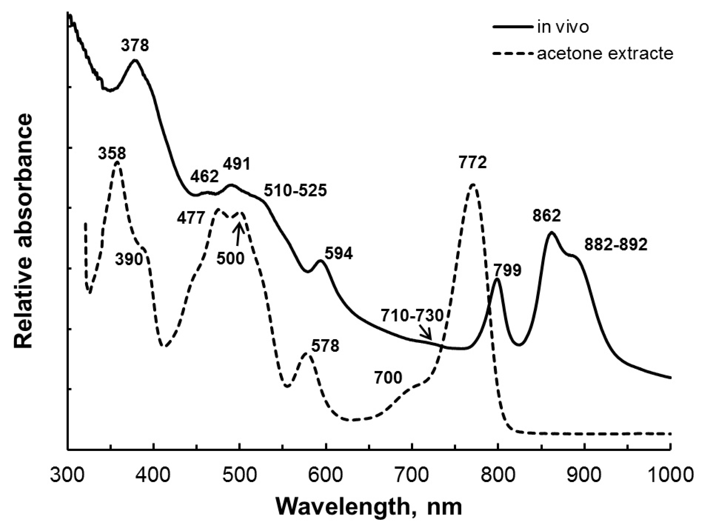
The cell morphology of all new strains was similar. Cells were oval- to rod-shaped and sometimes slightly bent (Figure 1A,B). The cells varied from 0.5 to 1.3 µm in width and 0.7 to 2.7 µm in length. Young cells of the strains B14B and A-7R were motile by means of a tuft of polar flagella (Figure 1B). Motility was not found in strain A-7Y. Cells multiplied by binary fission. In the later stages of growth, the cells formed a mucous capsule (Figure 1C). Ultrathin sections of cells of strain B14B showed a Gram-negative type of cell wall (Figure 1D). The internal photosynthetic membranes were lamellae and located in ordered stacks, as in other species of the Ectothiorhodospiraceae (Figure 1C,D). Dense oval inclusions, characteristic of polyphosphates, were seen regularly in the cells (Figure 1C).

3.2. Pigments

Colonies on an agarized medium and cell suspensions of the studied strains B14B, A-7R, and A-7Y were bright red. The in-vivo absorption spectrum had maxima at 378, 462, 491, 594, 799, and 862 nm and shoulders (indicated in parentheses) at (510–525), (710–730), and (882–892) nm (for different strains ± 2 nm; Figure 2). The absorption spectrum of cells extracted with acetone showed peaks at 358, (390), 477, 500, 578, (700), and 772 nm (Figure 2).
The main pigment was bacteriochlorophyll a. Its presence on the absorption spectrum of living cells was indicated by absorption maxima at 378, 594, 799, 862 nm and a shoulder at (882–892) nm (Figure 2). On the absorption spectrum of the acetone extract, bacteriochlorophyll a had a major maximum at 772 nm (Figure 2). Bacteria contained carotenoids of the spirilloxanthin series, the presence of which was confirmed by the maximum at 491 and the shoulder (510–525) nm on the absorption spectrum of whole-cell pigments (Figure 2). All known representatives of the genus Ectothiorhodospira are characterized by the presence of bacteriochlorophyll a and carotenoids of the spirilloxanthin series.

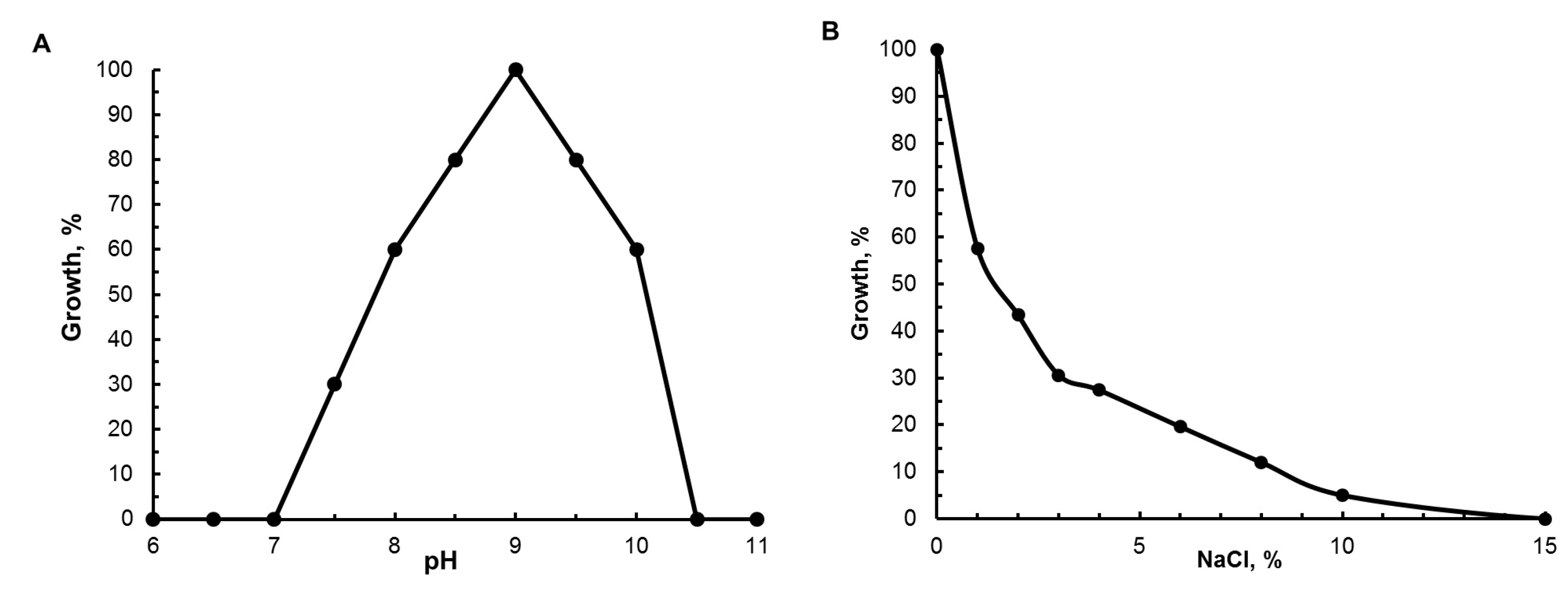
3.3. Physiological Characteristics

Bacteria of the studied strains were anaerobic phototrophs. Photoautotrophic growth with sulfide was weak (growth yields were roughly half that of photoheterotrophically grown cultures). Photoautotrophically grown cells could be subcultured several times. Sulfide was oxidized to sulfate with the formation of S° as an oxidation intermediate, which was deposited outside the cells. Thiosulfate and sulfite were not oxidized under photoautotrophic conditions. Growth on thiosulfate and sulfite was possible only in the presence of organic substrates. Bacteria grew photoheterotrophically, with sulfide, thiosulfate and sulfite as a sulfur source for biosynthesis. Assimilatory sulfate reduction was not possible. Under microaerobic conditions in the dark, growth was only possible on organic substrates with sulfide as a source of reduced sulfur (Table 1).
Under phototrophic conditions on a medium with sulfide as a sulfur source, strain B14B used acetate, butyrate, yeast extract, lactate, malate, pyruvate, succinate, and fumarate. In the presence of arginine, aspartate, glycerol, glucose, glutamate, lactose, propionate, peptone, propanol, sucrose, sorbitol, formate, fructose, citrate, ethanol growth was not observed. Nitrogen sources were ammonium, nitrate, and urea (Table 1). Growth factors were not required, but vitamin complex and yeast extract stimulated growth.
The best growth occurred at pH 9.0; the growth range was between pH 7.5 and 10.2 (Figure 3). Growth was possible at NaCl concentrations ranging from 0% (maximum growth) to 8%; the best growth occurred at 0% NaCl (Figure 3, Table 1). The studied strains were mesophiles. Optimum growth was at 25–35 °C; the temperature limits of growth were not determined.

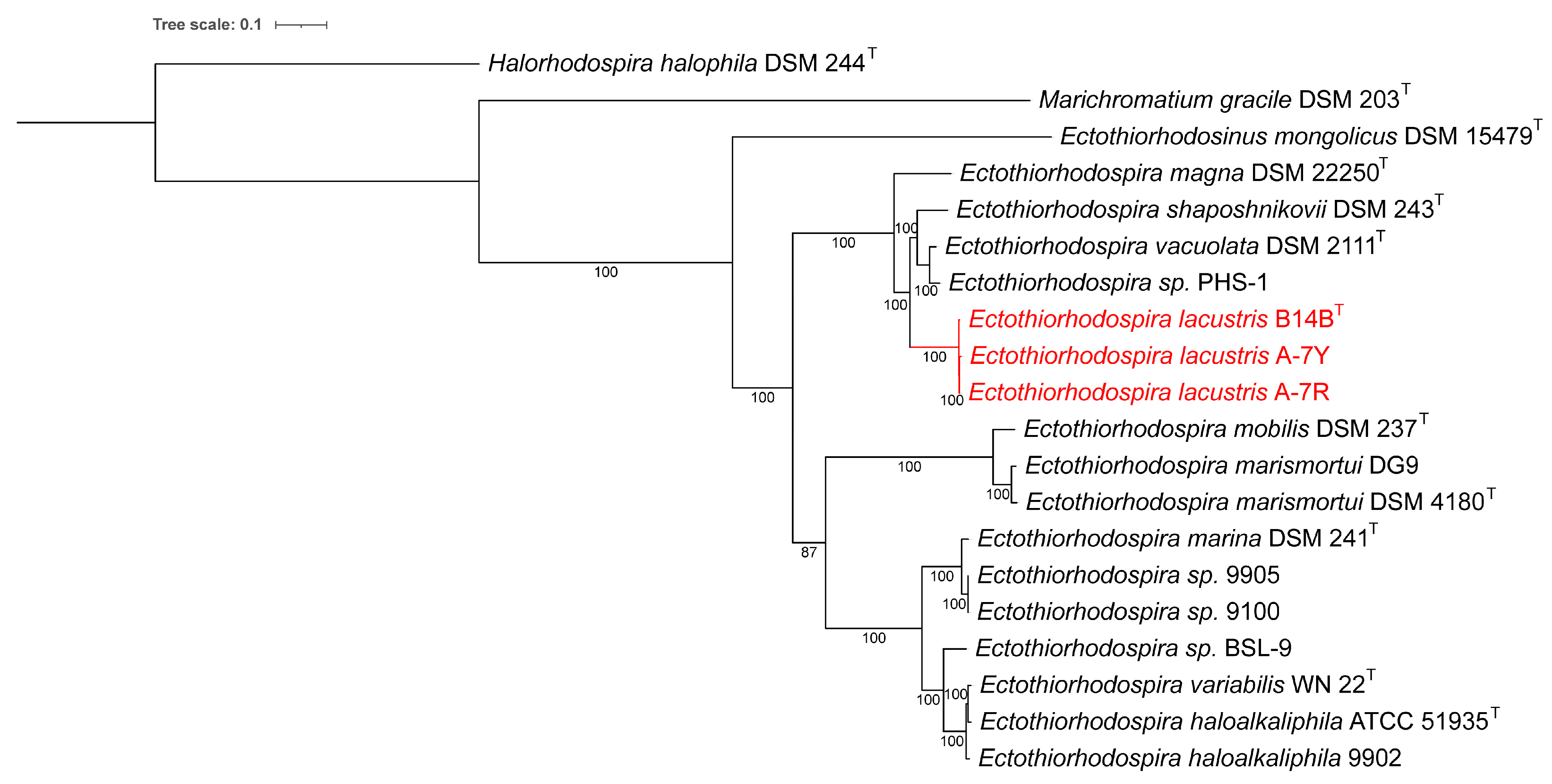
3.4. Taxonomic Position

According to the results of whole-genome sequencing, the G + C content in the DNA of the studied strains B14B, A-7R, and A-7Y was 62.9–63.0 mol% [1]. Phylogenetic analysis based on the comparison of 16S rRNA gene sequences showed that these strains belonged to the genus Ectothiorhodospira and revealed the greatest affinity with species Ect. shaposhnikovii and Ect. vacuolata but places the new strains on a separate clade on the 16S rRNA tree (Figure 4) [1]. Although an LALIGN analysis (https://www.ebi.ac.uk/Tools/psa/lalign/ accessed on 6 May 2023) showed the highest 16S rRNA identity with Ect. shaposhnikovii DSM 243T (99.4%; 1530/1540 bp); Ect. vacuolata DSM 2111T (99.0%; 1511/1526 bp), Ect. haloalkaliphila ATCC 51935T (99.0%; 1445/1459 bp), and Ect. magna DSM 22250T (98.8%; 1454/1471 bp), the dDDH and ANI results revealed low similarity between the studied strains and the related species that did not exceed accepted thresholds for bacterial species [14,24]. It is generally well known that whole genome-based comparisons of dDDH and ANI provide a higher resolution for species differentiation than single gene-based comparisons. dDDH values between strain B14B and the known Ectothiorhodospiraceae species varied from 19.7% with Ets. mongolicus DSM 15479T to 38.8% with Ectothiorhodospira vacuolata DSM 2111T (Table 2).
On the total genome phylogenetic tree, strains B14B, A-7R, and A-7Y formed a separate branch (Figure 5) and had ANI values <90% (75.0–89.4%) relative to any of the other strains of the genus Ectothiorhodospira, but high ANI values between themselves (99.0–99.9%) [1]. ANI percentages > 95% have been suggested to recognize bacteria as belonging to the same species, while bacteria with ANI <90% would be recognized in most cases as separate species [14]. The ANI with the most closely related species Ect. shaposhnikovii and Ect. vacuolata was 87.8–89.4%, which are all well below the arbitrary species cutoff of 95%.
A comparative systems analysis in BV-BRC between genomes of all known species of Ectothiorhodospiraceae and Halorhodospiraceae revealed that strains B14B, A-7R, and A-7Y had 15 unique PGFams that are not found in any other Ectothiorhodospiraceae genome, and they all contained a gene cluster for nitric oxide reductase (NOR), which is absent in all other species. The norCBQD cluster contains two NO reductase activation proteins as well as small and large NOR subunits (C and B) that show similarity to cytochrome oxidases. Nitric oxide reductase catalyzes bacterial denitrification by reducing nitric oxide (NO) to nitrous oxide (N2O), an obligatory step involved in the sequential reduction of nitrate to dinitrogen. The presence of norCB subunits is often used as a functional marker gene to identify denitrifying bacteria [25]. In addition to the PGFam analysis, a BLAST gene search using each of the 4 nor genes confirmed that they were only present in strains B14B, A-7R, and A-7Y and absent in all other Ectothiorhodospiraceae strains. There are no known studies of nitric oxide reduction in Ectothiorhodospiraceae, but strains B14B, A-7R, and A-7Y at least have the genetic capacity for nitric oxide reduction. An NCBI protein BLAST search of the NorQ protein showed Wenzhouxiangella sediminis (81.44% identity), Halomonas korlensis (76.03% identity), and Halomonas nitroreducans (74.53% identity) as the closest named relatives. All three of these are moderately halophilic, denitrifying Gammaproteobacteria isolated from saline, alkalophilic sediments, or soils [26,27,28].
The norCBQD gene cluster is found upstream from a ‘periplasmic sensor signal transduction histidine kinase’ (PGF_03758479) and a ‘two-component transcriptional response regulator’ (PGF_09666309). A synteny analysis in BV-BRC showed that these two genes are only conserved in six other Ectothiorhodospiraceae strains (Figure 6), which, interestingly, are not the closest related to the B14B clade on the whole genome and 16S rRNA trees (Figure 4 and Figure 5). This indicates that the norCBQD gene cluster in the three strains, B14B, A-7R, and A-7Y may have been obtained in a more recent evolutionary event. In addition to the general genomic difference, these genetic differences are another reason to consider these strains as a separate species [1].
Based on the combination of phenotypic and phylogenetic properties of strains B14B, A-7R, and A-7Y, it was proposed to classify them as a new species for which the name Ectothiorhodospira lacustris sp. nov. is proposed with strain B14BT (=DSM 116064T =KCTC 25542T = UQM 41491T) as type strain.

4. Description of Ectothiorhodospira lacustris sp. nov.

Ectothiorhodospira lacustris (la.cus’tris. M.L. fem. adj. lacustris (L. n. lacus a lake) inhabiting lakes, growing in lakes).
Cells are oval- to rod-shaped, sometimes slightly bent rods, 0.5–1.3 × 0.7–2.7 µm in size. Young cells are motile by means of polar flagella. Some strains are not mobile. Reproduction occurs by binary fission. Internal photosynthetic membranes are lamellar stacks. The color of cell suspensions is deep red. Photosynthetic pigments are bacteriochlorophyll a and carotenoids of the spirilloxanthin series. Absorption spectra of living cells have maxima at 378, 462, 491, 594, 799, and 862 nm, with shoulders at (510–525), (710–730), and (882–892) nm.
Growth occurs anaerobic in the light. Photoautotrophic growth with sulfide is weak (growth yield is roughly half that of photoheterotrophically grown cultures). Photoautotrophically grown cells can be subcultured several times. Sulfide is oxidized to sulfate, with S°, which is deposited outside the cells, as an intermediary product. Thiosulfate and sulfite are not oxidized under photoautotrophic conditions. Photoheterotrophic growth is possible with sulfide, thiosulfate, and sulfite as a sulfur source for biosynthesis under anoxic conditions. Assimilatory sulfate reduction is not possible. Chemolithotrophic growth is not possible under microoxic conditions in the dark in the presence of sulfide. Chemoheterotrophic growth is possible under microoxic conditions in the dark in the presence of sulfide.
Under phototrophic conditions and in the presence of sulfide, acetate, butyrate, yeast extract, lactate, malate, pyruvate, succinate, and fumarate are used. Arginine, aspartate, glycerol, glucose, glutamate, lactose, propionate, peptone, propanol, sucrose, sorbitol, formate, fructose, citrate, and ethanol are not used. Ammonia, nitrate, and urea are used as nitrogen sources and contain genes encoding a nitrogenase, so should be able to grow on N2. Growth factors are not required, but vitamin complex and yeast extract stimulate growth.
NaCl is not required for growth but up to 8% NaCl are tolerated. Optimum growth is at pH 9.0 (range pH 7.5–10.2), 0% NaCl (range 0–8%).
The bacterium is a mesophile with optimum growth at 25–35 °C. The temperature limits of growth have not been determined.
Habitat: low-mineralized soda lakes. The type strain B14B was isolated from biofilms of the shallow-water soda lake Bezymyannoye in central Mongolia (pH 9.0, mineralization 1–2 g/L, of which 80% sodium bicarbonate and 20% NaCl from the total mineralization of water).
The DNA G + C content of the species is 62.9–63.0 mol% according to genomic DNA data. The DNA G + C content of the type strain B14B is 62.9 mol% according to genomic DNA data.
The type strain B14B is deposited in the German Collection of Microorganisms and Cell Cultures GmbH (DSMZ) under accession number DSM 116064, in the Korean Collection for Type Cultures under accession number KCTC 25542, and in the Collection of Unique and Extremophilic Microorganisms under accession number UQM 41491.
The full genome DNA sequence of the type strain B14BT is registered under accession no. JAJNQM000000000.
The 16S rRNA gene sequence of the type strain B14BT is deposited at GenBank under accession no. OQ618219.

Author Contributions

Conceptualization, I.A.B., J.A.K., V.M.G. and J.F.I.; methodology, I.A.B., J.A.K., V.M.G. and J.F.I.; software, J.A.K.; cultivation of bacterial culture, I.A.B.; validation, I.A.B., J.A.K. and J.F.I.; formal analysis, J.F.I.; investigation, I.A.B., J.A.K., V.M.G. and J.F.I.; data curation, I.A.B., J.A.K., V.M.G. and J.F.I.; writing—original draft preparation, I.A.B.; writing—review and editing, I.A.B., J.A.K., V.M.G. and J.F.I. All authors have read and agreed to the published version of the manuscript.

Funding

I.A.B. and V.M.G. were supported by the Ministry of Science and Higher Education of the Russian Federation.

Data Availability Statement

All sequence data are available in a publicly accessible repository of NCBI Genbank.

Acknowledgments

The authors thank N. A. Kostrikina for preparation of whole cell preparations and L. L. Mityushina for ultrathin sections for electron microscopy. Electron microscopic studies were carried out on the basis of the UNIQEM Collection Core Facility, Research Center of Biotechnology of the Russian Academy of Science.

Conflicts of Interest

The authors declare no conflict of interest.

References

  1. Imhoff, J.F.; Kyndt, J.A.; Meyer, T.E. Genomic comparison, phylogeny and taxonomic reevaluation of the Ectothiorhodospiraceae and description of Halorhodospiraceae fam. nov. and Halochlorospira gen. nov. Microorganisms 2022, 10, 295. [Google Scholar] [CrossRef]
  2. Imhoff, J.F. Genus ectothiorhodospira. In Bergey’s Manual of Systematic Bacteriology, 2nd ed.; Brenner, D.J., Krieg, N.R., Staley, J.T., Eds.; Springer: New York, NY, USA, 2005; Volume 2, pp. 43–48. [Google Scholar]
  3. Frigaard, N.U.; Dahl, C. Sulfur metabolism in phototrophic sulfur bacteria. Adv. Microb. Physiol. 2009, 54, 103–200. [Google Scholar] [CrossRef] [PubMed]
  4. Dahl, C. Sulfur Metabolism in Phototrophic Bacteria. In Modern Topics in the Phototrophic Prokaryotes; Hallenbeck, P.C., Ed.; Springer: Cham, Switzerland, 2017; pp. 27–66. [Google Scholar] [CrossRef]
  5. Oren, A. The Family Ectothiorhodospiraceae. In The Prokaryotes; Rosenberg, E., DeLong, E.F., Lory, S., Stackebrandt, E., Thompson, F., Eds.; Springer: Berlin/Heidelberg, Germany, 2014; pp. 199–222. [Google Scholar] [CrossRef]
  6. Bryantseva, I.; Gorlenko, V.M.; Kompantseva, E.I.; Imhoff, J.F.; Süling, J.; Mityushina, L. Thiorhodospira sibirica gen. nov., sp. nov., a new alkaliphilic purple sulfur bacterium from a Siberian soda lake. Int. J. Syst. Bacteriol. 1999, 49, 697–703. [Google Scholar] [CrossRef]
  7. Gorlenko, V.M.; Bryantseva, I.A.; Panteleeva, E.E.; Tourova, T.P.; Kolganova, T.V.; Makhneva, Z.K.; Moskalenko, A. Ectothiorhodosinus mongolicum gen. nov., sp. nov., a new purple bacterium from a soda lake in Mongolia. Microbiology 2004, 73, 66–73. [Google Scholar] [CrossRef]
  8. Euzeby, J.P. Validation list no. 116. List of new names and new combinations previously effectively, but not validly, published. Int. J. Syst. Evol. Microbiol. 2007, 57, 1371–1373. [Google Scholar] [CrossRef]
  9. Imhoff, J.F.; Rahn, T.; Künzel, S.; Keller, A.; Neulinger, S.C. Osmotic adaptation and compatible solute biosynthesis of phototrophic bacteria as revealed from genome analyses. Microorganisms 2019, 9, 46. [Google Scholar] [CrossRef]
  10. Gorlenko, V.M.; Imhoff, J.F. Biodiversity of alkaliphilic purple sulphur bacteria of Ectothiorhodospiraceae family. In Proceedings of the Program and Abstracts of the 12th International Symposium on Phototrophic Prokaryotes, Pau, France, 27 August–1 September 2006; p. 74. [Google Scholar]
  11. Pfennig, N.; Lippert, K.D. Über das Vitamin B12-Bedürfnis phototropher Schwefelbakterien. Arch. Mikrobiol. 1966, 55, 245–256. [Google Scholar] [CrossRef]
  12. Ryter, A.; Kellenberger, E.; Birch-Andersen, A.; Maaløe, O. Etude au microscope électronique des plasmes contenant de l’acide déoxyribonucléique 1 Les nucléoides des bactéries en croissance active. Z. Naturforsch. 1958, 13, 597–605. [Google Scholar] [CrossRef]
  13. Reynolds, E.S. The use of lead citrate at high pH as an electron opaque stain in electron microscopy. J. Cell Biol. 1963, 17, 208–218. [Google Scholar] [CrossRef]
  14. Richter, M.; Rosselló-Móra, R.; Glöckner, F.O.; Peplies, J. JSpeciesWS: A web server for prokaryotic species circumscription based on pairwise genome comparison. Bioinformatics 2016, 32, 929–931. [Google Scholar] [CrossRef]
  15. Meier-Kolthoff, J.P.; Göker, M. TYGS is an automated high-throughput platform for state-of-the-art genome-based taxonomy. Nat. Commun. 2019, 10, 2182. [Google Scholar] [CrossRef]
  16. Wattam, A.R.; Davis, J.J.; Assaf, R.; Boisvert, S.; Brettin, T.; Bun, C.; Conrad, N.; Dietrich, E.M.; Disz, T.; Gabbard, J.L.; et al. Improvements to PATRIC, the all-bacterial Bioinformatics Database and Analysis Resource Center. Nucleic Acids Res. 2017, 45, D535–D542. [Google Scholar] [CrossRef]
  17. Stamatakis, A.; Hoover, P.; Rougemont, J.J.S. A rapid bootstrap algorithm for the RAxML web servers. Syst. Biol. 2008, 57, 758–771. [Google Scholar] [CrossRef] [PubMed]
  18. Stamatakis, A. RAxML version 8: A tool for phylogenetic analysis and post-analysis of large phylogenies. Bioinformatics 2014, 30, 1312–1313. [Google Scholar] [CrossRef]
  19. Letunic, I.; Bork, P. Interactive Tree Of Life (iTOL) v4: Recent updates and new developments. Nucleic Acids Res. 2019, 47, 256–259. [Google Scholar] [CrossRef]
  20. Edgar, R.C. MUSCLE: Multiple sequence alignment with high accuracy and high throughput. Nucleic Acids Res. 2004, 32, 1792–1797. [Google Scholar] [CrossRef] [PubMed]
  21. Tamura, K.; Nei, M. Estimation of the number of nucleotide substitutions in the control region of mitochondrial DNA in humans and chimpanzees. Mol. Biol. Evol. 1993, 10, 512–526. [Google Scholar] [CrossRef]
  22. Kumar, S.; Stecher, G.; Li, M.; Knyaz, C.; Tamura, K. MEGA X: Molecular Evolutionary Genetics Analysis across computing platforms. Mol. Biol. Evol. 2018, 35, 1547–1549. [Google Scholar] [CrossRef]
  23. Bryantseva, I.A.; Tourova, T.P.; Kovaleva, O.L.; Kostrikina, N.A.; Gorlenko, V.M. Ectothiorhodospira magna sp. nov., a new large alkaliphilic purple sulfur bacterium. Microbiology 2010, 79, 780–790. [Google Scholar] [CrossRef]
  24. Chun, J.; Oren, A.; Ventosa, A.; Henrik, C.; Arahal, D.R.; da Costa, M.S.; Rooney, A.P.; Yi, H.; Xu, X.; De Meyer, S.; et al. Proposed minimal standards for the use of genome data for the taxonomy of prokaryotes. Int. J. Syst. Evol. Microbiol. 2018, 68, 461–466. [Google Scholar] [CrossRef] [PubMed]
  25. Braker, G.; Tiedje, J.M. Nitric oxide reductase (norB) genes from pure cultures and environmental samples. Appl. Environ. Microbiol. 2003, 69, 3476–3483. [Google Scholar] [CrossRef] [PubMed]
  26. Guo, L.Y.; Dunlap, C.A.; Rooney, A.P.; Chen, G.J.; Du, Z.J. Wenzhouxiangella sediminis sp. nov., isolated from coastal sediment. Int. J. Syst. Evol. Microbiol. 2016, 66, 4575–4579. [Google Scholar] [CrossRef] [PubMed]
  27. Li, H.B.; Zhang, L.P.; Chen, S.F. Halomonas korlensis sp. nov., a moderately halophilic, denitrifying bacterium isolated from saline and alkaline soil. Int. J. Syst. Evol. Microbiol. 2008, 58, 2582–2588. [Google Scholar] [CrossRef] [PubMed]
  28. González-Domenech, C.M.; Béjar, V.; Martínez-Checa, F.; Quesada, E. Halomonas nitroreducens sp. nov., a novel nitrate-and nitrite-reducing species. Int. J. Syst. Evol. Microbiol. 2008, 58, 872–876. [Google Scholar] [CrossRef]
Figure 1. Morphology and the fine structure of strain B14B cells: (A) phase contrast light microscopy, (B) transmission electron microscopy of cells contrasted with phosphotungstic acid, and (C,D) ultrathin cell sections. Scale bar: 5 μm (A) and 0.5 μm (BD). Designations: L, lamellar photosynthetic membranes; C, capsule; PP, polyphosphates; CW, cell wall.
Microorganisms 11 01336 g001
Figure 2. Absorption spectra of whole cells (in vivo) in glycerol (1:1, v/v) and of the acetone extract of strain B14B.
Microorganisms 11 01336 g002
Figure 3. Influence of pH (A) and of concentrations of NaCl (B) on growth of strain B14B.
Microorganisms 11 01336 g003
Figure 4. A 16S rRNA-derived phylogenetic tree for the phototrophic Ectothiorhodospiraceae species. Halorhodospira halophila DSM 244T and Marichromatium gracile DSM 230T sequences were included in the tree as outgroups. The phylogenetic tree was calculated in MegaX, and iTOL was used to draw the phylogenetic tree. Bootstrap values were generated from 1000 bootstrapping rounds. The new strains are indicated in red.
Microorganisms 11 01336 g004
Figure 5. Whole genome-based phylogenetic tree of the Ectothiorhodospiraceae. The support values for the phylogenetic tree are generated using 100 rounds of the ‘rapid bootstrapping’ option of RaxML. The branch-length tree scale is defined as the mean number of substitutions per site, which is an average across both nucleotide and amino acid changes. The new strains are indicated in red, and Halorhodospira halophila DSM 244T and Marichromatium gracile DSM 230T were included in the tree as outgroups.
Microorganisms 11 01336 g005
Figure 6. Comparison of the norCBQD genomic region between Ectothiorhodospira species. Genes are colored based on their family membership.
Microorganisms 11 01336 g006
Table 1. Comparison of the properties of Ectothiorhodospira lacustris and related species.
PropertyEctothiorhodospira
lacustris
Ectothiorhodospira
vacuolata
Ectothiorhodospira shaposhnikoviiEctothiorhodospira magna
Cell shapeOval- to rod-shaped, sometimes slightly bentRod-shaped, sometimes slightly bentRod-shaped, usually slightly bent vibrios or short spirillaVibrioid or spirilloid
Size, µm0.5–1.3 × 0.7–2.71.5 × 2–40.8–0.9 × 1.5–2.52–3.2 × 9.6–20
Motility±, Polar tuft of flagella+, Polar tuft of flagella+, Polar tuft of flagella+, Polar tuft of flagella
Gas vesicles+
Internal membranesLamellar stacksLamellar stacksLamellar stacksLong strands of lamellae
Color of the cell suspensionsDark redPink to redRedRed to brownish red
PigmentsBchl a, carotenoids of the spirilloxanthin seriesBchl a, carotenoids of the spirilloxanthin seriesBchl a, carotenoids of the spirilloxanthin seriesBchl a, carotenoids of the spirilloxanthin series
Absorption spectra of the whole cells378, 462, 491, (510–525), 594, (710–730), 799, 862, (882–892)378, 486, 513, 555, 593, 798, 855, (893)378, 481, 513, 556, 593, 798, 854, (892)373, 485–514, (545), 593, 797, 858, (885)
NaCl optimum (range), %0 (0–8)1–6 (0.5–10)3 (0–7)0.5–1.5 (0–8)
pH optimum (range)9.0 (7.5–10.2)7.5–9.58.0–8.59–10 (8–11)
Nitrogen sourcesAmmonia, nitrate, urea, dinitrogen (genomic data)Ammonia, dinitrogenAmmonia, dinitrogen, nitrate, some amino acidsAmmonia
Growth factorsNot requiredNot requiredNot requiredYeast extract satisfies all requirements
Photoautotrophic growth+ *+++ *
Photoheterotrophic growth++uu
Chemolithotrophic growthu+
Sulfate assimilation+
Substrates used:
HydrogenND+++ *
Sulfide++++
Thiosulfate++
Sulfur++++
Sulfiteu+
Organic substancesAcetate, butyrate, yeast extract, lactate, malate, pyruvate, succinate, fumarateAcetate, malate, propionate, pyruvate, succinate, (fructose), fumarateAcetate, butyrate, lactate, malate, propionate, pyruvate, succinate, fructose, fumarateAcetate, malate, yeast extract *, propionate *, pyruvate *, succinate, fumarate
DNA G + C content of the species, mol%62.9–63.0 (genomic DNA data)61.4–63.6 (Tm)62.0–64.0 (Tm)59.2 (Tm)
61.5–62.1 (Tm)
DNA G+C content of the type strain, mol%62.9 (genomic DNA data)
61.5 (Tm)
63.4 (genomic DNA data)
63.6 (Tm)
62.4 (genomic DNA data)
62.0 (Tm)
60.9 (genomic DNA data)
59.2 (Tm)
Symbols: +, characteristic positive or substrate utilized by most strains; –, characteristic negative or substrate not utilized by most strains; *, weak growth; (fructose), substrate utilized by some strains; ND, not determined; u, no data available; Tm, thermal denaturation. Data for reference species were taken from [1,2,23].
Table 2. Comparison of the properties of Ectothiorhodospira lacustris and related species. Comparison of the genomic sequences of Ectothiorhodospira lacustris B14BT and related species based on digital DNA-DNA hybridization (dDDH) and relative G+C content differences. C.I. stands for confidence intervals.
Subject StraindDDH (d4, in %)C.I. (d4, in %)G + C%
Difference
Ectothiorhodospira vacuolata DSM 2111T38.8[36.4–41.3]0.52
Ectothiorhodospira shaposhnikovii DSM 243T36.6[34.1–39.1]0.53
Ectothiorhodospira magna B7-7T = DSM 22250T30[27.7–32.6]1.97
Thiorhodospira sibirica A12T = ATCC 700588T22.3[20.1–24.8]6.2
Ectothiorhodospira marina DSM 241T21.5[19.2–23.9]0.63
Ectothiorhodospira variabilis WN22T = DSM 21381T 21.4[19.1–23.8]0.08
Ectothiorhodospira marismortui DSM 4180T20.8[18.6–23.2]5.33
Thioalkalivibrio sulfidiphilus HL-EbGr7T20.7[18.5–23.1]2.16
Ectothiorhodospira mobilis DSM 237T20.7[18.5–23.1]5.42
Thioalkalivibrio denitrificans ALJDT = DSM 13742T20.2[18.0–22.7]1.67
Ectothiorhodosinus mongolicus M9T = DSM 15479T19.7[17.5–22.1]7.29
Disclaimer/Publisher’s Note: The statements, opinions and data contained in all publications are solely those of the individual author(s) and contributor(s) and not of MDPI and/or the editor(s). MDPI and/or the editor(s) disclaim responsibility for any injury to people or property resulting from any ideas, methods, instructions or products referred to in the content.

