Article

Effect of Methionine on Gene Expression in Komagataella phaffii Cells

1
Laboratory of Biochemical Genetics, Department of Genetics and Biotechnology, Saint Petersburg State University (SPBU), Saint Petersburg 199034, Russia
2
Institute of Chemistry, Saint Petersburg State University (SPBU), Petergof, Saint Petersburg 198504, Russia
*
Authors to whom correspondence should be addressed.
Microorganisms 2023, 11(4), 877; https://doi.org/10.3390/microorganisms11040877
Submission received: 1 March 2023 / Revised: 27 March 2023 / Accepted: 27 March 2023 / Published: 29 March 2023
(This article belongs to the Special Issue Yeasts Biochemistry and Biotechnology)

Abstract

:
Komagataella phaffii yeast plays a prominent role in modern biotechnology as a recombinant protein producer. For efficient use of this yeast, it is essential to study the effects of different media components on its growth and gene expression. We investigated the effect of methionine on gene expression in K. phaffii cells using RNA-seq analysis. Several gene groups exhibited altered expression when K. phaffii cells were cultured in a medium with methanol and methionine, compared to a medium without this amino acid. Methionine primarily affects the expression of genes involved in its biosynthesis, fatty acid metabolism, and methanol utilization. The AOX1 gene promoter, which is widely used for heterologous expression in K. phaffii, is downregulated in methionine-containing media. Despite great progress in the development of K. phaffii strain engineering techniques, a sensitive adjustment of cultivation conditions is required to achieve a high yield of the target product. The revealed effect of methionine on K. phaffii gene expression is important for optimizing media recipes and cultivation strategies aimed at maximizing the efficiency of recombinant product synthesis.

1. Introduction

The yeast Komagataella phaffii (previously known as Pichia pastoris) is one of the most efficient and robust systems for recombinant protein production. Powerful promoters, the high preference for respiratory growth, and the ability to rewire metabolic pathways for maximizing recombinant protein synthesis make K. phaffii a promising expression host [1,2,3]. K. phaffii performs all eukaryotic posttranslational modifications, and a weak tendency to protein hypermannosylation makes it a preferable producer in pharmacology to the model yeast Saccharomyces cerevisiae [4]. This feature goes hand in hand with successes in engineering its glycosylation pathways, and allows the production of proteins with modifications highly similar to mammalian cells [4,5]. Another beneficial feature is the ability of this yeast to efficiently synthesize and secrete proteins with a relatively easy subsequent purification procedure [6]. The availability of simple and robust high-cell density cultivation procedures and well-developed tools for strain engineering contribute to expanding the range of K. phaffii applications. Although many efficient molecular tools and techniques are now available to work on K. phaffii, achieving a high yield of the target product requires a sensitive adjustment of its cultivation conditions, which differ according to the target protein [7,8,9].
Thus, unique biological features, combined with long-term work on improving productivity, have made K. phaffii highly prominent for biotechnological and pharmaceutical applications [7]. To date, this yeast is applied for synthesizing thousands of recombinant proteins, both on a laboratory and industrial scale [9]. K. phaffii is used to produce recombinant subunit vaccines [10], various membrane proteins [11,12,13], and several nonprotein substances [14,15]. This practical importance stimulates the consideration of K. phaffii as a new model object for fundamental research, apart from being a production host [16].
Being methylotrophic, K. phaffii can grow on methanol as a sole carbon and energy source. The first reaction of the methanol utilization pathway (MUP) is the oxidation of methanol into formaldehyde by the enzyme alcohol oxidase, encoded by the AOX1 and AOX2 genes [17].
Part of formaldehyde is oxidized to CO2 in a series of reactions controlled by formaldehyde dehydrogenase, S-formylglutathione hydrolase, and formate dehydrogenase. This dissimilative branch of the MUP produces energy in the form of NADH, and prevents cell damage from the harmful effects of formaldehyde [18,19].
Another part of formaldehyde enters the assimilative branch of the MUP. Dihydroxyacetone synthase catalyzes the synthesis of dihydroxyacetone and glyceraldehyde-3-phosphate from formaldehyde and xylulose-5-phosphate. In further reactions, the enzymes dihydroxyacetone kinase, fructose-1,6-bisphosphate aldolase, and fructose-1,6-bisphosphatase result the formation of glyceraldehyde 3-phosphate, which is used for the generation of biomass and energy [18].
The product of the AOX1 gene primarily has methanol-oxidizing activity in methanol-grown K. phaffii cells [17]. This gene expression depends on the type of carbon source in the medium [20]. For instance, glycerol or glucose both fully inhibit the AOX1 promoter activity. On the contrary, AOX1 gene expression is induced under conditions when methanol is the sole carbon source in the medium, and the level of AOX1 mRNA reaches 5% of the total cell mRNA [17,18,19,20,21]. The strict regulation and high activity of the AOX1 promoter determine its wide use for recombinant protein production [1]. This promoter is a part of plasmid vectors that are widely used for heterologous expression in K. phaffii (e.g., pPICZ series and pPIC9). Such practical importance drives molecular mechanism studies of the regulation of the AOX1 gene and other methanol metabolism genes (methanol utilization, MUT genes) [22].
We have previously shown that the expression of AOX1 and other MUT genes in K. phaffii cells is downregulated when certain amino acids are the sole medium nitrogen source [23,24]. This may be explained by the fact that this yeast can use several amino acids as the sole carbon, nitrogen, and energy sources [25,26]. Transcriptome analysis revealed differential expression of about 18.9% of all protein-coding genes when K. phaffii was grown on proline-containing media. It was shown that K. phaffii catabolizes proline, even when other nitrogen, carbon, and energy sources are available. As a result, the utilization genes of other sources, in particular methanol, are repressed [27]. Here, we continue these studies and investigate the effects of methionine as another amino acid.
Methionine and its derivatives implement various cell functions, such as DNA methylation, lipid homeostasis, and polyamine synthesis [28,29]. In yeast and mammalian cells, S-adenosylmethionine (a methionine derivative) has been shown to directly activate the target of rapamycin (TOR) complex I [30]. Methionine is of particular interest for its effect on longevity, given a wide range of studies showing that methionine restriction prolongs the lifespan in most model organisms [31].
Methionine plays an important role in the oxidative stress response, as it scavenges free radicals and protects the cell from reactive oxygen species (ROS) through participating in the activation of antioxidant enzymes [32]. Methionine derivatives are also a part of the biosynthesis of glutathione, the functions of which include counteracting oxidative stress [33].
Methionine stands out from other amino acids, since it acts as a translation initiation codon. Methionyl-tRNAi is required for translation initiation complex assembly [34]. In yeast cells, methionine activates the expression of genes involved in translation and ribosomal biogenesis [35].
Methionine metabolism has been studied in detail in Saccharomyces cerevisiae budding yeast. This microorganism can absorb methionine by specific and nonspecific permeases, and use the amino acid as the sole carbon source [36]. Additionally, S. cerevisiae can synthesize methionine de novo. The biosynthesis of this sulfur-containing amino acid involves three parts: construction of the methionine carbon chain, production of the sulfur anion, and incorporation of the sulfur anion into the carbon skeleton [37].
Aspartate is the precursor of the methionine carbon chain. Aspartokinase and aspartate-βsemialdehyde dehydrogenase catalyze the production of homoserine from aspartate. Next, homoserine O-acetyltransferase catalyzes the esterification reaction of homoserine and acetyl-CoA to produce O-acetylhomoserine [38].
Further addition of the sulfur atom is possible only in the form of sulfide, so sulfate and sulfite from the external environment are first reduced in the highly conservative pathway [39]. Once external sulfate enters the cell, it is metabolized into active adenosyl sulfate by ATP sulfurylase, and further converted to phosphoadenylsulfate (APS) by APS kinase. The enzymes PAPS reductase and sulfite reductase reduce APS to sulfite and sulfide, respectively [40].
Finally, homocysteine synthase integrates sulfide into O-acetylhomoserine to produce homocysteine. In yeast, this is the only reaction to incorporate sulfur into the carbon chain [37]. Homocysteine is a precursor of both methionine and cysteine, and the latter can be converted back into homocysteine via two transsulfuration pathways. To form methionine, the enzyme homocysteine methyltransferase removes the methyl group from 5-methyl-tetrahydrofolate (a one-carbon group compound) to methylate homocysteine [37].
One of the most essential methionine derivatives is S-adenosylmethionine (SAM), a major and vital cellular methylating agent. SAM synthetase catalyzes the SAM formation from ATP and methionine [41]. The methyl group transfer from SAM to the acceptor leads to S-adenosylhomocysteine (SAH) formation, which is further hydrolyzed to homocysteine. The resulting cycle, involving homocysteine, methionine, SAH, and SAM, is called the methyl cycle [40]. In addition, after SAM decarboxylation, the aminopropyl group can be involved in polyamine synthesis. As a result, methylthioadenosine (MTA) is formed, which can be converted into methionine again, closing the methylthioadenosine cycle [37]. In addition, SAM serves as an amino group donor in the synthesis of the biotin precursor pelargonium [42], and participates in the synthesis of modified nucleotides in tRNA and rRNA [43].
In S. cerevisiae yeast, methionine in the medium acts as a strong growth signal, triggering anabolism by activating the pentose phosphate pathway, amino acids, and nucleotide biosynthesis [35,44]. Thus, methionine and its derivatives play an important role in cellular metabolism. This research aims to study the complex effect of methionine on gene expression in the biotechnologically important yeast K. phaffii.

2. Materials and Methods

2.1. Media and Cultivation Conditions

Low salt LB medium was used for Escherichia coli manipulations. One L of low salt LB contained (here and further w/v): 1% tryptone, 0.5% yeast extract, 0.5% NaCl, 2.4% agar, and 20 mg zeocin for plasmid selection. Bacterial cells were grown at 37 °C.
YPD medium was used for K. phaffii manipulations. One L of YPD contained: 2% glucose, 2% peptone, 1% yeast extract, and 2.4% agar. YPDS media was used for zeocin selection. One L of YPDS contained: 2% glucose, 2% peptone, 1% yeast extract, 18.2% sorbitol (1 M), 2.4% agar, and 200 mg zeocin.
Modifications of standard buffered complex media were used for K. phaffii cultivation, transcriptome analysis, and quantitative enzymatic assays. One L of each media type contained: 100 mM potassium phosphate buffer (pH 6.0), 4 × 10⁵% biotin, and 1.34% Yeast Nitrogen Base without amino acids (#Y0626-1 KG, Sigma-Aldrich, St. Louis., MO, USA). BMG medium contained 1% glycerol. BMM medium contained 0.5% methanol. BMM and Met medium contained 0.5% methanol and 0.46% methionine. Yeast cells were grown at 30 °C.

2.2. Yeast and Bacterial Strains

The bacterial strain Escherichia coli DH5α F φ80lacZΔM15 Δ(lacZYA-argF)U169 recA1 endA1 hsdR17(rK, mK+) phoA supE44 λthi-1 gyrA96 relA1 (Thermo Fisher Scientific, Waltham, MA, USA) was used for the construction of the pPICZ-PHO1 plasmid.
K. phaffii X-33 strain (Thermo Fisher Scientific, Waltham, MA, USA) was used for growth studies and transcriptome analysis.
K. phaffii PAP1-X33 strain was generated in this study. A DNA fragment corresponding to the coding sequence of K. phaffii PHO1 acid phosphatase (ACP) gene (PAS_chr2-1_0103) was amplified by PCR using the Thersus Polymerase Kit (Evrogen, Moscow, Russia). Twenty picoM of primers PHO1-BstBI-F (5′-ATTACATTCGAAACGATGTTTTCTCCTATTCTAAGTCTGG-3′) and PHO1-AgeI-R (5′-ATTACTACCGGTTTATGACAAGTCATCCCAGAAG-3′) was added per reaction mix. The template was 500 ng of genomic DNA of X-33 strain. The reaction parameters were as follows: initial denaturation at 95 °C for 3 min, then 30 cycles 95 °C for 30 s, 53 °C for 30 s, 72 °C for 2 min. The resulting fragment was cloned into the pPICZαC plasmid using AgeI and BstBI restriction sites. The structure of the obtained pPICZ-PHO1 plasmid was confirmed by digestion with restriction enzymes and PCR analysis (Figure S1a in Supplementary Materials). The map of the pPICZ-PHO1 plasmid is depicted in Figure S1b in Supplementary Materials. The plasmid was linearized with the restriction enzyme SacI and transformed into K. phaffii X-33 strain. The resulting PAP1-X33 strain carries a PHO1 reporter gene under the control of the AOX1 gene promoter.

2.3. Molecular Methods

Yeast genomic DNA was extracted using LumiPure from an AnySample Kit (Lumiprobe, Moscow, Russia). Plasmids were isolated using the Plasmid Miniprep kit (Evrogen, Moscow, Russia). DNA was purified from agarose gels using a Cleanup Standard kit (Evrogen, Moscow, Russia).
DNA hydrolysis by restriction endonucleases, vector dephosphorylation, and DNA ligation were performed using the buffers and conditions recommended by the enzyme manufacturer (SibEnzyme, Moscow, Russia). PCR was performed using the Thersus Polymerase Kit (Evrogen, Moscow, Russia).
Yeast and bacterial cell transformations were performed using electroporation in accordance with the protocols [45,46].

2.4. Enzymatic Assays

A quantitative assay of acid phosphatase (ACP) activity was performed according to the previous research [47]. In brief, 100 μL of the cell suspension was taken and added into 800 μL of 0.1 M Na-citrate buffer, pH 4.6. Next, 100 μL of 0.15 M p-nitrophenyl phosphate substrate was added. The reaction mixture was stirred and incubated for 20 min at 30 °C in a water bath. The reaction was stopped by adding 500 μL of 1 M NaOH. The light absorption of the solutions was measured at a wavelength of 410 nm using a spectrophotometer. The specific activity of ACP was determined as the ratio of the absorption of light by the reaction mixture at a wavelength of 410 nm to the optical density of the initial cell suspension at a wavelength of 600 nm.

2.5. RNA-Sequencing

The RNeasy Mini Kit (Quiagen, Germantown, ML, USA) was used for the total RNA isolation from yeast cells. DNA molecules were removed from the sample by DNase (Thermo Fisher Scientific, Waltham, MO, USA) treatment. RNA purification was performed using the CleanRNA Standard Kit (Evrogen, Moscow, Russia). The quality of the isolated RNA molecules was assessed using agarose gel electrophoresis.
RNA-Seq library preparation was performed using the QuantSeq 3′ mRNA-Seq Library Prep Kit FWD (Lexogen, Vienna, Austria). The quality of the resulting libraries was tested with the Fragment Analyzer. The pool of libraries was sequenced on an Illumina MiSeq (single read, 300 bp) at Evrogen, Moscow, Russia.

2.6. Bioinformatic Analysis

The quality of the raw and trimmed reads was estimated using the FastQC program [48]. Illumina indices were removed and reads were filtered using the Trimmomatic program [49]. The quality-tested reads were aligned against the reference genome of K. phaffii yeast (ASM2700v1). The reference genome and annotations were retrieved from the NCBI database. Reads per genome were aligned using the hisat-2 program with standard parameters [50]. After alignment, reads were sorted and indexed using the samtools program [51]. The number of gene-aligned reads was calculated using the featureCounts program [52]. The number of resulting reads for each sample analyzed by RNA-sequencing is presented in Table S2 in the Supplementary Materials.
Differential gene expression analysis was performed according to the standard protocol in the DESeq2 package (version 1.24.0) in the R Studio program using R language (version 3.6.3) [53,54]. Further analysis included differentially expressed genes with corrected p-value less than 0.05 and log2FoldChange greater than 0.5 modulo. The results of the DESeq2 analysis are presented in Table S3 in the Supplementary Materials.

2.7. Gene Nomenclature

Names of K. phaffii genes with known annotation genes (AOX1, AOX2, etc.) were taken from other studies, especially [55]. For K. phaffii genes that were analyzed in this study as orthologs of the S. cerevisiae genes, “Kp” index was added to the name to distinguish them from S. cerevisiae ones.

3. Results

3.1. Addition of Methionine Does Not Enhance Growth of K. phaffii X-33 Strain

It was shown that some amino acids (e.g., proline and glutamate) stimulate K. phaffii growth in media containing methanol [23]. This is because the utilization of these amino acids is enough to provide K. phaffii cells with carbon, nitrogen, and energy [25,26]. As for methionine, it was shown that K. phaffii is not able to grow in media with this amino acid as the sole carbon, nitrogen, and energy source [56]. However, it was revealed that external methionine triggers anabolism and proliferation in S. cerevisiae [35]. Thus, we investigated if methionine affects growth of K. phaffii in media with glycerol and methanol as carbon and energy sources.
The starting culture of K. phaffii wild-type X-33 strain was grown overnight in 20 mL of liquid YPD media. Volumes of 100 mL of BMG, BMG and Met, BMM, BMM and Met, and BM and Met media were inoculated with 0.1% (100 μL) of starting culture. The optical density of yeast cultures was measured with a spectrophotometer at a wavelength of 600 nm (Figure 1).
K. phaffii X-33 strain did not grow in BM and Met media with methionine as a sole source of carbon and energy, which supports previous data [56]. In glycerol-containing BMG media, addition of methionine did not significantly influence the growth of K. phaffii. However, in methanol-containing BMM media, addition of methionine resulted in a slight decrease in cell density. Thereby, we also investigated how methionine affects gene expression in K. phaffii cells.

3.2. Methionine Influences the Expression of Genes Involved in Its Synthesis and Degradation in K. phaffii Cells

For the experiment, K. phaffii cells were grown according to a two-stage cultivation scheme, where at the first stage the cells grew biomass in a medium with glycerol, and then were transferred to a medium with methanol, which leads to the activation of MUT genes. Thus, the K. phaffii X-33 strain was grown in 100 mL of BMG medium with glycerol. After 40 h of growth, the cells were transferred to 100 mL of the medium with methanol and methionine, or without methionine (BMM and Met, or BMM). After 15 h of growth in the methanol-containing medium, total RNA was isolated from the cells and further used to prepare libraries for RNA sequencing.
Differential gene expression analysis revealed 142 differentially expressed genes (DEGs), which is 3% of the total number of transcribed genes detected (4687 protein-coding genes) (Table S3 in the Supplementary Materials). Among them, 75 genes increased and 67 genes decreased expression in the methionine-containing medium compared with gene expression in the medium without methionine, respectively.
One of the main groups of DEGs comprises genes involved in methionine biosynthesis. Gene homology and metabolic flux profiling studies revealed that amino acids are synthesized with high similarity in K. phaffii and S. cerevisiae [57,58]. Therefore, we used S. cerevisiae data to match these DEGs to specific parts of the methionine metabolism pathway (Figure 2).
Methionine biosynthesis in S. cerevisiae comprises three main parts: construction of the methionine carbon chain, production of the sulfur anion, and incorporation of the sulfur anion into the carbon skeleton [37].
Our data demonstrate the repression of the sulfur assimilation branch genes in the methionine presence. The expression level of adenylylsulfate kinase (KpMET14), sulfite reductase β-subunit (KpMET5), and sulfite reductase α-subunit (KpMET10) was significantly decreased in methionine-containing media. On the other hand, expression of genes involved in O-acetylhomoserine biosynthesis did not change significantly in response to methionine.
The expression of the methionine and cysteine synthase gene (KpMET25), which connects the sulfur assimilation branch and O-acetylhomoserine, was reduced in media with methionine. The methionine synthase gene KpMET6 did not change expression in methionine-containing media.
The expression level was significantly reduced for the folylpolyglutamate synthase gene KpMET7. This gene encodes an enzyme that controls the synthesis of tetrahydrofolate in the folate cycle. A derivative of this metabolite is involved in methionine synthesis. The cysteine biosynthesis gene encoding cystathionine gamma-lyase KpCYS3 showed reduced expression in response to methionine in the medium.

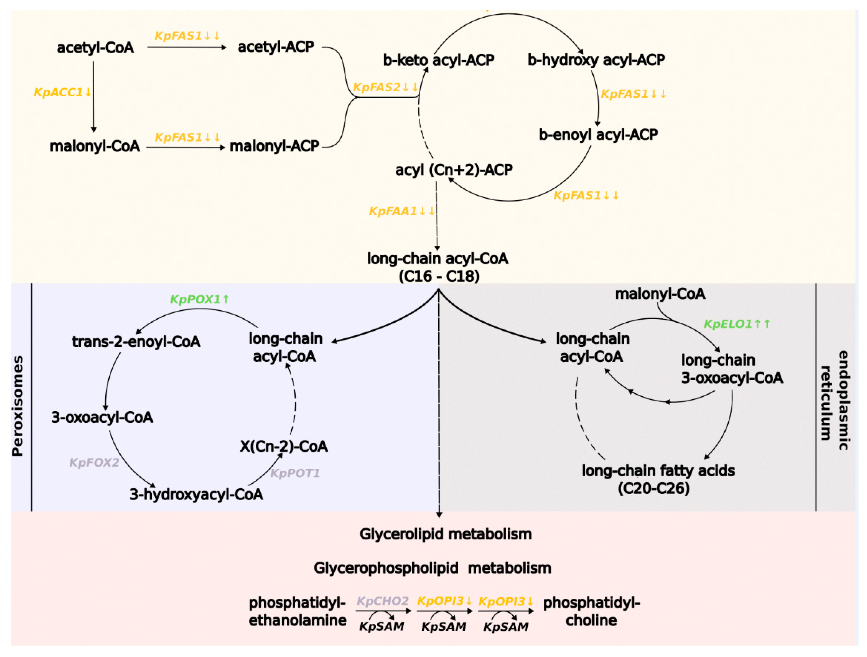
3.3. Methionine Influences the Expression of Genes Involved in Fatty Acid Metabolism in K. phaffii Cells

Differential expression analysis revealed the suppression of de novo fatty acid (FA) synthesis by methionine in the culture media (Figure 3). In S. cerevisiae, the first and rate-limiting step in this biosynthesis process is a reaction catalyzed by acetyl-coenzyme A carboxylase [60]. This reaction requires a biotin cofactor that attaches to the apoprotein ACC1p [61]. Fatty acid chain synthesis continues by attaching acetyl-CoA and malonyl-CoA to the fatty acid synthase (FAS) complex domain, called acyl-carrier protein (Acp). Then acetoacetyl is formed in the decarboxylation reaction, and like the other acyl residues, it is bounded to Acp at every biosynthetic step. The FAS complex consists of two polyfunctional proteins, α and β, which are encoded by the FAS2 and FAS1 genes, respectively [62]. The FAS complex works throughout the fatty acids biosynthesis by adding two residues of malonyl-CoA to the growing chain.
According to our results, for K. phaffii cells grown in a methionine-containing medium, gene expression was reduced for the enzymes acetyl-CoA carboxylase (KpACC1) and fatty acid synthetase (KpFAS1 and KpFAS2), which are crucial for fatty acid biosynthesis. The gene expression of the enzyme long-chain fatty acyl-CoA synthetase, with a preference for C12:0-C16:0 fatty acids (KpFAA1), which makes long-chain acyl-CoA, including palmitoyl-CoA, was also reduced.
On the contrary, the expression of the elongase enzyme gene (KpELO) of the elongation pathway (C20 residues and above) of FAs was significantly increased. This pathway occurs in the endoplasmic reticulum, where FAs up to 26 residues form. The reactions carried out by elongase are similar to those occurring in de novo biosynthesis, and require malonyl-CoA. FA-acyl-CoA to Acp bonding is not required for elongation pathway reactions [61].
Expression of the gene for the fatty acid β-oxidation enzyme fatty-acyl coenzyme A oxidase (KpPOX1) was also increased. In yeast, β-oxidation is performed only in the peroxisomes [64]. This process produces acetyl-CoA, which can further enter the glyoxylate cycle or pass into the mitochondria. However, the expression of the peroxisomal membrane protein required for the medium-chain fatty acid oxidation gene (PEX11), which is involved in β-oxidation and peroxisome proliferation, was reduced.
One of the fatty acid derivatives are phospholipids, components of cell membranes. They are formed from glycerol-3-phosphate backbone and fatty acid residues [65]. Our results show decreased expression of the KpOPI3 gene. In S. cerevisiae, the OPI3 gene product synthesizes phosphatidylcholine (PC) from phosphatidylethanolamine (PE) in reactions coupled with the consumption of SAM [66].

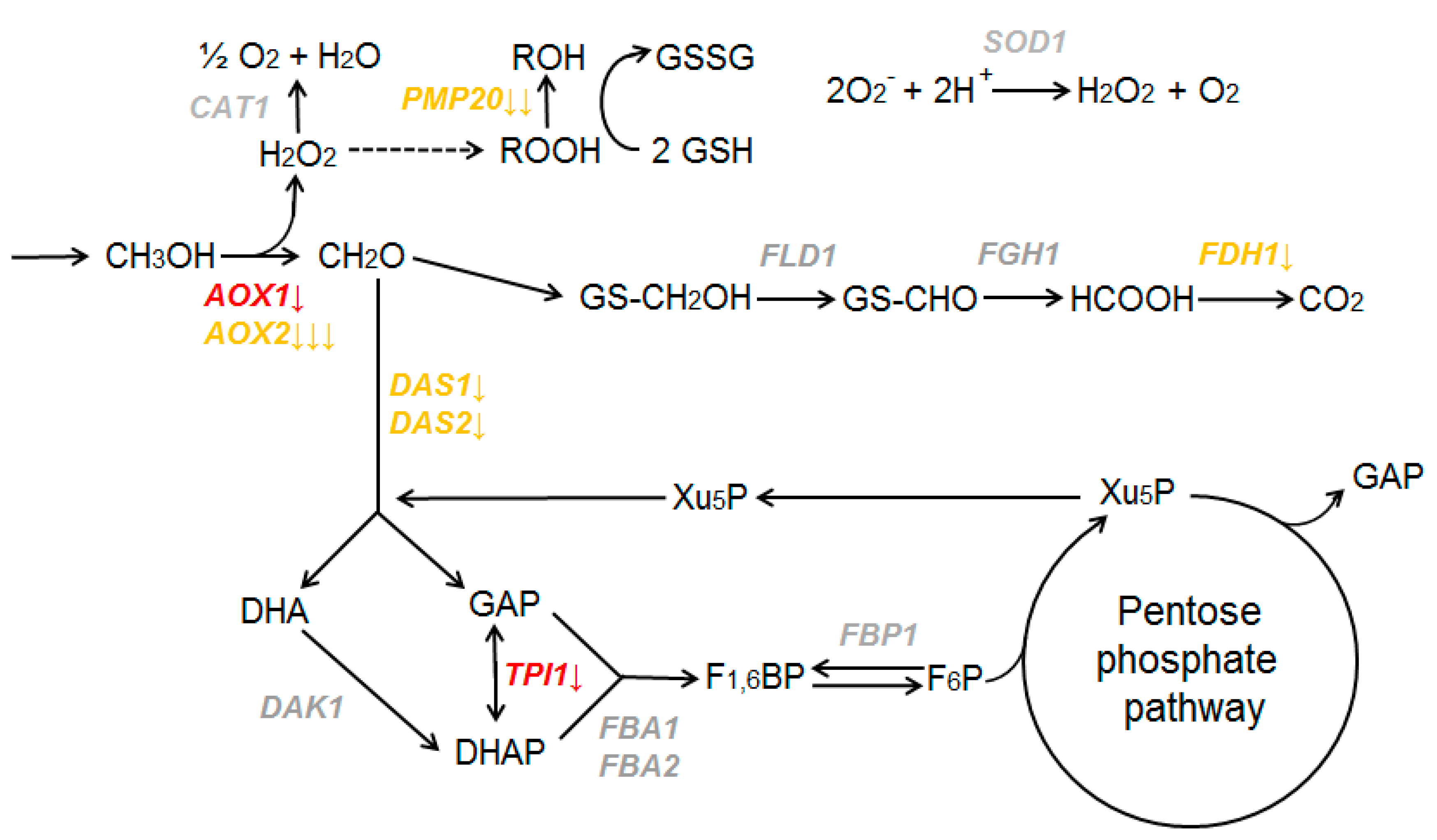
3.4. Methionine Influences the Expression of Genes Involved in Methanol Utilization in K. phaffii Cells

The activity of some methanol utilization (MUT) genes was decreased when K. phaffii grew in methionine-containing media (Figure 4).
It was shown that in a methionine-containing medium, the expression of the alcohol oxidase AOX2 gene is decreased. Repression of the AOX1 gene, which is responsible for 90% of alcohol oxidase, may be considered if a less strict cutoff for DEG is applied (0.05 < p adj < 0.1). Genes involved in first reactions of the dissimilative branch (FLD1 and FGH1) do not change their expression. The formate dehydrogenase gene FDH1 is repressed by methionine.
Genes encoding the enzymes of the assimilative branch of methanol metabolism also demonstrate different expression. Dihydroxyacetone synthase genes DAS1 and DAS2 are repressed in response to methionine in the media. Among other main genes, only TPI1 may be considered repressed by methionine if a less strict cutoff for DEG is applied (0.05 < p adj < 0.1). Dihydroxyacetone kinase (DAK1), fructose-1,6-bisphosphate aldolase (FBA1 and FBA2), and fructose-1,6-bisphosphatase (FBP1) did not significantly change their mRNA levels in response to methionine.
The expression level was reduced for genes involved in cellular defense mechanisms against reactive oxygen species that are usually produced in methanol oxidation processes. While catalase and superoxide dismutase genes (CAT1, SOD1) did not significantly change their expression, peroxisomal glutathione peroxidase gene PMP20 was repressed by methionine.
Thus, in K. phaffii cells, MUT genes demonstrate different regulation in response to methionine. Some of them are downregulated when this amino acid is added to the media, but others do not significantly change their expression levels. Interestingly, the expression of the KpMIG1 gene, which is implicated in the repression of MUT genes [67], was upregulated when methionine was added to the medium.

3.5. AOX1 Gene Promoter Is Repressed in Medium with Methionine

Since transcriptome analysis demonstrated borderline results for AOX1 gene expression, a reporter system was used to precisely analyze its expression in dependence on methionine. A K. phaffii PAP1-X33 strain carrying a PHO1 reporter gene under the AOX1 gene promoter control was generated. The resulting strain synthesizes and secretes acid phosphatase in response to activation of the AOX1 promoter by methanol (Figure S1c in the Supplementary Materials).
PAP1-X33 strain was grown in 100 mL BMG media with glycerol. After 40 h of growth, cells were transferred to 100 mL of media with methanol, and with or without methionine (BMM and Met, or BMM). After 24 h of methanol induction, ACP specific activity in the cultures was measured (Figure 5).
It was shown that the activity of the AOX1 promoter is indeed decreased during methanol induction if methionine is present in the medium.

4. Discussion

K. phaffii yeast plays a prominent role in modern biotechnology as a workhorse for the production of recombinant proteins and various chemical substances. For efficient use of this yeast, it is important to study the effects of different media components on its growth and gene expression. The amino acid methionine is not only a block for protein synthesis, but is also crucial for a large number of cell processes. For S. cerevisiae yeast, the presence of methionine in the media can induce a hierarchically organized anabolic program enabling proliferation [35].
K. phaffii can metabolize several amino acids (e.g., proline) as a sole carbon and energy source. The presence of such amino acids in the media stimulates the growth of K. phaffii cells and strongly regulates its gene expression [27]. K. phaffii cells do not use methionine as a carbon and energy source [55]. Our results demonstrate that it also does not significantly influence the growth of K. phaffii X-33 strain in media containing glycerol. In media with methanol, its growth was slightly impaired when methionine was added at a concentration of 0.46%. This coincides with the results of gene expression analysis that demonstrate repression of some MUT genes if methionine is present in the media during methanol induction.
Our previous results demonstrate that the presence of proline in the media results in the repression of all main MUT and PEX genes in K. phaffii cells [27]. Thus, a signal system downregulates the expression of MUT and PEX genes to an optimal level when proline and methanol are concurrently metabolized by K. phaffii cells. In this case, these genes are regulated as a unit, and form a regulon. However, for methionine the picture is different—only some of the MUT genes are downregulated, while expression of others does not depend on the presence of methionine in the media.
Several proteins were previously found to be directly involved in AOX1 and MUT gene regulation. Here, we demonstrate that expression of the KpMIG2 gene (PAS_chr1-4_0526), which is involved in the repression of AOX1 and other MUT genes [67], is significantly upregulated when K. phaffii is grown in methionine-containing media. Interestingly, the expression of this gene is also activated by proline [27]. It might be suggested that this gene represents an important node that intertwines the regulation of methanol utilization and other metabolic processes in K. phaffii cells.
The effect of methionine on fatty acid and lipid metabolism in different cells has been reported, but the mechanism of this relationship has not yet been identified [29,68]. Our results demonstrate the complex effect of excess methionine on fatty acid metabolism in K. phaffii cells. According to the differential expression analysis, fatty acid de novo biosynthesis was suppressed in a medium with methionine. However, the genes of the first enzymes of the elongation and fatty acid β-oxidation pathway were upregulated under the influence of methionine. These results suggest that methionine redirects fatty acid metabolism.
It has been previously suggested that the tight regulation of fatty acid metabolism may occur because of the restricted availability of acetyl-CoA [69]. However, our data show a statistically significant increase in gene expression of the acetyl-coenzyme A synthetase enzyme (PAS_chr3_0403), as well as suppression of gene expression of the acetyl-CoA carboxylase (PAS_chr1-4_0249) that produces malonyl-CoA from acetyl-CoA, which is repressed by acyl-CoAs, including malonyl-CoA and palmitoyl-CoA [70]. In addition, in our case, there was increased expression of the fatty acid β-oxidation enzyme gene (PAS_chr1-4_0538), which results in the acetyl-CoA synthesis. All of the above suggests that acetyl-CoA deficiency is unlikely to cause a strong decrease in the gene expression of the fatty acid de novo biosynthesis pathway. Overall, finding a direct link between methionine and fatty acid metabolism seems to be problematic, based on transcriptomic data alone.
In S. cerevisiae cells, transcription of methionine biosynthesis pathway genes is suppressed by the presence of large amounts of methionine or cysteine in the media [71]. Our results showed that for K. phaffii, methionine presence in the media results in the downregulation of genes involved in the sulfur assimilation pathway. We also observed a decrease in expression of the KpMET25 gene, which is responsible for a binding reaction between two branches of methionine biosynthesis: sulfur assimilation, and construction of methionine carbon backbone. The genes of the latter branch did not significantly change in their expression levels. This may occur because aspartate and homoserine are indirect precursors for other amino acids, e.g., threonine and isoleucine [72].
Media recipes for K. phaffii cultivation often contain peptone and amino acid mixes, and methionine is an essential part of these ingredients. Our results revealed the methionine effect on gene expression in K. phaffii cells, which is important for further optimization of this yeast cultivation. For instance, K. phaffii MutS strains have previously been shown to be prone to inappropriately incorporate O-methyl-L-homoserine instead of methionine residues when cells grew in culture media with methanol as the sole carbon source. Supplementation of the cultivation media with methionine is proposed to eliminate this negative effect [73]. Our results demonstrate that such supplementation should be carefully optimized to balance between reducing O-methyl-L-homoserine misincorporation and repressing activity of the AOX1 gene promoter, which is often used for recombinant protein production.
K. phaffii yeast is now considered a prospective model object for fundamental research. This trend is stimulated by the practical importance K. phaffii and development of modern research techniques, such as methods of transcriptome, proteome, and metabolome analysis. K. phaffii possesses a sophisticated mechanism that regulates the expression of AOX1 and other MUT genes. This mechanism is inextricably linked to regulation of different metabolic pathways and cellular processes. It allows K. phaffii to efficiently coordinate expression of MUT genes in response to various factors. Interestingly, most of the proteins involved in this regulation have orthologues in other yeast species that do not possess methanol utilization pathways. It may be proposed that, during the evolution of methylotrophic yeasts, preexisting regulatory elements rearranged and took under control the emerging unique pathway of methanol utilization. Thus, further investigation of MUT genes regulation in K. phaffii, in comparison to other yeast species, becomes important for understanding the evolution of regulatory systems. In this study, we demonstrate novel aspects of such regulation.

Supplementary Materials

The following supporting information can be downloaded at: https://www.mdpi.com/article/10.3390/microorganisms11040877/s1, Figure S1: Analysis of pPICZ-PHO1 plasmid and PAP1-X33 K. phaffii strain generated in this study; Table S1: Growth curves of K. phaffii X-33 strain in different media; Table S2. Number of reads for each sample analyzed by RNA-sequencing; Table S3. The results of the differential gene expression analysis using DESeq2; Table S4. Differentially expressed genes discussed in this paper; Table S5. Analysis of ACP specific activity of K. phaffii PAP1-X33 strain carrying reporter gene PHO1 under control of AOX1 promoter.

Author Contributions

Conceptualization, T.I. and A.R.; methodology, M.P. and E.S.; software, T.I. and A.S.; investigation, T.I., K.P., M.S. and A.M.; writing—original draft preparation, T.I., A.G., M.P., E.S. and A.R.; writing—review and editing, M.P., E.S. and A.G. All authors have read and agreed to the published version of the manuscript.

Funding

This research was funded by the Russian Science Foundation, grant number 19-73-10077-Π.

Data Availability Statement

The data presented in this study are available in Supplementary Materials.

Conflicts of Interest

The authors declare no conflict of interest.

References

  1. Athmaram, T.N.; Singh, A.K.; Saraswat, S.; Srivastava, S.; Misra, P.; Kameswara Rao, M.; Gopalan, N.; Rao, P.V.L. A Simple Pichia pastoris Fermentation and Downstream Processing Strategy for Making Recombinant Pandemic Swine Origin Influenza a Virus Hemagglutinin Protein. J. Ind. Microbiol. Biotechnol. 2013, 40, 245–255. [Google Scholar] [CrossRef]
  2. Liu, W.-C.; Gong, T.; Wang, Q.-H.; Liang, X.; Chen, J.-J.; Zhu, P. Scaling-up Fermentation of Pichia pastoris to Demonstration-Scale Using New Methanol-Feeding Strategy and Increased Air Pressure Instead of Pure Oxygen Supplement. Sci. Rep. 2016, 6, 18439. [Google Scholar] [CrossRef] [PubMed] [Green Version]
  3. Liu, W.-C.; Inwood, S.; Gong, T.; Sharma, A.; Yu, L.-Y.; Zhu, P. Fed-Batch High-Cell-Density Fermentation Strategies for Pichia pastoris Growth and Production. Crit. Rev. Biotechnol. 2019, 39, 258–271. [Google Scholar] [CrossRef]
  4. Balamurugan, V.; Reddy, G.R.; Suryanarayana, V.V.S. Pichia pastoris: A Notable Heterologous Expression System for the Production of Foreign Proteins—Vaccines. 1 April 2007. Available online: http://nopr.niscpr.res.in/handle/123456789/3021 (accessed on 28 February 2023).
  5. Jacobs, P.P.; Geysens, S.; Vervecken, W.; Contreras, R.; Callewaert, N. Engineering Complex-Type N-Glycosylation in Pichia pastoris Using GlycoSwitch Technology. Nat. Protoc. 2009, 4, 58–70. [Google Scholar] [CrossRef]
  6. Li, P.; Anumanthan, A.; Gao, X.-G.; Ilangovan, K.; Suzara, V.V.; Düzgüneş, N.; Renugopalakrishnan, V. Expression of Recombinant Proteins in Pichia pastoris. Appl. Biochem. Biotechnol. 2007, 142, 105–124. [Google Scholar] [CrossRef]
  7. Ahmad, M.; Hirz, M.; Pichler, H.; Schwab, H. Protein Expression in Pichia pastoris: Recent Achievements and Perspectives for Heterologous Protein Production. Appl. Microbiol. Biotechnol. 2014, 98, 5301–5317. [Google Scholar] [CrossRef] [PubMed] [Green Version]
  8. Baghban, R.; Farajnia, S.; Ghasemi, Y.; Mortazavi, M.; Zarghami, N.; Samadi, N. New Developments in Pichia pastoris Expression System, Review and Update. Curr. Pharm. Biotechnol. 2020, 19, 451–467. [Google Scholar] [CrossRef]
  9. Karbalaei, M.; Rezaee, S.A.; Farsiani, H. Pichia pastoris: A Highly Successful Expression System for Optimal Synthesis of Heterologous Proteins. J. Cell Physiol. 2020, 235, 5867–5881. [Google Scholar] [CrossRef] [PubMed]
  10. Wang, M.; Jiang, S.; Wang, Y. Recent Advances in the Production of Recombinant Subunit Vaccines in Pichia pastoris. Bioengineered 2016, 7, 155–165. [Google Scholar] [CrossRef] [PubMed] [Green Version]
  11. Jahic, M.; Veide, A.; Charoenrat, T.; Teeri, T.; Enfors, S.-O. Process Technology for Production and Recovery of Heterologous Proteins with Pichia pastoris. Biotechnol. Prog. 2006, 22, 1465–1473. [Google Scholar] [CrossRef]
  12. Byrne, B. Pichia pastoris as an Expression Host for Membrane Protein Structural Biology. Curr. Opin. Struct. Biol. 2015, 32, 9–17. [Google Scholar] [CrossRef] [Green Version]
  13. Hirz, M.; Richter, G.; Leitner, E.; Wriessnegger, T.; Pichler, H. A Novel Cholesterol-Producing Pichia pastoris Strain Is an Ideal Host for Functional Expression of Human Na,K-ATPase A3β1 Isoform. Appl. Microbiol. Biotechnol. 2013, 97, 9465–9478. [Google Scholar] [CrossRef]
  14. Tanaka, K.; Ishii, Y.; Ogawa, J.; Shima, J. Enhancement of Acetic Acid Tolerance in Saccharomyces cerevisiae by Overexpression of the HAA1 Gene, Encoding a Transcriptional Activator. Appl. Environ. Microbiol. 2012, 78, 8161–8163. [Google Scholar] [CrossRef] [Green Version]
  15. Carneiro, C.V.G.C.; Serra, L.A.; Pacheco, T.F.; Ferreira, L.M.M.; Brandão, L.T.D.; Freitas, M.N.d.M.; Trichez, D.; Almeida, J.R.M.d. Advances in Komagataella phaffii Engineering for the Production of Renewable Chemicals and Proteins. Fermentation 2022, 8, 575. [Google Scholar] [CrossRef]
  16. Bernauer, L.; Radkohl, A.; Lehmayer, L.G.K.; Emmerstorfer-Augustin, A. Komagataella phaffii as Emerging Model Organism in Fundamental Research. Front. Microbiol. 2021, 11, 607028. [Google Scholar] [CrossRef] [PubMed]
  17. Cregg, J.M.; Madden, K.R.; Barringer, K.J.; Thill, G.P.; Stillman, C.A. Functional Characterization of the Two Alcohol Oxidase Genes from the Yeast Pichia pastoris. Mol. Cell Biol. 1989, 9, 1316–1323. [Google Scholar] [CrossRef]
  18. Rußmayer, H.; Buchetics, M.; Gruber, C.; Valli, M.; Grillitsch, K.; Modarres, G.; Guerrasio, R.; Klavins, K.; Neubauer, S.; Drexler, H.; et al. Systems-Level Organization of Yeast Methylotrophic Lifestyle. BMC Biol. 2015, 13, 80. [Google Scholar] [CrossRef] [Green Version]
  19. Berrios, J.; Theron, C.W.; Steels, S.; Ponce, B.; Velastegui, E.; Bustos, C.; Altamirano, C.; Fickers, P. Role of Dissimilative Pathway of Komagataella phaffii (Pichia pastoris): Formaldehyde Toxicity and Energy Metabolism. Microorganisms 2022, 10, 1466. [Google Scholar] [CrossRef] [PubMed]
  20. Tschopp, J.F.; Brust, P.F.; Cregg, J.M.; Stillman, C.A.; Gingeras, T.R. Expression of the LacZ Gene from Two Methanol-Regulated Promoters in Pichia pastoris. Nucleic Acids Res. 1987, 15, 3859–3876. [Google Scholar] [CrossRef] [PubMed] [Green Version]
  21. SEllis, S.B.; Brust, P.F.; Koutz, P.J.; Waters, A.F.; Harpold, M.M.; Gingeras, T.R. Isolation of alcohol oxidase and two other methanol regulatable genes from the yeast Pichia pastoris. Mol. Cell Biol. 1985, 5, 1111–1121. [Google Scholar] [CrossRef]
  22. Kalender, Ö.; Çalık, P. Transcriptional Regulatory Proteins in Central Carbon Metabolism of Pichia pastoris and Saccharomyces cerevisiae. Appl. Microbiol. Biotechnol. 2020, 104, 7273–7311. [Google Scholar] [CrossRef]
  23. Rumjantsev, A.M.; Padkina, M.V.; Sambuk, E.V. Effect of Nitrogen Source on Gene Expression of First Steps of Methanol Utilization Pathway in Pichia pastoris. Russ. J. Genet. 2013, 49, 394–400. [Google Scholar] [CrossRef]
  24. Rumjantsev, A.M.; Bondareva, O.V.; Padkina, M.V.; Sambuk, E.V. Effect of Nitrogen Source and Inorganic Phosphate Concentration on Methanol Utilization and PEX Genes Expression in Pichia pastoris. Sci. World J. 2014, 743615. [Google Scholar] [CrossRef] [Green Version]
  25. Sahu, U.; Rangarajan, P.N. Methanol Expression Regulator 1 (Mxr1p) Is Essential for the Utilization of Amino Acids as the Sole Source of Carbon by the Methylotrophic Yeast, Pichia pastoris. J. Biol. Chem. 2016, 291, 20588–20601. [Google Scholar] [CrossRef] [PubMed] [Green Version]
  26. Rumyantsev, A.M.; Soloviev, G.A.; Slepchenkov, A.V.; Sambuk, E.V. Effects of Deletions in Pichia pastoris RTG Genes on Phenotype and AOX1 Expression. Adv. Microbiol. 2018, 8, 439–450. [Google Scholar] [CrossRef] [Green Version]
  27. Rumyantsev, A.; Sidorin, A.; Volkov, A.; Al Shanaa, O.; Sambuk, E.; Padkina, M. Transcriptome Analysis Unveils the Effects of Proline on Gene Expression in the Yeast Komagataella phaffii. Microorganisms 2021, 10, 67. [Google Scholar] [CrossRef]
  28. Wang, H.; Li, Y.; Che, Y.; Yang, D.; Wang, Q.; Yang, H.; Boutet, J.; Huet, R.; Yin, S. Production of L-Methionine from 3-Methylthiopropionaldehyde and O-Acetylhomoserine by Catalysis of the Yeast O-Acetylhomoserine Sulfhydrylase. J Agric. Food. Chem. 2021, 69, 7932–7937. [Google Scholar] [CrossRef] [PubMed]
  29. Rajakumar, S.; Suriyagandhi, V.; Nachiappan, V. Impairment of MET Transcriptional Activators, MET4 and MET31 Induced Lipid Accumulation in Saccharomyces cerevisiae. FEMS Yeast Res. 2020, 20, foaa039. [Google Scholar] [CrossRef] [PubMed]
  30. Walvekar, A.S.; Kadamur, G.; Sreedharan, S.; Gupta, R.; Srinivasan, R.; Laxman, S. Methylated PP2A stabilizes Gcn4 to enable a methionine-induced anabolic program. J. Biol. Chem. 2020, 295, 18390–18405. [Google Scholar] [CrossRef]
  31. Wu, Z.; Song, L.; Liu, S.Q.; Huang, D. Independent and Additive Effects of Glutamic Acid and Methionine on Yeast Longevity. PLoS ONE 2013, 8, e79319. [Google Scholar] [CrossRef] [Green Version]
  32. Campbell, K.; Vowinckel, J.; Keller, M.A.; Ralser, M. Methionine Metabolism Alters Oxidative Stress Resistance via the Pentose Phosphate Pathway. Antioxid. Redox Sign. 2016, 24, 543–547. [Google Scholar] [CrossRef] [PubMed] [Green Version]
  33. Martínez, Y.; Li, X.; Liu, G.; Bin, P.; Yan, W.; Más, D.; Valdivié, M.; Hu, C.-A.A.; Ren, W.; Yin, Y. The Role of Methionine on Metabolism, Oxidative Stress, and Diseases. Amino Acids 2017, 49, 2091–2098. [Google Scholar] [CrossRef] [PubMed]
  34. Zou, K.; Ouyang, Q.; Li, H.; Zheng, J. A Global Characterization of the Translational and Transcriptional Programs Induced by Methionine Restriction through Ribosome Profiling and RNA-Seq. BMC Genom. 2017, 18, 189. [Google Scholar] [CrossRef] [PubMed] [Green Version]
  35. Walvekar, A.S.; Srinivasan, R.; Gupta, R.; Laxman, S. Methionine Coordinates a Hierarchically Organized Anabolic Program Enabling Proliferation. Mol. Biol. Cell 2018, 29, 3183–3200. [Google Scholar] [CrossRef]
  36. Wiame, J.M.; Grenson, M.; Arst, H.N. Nitrogen Catabolite Repression in Yeasts and Filamentous Fungi. Adv. Microb. Physiol. 1985, 26, 1–88. [Google Scholar] [CrossRef]
  37. Thomas, D.; Surdin-Kerjan, Y. Metabolism of Sulfur Amino Acids in Saccharomyces cerevisiae. Microbiol Mol. Biol. Rev. 1997, 61, 503–532. [Google Scholar] [CrossRef]
  38. Cherest, H.; Eichler, F.; de Robichon-Szulmajster, H. Genetic and Regulatory Aspects of Methionine Biosynthesis in Saccharomyces cerevisiae. J. Bacteriol. 1969, 97, 328–336. [Google Scholar] [CrossRef] [Green Version]
  39. Linder, T. ATP Sulfurylase Is Essential for the Utilization of Sulfamate as a Sulfur Source in the Yeast Komagataella pastoris (Syn. Pichia pastoris). Curr. Microbiol. 2017, 74, 1021–1025. [Google Scholar] [CrossRef] [Green Version]
  40. Thomas, D.; Barbey, R.; Surdin-Kerjan, Y. Gene-Enzyme Relationship in the Sulfate Assimilation Pathway of Saccharomyces cerevisiae. Study of the 3’-Phosphoadenylylsulfate Reductase Structural Gene. J. Biol. Chem. 1990, 265, 15518–15524. [Google Scholar] [CrossRef] [PubMed]
  41. Hayakawa, K.; Kajihata, S.; Matsuda, F.; Shimizu, H. (13)C-Metabolic Flux Analysis in S-Adenosyl-L-Methionine Production by Saccharomyces cerevisiae. J. Biosci. Bioeng. 2015, 120, 532–538. [Google Scholar] [CrossRef]
  42. Stoner, G.L.; Eisenberg, M.A. Biosynthesis of 7, 8-Diaminopelargonic Acid from 7-Keto-8-Aminopelargonic Acid and S-Adenosyl-L-Methionine. The Kinetics of the Reaction. J. Biol. Chem. 1975, 250, 4037–4043. [Google Scholar] [CrossRef] [PubMed]
  43. Slany, R.K.; Bösl, M.; Crain, P.F.; Kersten, H. A New Function of S-Adenosylmethionine: The Ribosyl Moiety of AdoMet Is the Precursor of the Cyclopentenediol Moiety of the TRNA Wobble Base Queuine. Biochemistry 1993, 32, 7811–7817. [Google Scholar] [CrossRef]
  44. Gupta, R.; Laxman, S. TRNA Wobble-Uridine Modifications as Amino Acid Sensors and Regulators of Cellular Metabolic State. Curr. Genet. 2020, 66, 475–480. [Google Scholar] [CrossRef] [PubMed]
  45. Hanahan, D. Studies on transformation of Escherichia coli with plasmids. J. Mol. Biol. 1983, 166, 557–580. [Google Scholar] [CrossRef]
  46. Wu, S.; Letchworth, G.J. High efficiency transformation by electroporation of Pichia pastoris pretreated with lithium acetate and dithiothreitol. BioTechniques 2004, 36, 152–154. [Google Scholar] [CrossRef] [PubMed]
  47. Padkina, M.V.; Krasnopevtseva, N.G.; Petrashen, M.G. Genetic and biochemical studies of acid phosphatases of Saccharomyces cerevisiae. Properties of acid phosphatases from different strains (Russian). Genetika 1974, 10, 100–110. [Google Scholar]
  48. Leggett, R.M.; Ramirez-Gonzalez, R.H.; Clavijo, B.J.; Ewaite, D.; Davey, R.P. Sequencing quality assessment tools to enable data-driven informatics for high throughput genomics. Front. Genet. 2013, 4, 288. [Google Scholar] [CrossRef]
  49. Bolger, A.M.; Lohse, M.; Usadel, B. Trimmomatic: A flexible trimmer for Illumina sequence data. Bioinformatics 2014, 30, 2114–2212. [Google Scholar] [CrossRef] [Green Version]
  50. Kim, D.; Paggi, J.M.; Park, C.; Bennett, C.; Salzberg, S.L. Graph-based genome alignment and genotyping with HISAT2 and HISAT-genotype. Nat. Biotechnol. 2019, 37, 907–915. [Google Scholar] [CrossRef]
  51. Danecek, P.; Bonfield, J.K.; Liddle, J.; Marshall, J.; Ohan, V.; Pollard, M.O.; Whitwham, A.; Keane, T.; McCarthy, S.A.; Davies, R.M.; et al. Twelve Years of SAMtools and BCFtools. GigaScience 2021, 10, giab008. [Google Scholar] [CrossRef]
  52. Liao, Y.; Smyth, G.K.; Shi, W. featureCounts: An efficient general purpose program for assigning sequence reads to genomic features. Bioinformatics 2014, 30, 923–930. [Google Scholar] [CrossRef] [PubMed] [Green Version]
  53. Love, M.I.; Huber, W.; Anders, S. Moderated estimation of fold change and dispersion for RNA-seq data with DESeq2. Genome Biol. 2014, 15, 550. [Google Scholar] [CrossRef] [PubMed] [Green Version]
  54. R Core Team. R: A Language and Environment for Statistical Computing; R Foundation for Statistical Computing: Vienna, Austria, 2018; Available online: https://www.R-project.org/ (accessed on 28 February 2023).
  55. Vogl, T.; Sturmberger, L.; Kickenweiz, T.; Wasmayer, R.; Schmid, C.; Hatzl, A.M.; Gerstmann, M.A.; Pitzer, J.; Wagner, M.; Thallinger, G.G.; et al. A Toolbox of Diverse Promoters Related to Methanol Utilization: Functionally Verified Parts for Heterologous Pathway Expression in Pichia pastoris. ACS Synth. Biol. 2016, 5, 172–186. [Google Scholar] [CrossRef] [PubMed]
  56. Dey, T.; Krishna Rao, K.; Khatun, J.; Rangarajan, P.N. The Nuclear Transcription Factor Rtg1p Functions as a Cytosolic, Post-Transcriptional Regulator in the Methylotrophic Yeast Pichia pastoris. J. Biol. Chem. 2018, 293, 16647–16660. [Google Scholar] [CrossRef] [Green Version]
  57. Solà, A.; Maaheimo, H.; Ylönen, K.; Ferrer, P.; Szyperski, T. Amino Acid Biosynthesis and Metabolic Flux Profiling of Pichia pastoris. Eur. J. Biochem. 2004, 271, 2462–2470. [Google Scholar] [CrossRef]
  58. Ferrer, P.; Albiol, J. 13C-Based Metabolic Flux Analysis of Recombinant Pichia pastoris. Methods Mol. Biol. 2014, 1191, 291–313. [Google Scholar] [CrossRef]
  59. Chan, S.Y.; Appling, D.R. Regulation of S-Adenosylmethionine Levels in Saccharomyces cerevisiae. J. Biol. Chem. 2003, 278, 43051–43059. [Google Scholar] [CrossRef] [Green Version]
  60. Hu, Y.; Zhu, Z.; Nielsen, J.; Siewers, V. Engineering Saccharomyces cerevisiae Cells for Production of Fatty Acid-Derived Biofuels and Chemicals. Open. Biol. 2019, 9, 190049. [Google Scholar] [CrossRef] [Green Version]
  61. Tehlivets, O.; Scheuringer, K.; Kohlwein, S.D. Fatty Acid Synthesis and Elongation in Yeast. Biochim. Biophys. Acta 2007, 1771, 255–270. [Google Scholar] [CrossRef]
  62. Johansson, P.; Wiltschi, B.; Kumari, P.; Kessler, B.; Vonrhein, C.; Vonck, J.; Oesterhelt, D.; Grininger, M. Inhibition of the fungal fatty acid synthase type I multienzyme complex. Proc. Natl. Acad. Sci. USA 2008, 105, 12803–12808. [Google Scholar] [CrossRef] [Green Version]
  63. Ye, C.; Sutter, B.M.; Wang, Y.; Kuang, Z.; Tu, B.P. A Metabolic Function for Phospholipid and Histone Methylation. Mol. Cell 2017, 66, 180–193.e8. [Google Scholar] [CrossRef] [Green Version]
  64. Câmara, A.d.A.; Maréchal, P.-A.; Tourdot-Maréchal, R.; Husson, F. Oxidative Stress Resistance during Dehydration of Three Non-Saccharomyces Wine Yeast Strains. Food Res. Int. 2019, 123, 364–372. [Google Scholar] [CrossRef] [PubMed]
  65. Carman, G.M.; Han, G.-S. Regulation of Phospholipid Synthesis in the Yeast Saccharomyces cerevisiae. Annu. Rev. Biochem. 2011, 80, 859–883. [Google Scholar] [CrossRef] [PubMed]
  66. Sadhu, M.J.; Moresco, J.J.; Zimmer, A.D.; Yates, J.R.; Rine, J. Multiple Inputs Control Sulfur-Containing Amino Acid Synthesis in Saccharomyces cerevisiae. Mol. Biol. Cell 2014, 25, 1653–1665. [Google Scholar] [CrossRef]
  67. Shi, L.; Wang, X.; Wang, J.; Zhang, P.; Qi, F.; Cai, M.; Zhang, Y.; Zhou, X. Transcriptome analysis of ∆mig1∆mig2 mutant reveals their roles in methanol catabolism, peroxisome biogenesis and autophagy in methylotrophic yeast Pichia pastoris. Genes Genom. 2018, 40, 399–412. [Google Scholar] [CrossRef] [PubMed]
  68. Chatterjee, M.T.; Khalawan, S.A.; Curran, B.P. Subtle Alterations in Growth Medium Composition Can Dramatically Alter the Percentage of Unsaturated Fatty Acids in the Yeast Saccharomyces cerevisiae. Yeast 2001, 18, 81–88. [Google Scholar] [CrossRef]
  69. Zhang, C.; Ma, Y.; Miao, H.; Tang, X.; Xu, B.; Wu, Q.; Mu, Y.; Huang, Z. Transcriptomic Analysis of Pichia pastoris (Komagataella phaffii) GS115 During Heterologous Protein Production Using a High-Cell-Density Fed-Batch Cultivation Strategy. Front. Microbiol. 2020, 11, 463. [Google Scholar] [CrossRef]
  70. Shirra, M.K.; Patton-Vogt, J.; Ulrich, A.; Liuta-Tehlivets, O.; Kohlwein, S.D.; Henry, S.A.; Arndt, K.M. Inhibition of Acetyl Coenzyme A Carboxylase Activity Restores Expression of the INO1 Gene in a Snf1 Mutant Strain of Saccharomyces cerevisiae. Mol. Cell Biol. 2001, 21, 5710–5722. [Google Scholar] [CrossRef] [Green Version]
  71. Mertz, J.E.; Spence, K.D. Methionine Adenosyltransferase and Ethionine Resistance in Saccharomyces cerevisiae. J. Bacteriol. 1972, 111, 778–783. [Google Scholar] [CrossRef] [Green Version]
  72. Farfán, M.J.; Aparicio, L.; Calderón, I.L. Threonine Overproduction in Yeast Strains Carrying the HOM3-R2 Mutant Allele under the Control of Different Inducible Promoters. Appl. Environ. Microbiol. 1999, 65, 110–116. [Google Scholar] [CrossRef] [Green Version]
  73. Schotte, P.; Dewerte, I.; De Groeve, M.; De Keyser, S.; De Brabandere, V.; Stanssens, P. Pichia pastoris MutS Strains Are Prone to Misincorporation of O-Methyl-l-Homoserine at Methionine Residues When Methanol Is Used as the Sole Carbon Source. Microb. Cell Fact. 2016, 15, 98. [Google Scholar] [CrossRef] [PubMed] [Green Version]
Figure 1. Growth curves of K. phaffii X-33 strain in different media. BMG medium contains 1% glycerol; BMG and Met—1% glycerol and 0.46% L-methionine; BMM—1% methanol; BMM and Met—1% glycerol and 0.46% L-methionine; BM and Met—only 0.46% L-methionine. Cells were grown in four separate cultures for each type of the medium. Each measurement was performed in two replicas for each culture. Mean OD600 values ± SEM are plotted. Raw data is presented in Table S1 in the Supplementary Materials.
Microorganisms 11 00877 g001
Figure 2. Scheme of proposed methionine metabolism pathways in K. phaffii cells (based on data for S. cerevisiae [37,59]). Genes are placed near the reactions catalyzed by the corresponding proteins. Genes that are repressed in media with methionine are marked in orange. Genes that do not change their expression are marked in grey. The number of arrows represents values of the log2FoldChange parameter: one arrow 0.5 < log2FoldChange < 1; two arrows 1 < log2FoldChange < 2; three arrows 2 < log2FoldChange.
Microorganisms 11 00877 g002
Figure 3. Scheme of the proposed fatty acid metabolism pathways in K. phaffii cells (based on data for S. cerevisiae [61,63]). Genes are placed near the reactions catalyzed by the corresponding proteins. Genes that are repressed in media with methionine are marked in orange. Genes that are activated in media with methionine are marked in green. Genes that do not change their expression are marked in grey. The number of arrows represents values of the log2FoldChange parameter: one arrow 0.5 < log2FoldChange < 1; two arrows 1 < log2FoldChange < 2; three arrows 2 < log2FoldChange.
Microorganisms 11 00877 g003
Figure 4. Scheme of the canonical methanol utilization pathway in K. phaffii cells (modified from [47]). Genes are placed near the reactions catalyzed by the corresponding proteins. Genes that are repressed in media with methionine are marked in orange (p adj < 0.05) and red (0.05 < p adj < 0.1). Genes that do not change their expression are marked in grey. The number of arrows represents values of the log2FoldChange parameter: one arrow 0.5 < log2FoldChange < 1; two arrows 1 < log2FoldChange < 2; three arrows 2 < log2FoldChange. Abbreviations of metabolites: DHA, dihydroxyacetone; DHAP, dihydroxyacetone phosphate; FRU1,6BP, fructose-1,6-bisphosphate; F6P, fructose-6-phosphate; GAP, glyceraldehyde-3-phosphate; GSH, glutathione; GSSG, oxidized glutathione self-dimer; Xu5P, xylulose 5-phosphate.
Microorganisms 11 00877 g004
Figure 5. ACP specific activity in the cultures of K. phaffii PAP1-X33 strain after methanol induction in media with or without methionine (BMM and Met, or BMM). The mean from four separate experiments with two technical replicates is plotted. Raw data is presented in Table S5 in the Supplementary Materials.
Microorganisms 11 00877 g005
Disclaimer/Publisher’s Note: The statements, opinions and data contained in all publications are solely those of the individual author(s) and contributor(s) and not of MDPI and/or the editor(s). MDPI and/or the editor(s) disclaim responsibility for any injury to people or property resulting from any ideas, methods, instructions or products referred to in the content.

