Article

Single-Stranded DNA-Binding Proteins Mediate DSB Repair and Effectively Improve CRISPR/Cas9 Genome Editing in Escherichia coli and Pseudomonas

College of Life Sciences, Henan Agricultural University, Key Laboratory of Enzyme Engineering of Agricultural Microbiology, Ministry of Agriculture and Rural Affairs, Zhengzhou 450046, China
*
Authors to whom correspondence should be addressed.
Microorganisms 2023, 11(4), 850; https://doi.org/10.3390/microorganisms11040850
Submission received: 18 February 2023 / Revised: 22 March 2023 / Accepted: 25 March 2023 / Published: 27 March 2023
(This article belongs to the Section Microbial Biotechnology)

Abstract

:
Single-stranded DNA-binding proteins (SSBs) are essential for all living organisms. Whether SSBs can repair DNA double-strand breaks (DSBs) and improve the efficiency of CRISPR/Cas9-mediated genome editing has not been determined. Here, based on a pCas/pTargetF system, we constructed pCas-SSB and pCas-T4L by replacing the λ-Red recombinases with Escherichia coli SSB and phage T4 DNA ligase in pCas, respectively. Inactivation of the E. coli lacZ gene with homologous donor dsDNA increased the gene editing efficiency of pCas-SSB/pTargetF by 21.4% compared to pCas/pTargetF. Inactivation of the E. coli lacZ gene via NHEJ increased the gene editing efficiency of pCas-SSB/pTargetF by 33.2% compared to pCas-T4L/pTargetF. Furthermore, the gene-editing efficiency of pCas-SSB/pTargetF in E. colirecA, ΔrecBCD, ΔSSB) with or without donor dsDNA did not differ. Additionally, pCas-SSB/pTargetF with donor dsDNA successfully deleted the wp116 gene in Pseudomonas sp. UW4. These results demonstrate that E. coli SSB repairs DSBs caused by CRISPR/Cas9 and effectively improves CRISPR/Cas9 genome editing in E. coli and Pseudomonas.

1. Introduction

CRISPR/Cas gene-editing technology has been rapidly adopted and widely used in microorganisms, plants, animals, and humans since it was established in 2012 [1]. CRISPR is an acronym for clustered regularly interspaced short palindromic repeats, which comprise a leader sequence, multiple repeats, and spacers [2,3]; these repeats are present in the genomes of 42.3% of bacteria and 85.2% of archaea [4]. CRISPR-adjacent regions bear CRISPR-associated (Cas) genes that encode a series of Cas nucleases. When a phage or exogenous gene invades a bacterium, the Cas protein can recognize the protospacer adjacent motif (PAM) sequence, excise its upstream protospacer, and insert it between the leader sequence and the adjacent repeat to form a new spacer [5]. The foreign homologous sequence subsequently invades, and the promoter in the leader sequence initiates transcription of the CRISPR array, producing a long-stranded RNA called the precursor transcript (pre-crRNA). The pre-crRNA is then processed by Cas proteins into mature crRNA, which contains a single spacer. crRNA guides the Cas protein, which cuts homologous DNA and eliminates the invading foreign DNA [6]. CRISPR/Cas gene-editing technology is derived from this adaptive immune defense mechanism that occurs in bacteria.
A small-molecular-weight Cas protein (SpCas9) from Streptococcus pyogenes can complete DNA splicing alone, requiring a relatively common PAM sequence [7]. However, to splice its target DNA, SpCas9 requires the guidance of guide RNA (gRNA) or single-translated guide RNA (sgRNA), which is formed by the complementary pairing of crRNA and the noncoding trans-activating CRISPR RNA (tracrRNA) gene transcript. The tracrRNA gene is also located in the CRISPR-adjacent region [8]. The reconstitution of SpCas9 and an artificial chimera of crRNA and tracrRNA, which is called synthetic-guide RNA (sgRNA), greatly simplifies the process of gene editing, thus increasing the application of the Cas9/sgRNA system.
In recipient cells, Cas9/sgRNA sufficiently cuts target genes to produce DNA double-strand breaks (DSBs); subsequently, the cells repair these DSBs by nonhomologous end joining (NHEJ) or homologous recombination (HR) repair, generating point mutations. Eukaryotes use canonical DNA-PKcs-dependent NHEJ (D-NHEJ) [9,10], alternative NHEJ (alt-NHEJ) [11], or RAD51/BRCA2-dependent HR to repair DSBs [12]. HR repair can only occur during the S/G2 phase and requires homologous donor DNA, while NHEJ repair is constitutively active throughout the entire cell cycle, does not require donor DNA, and generates nucleotide insertions or deletions at the cleavage site. Therefore, NHEJ repair is the dominant pathway that is used for DSB repair in eukaryotic cells [13,14].
To repair DSBs during the logarithmic phase of growth, prokaryotes use RecA-dependent HR repair [15]; during the stationary phase of growth, several bacteria, excluding E. coli [16], use Ku- and Ligase D-dependent NHEJ repair [17,18], while E. coli uses RecBCD and LigA-dependent NHEJ, which is called alternative end-joining (A-EJ) [19]. However, the use of the CRISPR/Cas9 system in bacteria faces challenges. The use of Cas9- and gRNA-expressing plasmids or the addition of homologous DNA, even in the absence of crRNA or inactive Cas9 (dCas9), results in few or no surviving colonies or in abnormal morphology and decreased growth rates [20,21,22,23,24]. It has been suggested that DSBs, transient PAM recognition, and enzymatic binding along the DNA have cytotoxic effects on prokaryotic cells [22,25], and DSB repair by native HR and NHEJ repair mechanisms exhibit very low efficiency in bacteria.
The use of inducible Cas9 expression systems [26,27] or the incorporation of λ-Red homologous recombination enzymes and homologous recombination templates [26,27], the mycobacteria-derived Ku- and Ligase D-dependent NHEJ pathway [28], E. coli-derived RecBCD + LigA [29], the λ phage-derived Redβ protein [30], the Pseudomonas putida-derived Ssr protein [31] and the T4 DNA ligase [32] produce surviving colonies and result in efficient editing. Jiang et al. developed a facile bacterial gene-editing system, namely, the pCas/pTargetF system [33]. In this system, the pCas plasmid harbors the λ-Red recombination system, and the IPTG-inducible expression of sgRNA-PMB1 facilitates the guiding of the pMB1-dependent replication of pTarget to eliminate pTargetF. pCas is later eliminated by incubation at 37 °C because it harbors a temperature-sensitive replicon. pTargetF carries the sgRNA expression cassette. The λ-Red recombination system consists of the Exo (Redα), Beta (Redβ), and Gam (Redγ) proteins. Exo is a 5’ to 3’ dsDNA-dependent exonuclease that generates a ssDNA intermediate. Beta is a ssDNA-binding protein that promotes pairing or annealing between complementary ssDNAs. Gam prevents the degradation of dsDNA to enhance recombination by inhibiting the cellular nucleases [34]. Employing the pCas/pTargetF system to edit the single gene locus in E. coli, the efficiency of gene editing is 6–92%, which very much depends on the homologous arm length of donor DNA and donor DNA supplied in pTarget or in PCR fragment [33]. The pCas/pTargetF system has been used in a variety of bacteria [35,36,37,38].
Single-stranded DNA-binding proteins (SSBs) are essential for all living organisms and viruses [39]. SSBs bind to single-stranded DNA (ssDNA) with high affinity and in a sequence-independent manner in order to prevent the transient formation of dsDNA from ssDNA and to protect ssDNA from undesirable damage by nucleases and chemicals [40,41]. Furthermore, SSBs interact with every protein that is involved in DNA metabolism and thus participate in DNA replication, recombination, and repair as well as genome maintenance [42,43]. Previously, an SSB in E. coli has been shown to possess the ability to catalyze both DNA HR and nonhomologous recombination (NHR) [44]. Whether this protein can promote bacterial DSB repair during CRISPR/Cas9-mediated gene editing and improve gene-editing efficiency has not been determined. In this study, SSB was used to replace the λ-Red recombination system in pCas and was confirmed to efficiently promote HR- and NHR-mediated gene editing by the pCas/pTargetF system in E. coli and Pseudomonas.

2. Material and Methods

2.1. Strains, Plasmids and Culture Conditions

The bacterial strains and plasmids that were used in this study are listed in Table 1. The strains were grown in Luria–Bertani broth (LB) medium (1% (w/v) tryptone, 0.5% (w/v) yeast extract, 1% (w/v) NaCl) at 37 °C or 30 °C. Kanamycin (25 μg/mL), spectinomycin (50 μg/mL), or tetracycline (15 μg/mL) were added as needed. E. coli DH5α was used for plasmid construction and maintenance, and E. coli MG1655 and Pseudomonas sp. UW4 were used for gene editing procedures.

2.2. Plasmid Construction

All the primers that were used in this study are listed in Table 2. To construct the pCasΔRed plasmid, the pCas plasmid was amplified by PCR using the pC01/pC02 primers to generate a linear DNA fragment without the λ-Red recombination system. The resulting linear DNA fragment was self-ligated to form a circular plasmid by using the ClonExpress® II One Step Cloning Kit (Vazyme, Nanjing, China), resulting in the generation of pCasΔRed. To construct the pCas-SSB plasmid, the SSB gene, which contained the sequences that are homologous to the upstream and downstream regions of the λ-Red recombination system in pCas, was amplified by PCR using E. coli DH5α genomic DNA as a template and the pC03/pC04 primers. The linear fragment of pCas was amplified by PCR using pCas as a template and the pC05/pC06 primers. The two resulting DNA fragments were processed by using a ClonExpress® II One Step Cloning Kit to generate pCas-SSB. The SSB gene sequence was recorded in GenBank (accession no. 948570). Similarly, the pCas-T4L plasmid was constructed by replacing the SSB gene with the T4 DNA ligase gene with the pC07/pC08 primers. The T4 DNA ligase gene-template DNA was synthesized by BGI (Shenzhen, China) according to the sequence in GenBank (accession no. 1258680).
pTargetF-lacZ and pTargetF-wp116 were constructed by primer site mutation of pTargetF using the Fast Mutagenesis System kit (TransGen Biotech, Beijing, China), following the manufacturer’s protocol. The whole pTargetF plasmid was amplified by using the pC09/pC10 and pC11/pC12 primers. After digestion with the DMT enzyme, the PCR products were transformed into E. coli DH5α. The plasmids were extracted, and sequencing confirmed that they were pTargetF-lacZ and pTargetF-wp116.

2.3. Donor DNA Construction

Donor DNAs were used to delete the lacZ gene in E. coli MG1655 and the wp116 gene in Pseudomonas sp. UW4 via HR-mediated genome editing. The donor DNAs had 200-bp and 140-bp sequences that were homologous to each side (upstream or downstream) of the target region in the genome. The donor DNA that was used to delete the lacZ gene was constructed by overlapping PCR of the two fragments that were amplified by using E. coli MG1655 genomic DNA as a template and the pC13/pC14 and pC15/pC16 primers. The donor DNA, which harbored a tetracycline-resistance gene cassette that was used to delete the wp116 gene, was constructed by overlapping PCR of the three fragments that were amplified using Pseudomonas sp. UW4 ΔacdS genomic DNA as a template and the pC17/pC18, pC19/pC20, and pC21/pC22 primers. The wp116 gene encodes a putative methyl-accepting chemotaxis protein of UW4 (GenBank accession no. WP_015093116).

2.4. Genome Editing

pCas/pTargetF, plasmids derived from pCas/pTargetF and donor DNA as needed, were co-transformed into E. coli strains according to a standard CaCl2 transformation procedure [48] and into Pseudomonas sp. UW4 by electroporation as previously described [49]; both procedures were conducted with slight modification. Two hundred nanograms of each plasmid and 400 ng of donor DNA as needed were transfected into a 100 μL suspension of competent cells. After heat shock or electroporation, the mixture was immediately added to 0.9 mL of fresh LB medium supplemented with 10 mM arabinose and, then, the cells were allowed to recover by incubation at 30 °C and 220 rpm for 2 h. Then, the culture was divided into three aliquot parts and spread on three LB plates supplemented with 1 mM IPTG, 40 μg/mL X-gal, and 25 μg/mL kanamycin, 50 μg/mL spectinomycin, or 15 μg/mL tetracycline as needed. The plates were incubated overnight at 37 °C. The transformation experiment was conducted thrice for replication.
Transformants were identified by blue and white plaque assay, colony PCR, and DNA sequencing. The white colonies of E. coli lacZ mutation transformants were identified by PCR using the pC23/pC24 primer pair for HR and the pC25/pC26 primer pair for HNEJ. The resulting PCR products were also subjected to sequencing using the primer pairs. The pC27/pC28 primer pair was used to identify the wp116 HR transformants of Pseudomonas sp. UW4 by PCR and sequencing the resulting PCR products.
The two CRISPR plasmids that were used in this study endowed their host bacteria with kanamycin and spectinomycin resistance. Thus, the transformation rate was calculated by dividing the number of colonies that survived on LB kanamycin and spectinomycin plates by the quantity of one plasmid DNA. The efficiency of E. coli lacZ gene mutation by genome editing via CRISPR/Cas9-mediated NHEJ indel or HR integration was determined by calculating the ratio of white colonies to total colonies (white + blue colonies) on LB plates that were supplemented with 40 μg/mL X-gal and 25 μg/mL kanamycin or 50 μg/mL spectinomycin. The efficiency of the Pseudomonas sp. UW4 wp116 gene mutation by genome editing is expressed as the percentage of colonies with an altered target gene sequence among 20 randomly selected colonies.

3. Results

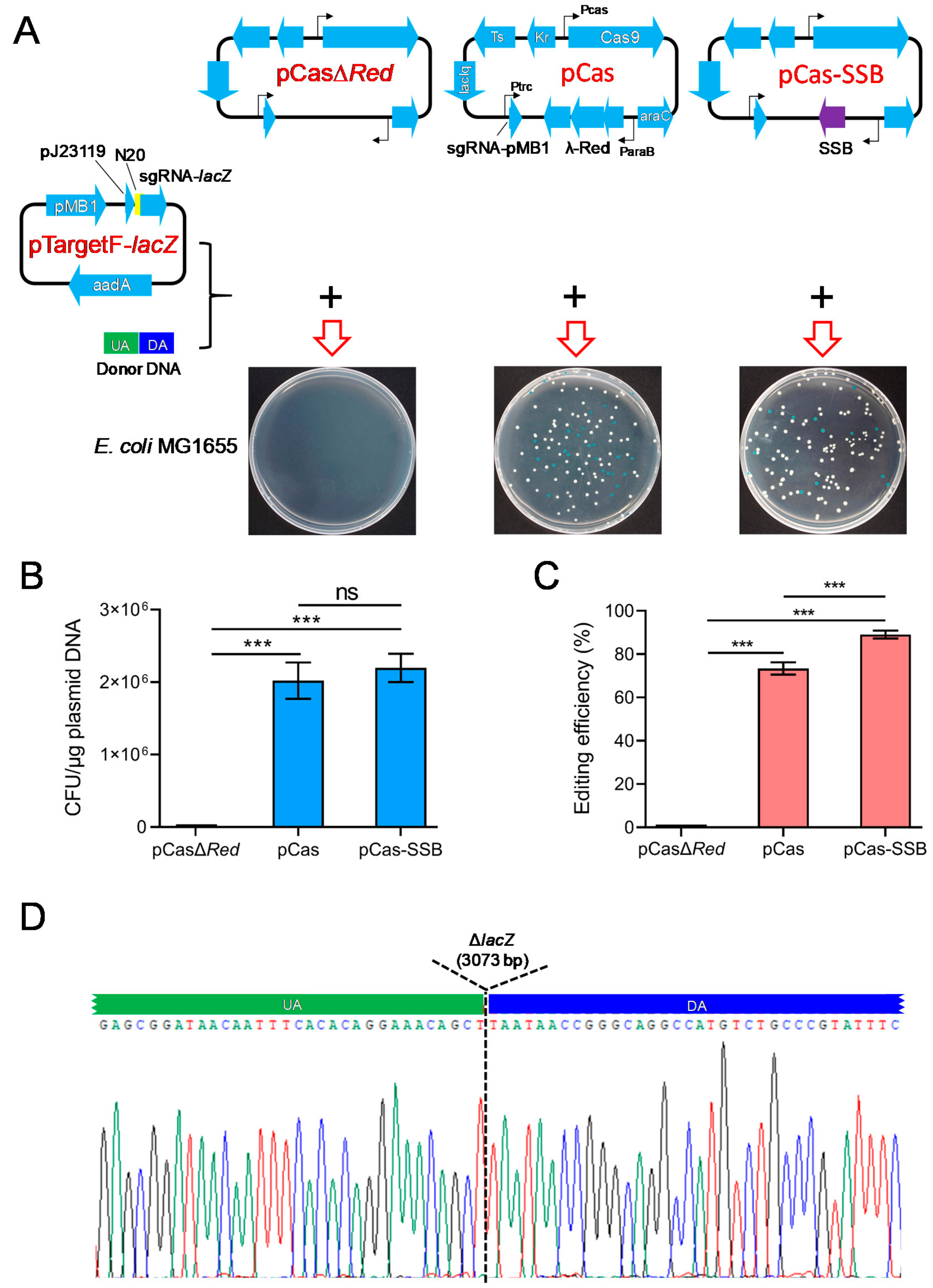
3.1. SSB Mediated the HR Repair of DSBs and Improved the Efficiency of CRISPR/Cas9 in Deleting the E. coli lacZ Gene

To investigate whether SSB effectively mediates the HR repair of DSBs and improves the efficiency of CRISPR/Cas9 gene editing in E. coli, donor DNA carrying sequences that were homologous to the upstream and downstream regions of the E. coli lacZ gene as well as pCasΔRed/pTargetF-lacZ, pCas/pTargetF-lacZ, or pCas-SSB/pTargetF-lacZ were transformed into E. coli MG1655. In the culture that was transformed with donor DNA and pCasΔRed/pTargetF-lacZ, no colonies grew on the LB plates supplemented with X-gal, IPTG, kanamycin, and spectinomycin. In the culture that was transformed with donor DNA as well as pCas/pTargetF-lacZ or pCas-SSB/pTargetF-lacZ, white and blue colonies grew on the plates (Figure 1A). White colonies represented colonies in which the lacZ gene had been mutated, and blue colonies represented colonies without lacZ gene mutation. The transformation rates of the two cultures were greater than 2 × 10⁶ CFU/μg plasmid DNA, and there was no difference between the transformation rates of the two cultures. The gene editing efficiency of the two transformed cultures was 73.4% and 89.1%, and the gene editing efficiency observed in the culture that was transformed with pCas-SSB/pTargetF-lacZ and donor DNA was 21.4% higher than that in the culture that was transformed with pCas/pTargetF-lacZ and donor DNA (p < 0.001) (Figure 1B,C). In each transformation group, 10 white colonies were randomly picked from the plates and were subjected to colony PCR sequencing for identification. In all the mutant transformants, the lacZ gene was correctly knocked out by HR (Figure 1D).

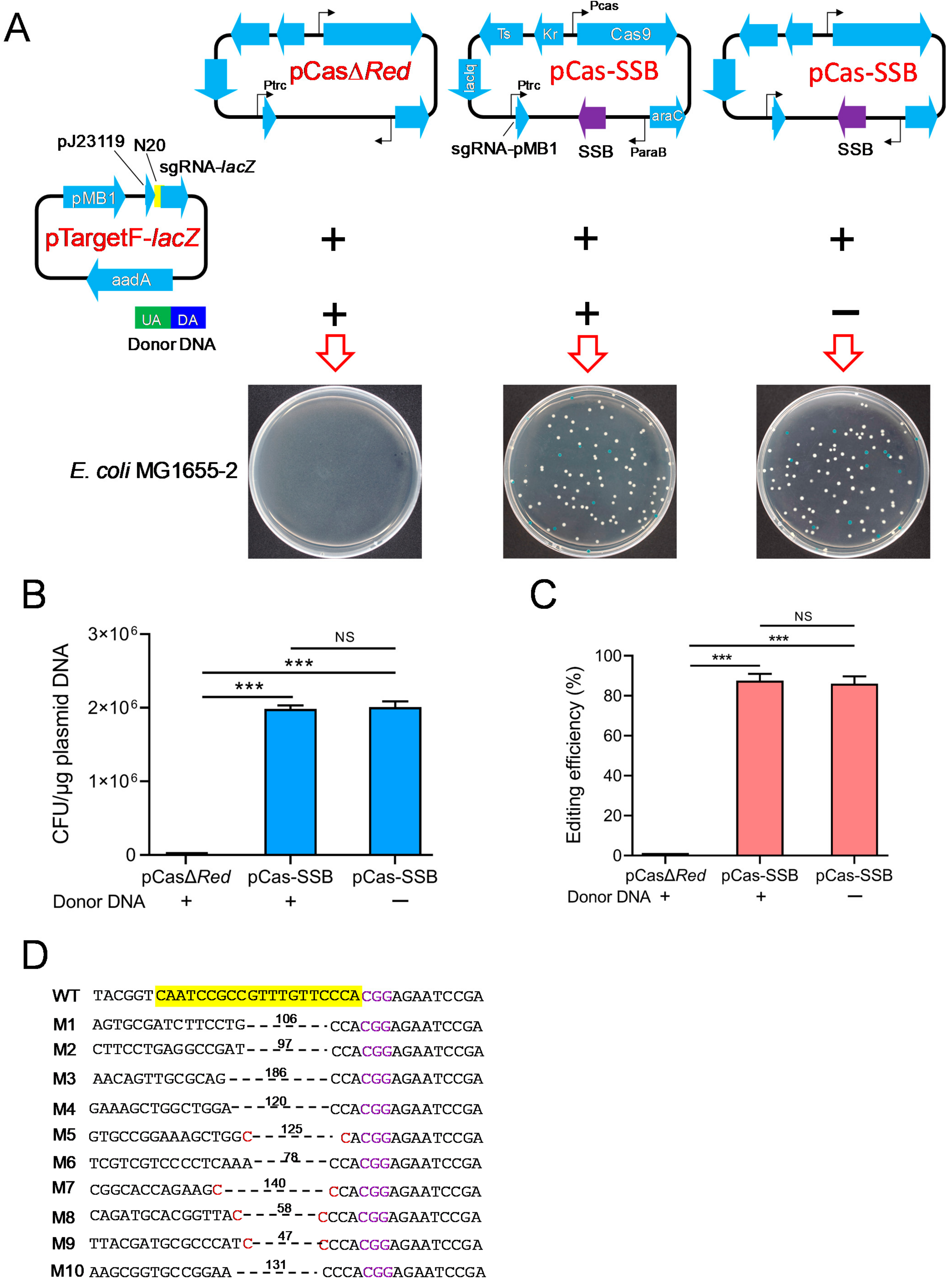
3.2. Cas9/SSB Mediated NHEJ to Delete the E. coli lacZ Gene

Phage T4 DNA ligase can repair the chromosomal DNA DSBs that are generated by CRISPR/Cas9 through NHEJ [32]. To determine whether SSB effectively mediates NHEJ to promote CRISPR/Cas9 gene editing in E. coli, pCasΔRed/pTargetF-lacZ, pCas-T4L/pTargetF-lacZ, and pCas-SSB/pTargetF-lacZ were transformed into E. coli MG1655. In the culture that was transformed with pCasΔRed/pTargetF-lacZ, no colonies grew on the LB plates supplemented with X-gal, IPTG, kanamycin, and spectinomycin. In the culture that was transformed with pCas-T4L/pTargetF-lacZ or pCas-SSB/pTargetF-lacZ, white and blue colonies grew on the plates (Figure 2A). The transformation rates of the two cultures were 1.3 × 10⁶ CFU/μg plasmid DNA and 2.0 × 10⁶ CFU/μg plasmid DNA. The pCas-SSB/pTargetF-lacZ transformation rate was 52.6% higher than the pCas-T4L/pTargetF-lacZ transformation rate (p < 0.001) (Figure 2B). The gene editing efficiency observed in the two transformed cultures was 64.8% and 86.3%, and the gene editing efficiency observed in the culture transformed with pCas-SSB/pTargetF-lacZ was 33.2% higher than that in the culture transformed with pCas-T4L/pTargetF-lacZ (p < 0.001) (Figure 2C). In each group, 10 white colonies were randomly picked from the plates and were subjected to colony PCR sequencing for identification. The indels in the mutant lacZ genes that were generated by pCas-T4L/pTargetF-lacZ transformation were characterized by a deletion of one–three bases upstream of the PAM sequence CGG. The deletion sizes in the transformants ranged from 32 to 252 bp, with an average of 104 bp. The deletion junction in five samples had a 1 or 2 bp microhomology region (Figure 2D). The indels in mutant lacZ genes that were generated by pCas-SSB/pTargetF-lacZ transformation were characterized by a deletion of one–three bases upstream of the PAM sequence CGG. The deletion sizes in the transformants ranged from 45 to 214 bp, with an average of 121 bp. The deletion junction in six samples had a 1–3 bp microhomology region (Figure 2E).

3.3. SSB Mediated DSB Repair Independently of RecA and RecBCD

To validate whether SSB mediated the DSB repair independently of RecA and RecBCD in E. coli, pCasΔRed/pTargetF-lacZ or pCas-SSB/pTargetF-lacZ as well as donor DNA as needed were transformed into E. coli MG1655-2 (ΔrecA, ΔrecBCD, ΔSSB). In the culture that was transformed with pCasΔRed/pTargetF-lacZ and donor DNA, no colonies grew on the LB plates supplemented with X-gal, IPTG, kanamycin, and spectinomycin. In the culture that was transformed with pCas-SSB/pTargetF-lacZ with donor DNA or pCas-SSB/pTargetF-lacZ without donor DNA, white and blue colonies grew on the plates (Figure 3A). The transformation rate of both cultures was 2.0 × 10⁶ CFU/μg plasmid DNA, and the gene editing efficiency of the two transformed cultures was 86.0% and 87.6%; that is, no difference was observed between these two cultures (Figure 3B,C). In each transformation group, 10 white colonies were randomly picked from the plates and were subjected to colony PCR sequencing for identification. In all the mutants that were generated by transformation with pCas-SSB/pTargetF-lacZ and donor DNA, the lacZ gene was correctly knocked out by HR, and this effect was also observed in E. coli MG1655 (Figure 1D). The indels in the mutant lacZ genes that were generated by transformation with pCas-SSB/pTargetF-lacZ without donor DNA were characterized by a deletion of two–three bases upstream of the PAM sequence CGG. The deletion sizes in the mutant transformants ranged from 47 to 186 bp, with an average of 109 bp. The deletion junction in four samples had a 1 bp microhomology region (Figure 3D).

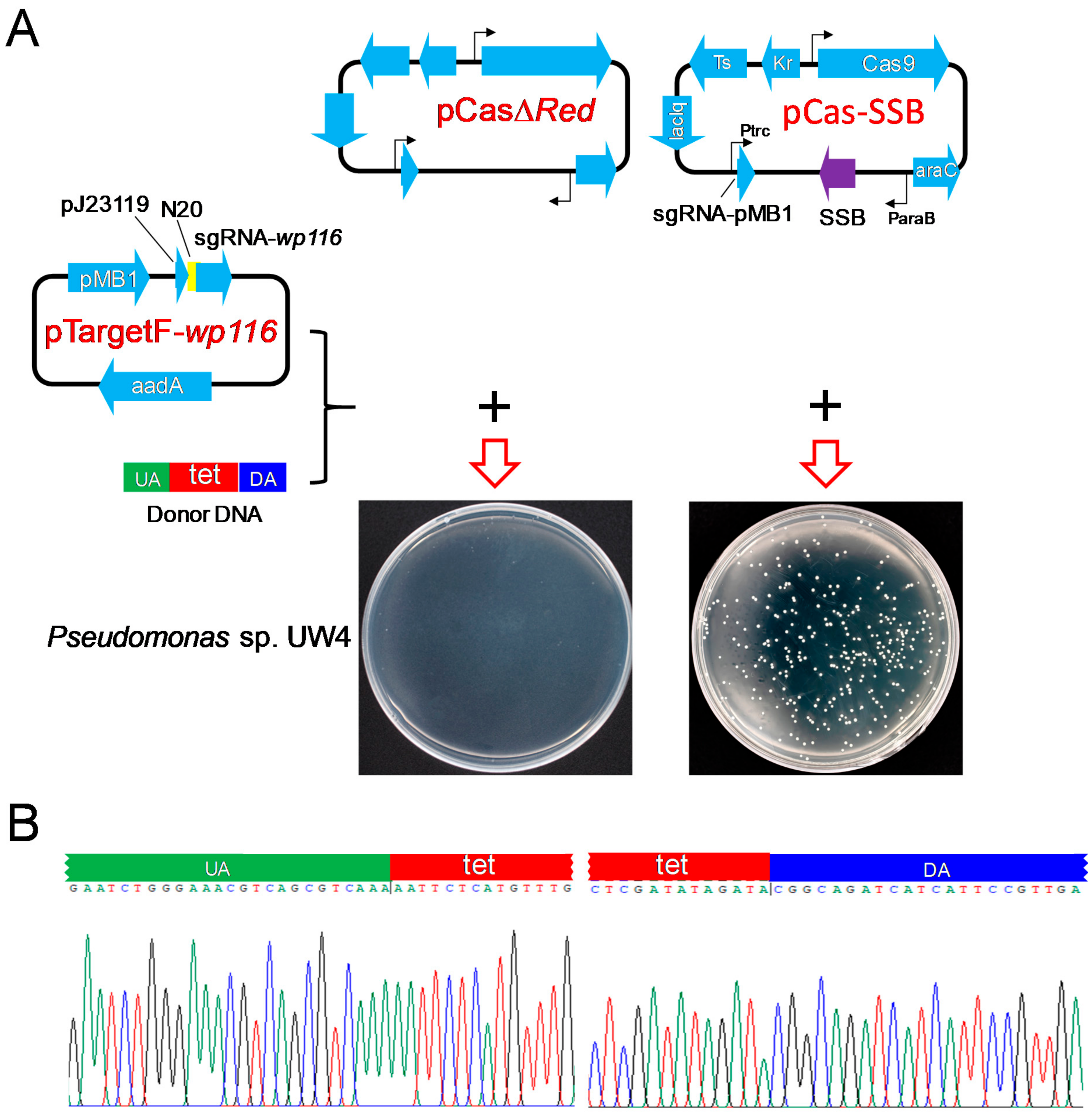
3.4. SSB-Mediated HR Promoted the Deletion of the Pseudomonas sp. UW4 wp116 Gene by CRISPR/Cas9

To examine whether SSB effectively mediates HR to promote CRISPR/Cas9 gene editing in bacteria other than E. coli, donor DNA harboring a tetracycline-resistance gene cassette and sequences located at both ends of the cassette, complementary sequences to the upstream and downstream regions of the Pseudomonas sp. UW4 wp116 gene, and pCasΔRed/pTargetF-wp116 or pCas-SSB/pTargetF-wp116 were transformed into Pseudomonas sp. UW4. When Pseudomonas sp. UW4 was transformed with donor DNA and pCasΔRed/pTargetF-wp116, no colonies grew on the LB plates supplemented with tetracycline, kanamycin, and spectinomycin. After transformation with donor DNA and pCas-SSB/pTargetF-wp116, colonies grew on the LB plates (Figure 4A), and the transformation rate was 3.3 × 10⁶ CFU/μg plasmid DNA. Twenty randomly selected colonies were identified by colony PCR sequencing. The wp116 gene in all the colonies was correctly deleted by HR (Figure 4B), revealing that the gene editing efficiency achieved 100%.

4. Discussion

All SSB proteins harbor at least one DNA-binding oligonucleotide/oligosaccharide-binding (OB) fold. The OB fold controls both ssDNA binding and oligomerization. Furthermore, SSBs also play key roles in recruiting SSB/ssDNA-processing enzymes that mediate DNA replication, recombination, and repair [42]. SSB and RecBCD from E. coli were demonstrated to be capable of catalyzing dsDNA HR in vitro. Both the HR and NHR efficiencies of E. coli expressing inactive SSB or recA were notably reduced. When SSB and recA were simultaneously lacking, cells lost their HR and NHR capabilities, revealing that SSB catalyzes HR and NHR with the assistance of nucleases [44]. In this study, we found that SSB-mediated HR and NHEJ efficiently repaired CRISPR/Cas9-induced DSBs, increased the number of viable colonies, and increased the gene editing efficiency in E. coli and Pseudomonas, even in recA-, recBCD- and SSB-deficient E. coli. It was further confirmed that SSB catalyzed HR and NHR with the aid of a nuclease.
The λ-Red recombinase system is commonly used to repair CRISPR/Cas9-induced DSBs and improve CRISPR/Cas9-assisted genome editing efficiency, and homologous donor ssDNA or dsDNA is required [26,27,33]. Compared with the λ-Red recombinase system, in this study, replacing the λ-Red recombinase system with SSB and providing donor dsDNA did not change the transformation rate but increased the gene editing efficiency by 21.4%. Surprisingly, the gene editing efficiency of SSB-mediated HR increased to 100% in Pseudomonas. SSB is composed of only 178 amino acids, and it is much smaller than λ-Red recombinases.
The efficiency of CRISPR/Cas9 genome editing via phage T4 DNA ligase-mediated repair of DSBs was markedly higher than that achieved via the mycobacteria-derived NHEJ pathway [28,32]. Compared to T4 DNA ligase-mediated repair of DSBs by NHEJ, the transformation rate and genome editing efficiency achieved via SSB-mediated repair of DSBs by NHEJ were 52.6% and 33.2% higher, respectively; however, the length of deleted chromosomal DNA and the deletion junction manner did not differ between these groups. Inducing the expression of Cas9 and assisting native A-EJ DNA repair could efficiently delete chromosomes in E. coli [29]; however, the process is complicated and time-consuming, and it has a high rate of off-target effects. These results suggested that the SSB-mediated HR and NHEJ mechanism for DSB repair that is described here provides an effective approach to improve CRISPR/Cas9 genome editing efficiency in E. coli and Pseudomonas.
It was reported that the Redβ protein of the λ Red recombination system alone can repair DSBs with ssDNA donors and that it can facilitate CRISPR/Cas9 genome editing [30]. Nevertheless, ssDNA donors must be artificially synthesized and are easily enzymatically digested. The Redβ protein contains a small OB fold [50], similar to SSB, which enables ssDNA binding, protects ssDNA from damage, reduces secondary structure formation in single-stranded DNA, and stimulates the formation of joint molecules by the RecA protein from linear duplex DNA and homologous circular single strands; however, unlike SSB, the Redβ protein cannot promote heteroduplex joint formation (dsDNA recombination) [51]. Furthermore, the Redβ protein can initiate single-strand annealing homologous DNA recombination and strand invasion [52,53]. Therefore, there is some difference between the SSB and Redβ proteins in terms of the mechanism by which they repair the DSBs that are induced by CRISPR/Cas9.
With diverse metabolic pathways and efficient metabolic rates, bacteria can synthesize novel and economically important products, such as enzymes, organic acids, vitamins, antibiotics, antibodies, hormones, carotenoids, steroids, alkaloids, alcoholic beverages, interferons, and vaccines [54]. CRISPR/Cas technology has been proven to be a very robust and effective technique for editing bacterial genomes and optimizing bacterial metabolic pathways. However, the first challenge of this approach is to ensure that the engineered cells survive CRISPR/Cas-induced DSBs [55]. The use of SSB for efficient repair of CRISPR/Cas-induced DSBs provides a better option for bacterial pathway engineering.
In summary, E. coli SSB-mediated HR and NHEJ efficiently repaired CRISPR/Cas9-induced DSBs to increase the number of viable colonies and the gene editing efficiency in E. coli and Pseudomonas. These processes occurred independently of RecA and RecBCD. The efficiency of CRISPR/Cas9 genome editing assisted by SSB was higher than that of the λ-Red recombinase system via HR and T4 DNA ligase via NHEJ. These results showed that SSB-assisted CRISPR/Cas9 genome editing is an alternative approach to facilitate the editing of bacterial genomes. To the best of our knowledge, this is the first report that E. coli SSB can repair the DSBs caused by CRISPR/Cas9 and effectively improve CRISPR/Cas9 genome editing in Escherichia coli and Pseudomonas.

Author Contributions

Methodology, R.C., Y.L. and Y.Q.; Formal analysis, Y.G.; Investigation, Q.Z., J.W. and Z.S.; Writing—review & editing, L.Q. All authors have read and agreed to the published version of the manuscript.

Funding

This work was supported by the Science and Technology Department of Henan Province (202102110044, 222102110302), and the National Natural Science Foundation of China (32102455).

Institutional Review Board Statement

Not applicable.

Informed Consent Statement

Not applicable.

Data Availability Statement

Not applicable.

Conflicts of Interest

The authors declare that they have no competing interest.

References

  1. Jinek, M.; Chylinski, K.; Fonfara, I.; Hauer, M.; Doudna, J.A.; Charpentier, E. A programmable dual-RNA–guided DNA endonuclease in adaptive bacterial immunity. Science 2012, 337, 816–821. [Google Scholar] [CrossRef]
  2. Grissa, I.; Vergnaud, G.; Pourcel, C. The CRISPRdb database and toolsto display CRISPRs and to generate dictionaries of spacers and repeats. BMC Bioinf. 2007, 8, 172. [Google Scholar] [CrossRef] [PubMed] [Green Version]
  3. Hille, F.; Richter, H.; Wong, S.P.; Bratovič, M.; Ressel, S.; Charpentier, E. The biology of CRISPR-Cas: Backward and forward. Cell 2018, 172, 1239–1259. [Google Scholar] [CrossRef] [PubMed] [Green Version]
  4. Makarova, K.S.; Wolf, Y.I.; Iranzo, J.; Shmakov, S.A.; Alkhnbashi, O.S.; Brouns, S.J.J.; Charpentier, E.; Cheng, D.; Haft, D.H.; Horvath, P.; et al. Evolutionary classification of CRISPR-Cas systems: A burst of class 2 and derived variants. Nat. Rev. Microbiol. 2020, 18, 67–83. [Google Scholar] [CrossRef]
  5. Bolotin, A.; Quinquis, B.; Sorokin, A.; Ehrlich, S.D. Clustered regularly interspaced short palindrome repeats (CRISPRs) have spacers of extrachromosomal origin. Microbiology 2005, 151, 2551–2561. [Google Scholar] [CrossRef] [Green Version]
  6. Brouns, S.J.J.; Jore, M.M.; Lundgren, M.; Westra, E.R.; Slijkhuis, R.J.H.; Snijders, A.P.L.; Dickman, M.J.; Makarova, K.S.; Koonin, E.V.; van der Oost, J. Small CRISPR RNAs guide antiviral defense in prokaryotes. Science 2008, 321, 960–964. [Google Scholar] [CrossRef] [Green Version]
  7. Kleinstiver, B.P.; Prew, M.S.; Tsai, S.Q.; Topkar, V.V.; Nguyen, N.T.; Zheng, Z.; Gonzales, A.P.; Li, Z.; Peterson, R.T.; Yeh, J.R.; et al. Engineered CRISPR-Cas9 nucleases with altered PAM specificities. Nature 2015, 523, 481–485. [Google Scholar] [CrossRef] [PubMed] [Green Version]
  8. Chyou, T.Y.; Brown, C.M. Prediction and diversity of tracrRNAs from type II CRISPR-Cas systems. RNA Biol. 2019, 16, 423–434. [Google Scholar] [CrossRef]
  9. Lieber, M.R. The mechanism of double-strand DNA break repair by the nonhomologous DNA end-joining pathway. Annu. Rev. Biochem. 2010, 79, 181–211. [Google Scholar] [CrossRef] [Green Version]
  10. Mladenov, E.; Iliakis, G. Induction and repair of DNA double strand breaks: The increasing spectrum of non-homologous end joining pathways. Mutat. Res. Fund. Mol. Mech. Mutagen. 2011, 711, 61–72. [Google Scholar] [CrossRef]
  11. Corneo, B.; Wendland, R.L.; Deriano, L.; Cui, X.; Klein, I.A.; Wong, S.; Arnal, S.; Holub, A.J.; Weller, G.R.; Pancake, B.A.; et al. Rag mutations reveal robust alternative end joining. Nature 2007, 449, 483–486. [Google Scholar] [CrossRef]
  12. Liu, J.; Majumdar, A.; Liu, J.; Thompson, L.H.; Seidman, M.M. Sequence conversion by single strand oligonucleotide donors via non-homologous end joining in mammalian cells. J. Biol. Chem. 2010, 285, 23198–23207. [Google Scholar] [CrossRef] [PubMed] [Green Version]
  13. Yang, H.; Ren, S.; Yu, S.; Pan, H.; Li, T.; Ge, S.; Zhang, J.; Xia, N. Methods favoring homology-directed repair choice in response to CRISPR/Cas9 induced-double strand breaks. Int. J. Mol. Sci. 2020, 21, 6461. [Google Scholar] [CrossRef] [PubMed]
  14. Pawelczak, K.S.; Gavande, N.S.; VanderVere-Carozza, P.S.; Turchi, J.J. Modulating DNA repair pathways to improve precision genome engineering. ACS Chem. Biol. 2018, 13, 389–396. [Google Scholar] [CrossRef]
  15. Pitcher, R.S.; Green, A.J.; Brzostek, A.; Korycka-Machala, M.; Dziadek, J.; Doherty, A.J. NHEJ protects mycobacteria in stationary phase against the harmful effects of desiccation. DNA Repair 2007, 6, 1271–1276. [Google Scholar] [CrossRef] [PubMed] [Green Version]
  16. Wilson, T.E.; Topper, L.M.; Palmbos, P.L. Non-homologous end-joining: Bacteria join the chromosome breakdance. Trends Biochem. Sci. 2003, 28, 62–66. [Google Scholar] [CrossRef]
  17. Stephanou, N.C.; Gao, F.; Bongiorno, P.; Ehrt, S.; Schnappinger, D.; Shuman, S.; Glickman, M.S. Mycobacterial nonhomologous end joining mediates mutagenic repair of chromosomal double-strand DNA breaks. J. Bacteriol. 2007, 189, 5237–5246. [Google Scholar] [CrossRef] [Green Version]
  18. Paris, Ü.; Mikkel, K.; Tavita, K.; Saumaa, S.; Teras, R.; Kivisaar, M. NHEJ enzymes LigD and Ku participate in stationary-phase mutagenesis in Pseudomonas putida. DNA Repair 2015, 31, 11–18. [Google Scholar] [CrossRef]
  19. Chayot, R.; Montagne, B.; Mazel, D.; Ricchetti, M. An end-joining repair mechanism in Escherichia coli. Proc. Natl. Acad. Sci. USA 2010, 107, 2141–2146. [Google Scholar] [CrossRef] [Green Version]
  20. Jiang, Y.; Qian, F.; Yang, J.; Liu, Y.; Dong, F.; Xu, C.; Sun, B.; Chen, B.; Xu, X.; Li, Y.; et al. CRISPR-Cpf1 assisted genome editing of Corynebacterium glutamicum. Nat. Commun. 2017, 8, 15179. [Google Scholar] [CrossRef] [Green Version]
  21. Yan, M.Y.; Yan, H.Q.; Ren, G.X.; Zhao, J.P.; Guo, X.P.; Sun, Y.C. CRISPR Cas12a-assisted recombineering in bacteria. Appl. Environ. Microbiol. 2017, 83, e00947-17. [Google Scholar] [CrossRef] [PubMed] [Green Version]
  22. Cho, S.; Choe, D.; Lee, E.; Kim, S.C.; Palsson, B.; Cho, B.K. High-level dCas9 expression induces abnormal cell morphology in Escherichia coli. ACS Synth. Biol. 2018, 7, 1085–1094. [Google Scholar] [CrossRef] [PubMed]
  23. Misra, C.S.; Bindal, G.; Sodani, M.; Wadhawan, S.; Kulkarni, S.; Gautam, S.; Mukhopadhyaya, R.; Rath, D. Determination of Cas9/dCas9 associated toxicity in microbes. biorXiv 2019, 848135. [Google Scholar] [CrossRef] [Green Version]
  24. Wang, Y.; Wang, S.; Chen, W.; Song, L.; Zhang, Y.; Shen, Z.; Yu, F.; Li, M.; Ji, Q. CRISPR-Cas9 and CRISPR-assisted cytidine deaminase enable precise and efficient genome editing in Klebsiella pneumoniae. Appl. Environ. Microb. 2018, 84, e1818–e1834. [Google Scholar] [CrossRef] [Green Version]
  25. Vento, J.M.; Crook, N.; Beisel, C.L. Barriers to genome editing with CRISPR in bacteria. J. Indus. Microbiol. Biotechnol. 2019, 46, 1327–1341. [Google Scholar] [CrossRef]
  26. Altenbuchner, J. Editing of the Bacillus subtilis genome by the CRISPR-Cas9 System. Appl. Environ. Microbiol. 2016, 82, 5421–5427. [Google Scholar] [CrossRef] [PubMed] [Green Version]
  27. Mougiakos, I.; Bosma, E.F.; Weenink, K.; Vossen, E.; Goijvaerts, K.; van der Oost, J.; van Kranenburg, R. Efficient genome editing of a facultative thermophile using mesophilic spCas9. ACS Synth. Biol. 2017, 6, 849–861. [Google Scholar] [CrossRef] [Green Version]
  28. Su, T.; Liu, F.; Gu, P.; Jin, H.; Chang, Y.; Wang, Q.; Liang, Q.; Qi, Q. A CRISPR-Cas9 assisted non-homologous end-joining strategy for one-step engineering of bacterial genome. Sci. Rep. 2016, 6, 37895. [Google Scholar] [CrossRef] [Green Version]
  29. Huang, C.; Ding, T.; Wang, J.; Wang, X.; Guo, L.; Wang, J.; Zhu, L.; Bi, C.; Zhang, X.; Ma, X.; et al. CRISPR-Cas9-assisted native end-joining editing offers a simple strategy for efficient genetic engineering in Escherichia coli. Appl. Microbiol. Biotechnol. 2019, 103, 8497–8509. [Google Scholar] [CrossRef]
  30. Wu, Z.; Chen, Z.; Gao, X.; Li, J.; Shang, G. Combination of ssDNA recombineering and CRISPR-Cas9 for Pseudomonas putida KT2440 genome editing. Appl. Microbiol. Biotechnol. 2019, 103, 2783–2795. [Google Scholar] [CrossRef]
  31. Aparicio, T.; de Lorenzo, V.; Martínez-García, E. CRISPR/Cas9-enhanced ssDNA recombineering for Pseudomonas putida. Microb. Biotechnol. 2019, 12, 1076–1089. [Google Scholar] [CrossRef] [PubMed] [Green Version]
  32. Su, T.; Liu, F.; Chang, Y.; Guo, Q.; Wang, J.; Wang, Q.; Qi, Q. The phage T4 DNA ligase mediates bacterial chromosome DSBs repair as single component non-homologous end joining. Synth. Syst. Biotechnol. 2019, 4, 107–112. [Google Scholar] [CrossRef] [PubMed]
  33. Jiang, Y.; Chen, B.; Duan, C.; Sun, B.; Yang, J.; Yang, S. Multigene editing in the Escherichia coli genome via the CRISPR-Cas9 system. Appl. Environ. Microbiol. 2015, 81, 2506–2514. [Google Scholar] [CrossRef] [PubMed] [Green Version]
  34. Egan, M.; Ramirez, J.; Xander, C.; Upreti, C.; Bhatt, S. Lambda red-mediated recombineering in the attaching and effacing pathogen Escherichia albertii. Biol. Proced. Online 2016, 18, 3. [Google Scholar] [CrossRef] [Green Version]
  35. Bai, L.; Gao, M.; Cheng, X.; Kang, G.; Cao, X.; Huang, H. Engineered butyrate-producing bacteria prevents high fat diet-induced obesity in mice. Microb. Cell Fact. 2020, 19, 94. [Google Scholar] [CrossRef]
  36. Li, Q.; Sun, B.; Chen, J.; Zhang, Y.; Jiang, Y.; Yang, S. A modified pCas/pTargetF system for CRISPR-Cas9-assisted genome editing in Escherichia coli. Acta Biochim. Biophys. Sin. 2021, 53, 620–627. [Google Scholar] [CrossRef]
  37. Figueredo, E.F.; Cruz, T.A.D.; Almeida, J.R.; Batista, B.D.; Marcon, J.; Andrade, P.A.M.; Hayashibara, C.A.A.; Rosa, M.S.; Azevedo, J.L.; Quecine, M.C. The key role of indole-3-acetic acid biosynthesis by Bacillus thuringiensis RZ2MS9 in promoting maize growth revealed by the ipdC gene knockout mediated by the CRISPR-Cas9 system. Microbiol. Res. 2022, 266, 127218. [Google Scholar] [CrossRef]
  38. Liu, X.; Tian, J.; Liu, L.; Zhu, T.; Yu, X.; Chu, X.; Yao, B.; Wu, N.; Fan, Y. Identification of an operon involved in fluoride resistance in Enterobacter cloacae FRM. Sci. Rep. 2017, 7, 6786. [Google Scholar] [CrossRef] [Green Version]
  39. Kur, J.; Olszewski, M.; Filipkowski, P. Single-stranded DNA-binding proteins (SSBs)-sources and applications in molecular biology. Acta Biochim. Pol. 2005, 52, 569–574. [Google Scholar] [CrossRef]
  40. Sigal, N.; Delius, H.; Kornberg, T.; Gefter, M.L.; Alberts, B. A DNA-unwinding protein isolated from Escherichia coli: Its interaction with DNA and with DNA polymerases. Proc. Natl. Acad. Sci. USA 1972, 69, 3537–3541. [Google Scholar] [CrossRef] [Green Version]
  41. Meyer, R.R.; Laine, P.S. The single-stranded DNA-binding protein of Escherichia coli. Microbiol. Rev. 1990, 54, 342–380. [Google Scholar] [CrossRef] [PubMed]
  42. Marceau, A.H. Functions of single-strand DNA-binding proteins in DNA replication, recombination, and repair. Methods Mol. Biol. 2012, 922, 1–21. [Google Scholar]
  43. Shereda, R.D.; Kozlov, A.G.; Lohman, T.M.; Cox, M.M.; Keck, J.L. SSB as an organizer/mobilizer of genome maintenance complexes. Crit. Rev. Biochem. Mol. Biol. 2008, 43, 289–318. [Google Scholar] [CrossRef] [PubMed] [Green Version]
  44. Chai, R.; Zhang, C.; Tian, F.; Li, H.; Yang, Q.; Song, A.; Qiu, L. Recombination function and recombination kinetics of Escherichia coli single-stranded DNA-binding protein. Sci. Bull. 2016, 61, 1594–1604. [Google Scholar] [CrossRef] [Green Version]
  45. Blattner, F.R.; Plunkett, G., 3rd; Bloch, C.A.; Perna, N.T.; Burland, V.; Riley, M.; Collado-Vides, J.; Glasner, J.D.; Rode, C.K.; Mayhew, G.F.; et al. The complete genome sequence of Escherichia coli K-12. Science 1997, 277, 1453–1462. [Google Scholar] [CrossRef] [Green Version]
  46. Shah, S.; Li, J.; Moffatt, B.A.; Glick, B.R. Isolation and characterization of ACC deaminase genes from two different plant growth-promoting rhizobacteria. Can. J. Microbiol. 1998, 44, 833–843. [Google Scholar] [CrossRef]
  47. Li, J.; Ovakim, D.H.; Charles, T.C.; Glick, B.R. An ACC deaminase minus mutant of Enterobacter cloacae UW4 no longer promotes root elongation. Curr. Microbiol. 2000, 41, 101–105. [Google Scholar] [CrossRef]
  48. Sambrook, J.; Fritsch, E.F.; Maniatis, T. Molecular Cloning: A Laboratory Manual, 2nd ed.; Cold Spring Harbor Laboratory Press: Cold Spring Harbor, NY, USA, 1989; pp. 16–688. [Google Scholar]
  49. Iwasaki, K.; Uchiyama, H.; Yagi, O.; Kurabayashi, T.; Ishizuka, K.; Takamura, Y. Transformation of Pseudomonas putida by electroporation. Biosci. Biotechnol. Biochem. 1994, 58, 851–854. [Google Scholar] [CrossRef] [Green Version]
  50. Newing, T.P.; Brewster, J.L.; Fitschen, L.J.; Bouwer, J.C.; Johnston, N.P.; Yu, H.; Tolun, G. Redβ177 an-nealase structure reveals details of oligomerization and λ Red-mediated homologous DNA recombination. Nat. Commun. 2022, 13, 5649. [Google Scholar] [CrossRef]
  51. Maresca, M.; Erler, A.; Fu, J.; Friedrich, A.; Zhang, Y.; Stewart, A. Single-stranded heteroduplex interme-diates in λ Red homologous recombination. BMC Mol. Biol. 2010, 11, 54. [Google Scholar] [CrossRef] [Green Version]
  52. Rybalchenko, N.; Golub, E.I.; Bi, B.; Radding, C.M. Strand invasion promoted by recombination protein β of coliphage λ. Proc. Natl. Acad. Sci. USA 2004, 101, 17056–17060. [Google Scholar] [CrossRef] [PubMed] [Green Version]
  53. Kuzminov, A. Recombinational repair of DNA damage in Escherichia coli and bacteriophage lambda. Microbiol. Mol. Biol. Rev. 1999, 63, 751–813. [Google Scholar] [CrossRef] [PubMed] [Green Version]
  54. Akoijam, N.; Kalita, D.; Joshi, S.R. Bacteria and their industrial importance. In Industrial Microbiology and Biotechnology; Verma, P., Ed.; Springer: Singapore, 2022; pp. 63–79. [Google Scholar]
  55. Zhao, D.; Zhu, X.; Zhou, H.; Sun, N.; Wang, T.; Bi, C.; Zhang, X. CRISPR-based metabolic pathway engineering. Metab. Eng. 2021, 63, 148–159. [Google Scholar] [CrossRef] [PubMed]
Figure 1. Comparison of CRISPR/Cas9 gene editing to delete the E. coli lacZ gene, assisted by SSB- and λ-Red recombination system-mediated HR repair of DSBs. (A) Schematic representation of the CRISPR-Cas9 two-plasmid system and the results of transforming the two plasmids into E. coli MG1655. LB plates were used containing X-gal, IPTG, kanamycin, and spectinomycin. pCas harbors Streptococcus pyogenes Cas9 under its native promoter Pcas, the λ-Red recombination system (λ-Red) with the arabinose-inducible promoter ParaB and arabinose-inducible transcription factor (araC), sgRNA-PMB1 with an IPTG-inducible promoter (Ptrc) guiding the pMB1 replication of pTarget for eliminating pTargetF, the lac repressor (lacIq), the temperature-sensitive origin of replication repA101 (Ts), and kanamycin resistance gene (Kr); pCasΔRed is derived from pCas by deletion of λ-Red; and pCas-SSB is derived from pCas by replacing λ-Red recombinases with E. coli SSB. pTargetF-lacZ contains sgRNA-lacZ with the efficient constitutive promoter pJ23119 targeting lacZ guided by N20 (Table 2), aadA conferring spectinomycin resistance and the pMB1 replicon; and the donor DNA contains an upstream homologous arm (UA) and a downstream homologous arm (DA) that are homologous to each side (upstream or downstream) of the lacZ gene in the E. coli genome. (B) The transformation rate of the CRISPR-Cas9 two-plasmid system into E. coli MG1665. (C) The editing efficiency of CRISPR-Cas9 to delete the E. coli lacZ gene. (D) Identification of the 10 randomly selected lacZ mutant colonies (white colonies) by colony PCR and DNA sequencing. The data in (B,C) represent the mean ± s.d. for n = 3 biologically independent samples. ***, p < 0.001 by one-way ANOVA followed by Dunnett’s test; ns, no significant differences.
Microorganisms 11 00850 g001
Figure 2. Effects of SSB- and T4 DNA ligase-mediated NHR repair of DSBs on CRISPR-Cas9 gene editing to delete the E. coli lacZ gene. (A) Illustration of Cas9 and sgRNA expression plasmids and their transformation into E. coli MG1655. pCas-T4L is derived from pCas by replacing λ-Red recombinases with phage T4 DNA ligase. (B) The transformation rate of the CRISPR-Cas9 plasmids into E. coli MG1665. (C) The editing efficiency of CRISPR-Cas9 to delete the E. coli lacZ gene. (D) Identification of the 10 randomly selected white colonies generated by pCas-T4L and pTargetF-lacZ transformation by colony PCR and sequencing. (E) Identification of the 10 randomly selected white colonies generated by pCas-SSB and pTargetF-lacZ transformation by colony PCR and sequencing. The sequences highlighted in yellow represent a 20-bp complementary region (N20) matching with the E. coli lacZ gene. The sequences colored in purple indicate the PAM sequence of Cas9. The sequences colored in red are micro-homology sequences. The dotted lines and associated numbers indicate the deleted sequences and deletion sizes (bp). WT, E. coli MG1665; C1-C10 or S1-S10, the 10 randomly selected white colonies. The data in (B,C) represent the mean ± s.d. for n = 3 biologically independent samples. *** p < 0.001 by one-way ANOVA followed by Dunnett’s test; ns, no significant differences.
Microorganisms 11 00850 g002
Figure 3. SSB repaired the DSBs and improved gene editing induced by CRISPR-Cas9 independent of RecA and RecBCD. (A) Schematic of the CRISPR-Cas9 two-plasmid system transformation into E. coli MG1655-2 (ΔrecA, ΔrecBCD, ΔSSB) to delete the lacZ gene with or without donor DNA. (B) The transformation rate of the CRISPR-Cas9 plasmids into E. coli MG1665-2 with or without donor DNA. (C) The editing efficiency of CRISPR-Cas9 to delete the lacZ gene in E. coli MG1655-2 with or without donor DNA. (D) Identification of the 10 randomly selected white colonies of E. coli MG1655-2, generated by pCas-SSB and pTargetF-lacZ transformation without donor DNA, by colony PCR and DNA sequencing. The sequences highlighted in yellow represent a 20-bp complementary region (N20) matching with the E. coli lacZ gene. The sequences colored in purple indicate the PAM sequence of Cas9. The sequences colored in red are micro-homology sequences. The dotted lines and associated numbers indicate the deleted sequences and deletion sizes (bp). UA, upstream homologous arm; DA, downstream homologous arm; WT, E. coli MG1665; M1-M10, the 10 randomly selected white colonies. The data in (B,C) represent the mean ± s.d. for n = 3 biologically independent samples. ***, p < 0.001 by one-way ANOVA followed by Dunnett’s test; ns, no significant differences.
Microorganisms 11 00850 g003
Figure 4. CRISPR-Cas9 editing of the Pseudomonas sp. UW4 wp116 gene assisted by SSB-mediated HR repair of DSBs. (A) Illustration of the plasmid expression of Cas9 and sgRNA targeting the wp116 gene and their transformation into Pseudomonas sp. UW4. (B) Identification of the 10 randomly selected transformant colonies of Pseudomonas sp. UW4 by colony PCR and DNA sequencing. UA, upstream homologous arm, homologous to the upstream region of the wp116 gene in the UW4 genome; DA, downstream homologous arm, homologous to the downstream region of the wp116 gene in the UW4 genome; tet, tetracycline resistance gene.
Microorganisms 11 00850 g004
Table 1. Strains and plasmids used in this study a.
Strain or PlasmidDescriptionSource or Reference
E. coli
DH5αF endA1 glnV44 thi-1 recA1 relA1 gyrA96 deoR nupG ϕ80dlacZΔM15 Δ(lacZYA-argF)U169 hsdR17 (rK mK+) λTaKaRa
MG1655K-12; F, λ, ilvG, rfb-50, rph-1[45]
MG1655-1MG1655 ΔlacZThis study
MG1655-2MG1655 ΔrecA ΔrecBCD ΔSSBThis study
Pseudomonas sp.
UW4Wild type[46]
UW4 ΔacdSΔacdS[47]
UW4 Δwp116Δwp116 This study
Plasmids
pCasrepA101(Ts) kan Pcas-cas9 ParaB-Red lacIq Ptrc-sgRNA-pMB1[33]
pCasΔRedrepA101(Ts) kan Pcas-cas9 ParaB lacIq Ptrc-sgRNA-pMB1This study
pCas-SSBrepA101(Ts) kan Pcas-cas9 ParaB-SSB lacIq Ptrc-sgRNA-pMB1This study
pCas-T4LrepA101(Ts) kan Pcas-cas9 ParaB-T4L lacIq Ptrc-sgRNA-pMB1This study
pTargetFpMB1 aadA sgRNA[33]
pTargetF-lacZpMB1 aadA sgRNA-lacZThis study
pTargetF-wp116pMB1 aadA sgRNA-wp116This study
a kan, kanamycin resistance gene; aadA, spectinomycin resistance gene; Pcas-cas9, the cas9 gene with its native promoter; ParaB-Red, the Red recombination genes with an arabinose-inducible promoter; Ptrc-sgRNA-pMB1, sgRNA with an N20 sequence for targeting the pMB1 region with a trc promoter; sgRNA-lacZ, sgRNA with an N20 sequence for targeting the lacZ locus; sgRNA-wp116, sgRNA with an N20 sequence for targeting the wp116 locus; wp116, the gene encoding the chemotactic receptor for 1-aminocyclopropane-1-carboxylic acid (ACC); SSB, the gene encoding the single-stranded DNA-binding protein of E. coli; T4L, T4 DNA ligase; acdS, 1-amino-cyclopropane-1-carboxylate deaminase.
Table 2. The primers used in this study a.
PrimerSequence (5′-3′)
pC01CGCATCCTCACGATAATATCCGGGTAGGCGCAATCACTTT
pC02GATATTATCGTGAGGATGCGTTTTTATAACCTCCTTAGAG
pC03TCGAGCTCTAAGGAGGTTATAAAAAATGGCCAGCAGAGGCGTAAACAAGGT
pC04ACCCGGATATTATCGTGAGGATGCGTCAGAACGGAATGTCATCATCAAAGT
pC05CGCATCCTCACGATAATATCCGGGT
pC06TTTTTATAACCTCCTCCTTAGAGCTCG
pC07TCGAGCTCTAAGGAGGTTATAAAAAATGATTCTTAAAATTCTGAACGAAAT
pC08ACCCGGATATTATCGTGAGGATGCGTCATAGACCAGTTACCTCATGAAAAT
pC09CAATCCGCCGTTTGTTCCCACGGGTTTTAGAGCTAGAAATAGCAAGTTAAAATAA
pC10TGGGAACAAACGGCGGATTGACTAGTATTATACCTAGGACTGAGCTAGCTG
pC11CCGCGGCCTGATCGAACAACGTTTTAGAGCTAGAAATAGCAAGTTAAAATAA
pC12GTTGTTCGATCAGGCCGCGGACTAGTATTATACCTAGGACTGAGCTAGCTG
pC13CGGTAGTGGGATACG
pC14CCTGCCCGGTTATTAAGCTGTTTCCTGTGT
pC15ACACAGGAAACAGCTTAATAACCGGGCAGG
pC16AAAAGCCTAGATAAA
pC17AGCTATTCGCCCATACATCG
pC18CCTGCCCGGTTATTAAGCTGTTTCCTGTGT
pC19GGAAACGTCAGCGTCAAAAATTCTCATGTTTGACAG
pC20CGGAATGATGATCTGCCGTATCTATATCGAGATGCG
pC21CGCATCTCGATATAGATACGGCAGATCATCATTCCG
pC22CTTGGGTCATGCTCTGCATGG
pC23CGGTAGTGGGATACGACGAT
pC24CGGTTGGAATAATAGCGAGA
pC25CCCAGGCTTTACACTTTATGC
pC26CAGATGAAACGCCGAGTT
pC27AGCTATTCGCCCAT
pC28CTTGGGTCATGCTCT
a The sequences in blue are N20 sequences.
Disclaimer/Publisher’s Note: The statements, opinions and data contained in all publications are solely those of the individual author(s) and contributor(s) and not of MDPI and/or the editor(s). MDPI and/or the editor(s) disclaim responsibility for any injury to people or property resulting from any ideas, methods, instructions or products referred to in the content.

