Article

Characterization of the Bacterial Microbiome in Natural Populations of Barley Stem Gall Midge, Mayetiola hordei, in Morocco

1
Laboratory of Innovative Technologies, National School of Applied Sciences of Tangier, Abdelmalek Essaâdi University, BP 1818 Tanger Principal, Tanger 90000, Morocco
2
Faculty of Sciences and Technology of Tangier, Abdelmalek Essâadi University, Tétouan 93000, Morocco
3
Laboratory of Systems Microbiology and Applied Genomics, Department of Sustainable Agriculture, University of Patras, 30100 Agrinio, Greece
4
AgroBioSciences Program, College for Sustainable Agriculture and Environmental Science, Mohammed VI Polytechnic University, Lot 660, Hay Moulay Rachid, Ben Guerir 43150, Morocco
*
Author to whom correspondence should be addressed.
Microorganisms 2023, 11(3), 797; https://doi.org/10.3390/microorganisms11030797
Submission received: 7 February 2023 / Revised: 13 March 2023 / Accepted: 15 March 2023 / Published: 21 March 2023

Abstract

:

Simple Summary

Barley stem gall midge, Mayetiola hordei (Kieffer), is one of the most devastating barley pests in many regions of the world, causing significant losses to agricultural output. In this work, we explore the presence of reproductive symbionts, including Wolbachia, Spiroplasma, Cardinium, and Arsenophonus, which can be employed as a substitute for chemical pesticides in pest management, in addition to the impact of their geographic origin on bacterial composition, as well as the diversity of the natural populations of barley stem gall midges detected in four Moroccan barley-growing regions. The screening revealed the presence of Wolbachia; in contrast, none of the samples analyzed had Spiroplasma, Cardinium, or Arsenophonus infections. Overall, 5 phyla, 7 classes, and 42 genera were detected throughout all samples, with a significant environmental influence on the taxonomic assortment of the microbiota.

Abstract

Mayetiola hordei (Kieffer), known as barley stem gall midge, is one of the most destructive barley pests in many areas around the world, inflicting significant qualitative and quantitative damage to crop production. In this study, we investigate the presence of reproductive symbionts, the effect of geographical origin on the bacterial microbiome’s structure, and the diversity associated with natural populations of M. hordei located in four barley-producing areas in Morocco. Wolbachia infection was discovered in 9% of the natural populations using a precise 16S rDNA PCR assay. High-throughput sequencing of the V3-V4 region of the bacterial 16S rRNA gene indicated that the native environments of samples had a substantial environmental impact on the microbiota taxonomic assortment. Briefly, 5 phyla, 7 classes, and 42 genera were identified across all the samples. To our knowledge, this is the first report on the bacterial composition of M. hordei natural populations. The presence of Wolbachia infection may assist in the diagnosis of ideal natural populations, providing a new insight into the employment of Wolbachia in the control of barley midge populations, in the context of the sterile insect technique or other biological control methods.

1. Introduction

Mayetiola hordei (Kieffer) (Diptera: Cecidomyiidae), the barley stem gall midge (BSGM), is a small fly (2–4 mm in length), and one of the most devastating pests of cereal crops. This insect is native to the Mediterranean regions in North Africa, including Morocco, Algeria, and Tunisia, and additionally to Southern Europe, Spain, and Italy [1]. Infestation rates and yield losses can become severe after BSGM is established in a region or country. For example, around 30–50% of barley crops in Libya and Tunisia are infested by this pest and cereal production in Morocco is decreased by about 35% due to the damage caused by M. hordei [2,3]. This insect has a long-lived larval stage with two feeding stages and a third non-feeding stage, during a generation time that is completed in 45 days at an average temperature of (18 ± 1 °C) [3]. Adverse effects on crops occur during this developmental stage. The principal symptom in seedlings is a yellowing of new growth, which can occasionally lead to plant mortality. BSGM puparia are always embedded in galls and associated with plant stunting [2,4]. The female can lay up to 400 [5]. The most practical approach to manage BSGM is prevention, including insecticide applications and planting adjustment, while the use of resistant cultivars is not common, although it is considered one of the most successful and cost-efficient strategies to control these insects [6,7].
Among the methods that can fulfill the projected increased need for novel insect pest management techniques brought on by expanding human populations and climate change are those based on symbiotic microorganisms [8]. Insects coexist in a variety of ways with many different microorganisms [9,10]. Numerous bacterial species and insect hosts have been shown to form complex symbiotic relationships that can be classified as commensalism, mutualism, or parasitism, which affect a variety of elements of the host’s biology, physiology, ecology, and evolution, including nutrition, reproduction, mating behavior, fitness, immunity, and status as a pest [11,12,13]. The interactions between insects and their endosymbionts, especially those related to mechanisms of insect reproduction, are considered as a rapid, environmentally friendly, and efficient biological approach to suppress or eliminate insect infestations [8]. In this way, insects that damage crops and spread diseases to people, plants, and domestic animals have been managed, and in many cases eliminated, resulting in improved standards of living and quality of life throughout the world [14]. Wolbachia is an α-proteobacterium, an intracellular symbiont discovered in 1924 [15], present in a wide variety of arthropod taxa, including insects, mites, spiders, springtails, crustaceans, and some nematodes. More than 20% of arthropod species may be Wolbachia-infected, according to several PCR-based assessments. Based on their main genetic changes across a variety of hosts, Wolbachia have recently been grouped into 17 supergroups, represented by the letters A to S [16,17,18]. Supergroup G, which was recently eliminated because it was likely a combination of supergroups A and B [19,20], and the cave spider supergroup R, which showed a strong affinity for supergroup A, are exceptions [21,22]. Wolbachia can infect and maintain itself in hosts by manipulating their reproduction [23,24]. Wolbachia-induced cytoplasmic incompatibility (CI) is one of several techniques that can be differentiated and advantageous in two ways: as a method for controlling insect pest populations, similar to the sterile insect technique, and as a drive system for spreading genotypes in host populations. Furthermore, pathogenic Wolbachia strains also have the ability to manage vector species by altering the age structure of their populations [9,25]. Wolbachia also plays an important role in aspects of its host ecology, such as reproduction, nutrition, pathogen resistance, and fecundity [26].
In addition to Wolbachia, other endosymbiotic bacteria that can affect the biology of their insect hosts exist, such as for Spiroplasma, an extracellular symbiont belonging to the class Mollicutes, which can kill males in a variety of insect hosts, including Drosophila flies, butterflies, and lady beetles. Moreover, these bacteria can confer protection to the infected host against natural adversaries [27,28]. The endosymbiont Cardinium belonging to the Bacteroidetes is also known to be involved in arthropod reproduction manipulation, causing cytoplasmic incompatibility, parthenogenesis, and feminization to its hosts [29,30]. Furthermore, the bacterial symbiont Arsenophonus can induce a male-killing phenotype, resulting in high-rate mortality of male embryos produced by infected females. The bacterium has no discernible effect on the host. Therefore, it can propagate throughout the ecological community of its hosts, such as filth flies and their parasitoids, colonize new species, and remain in the population for lengthy periods of time [31,32].
To the best of our knowledge, this is the first study about the BSGM bacterial microbiome, although a few studies have recently been published on the characterization of the bacterial community associated with the Moroccan Hessian fly (Mayetiola destructor), an insect that shares a similar distribution to BSGM, similar morphology, and comparable symptoms concerning host plants [33]. According to Bel Mokhtar et al. (2020), the sequencing of the 16S rRNA bacterial gene (V3-V4 region) revealed various taxa at the phylum level, including Proteobacteria, Bacteroidetes, Actinobacteria, Cyanobacteria, Deinococcus-Thermus, and Firmicutes. At the genus level, different frequencies were observed between collection regions in Morocco for Empedobacter, Ralstonia, Afipia, and Pseudomonas. Regarding Wolbachia-infection, it was detected in all Moroccan regions with a different relative abundance [34]. Additionally, Bansal et al. (2011) identified four phyla in Hessian fly adults based on classical sequencing: Proteobacteria, Firmicutes, Actinobacteria, and Bacteroidetes. At the genus level, various types of bacteria were detected, including Enterobacter, Pantoea, Stenotrophomonas, Pseudomonas, Bacillus, Ochrobactrum, Acinetobacter, Alcaligenes, Nitrosomonas, Arcanobacterium, Microbacterium, Paenibacillus, and Klebsiella [35].
The aim of this study is to screen natural populations of BSGM midge for the presence of reproductive symbionts, Wolbachia, Spiroplasma, Arsenophonus, and Cardinium, using the 16S rRNA gene. In addition, a high-throughput sequencing (HTS) approach is performed using the Illumina-MiSeq technology to investigate the bacterial symbionts associated with natural populations of BSGM larvae to elucidate the relationship between the insects’ bacterial community profile and their habitat.

2. Materials and Methods

2.1. Barley Stem Gall Midge Collection and DNA Isolation

Plants infected with BSGM were obtained from four major barley-producing areas in Morocco: Abda, Rabat-Salé-Zemmour-Zaër, Fès-Meknes, and Doukkala. All infested plant samples were kept at the International Center of Agricultural Research in Dry Areas (ICARDA) in Rabat, Morocco (Table 1), where the BSGM larvae and pupae were collected during the winter of 2020. Larvae were stored in absolute ethanol at −20 °C, whereas pupae protected in infested plant tissues were reared in a growth room under constant conditions (temperature 23 ± 2 °C, relative humidity 65 ± 5%). Once the adults emerged, they were stored in 100% ethanol at −20 °C. Before DNA extraction, each sample was surface sterilized with a 70% v/v ethanol solution, rinsed with sterile deionized water to remove traces of ethanol, left to dry on a sterile surface, and placed in 1.5 mL centrifuge tubes. After surface sterilization, whole-fly DNA was isolated using a modified CTAB (cetyl trimethyl ammonium bromide) method [36]. The quantity and quality of the DNA preparations and the concentration of double-stranded DNA were both analyzed by a Q5000 micro-volume UV-Vis spectrophotometer (Quawell Technology, San Jose, CA, USA). DNA samples were stored in Eppendorf tubes at −20 °C until further analysis.

2.2. Screening and Identification of Bacterial Symbionts

From the BSGM samples collected, 152 insects were selected (Table 1), including 108 that were acquired from 4 different areas, whereas only 2 regions (Rabat and Fez Meknes) were the source of the 44 adult specimens. However, the larvae from Doukkala and Abda were unable to develop into adults in the laboratory setting. The detection of Wolbachia, Spiroplasma, Cardinium, and Arsenophonus was performed by PCR assays using 16S rDNA genus specific primers. Blank and negative controls were incorporated during DNA extractions, and the PCRs were conducted using identical conditions. However, no amplicons were produced from these samples. The quality of the extracted DNA was tested by amplifying part of the mitochondrial 12S rRNA gene (Table S2). Amplification was performed in 25 µL reaction mixtures containing 2.5 µL KAPA Taq buffer (KAPA BioSystems, Wilmington, MA, USA) 10×, 0.25 µL dNTPs (25 mM), 0.25 µL of KAPA Taq DNA polymerase, 0.5 µL of the forward primer (25 µM), 0.5 µL of the reverse primer (25 µM), 1 µL of template DNA solution, and was finalized with 20 µL sterile deionized water. The PCR temperature profile was 95 °C for 5 min followed by 35 cycles of 95 °C for 30 s, 30 s at the optimum annealing temperature for each pair of primers, 1 min at 72 °C, and a final extension step of 72 °C for 5 min. PCR products were electrophoresed on a 1.5% agarose gel in order to examine the presence and size of the amplified fragments. The primer sequences used in this study along with the product size and annealing temperature are summarized in Table S2. The PCR-positive products were purified using polyethylene glycol (20% PEG, 2.5 M NaCl) [37] and resuspended in 15 µL of water. Sanger sequencing was performed on purified products with the BigDye Terminator v3.1 Cycle Sequencing Kit following the manufacturer’s recommendations (Applied Biosystems, Waltham, MA, USA). Reaction products were purified using an ethanol/EDTA protocol according to the manufacturer’s instructions (Applied Biosystems, Waltham, MA, USA) and were analyzed in an ABI PRISM 3500 Genetic Analyzer (Applied Biosystems, Waltham, MA, USA).

2.3. Amplification of the V3-V4 Region, Index PCR, Purification, and Illumina Sequencing

For the high-throughput amplicon sequencing analysis, 78 samples of larvae were selected. However, 3 of these larvae (1 from Abda, 1 from Doukkala, and 1 from Rabat) were disregarded because they had low read counts (less than 1000) to prevent issues related to infinite values that may arise when applying the logarithmic transformation. Therefore, the final number of larvae analyzed was 75, including 19 larvae from Rabat, 17 from Abda, 19 from Doukkala, and 20 from Fes-Meknes (Table 1). The hypervariable V3-V4 region of the bacterial 16S rRNA gene was amplified using MiSeq universal primers 341F and 805R (Table S2). The PCR amplification was performed in two steps. The first amplification was performed using the bacterial universal primers 27F-1492R (Table S2) in a 20 μL reaction comprising 2 μL of KAPA Taq buffer 10×, 0.16 μL of dNTPs (25 mM), 0.16 μL of 200 KAPA Taq DNA polymerase (Roche, Basel, Switzerland), 0.4 μL of forward primer (25 μM), 0.4 μL of reverse primer (25 μM), 1 μL of the template DNA solution, and 15.88 μL of water. A 3 min incubation time at 95 °C was used for DNA denaturation, followed by 20 cycles of 95 °C for 30 s, 53 °C for 30 s, and 72 °C for 2 min, with a final 5 min extension at 72 °C. The second amplification included a nested PCR using MiSeq universal primers 341F and 805R. The amplification was performed in 25 µL reaction mixtures containing 2.5 µL KAPA Taq buffer 10×, 0.2 µL dNTPs (25 mM), 0.1 µL of KAPA Taq DNA polymerase, 1 µL of the forward primer (25 µM), 1 µL of the reverse primer (25 µM), 1 µL of the first-step reaction as template, and was finalized with 19.2 µL of sterile deionized water. The PCR amplifications were performed with incubation at 95 °C for 3 min, followed by 21 cycles of 95 °C for 30 s, 54 °C for 30 s and 72 °C for 1 min, and a final 5 min extension at 72 °C. For the Illumina sequencing, the resulting PCR amplicons were used as templates for further amplification, to include the indexes (barcodes) as well as the Illumina adaptors, in a 50 µL volume containing 5 µL KAPA Taq buffer 10×, 0.4 µL dNTPs (25 mM), 0.2 µL of KAPA Taq DNA polymerase, 5 µL of the forward index primer (10 µM), 5 µL of the reverse index primer (10 µM), 2 µL of the cleaned PCR product diluted up to 10 ng/µL, and 32.4 µL of sterile deionized water. The PCR amplifications were performed with incubation at 95 °C for 3 min followed by 8 cycles of 95 °C for 30 s, 30 s at 55 °C, 30 s at 72 °C, and a final extension step of 72 °C for 3 min. The resulting amplicons from the indexing PCR were cleaned using the NucleoMag NGS (next-generation sequencing) clean-up and size selection kit (Macherey-Nagel, Düren, Germany), according to the manufacturer’s recommendations. From all samples examined, the indexed amplicons were mixed in an equimolar ratio (8 nM) and the sequencing was performed by Macrogen using a 2 × 300 bp pair-end kit on a MiSeq platform [38]. Raw sequencing reads were demultiplexed, converted to FASTQ, and the Illumina adapters were trimmed using Illumina standard algorithms.

2.4. NGS Sequencing and Bioinformatics Analysis

Bioinformatics analysis of raw sequencing reads was performed using USEARCH v. 11 and QIIME2 distribution [39,40]. Paired-end reads were assembled and trimmed by length using the usearch -fastq_mergepairs command. The quality of assembled sequences was improved using -fastq_filter, followed by the -fastx_uniques command to detect unique read sequences and their frequencies. Sequences were clustered into operational taxonomic units (OTUs) using the -cluster_otus command at 90% OTU clustering based on the UPARSE algorithm [41]. Crosstalk errors were identified and filtered using the -uncross command based on the UNCROSS2 algorithm [42]. The taxonomy was assigned with Qiime2 using the BLAST + algorithm against the SILVA 138 release database [43,44]. The diversity of individual samples was identified by calculating alpha diversity metrics (evenness, Richness, Shannon, and Simpson indexes) using the “diversity” function of the R package “vegan”. Alpha diversity indexes were plotted with the ggplot function from the “ggplot2” package. Statistical differences in bacterial compositions between populations were tested using the nonparametric Kruskal and Wallis and Wilcoxon rank-sum tests [45]. To assess the similarity of bacterial communities in different locations, a beta diversity analysis was performed using the generalized UniFrac distance and visualized by non-metric multidimensional scale (NMDS) maps [46]. Permutational multivariate analysis of variance (PERMANOVA) using distance matrices was performed using the “adonis” function from the R package “vegan” to identify significant differences between isolated groups. A p-value < 0.05 was considered indicative of statistical significance.
A network analysis of OTUs was performed to investigate and visualize the interactions between microorganisms, which could be explained by similar or complementary functions and/or sharing similar environmental conditions, but not necessarily having physical interactions [47,48]. Networks were obtained using the CoNet plugin in Cytoscape 3.8.2 (Institute for System Biology, Seattle, WA, USA) and visualized with Gephi 0.9.2 (Institute for System Biology, Seattle, WA, USA) (Gephi, WebAtlas, Paris, France) [49]. The network was built using Pearson’s and Spearman’s rank correlation coefficients, mutual information, and the Bray–Curtis and Kullback–Leibler dissimilarity indices. The statistical significance of copresence/mutual exclusion, edge-specific permutation, and bootstrap score distributions were calculated using 1000 iterations. Edges with original scores outside the 0.95 range of their bootstrap distribution were rejected, and p-values were corrected using the Benjamini–Hochberg method.

2.5. Phylogenetic Analysis

Phylogenetic analysis was based on partial 16S rRNA gene sequences obtained from Wolbachia-infected specimens and sequences representing Wolbachia-associated OTUs obtained by Illumina MiSeq sequencing. Multiple alignments were performed with MUSCLE, implemented in MEGA 7 software, using standard algorithm parameters [50,51]. Alignments were manually edited and trimmed to adjust the sequence length. Phylogenetic tree reconstruction was based on the maximum likelihood statistical model and was performed using MEGA 7 software. The (GTR + G + I) substitution model was used to estimate nucleotide evolution [52,53]. A bootstrap test with 1000 iterations was used to assess the reliability of the produced phylogeny [54]. The entirety of the 16S rRNA gene sequences produced in this work were submitted to the GenBank database of NCBI with accession numbers OQ189903-OQ189915.

2.6. Core Microbiome

Core microbiome analyses were performed using microbiome R package based on the M. hordei dataset [55]. The analysis was performed to detect major bacterial taxa that were recognized in 75% of the samples with a relative abundance threshold value greater than 0.01%, as well as to determine the taxa that were common between the different population regions, and to differentiate them from those which were specific to some regions.

3. Results

3.1. Infection Status of Reproductive Symbionts in Natural Populations of BSGM

3.1.1. Infection Prevalence

PCR screening was used to assay the presence of four reproductive symbionts (Wolbachia, Spiroplasma, Cardinium, and Arsenophonus) in four natural BSGM populations. In total, 152 BSGMs were screened for reproductive symbionts (Table S1). The screening results indicate that the barley midge is infected only by Wolbachia with a prevalence of 9%. Interestingly, the Wolbachia percentage infection in natural populations did not reveal an even distribution among the different locations. In fact, only BSGMs from Rabat and Fes-Meknes were found to be infected with Wolbachia. In total, 13 flies were infected, five larvae, two females, and three males, out of 44 samples examined from Rabat (22%), and one larva, one female, and one male, out of 52 samples examined from Fes-Meknes (6%) (Table S1, Figure 1). On the contrary, none of the BSGM populations examined were infected with Spiroplasma, Cardinium, or Arsenophonus.

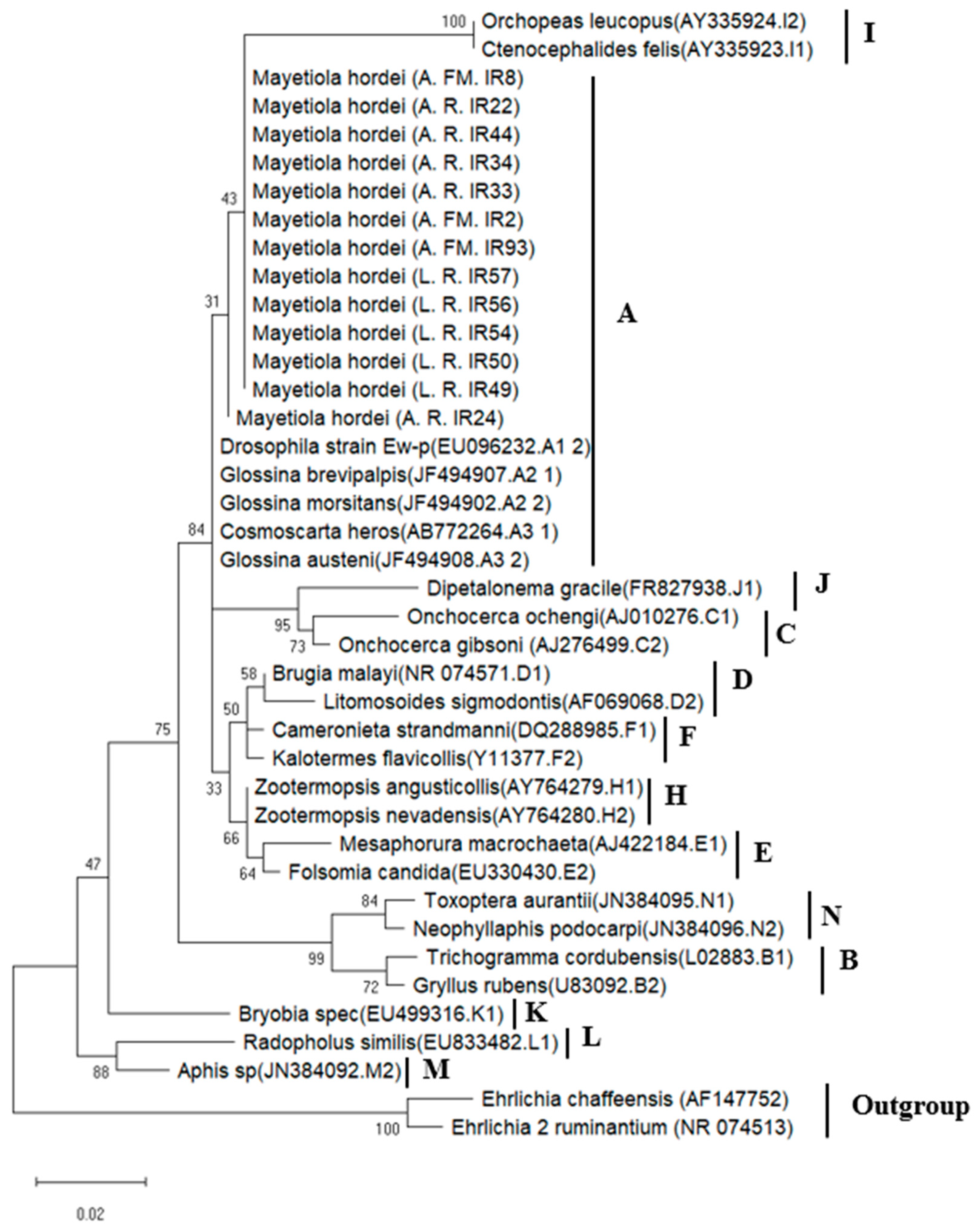
3.1.2. Phylogenetic Analysis of Wolbachia Sequences in BSGM Populations

The Wolbachia phylogenetic analysis was performed on the thirteen Wolbachia-infected samples based on the partial 16S rRNA gene sequences, using a total of 325 bp (File S1) remaining in the thirteen Wolbachia sequences after manually removing the low-quality bases. The 325 bp still contained enough informative data to establish a distinct phylogenetic signal for Wolbachia found in insects, thus allowing an accurate inference of their evolutionary relationships [56,57,58]. According to the results, the Wolbachia strains detected in BSGM populations belonged to supergroup A, exhibiting a high sequence similarity of pairwise distances (98.5%) with Wolbachia sequences isolated from Glossina and Drosophila species (Figure 1).

3.2. 16S rRNA Amplicon Sequencing

Applying Illumina high-throughput sequencing of 16S rRNA gene amplicons, the bacterial community composition and diversity of 75 natural barley stem gall midge samples from Rabat, Doukkala, Fes-Meknes, and Abda locations were examined. After sequencing and quality filtering, a total of 1,757,130 qualified paired-end reads with an average count per sample of 44,504 reads were divided into 638 OTUs. Based on a 90% sequence similarity, 61 OTUs were classified in five phyla, that is, for Proteobacteria, Firmicutes, Dependentiae (a recently characterized phylum, Table S3), Bacteroidetes, and Actinobacteria. Seven classes, Gammaproteobacteria, Alphaproteobacteria, Bacilli, Bacterodia, Actinobacteria, Negativicutes (a new class, Table S4), and Babeliae, plus 42 genera were procured across all the samples (Table S5).

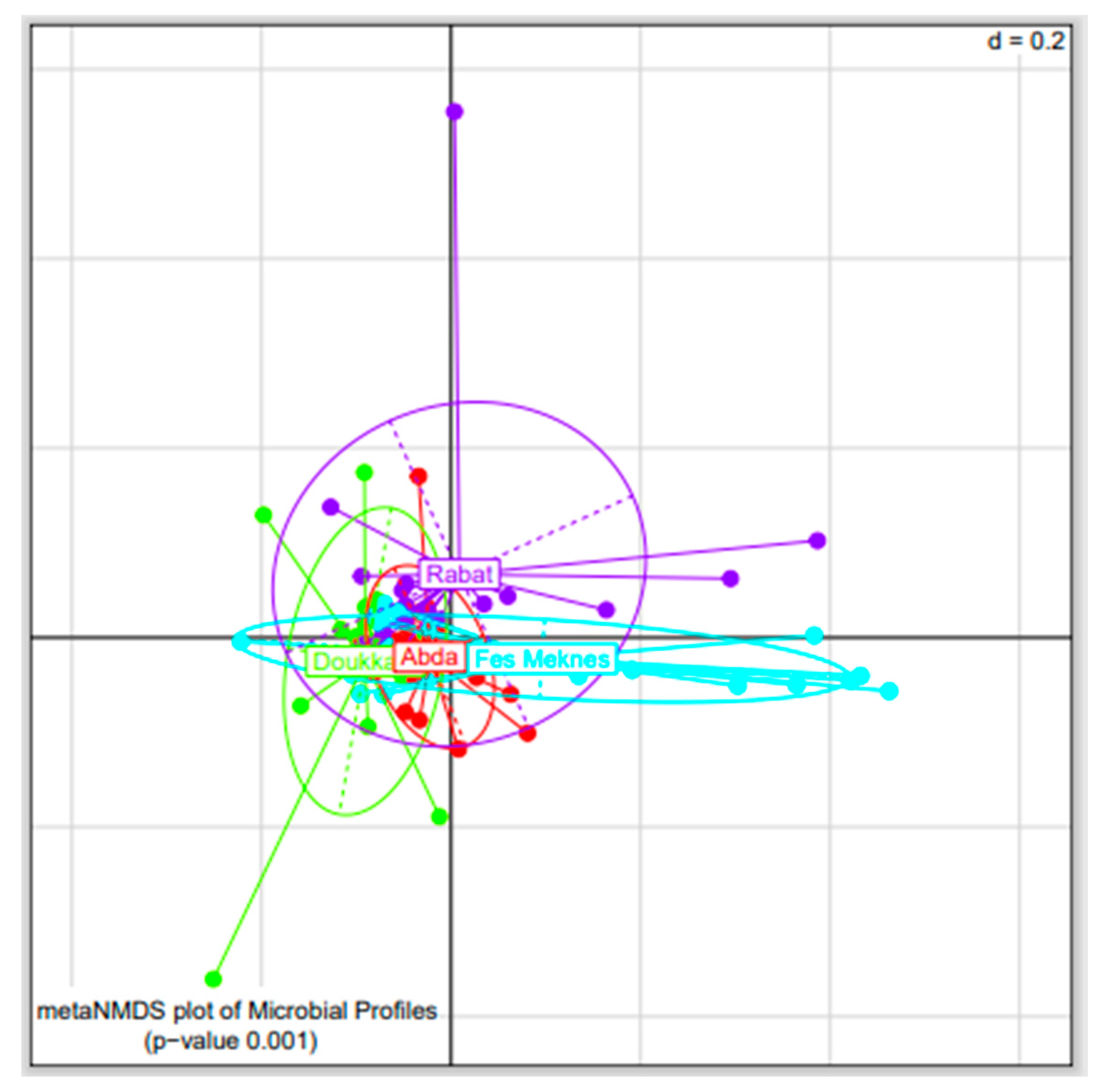
3.2.1. Bacterial Diversity among BSGM Natural Populations

The bacterial communities of BSGM were clustered according to their geographic origins. The NMDS plots based on the generalized UniFrac distance revealed that samples from Rabat, Fes-Meknes, Abda, and Doukkala were significantly separated (PERMANOVA, p < 0.001, Figure 2), with similar results also obtained with pairwise analyses (Supplementary Figure S1).
Based on the number and relative abundance of OTUs and Simpson and Shannon indices (Figure S2), the four natural populations of BSGM were characterized by different species’ richness and diversity. The samples from Abda, Doukkala, and Fes-Meknes contained most of the bacterial species. Rabat samples, on the other hand, had statistically lower species richness and diversity than the other locations. Furthermore, based on the Shannon score, samples from the Doukkala region had statistically higher diversity than samples from the Rabat region (pairwise ANOVA: p < 0.05).

3.2.2. Bacterial Composition of BSGM Natural Populations

The most abundant phylum detected in the four regions of Abda, Doukkala, Fes-Meknes, and Rabat was Proteobacteria (92.33 ± 1.01%, 79.05 ± 4.61%, 91.75 ± 1.8%, 89.41 ± 2.75%, respectively), while the classification of the rest of the phyla was different from one region to another. In Abda and Fes-Meknes, the second phylum was Firmicutes (4.34 ± 1.08%, 4.43 ± 1.4%, respectively), followed by Bacteroidetes (2.78 ± 0.42%, 2.08 ± 0.58%), Actinobacteria (0.31 ± 0.1%, 1.48 ± 0.75%), and Dependentiae (0.24 ± 0.1%, 0.26 ± 0.13%). In Doukkala, the second phylum was Firmicutes (11.00 ± 3.79%), followed by Actinobacteria (6.80 ± 3.58%), Bacteroidetes (2.94 ± 0.46%), and Dependentiae (0.21 ± 0.09%), while in Rabat, the second phylum was Actinobacteria (6.26 ± 2.83%), followed by Bacteroidetes (2.18 ± 0.4%), Firmicutes (2 ± 0.69%), and Dependentiae (0.15 ± 0.1%) (Figure 3a). The evaluation at the class level revealed that Gammaproteobacteria and Alphaproteobacteria were the most dominant members in all samples examined, followed by Bacilli in Abda, Doukkala, and Fes-Meknes. By contrast, in Rabat, the third class was Actinobacteria. Interestingly, members of Negativicutes and Babeliae were also detected, but to a much smaller degree (Figure 3b). At the genus level, the most abundant sequences in Abda, Doukkala, and Rabat were affiliated with Pseudomonas (27.02 ± 2.25%, 28.67 ± 2.44%, 45.24 ± 5.55%, respectively), followed by Stenotrophomonas (14.15 ± 1.44%, 17.68 ± 2.17%, 10.61 ± 1.42%, respectively), while the individuals from Fes-Meknes had a high relative abundance of Pantoea (24.80 ± 7.79%), followed by Pseudomonas (19.51 ± 3.34%). Different frequencies were observed for the remaining genera between the collection regions. In Abda, the third most prevalent OTU was Acinetobacter, followed by Phyllobacterium and Ochrobactrum (15.59 ± 3.53%, 6.72 ± 1.16%, 4.14 ± 0.84%, respectively); in Doukkala, the third abundant OTU was Phyllobacterium, followed by Paenarthrobacter and Streptococcus (7.37 ± 1.12%, 5.15 ± 3.08%, 3.85 ± 2.85%, respectively); in Fes-Meknes, the third genus was Stenotrophomonas, followed by Phyllobacterium and Ochrobactrum (8.78 ± 1.57%, 7.41 ± 1.32%, 6.92 ± 1.42%, respectively); and in Rabat, the third OTU was Phyllobacterium, followed by Ochrobactrum and Allorhizobium-Neorhizobium-Pararhizobium-Rhizobium (8.78 ± 1.18%, 6.93 ± 1.26%, 3.70 ± 2.98%, respectively). The frequencies of the rest of the genera were less than 0.1 (Figure 3c). Wolbachia was detected in samples from Rabat and Abda (Table S6) with a low relative abundance (0.75 ± 0.48%, 0.001 ± 0%, respectively). The absence of Spiroplasma, Cardinium, and Arsenophonus in the bacterial microbiome of BSGM populations was also confirmed by amplicon sequencing data.

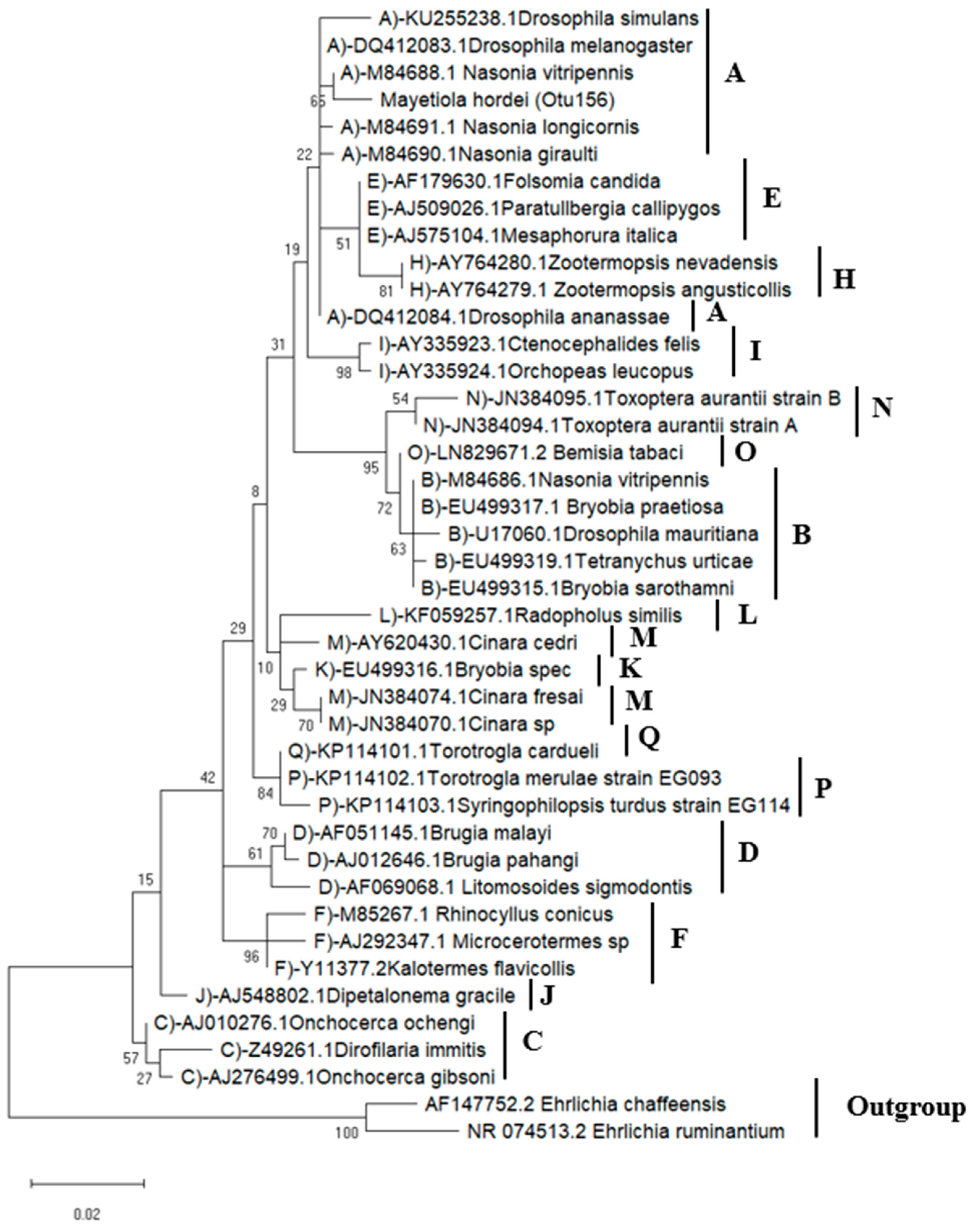
3.2.3. Wolbachia Phylogenetic Analysis

Using conventional sequence clustering into OTUs, the Wolbachia infection status was determined from normalized libraries (90% sequence identity). Wolbachia-related reads were clustered into a single OTU (OTU 156), with prevalence discrepancies between the two regions investigated. In Rabat, seven samples out of 20 were infected, whereas, in Abda, one sample out of 18 was found to be infected with Wolbachia. The sequence of the Wolbachia-related OTU (443 bp) was positioned within the supergroup A sequences according to the phylogenetic analyses based on the 16S rRNA gene (Figure 4). These results indicate that the BSMG derived from different regions most likely carries the same Wolbachia strain.

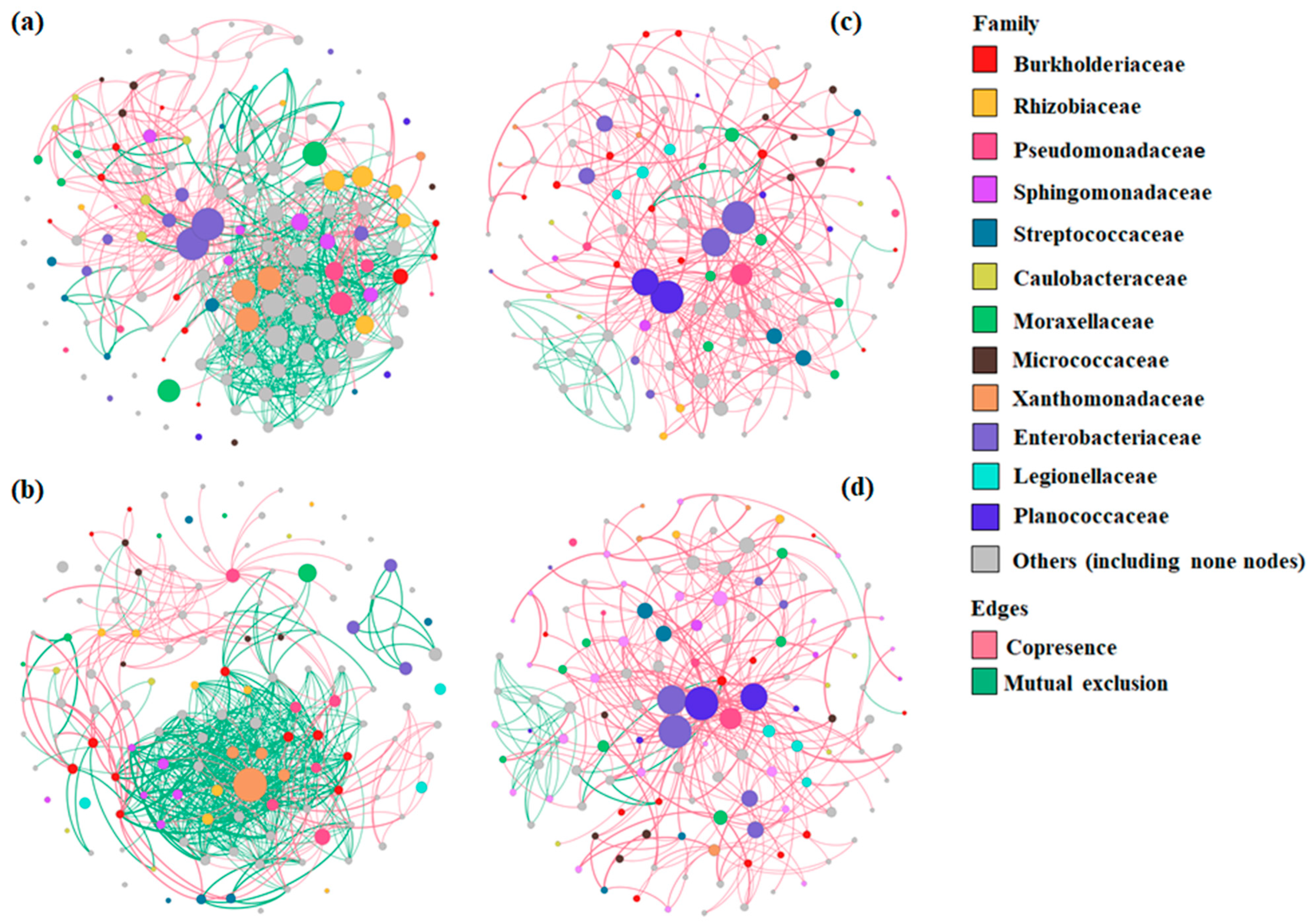
3.2.4. Bacterial Co-Occurrence/Mutual Exclusion Network Analysis of BSGM Natural Populations

Co-occurrence and mutual exclusion network analysis was performed to examine the potential interactions between bacterial partners in each region separately. The networks for each region were shown at the family (Figure 5) and genus (Figure S3) levels. The number of OTUs (nodes), interactions (edges), and clustering coefficients varied amongst the four regions. The Fes-Meknes region had the highest number of nodes (144), followed by Rabat, Doukkala, and Abda (137, 132, and 122, respectively). Bacterial communities from Fes-Meknes had more interactions (1179) and a lower clustering coefficient (0.135) than Rabat (1177 edges and 0.215 coefficient), Doukkala, and Abda (395 edges and 0.215 coefficient, 350 edges and 0.041 coefficient, respectively). At the family level, mutual exclusions accounted for the majority of interactions in Doukkala and Abda (91.39% and 75.31%, respectively) (Figure 5c,d), while in the two left regions, Fes-Meknes and Rabat (Figure 5a,b), copresence accounted for the majority of the interactions (72.87% and 64.03%, respectively), with Proteobacteria dominating interactions in all four regions. At the genus level, Stenotrophomonas showed the highest degree of interactions in Rabat, Erwinia in Fes-Meknes and Doukkala, and Lysinibacillus in Abda.

3.2.5. Core Microbiome Analysis of BSGM Populations

The core bacterial community was composed of 20 OTUs out of 61 (33%). Among the four regions of Abda, Doukkala, Fes-Meknes, and Rabat, a core microbiome was detected, which included 11 OTUs (55%): Stenotrophomonas [OTU6], three variants of Pseudomonas [OTU2, OTU39, and OTU18], Phyllobacterium [OTU4], Sphingomonas [OTU26], Ochrobactrum [OTU30], Chryseobacterium [OTU21], Delftia [OTU14], Candidatus Nucleicultrix [OTU19], and Novosphingobium [OTU23]. Additionally, a core microbiome was observed exclusively in three regions: the case of two OTUs (10%): Achromobacter [OTU25] in Doukkala Rabat and Fes-Meknes, and Cutibacterium [OTU43] in Doukkala, Abda, and Fes-Meknes. Two OTUs (10%) were found in two regions: Paracoccus [OTU24] in Doukkala and Fes-Meknes, and Cupriavidus [OTU24] in Abda and Rabat. Finally, five OTUs (25%) were found only in one region, four in Abda (Paracoccus [OTU22], two variants of Acinetobacter [OTU12, OTU13], and Brevundimonas [OTU20]), plus one OTU from Rabat (Bradyrhizobium [OTU33]) (Figure 6) (see Table S7 for more details about the overall prevalence of the core microbiome).

4. Discussion

In the current study, we evaluated the prevalence of reproductive symbionts, the bacterial diversity of natural populations, and the impact of geographic origin on the bacterial community structure of barley stem gall midges, located in four different producing regions in Morocco. The bacterial microbiome analysis was performed using next-generation, high-throughput sequencing of the V3–V4 region of the bacterial 16S rRNA gene.
To analyze the existence of reproductive symbionts, a genus specific 16S rRNA PCR test was used. The screening results revealed that BSGM was only infected with Wolbachia. Surprisingly, Wolbachia infection was only found in BSGM populations from Rabat and Fez-Meknes, five larvae, two females, and three males from 44 samples from Rabat (22%), and one larva, one female, and one male from 52 individuals from Fez-Meknes (6%); in total 13 samples out of 152 BSGMs were infected, with a prevalence of 9%. However, none of the BSGM populations studied contained Spiroplasma, Cardinium, or Arsenophonus. The HTS DNA sequence datasets also revealed the presence of Wolbachia infections, which were detected in two out of four regions studied. Seven samples out of 20 were infected in Rabat (35%), while in Abda, one sample out of 18 (5%) was found to be infected with Wolbachia (Table S6). The discrepancy in prevalence between the two methods may be attributable to the low Wolbachia infection density, as well as to the exclusion of some samples from the HTS sequence datasets due to their low read counts (less than 1000) and the higher sensitivity of HTS compared to conventional PCR screening [54,59]. To the best of our knowledge, Wolbachia had never been identified in BSGM populations. However, it was detected in a recent study of Hessian fly originating from Morocco, with a total of 13 samples out of 244 (5%) carrying Wolbachia infections: three females and ten males. Concerning HTS DNA sequence datasets, Wolbachia infections were also detected: 31 individuals out of 40 (78%) in various Hessian fly populations [34]. The Hessian fly shares with BSGM a similar distribution, similar morphology, and comparable symptoms in host plants [29]. On the other hand, no Wolbachia was found in the few other Hessian fly studies [35,60,61]. Wolbachia was also reported in the Asian rice gall midge (Orseolia oryzae) in India, where the sequencing of the 16S rRNA bacterial gene (V3-V4 region) revealed that Wolbachia was the predominant bacterium in the pupae and adults of both genders, with prevalences of 97.3%, 89.8%, and 79.6%, respectively [62]. In addition, Wolbachia is the most widespread intracellular bacterial genus, infecting filarial nematodes and a wide range of arthropod groups, including almost 65% of insect species [63,64,65], such as aphids [36,66,67,68] and fruit flies (Diptera:Tephritidae) [69,70,71].
Our efforts to identify Wolbachia strains found in natural populations of BSGM were limited by the difficulty in amplifying MLST (multi-locus sequence typing) and wsp genes, which could be attributed to the low infection density of Wolbachia [72,73]. The phylogenetic analyses were limited to 16S rRNA sequences amplified using Wolbachia-specific primers and the Wolbachia-related sequences acquired using HTS. All Wolbachia sequences found in this investigation exhibited the highest homology to strains from supergroup A (Figure 1 and Figure 6), indicating that the provenance of the BSGM samples did not contribute to the diversification of Wolbachia strains. It was previously discovered that the density of Wolbachia in a host influences the level of CI [74]. In light of this, the low infection rate found in our study implies that Wolbachia is unlikely to cause CI in the studied BSGM populations. Additionally, the fact that both males and females carry Wolbachia may indicate that the infection should not have any consequences on the sex ratio in favor of females. Even though Wolbachia are typically transmitted vertically, the population of barley midges in Rabat, Fes-Meknes, and Abda most likely became infected through horizontal transmission, which is common among insects [75,76] and may be mediated by parasitoids or host plants [77,78]. However, our results provide a fresh insight into the BSGM capacity to harbor the Wolbachia infection that may allow us to use the transinfection method to establish a reliable Wolbachia-infected lineage, by directing symbiont strains to be transferred to new hosts either within the same species or between species. Since it has been previously accomplished, the case of Wolbachia transinfections includes transporting strains between Drosophila species [79,80], Aedes aegypti [81,82,83], Culex quinquefasciatus [84], Bactrocera oleae [85], and Ceratitis capitata [86,87,88,89].
In order to investigate the effect of geographical origin on bacterial structure, HT sequencing was applied on 75 natural barley stem gall midge samples, based on a 90% sequence similarity. The results reveal a high number of distinct OTUs from four separate BSGM populations (Table S5 and Figure 3c). Our findings demonstrate that native environments have a major impact on the microbiota. The four BSGM natural populations were distinguished by various levels of species richness and diversity. Most bacterial species were found in the samples from Abda, Doukkala, and Fes-Meknes. In contrast, Rabat samples had statistically lower species richness and diversity. Additionally, samples from the Doukkala region displayed significantly higher diversity than samples from the Rabat region, according to the Shannon index. Since the research on the bacterial microbiome of this insect is still limited, it is the first time that changes in the structure of microbial communities have been observed in populations of BSGM with various geographic distributions. However, it has also been reported in other fly or insect species, including cases of the Hessian fly (M. destructor) [34,61], oriental fruit fly (Bactrocera dorsalis) [90,91], and melon fly (Zeugodacus cucurbitae) [92,93].
Proteobacteria was the predominant phylum detected in the four regions of Abda, Doukkala, Fes-Meknes, and Rabat. This phylum was also the most abundant in other insects, such as the Hessian fly from Morocco (three out of four regions) [34], the Asian rice gall midge (O. oryzae) from India [63], and the melon fly from Bangladesh (Z. cucurbitae) [92,93]. However, the classification of the rest of the phyla, Actinobacteria, Bacteroidetes, and Firmicutes, was different from one region to the other, in agreement with the results for other insects [34,62,92]. Additionally, a recently characterized phylum was detected in our study as the fifth most-common phylum among the four studied regions, with a prevalence of 32% (24 out of 75 samples), that is, candidate phylum Dependentiae (also known as TM6). The identification of this phylogenetic group of bacteria was based on isolates infecting free-living amoebae and on metagenomic studies. This phylum is common in a variety of habitats, including hospital biofilms, soil, waste water, and peat bogs; it is characterized by a relatively limited metabolic potential, but encodes a vast array of transporters, including ATP transporters and genes with bacterial endosymbiont-enriched activities [94,95,96,97,98]. At the class level, Gammaproteobacteria and Alphaproteobacteria were the most prevalent members in all samples analyzed, followed by Bacilli in Abda, Doukkala, and Fes-Meknes, and Actinobacteria in Rabat. However, in the Hessian fly populations from Morocco, the most frequent classes were Gammaproteobacteria, Alphaproteobacteria, and Betaproteobacteria [34]. Interestingly, members of Negativicutes were also detected but at a much lesser degree (Figure 3b), with a prevalence of 9.33% (seven out of 75 samples). This class was recently certified in the phylum Firmicutes that has generally been defined by having a low genomic GC content, Gram-positive (monoderm) cells, and an envelope composed of a cytoplasmic membrane and a thick coating of peptidoglycan [99]. However, Negativicutes are diderms, characterized by an inner and outer membrane containing lipopolysaccharides [100,101]. In addition, Negativicutes have colonized a variety of hosts, including insects, animals, soil, ocean, and sediments. They mainly rely on fermentation because they cannot grow in the presence of oxygen. Some members of Negativicutes are known by their ability to ferment lactate, as in the case of Veillonella, the genus that was present in our results, while other Gram-negative members can produce endospores [102,103]. At the genus level, Pseudomonas exhibited the highest relative abundances in Abda, Doukkala, and Rabat; this bacterial genus is known from a wide range of habitats and hosts and exhibits remarkable metabolic diversity [104,105]. The genus has adapted to interact specifically in both useful and harmful ways, mostly with plants [106,107], humans [108], and insects [109,110]. In the same three regions, Pseudomonas was followed by Stenotrophomonas, a genus reported from a variety of habitats that is frequently characterized as a multidrug-resistant, opportunistic human pathogen [111]. However, they are more usually discovered in soils or in close proximity to plants [112], where they can create symbiotic relationships [113]. Stenotrophomonas is also linked to a variety of insect species [114,115]. On the other hand, Pantoea was prevalent in the region of Fes-Meknes; this genus has regularly been isolated from a variety of terrestrial and aquatic ecosystems, in addition to connections with humans, animals, and insects [116,117]. Additionally, Pantoea isolates have been used as immunopotentiators to create supportive medications for melanoma, infections, allergies, and the reversal of immunosuppression [118,119,120]. Furthermore, several Pantoea isolates have been reported to produce antimicrobials and have been converted into commercial biocontrol treatments [121,122,123]. Pantoea was followed by Pseudomonas in the region of Fes-Meknes; this genus was also present in Hessian flies with different frequencies among different populations [34]. The reported variation in the microbiome of BSGM populations could be influenced by barley cultivars, insect pressure, environmental factors, including annual mean temperature, precipitation, and maximum snowfall, as well as the effects of host plants and insect developmental stages and sexes, plus the effect of Wolbachia infection on the microbiome [124,125].

5. Conclusions

The bacterial composition of populations of BSGM was found to be significantly influenced by the geographic location. With the highest number of OTUs, Proteobacteria were found to be the most abundant phylum in flies from the four sites. To the best of our knowledge, this is the first study of the BSGM bacterial microbiome that reports Wolbachia infection in this insect. The low infection rate identified in our study led us to suspect that Wolbachia is unlikely to be the source of cytoplasmic incompatibility in BSGM reproduction. On the other hand, our findings may offer a new perspective on the application of Wolbachia in the control of BSGM.

Supplementary Materials

The following supporting information can be downloaded at https://www.mdpi.com/article/10.3390/microorganisms11030797/s1, Table S1. Prevalence of bacterial endosymbionts screened in populations of barley stem gall midges. Table S2. List of bacterial primers and annealing temperatures used; Table S3. Distribution of Dependencia family (Otu 45); Table S4. Distribution of Negativicutes class (Otu 106); Table S5. Relative abundance (%) of different bacteria detected in Barley stem gall midge regions of Abda, Doukkala, Fes Meknes, and Rabat; File S1. Wolbachia sequence alignments; Figure S1. Pairwise non-metric multidimensional scaling (NMDS) plot of bacterial communities in barley stem gall midge samples collected from Rabat (purple), Fes-Meknes (blue), Abda (red), and Doukkala (green) (p < 0.001). The ’d’ stands for the grid’s dissimilarity scale (d = 0.2 implies that the distance between two grid lines represents approximately 20% dissimilarity between the regions); Figure S2. Species richness and diversity indices with significance differences in natural populations of barley stem gall midge samples collected from the regions of Rabat, Fes Meknes, Abda, and Doukkala. The interquartile range (IQR) is represented by boxes, the median is represented by a line within the boxes, and samples are represented by dots (* 0.01 < p < 0.05, ** p ≤ 0.01); Figure S3. Co-occurrence and mutual exclusion networks at the genus level for OTUs that compose the bacterial communities of Mayetiola hordei natural populations from the four regions of (a) Fes-Meknes, (b) Rabat, (c) Doukkala, and (d) Abda. The degree of interaction determines the size of each node. Cases of copresence are represented by green edges, while mutual exclusion is represented by red edges. Table S6. Distribution of Wolbachia genus (Otu 156). Table S7. Overall core prevalence.

Author Contributions

Conceptualization, A.M., M.R.B., G.T. and P.S.; methodology, E.A., G.T. and P.S.; collection of samples, K.E.F.; analysis, I.R., N.B.M., E.A. and G.T.; resources, G.T.; writing—original draft preparation, I.R.; writing—review and editing, I.R., N.B.M., A.M., M.R.B., E.A, K.E.F., G.T. and P.S.; supervision, A.M., G.T. and P.S. All authors have read and agreed to the published version of the manuscript.

Funding

This research was conducted with the assistance of the European Union’s Erasmus + Programme. Additionally, this study has been co-financed by the European Regional Development Fund of the European Union and Greek national funds through the Operational Program Competitiveness, Entrepreneurship and Innovation, under the call RESEARCH–CREATE–INNOVATE (project code: T2EDK-2020).

Data Availability Statement

All the data that support the findings of this study are available in NCBI under BioProject PRJNA915357.

Acknowledgments

The authors are thankful to International Center for Agricultural Research in the Dry Areas (ICARDA) for collecting and rearing barley stem gall midge populations from Morocco’s several barley areas.

Conflicts of Interest

The authors certify that this work contains no conflict of interest.

References

  1. Cherif, A.; Mediouni Ben Jemâa, J. Distribution, population dynamics and damage potential of barley stem gall midge, Mayetiola hordei (Diptera: Cecidomyiidae) on cultivated barley in two semi-arid areas of North Tunisia. Crop Prot. 2018, 112, 295–303. [Google Scholar] [CrossRef]
  2. Malipatil, M. Industry Biosecurity Plan for the Grains Industry. Threat Specific Contingency Plan Barley Stem Gall Midge. Mayetiola hordei; Plant Health Australia: Canberra, Australia, 2008; p. 23. [Google Scholar]
  3. Lhaloui, S.; Bouhssini, M.E.; Otmane, R.; Ouriniche, S.; Alami, A. Comparative Biology and Life Cycle of The Barley Stem Gall Midge and Hessian fly (Diptera: Cecidomyiidae) in Morocco. Rev. Maroc. Prot. Plantes 2016, 17–37. Available online: https://revues.imist.ma/index.php/RMPP/article/view/6420 (accessed on 12 March 2023).
  4. Parker, B.; El-Bouhssini, M.; Skinner, M. Field Guide: Insect Pests of Wheat and Barley in North Africa, West and Central Asia; ICARDA: Beirut, Lebanon, 2001; ISBN 978-92-9127-114-6. [Google Scholar]
  5. Hardie, J.; Minks, A.K. Pheromones of Non-Lepidopteran Insects Associated with Agricultural Plants; CABI Publishing: Wallingford, UK, 1999. [Google Scholar]
  6. Lhaloui, S.; Bouhssini, M.E.; Naserlhaq, N.; Amri, A.; Nachit, M.; Haddoury, J.E. Les Cecidomyies Des Cereales Au Maroc; INRA: Rabat, Morocco, 2005; 52p, ISBN 9981-1994-7-8. [Google Scholar]
  7. Schmid, R.B.; Knutson, A.; Giles, K.L.; McCornack, B.P. Hessian Fly (Diptera: Cecidomyiidae) Biology and Management in Wheat. J. Integr. Pest Manag. 2018, 9, 14. [Google Scholar] [CrossRef] [Green Version]
  8. Douglas, A.E. Lessons from Studying Insect Symbioses. Cell Host Microbe 2011, 10, 359–367. [Google Scholar] [CrossRef] [Green Version]
  9. Bourtzis, K.; Miller, T.A. (Eds.) Insect symbiosis; Contemporary Topics in Entomology Series; CRC Press: Boca Raton, FL, USA, 2003; ISBN 978-0-8493-1286-1. [Google Scholar]
  10. Sanchez-Contreras, M.; Vlisidou, I. The Diversity of Insect-bacteria Interactions and its Applications for Disease Control. Biotechnol. Genet. Eng. Rev. 2008, 25, 203–244. [Google Scholar] [CrossRef] [PubMed] [Green Version]
  11. Baumann, P. Biology bacteriocyte-associated endosymbionts of plant sap-sucking insects. Annu. Rev. Microbiol. 2005, 59, 155–189. [Google Scholar] [CrossRef]
  12. Zchori-Fein, E.; Bourtzis, K. Manipulative Tenants: Bacteria Associated with Arthropods; CRC Press: Boca Raton, FL, USA, 2011; ISBN 978-1-4398-2749-9. [Google Scholar]
  13. Buchner, P.; Buchner, P. Endosymbiosis of animals with plant microorganisms; Interscience: New York, NY, USA, 1965. [Google Scholar]
  14. Douglas, A.E. Symbiotic microorganisms: Untapped resources for insect pest control. Trends Biotechnol. 2007, 25, 338–342. [Google Scholar] [CrossRef] [PubMed]
  15. Hertig, M.; Wolbach, S.B. Studies on Rickettsia-Like Micro-Organisms in Insects. J. Med. Res. 1924, 44, 329–374.7. [Google Scholar] [PubMed]
  16. Lefoulon, E.; Clark, T.; Guerrero, R.; Cañizales, I.; Cardenas-Callirgos, J.M.; Junker, K.; Vallarino-Lhermitte, N.; Makepeace, B.L.; Darby, A.C.; Foster, J.M.; et al. Diminutive, degraded but dissimilar: Wolbachia genomes from filarial nematodes do not conform to a single paradigm. Microb. Genom. 2020, 6, e000487. [Google Scholar] [CrossRef]
  17. Fenn, K.; Conlon, C.; Jones, M.; Quail, M.A.; Holroyd, N.E.; Parkhill, J.; Blaxter, M. Phylogenetic Relationships of the Wolbachia of Nematodes and Arthropods. PLoS Pathog. 2006, 2, e94. [Google Scholar] [CrossRef] [Green Version]
  18. Lefoulon, E.; Clark, T.; Borveto, F.; Perriat-Sanguinet, M.; Moulia, C.; Slatko, B.E.; Gavotte, L. Pseudoscorpion Wolbachia symbionts: Diversity and evidence for a new supergroup S. BMC Microbiol 2020, 20, 188. [Google Scholar] [CrossRef] [PubMed]
  19. Rowley, S.M.; Raven, R.J.; McGraw, E.A. Wolbachia pipientis in Australian Spiders. Curr Microbiol 2004, 49, 208–214. [Google Scholar] [CrossRef] [PubMed]
  20. Baldo, L.; Werren, J.H. Revisiting Wolbachia Supergroup Typing Based on WSP: Spurious Lineages and Discordance with MLST. Curr. Microbiol. 2007, 55, 81–87. [Google Scholar] [CrossRef] [PubMed]
  21. Wang, G.-H.; Jia, L.-Y.; Xiao, J.-H.; Huang, D.-W. Discovery of a new Wolbachia supergroup in cave spider species and the lateral transfer of phage WO among distant hosts. Infect. Genet. Evol. 2016, 41, 1–7. [Google Scholar] [CrossRef] [PubMed] [Green Version]
  22. Gerth, M. Classification of Wolbachia (Alphaproteobacteria, Rickettsiales): No Evidence for a Distinct Supergroup in Cave Spiders. bioRxiv 2016. [Google Scholar] [CrossRef] [PubMed]
  23. Bourtzis, K. Wolbachia-Based Technologies for Insect Pest Population Control. In Transgenesis and the Management of Vector-Borne Disease; Aksoy, S., Ed.; Advances in Experimental Medicine and Biology; Springer: New York, NY, USA, 2008; pp. 104–113. ISBN 978-0-387-78225-6. [Google Scholar]
  24. Saridaki, A.; Bourtzis, K. Wolbachia: More than just a bug in insects genitals. Curr. Opin. Microbiol. 2010, 13, 67–72. [Google Scholar] [CrossRef]
  25. Vreysen, M.J.B.; Robinson, A.S.; Hendrichs, J. Area-Wide Control of Insect Pests: From Research to Field Implementation; Springer: Dordrecht, The Netherlands, 2007; ISBN 978-1-4020-6058-8. [Google Scholar]
  26. Correa, C.C.; Ballard, J.W.O. Wolbachia Associations with Insects: Winning or Losing Against a Master Manipulator. Front. Ecol. Evol. 2016, 3, 153. [Google Scholar] [CrossRef] [Green Version]
  27. Xie, J.; Butler, S.; Sanchez, G.; Mateos, M. Male killing Spiroplasma protects Drosophila melanogaster against two parasitoid wasps. Heredity 2014, 112, 399–408. [Google Scholar] [CrossRef]
  28. Anbutsu, H.; Fukatsu, T. Spiroplasma as a model insect endosymbiont. Environ. Microbiol. Rep. 2011, 3, 144–153. [Google Scholar] [CrossRef]
  29. Zhang, Y.-K.; Chen, Y.-T.; Yang, K.; Hong, X.-Y. A review of prevalence and phylogeny of the bacterial symbiont Cardinium in mites (subclass: Acari). Syst. Appl. Acarol. 2016, 21, 978. [Google Scholar] [CrossRef] [Green Version]
  30. Zchori-Fein, E.; Perlman, S.J.; Kelly, S.E.; Katzir, N.; Hunter, M.S.Y. Characterization of a ‘Bacteroidetes’ symbiont in Encarsia wasps (Hymenoptera: Aphelinidae): Proposal of ‘Candidatus Cardinium hertigii’. Int. J. Syst. Evol. Microbiol. 2004, 54, 961–968. [Google Scholar] [CrossRef] [PubMed]
  31. Ferree, P.M.; Avery, A.; Azpurua, J.; Wilkes, T.; Werren, J.H. A Bacterium Targets Maternally Inherited Centrosomes to Kill Males in Nasonia. Curr. Biol. 2008, 18, 1409–1414. [Google Scholar] [CrossRef] [Green Version]
  32. Taylor, G.P.; Coghlin, P.C.; Floate, K.D.; Perlman, S.J. The host range of the male-killing symbiont Arsenophonus nasoniae in filth fly parasitioids. J. Invertebr. Pathol. 2011, 106, 371–379. [Google Scholar] [CrossRef]
  33. Mezghani Khemakhem, M.; Makni, H.; Marrakchi, M. PCR-RFLP identification of mitochondrial markers of Mayetiola (Diptera: Cecidomyiidae) on cereals. Ann. Soc. Entomol. Fr. 2002, 38, 277–282. [Google Scholar]
  34. Bel Mokhtar, N.; Maurady, A.; Britel, M.R.; El Bouhssini, M.; Batargias, C.; Stathopoulou, P.; Asimakis, E.; Tsiamis, G. Detection of Wolbachia Infections in Natural and Laboratory Populations of the Moroccan Hessian Fly, Mayetiola destructor (Say). Insects 2020, 11, 340. [Google Scholar] [CrossRef]
  35. Bansal, R.; Hulbert, S.; Schemerhorn, B.; Reese, J.C.; Whitworth, R.J.; Stuart, J.J.; Chen, M.-S. Hessian Fly-Associated Bacteria: Transmission, Essentiality, and Composition. PLoS ONE 2011, 6, e23170. [Google Scholar] [CrossRef] [Green Version]
  36. Augustinos, A.A.; Santos-Garcia, D.; Dionyssopoulou, E.; Moreira, M.; Papapanagiotou, A.; Scarvelakis, M.; Doudoumis, V.; Ramos, S.; Aguiar, A.F.; Borges, P.A.V.; et al. Detection and Characterization of Wolbachia Infections in Natural Populations of Aphids: Is the Hidden Diversity Fully Unraveled? PLoS ONE 2011, 6, e28695. [Google Scholar] [CrossRef] [PubMed] [Green Version]
  37. Hartley, J.L.; Bowen, H. PEG precipitation for selective removal of small DNA fragments. Focus 2003, 25, 18. [Google Scholar]
  38. Klindworth, A.; Pruesse, E.; Schweer, T.; Peplies, J.; Quast, C.; Horn, M.; Glöckner, F.O. Evaluation of general 16S ribosomal RNA gene PCR primers for classical and next-generation sequencing-based diversity studies. Nucleic Acids Res. 2013, 41, e1. [Google Scholar] [CrossRef] [PubMed]
  39. Bolyen, E.; Rideout, J.R.; Dillon, M.R.; Bokulich, N.A.; Abnet, C.C.; Al-Ghalith, G.A.; Alexander, H.; Alm, E.J.; Arumugam, M.; Asnicar, F.; et al. Reproducible, interactive, scalable and extensible microbiome data science using QIIME 2. Nat. Biotechnol. 2019, 37, 852–857. [Google Scholar] [CrossRef] [PubMed]
  40. Edgar, R.C. Search and clustering orders of magnitude faster than BLAST. Bioinformatics 2010, 26, 2460–2461. [Google Scholar] [CrossRef] [PubMed] [Green Version]
  41. Edgar, R.C. UPARSE: Highly accurate OTU sequences from microbial amplicon reads. Nat. Methods 2013, 10, 996–998. [Google Scholar] [CrossRef] [PubMed]
  42. Edgar, R.C. UNCROSS2: Identification of cross-talk in 16S rRNA OTU tables. bioRxiv 2018. [Google Scholar] [CrossRef]
  43. Camacho, C.; Coulouris, G.; Avagyan, V.; Ma, N.; Papadopoulos, J.; Bealer, K.; Madden, T.L. BLAST+: Architecture and applications. BMC Bioinform. 2009, 10, 421. [Google Scholar] [CrossRef] [PubMed] [Green Version]
  44. Quast, C.; Pruesse, E.; Yilmaz, P.; Gerken, J.; Schweer, T.; Yarza, P.; Peplies, J.; Glöckner, F.O. The SILVA ribosomal RNA gene database project: Improved data processing and web-based tools. Nucleic Acids Res. 2013, 41, D590–D596. [Google Scholar] [CrossRef]
  45. Hollander, M.; Wolfe, D.A.; Chicken, E. Nonparametric Statistical Methods, 3rd ed.; John Wiley & Sons, Inc.: Hoboken, NJ, USA, 2014; ISBN 978-0-470-38737-5. [Google Scholar]
  46. Chen, J.; Bittinger, K.; Charlson, E.S.; Hoffmann, C.; Lewis, J.; Wu, G.D.; Collman, R.G.; Bushman, F.D.; Li, H. Associating microbiome composition with environmental covariates using generalized UniFrac distances. Bioinformatics 2012, 28, 2106–2113. [Google Scholar] [CrossRef] [Green Version]
  47. Steele, J.A.; Countway, P.D.; Xia, L.; Vigil, P.D.; Beman, J.M.; Kim, D.Y.; Chow, C.-E.T.; Sachdeva, R.; Jones, A.C.; Schwalbach, M.S.; et al. Marine bacterial, archaeal and protistan association networks reveal ecological linkages. ISME J. 2011, 5, 1414–1425. [Google Scholar] [CrossRef] [Green Version]
  48. Zhou, J.; Deng, Y.; Luo, F.; He, Z.; Yang, Y. Phylogenetic Molecular Ecological Network of Soil Microbial Communities in Response to Elevated CO2. mBio 2011, 2, e00122-11. [Google Scholar] [CrossRef] [Green Version]
  49. Faust, K.; Raes, J. CoNet app: Inference of biological association networks using Cytoscape. F1000Res 2016, 5, 1519. [Google Scholar] [CrossRef] [Green Version]
  50. Edgar, R.C. MUSCLE: Multiple sequence alignment with high accuracy and high throughput. Nucleic Acids Res. 2004, 32, 1792–1797. [Google Scholar] [CrossRef] [Green Version]
  51. Kumar, S.; Stecher, G.; Tamura, K. MEGA7: Molecular Evolutionary Genetics Analysis Version 7.0 for Bigger Datasets. Mol. Biol. Evol. 2016, 33, 1870–1874. [Google Scholar] [CrossRef] [PubMed] [Green Version]
  52. Lefort, V.; Longueville, J.-E.; Gascuel, O. SMS: Smart Model Selection in PhyML. Mol. Biol. Evol. 2017, 34, 2422–2424. [Google Scholar] [CrossRef] [PubMed] [Green Version]
  53. Akaike, H. Autoregressive model fitting for control. Ann. Inst. Stat. Math. 1971, 23, 163–180. [Google Scholar] [CrossRef]
  54. Comar, M.; D’Accolti, M.; Cason, C.; Soffritti, I.; Campisciano, G.; Lanzoni, L.; Bisi, M.; Volta, A.; Mazzacane, S.; Caselli, E. Introduction of NGS in Environmental Surveillance for Healthcare-Associated Infection Control. Microorganisms 2019, 7, 708. [Google Scholar] [CrossRef] [Green Version]
  55. Anderson, M.J. Permutation tests for univariate or multivariate analysis of variance and regression. Can. J. Fish. Aquat. Sci. 2001, 58, 626–639. [Google Scholar] [CrossRef]
  56. Vandekerckhove, T.T.M.; Watteyne, S.; Willems, A.; Swings, J.G.; Mertens, J.; Gillis, M. Phylogenetic analysis of the 16S rDNA of the cytoplasmic bacterium Wolbachia from the novel host Folsomia candida (Hexapoda, Collembola) and its implications for Wolbachial taxonomy. FEMS Microbiol. Lett. 1999, 180, 279–286. [Google Scholar] [CrossRef] [Green Version]
  57. Carvajal, T.M.; Hashimoto, K.; Harnandika, R.K.; Amalin, D.M.; Watanabe, K. Detection of Wolbachia in field-collected Aedes aegypti mosquitoes in metropolitan Manila, Philippines. Parasites Vectors 2019, 12, 361. [Google Scholar] [CrossRef] [Green Version]
  58. Lo, N.; Casiraghi, M.; Salati, E.; Bazzocchi, C.; Bandi, C. How Many Wolbachia Supergroups Exist? Mol. Biol. Evol. 2002, 19, 341–346. [Google Scholar] [CrossRef] [Green Version]
  59. Cao, Y.; Fanning, S.; Proos, S.; Jordan, K.; Srikumar, S. A Review on the Applications of Next Generation Sequencing Technologies as Applied to Food-Related Microbiome Studies. Front. Microbiol. 2017, 8, 1829. [Google Scholar] [CrossRef] [Green Version]
  60. Johnson, A.J.; Schemerhorn, B.J.; Shukle, R.H. A First Assessment of Mitochondrial DNA Variation and Geographic Distribution of Haplotypes in Hessian fly (Diptera: Cecidomyiidae). Ann. Entomol. Soc. Am. 2004, 97, 940–948. [Google Scholar] [CrossRef]
  61. Johnson, A.J.; Morton, P.K.; Schemerhorn, B.J.; Shukle, R.H. Use of a Nuclear Marker to Assess Population Structure in Hessian Fly (Diptera: Cecidomyiidae). Ann. Entomol. Soc. Am. 2011, 104, 666–674. [Google Scholar] [CrossRef]
  62. Ojha, A.; Sinha, D.K.; Padmakumari, A.P.; Bentur, J.S.; Nair, S. Bacterial Community Structure in the Asian Rice Gall Midge Reveals a Varied Microbiome Rich in Proteobacteria. Sci. Rep. 2017, 7, 9424. [Google Scholar] [CrossRef] [PubMed] [Green Version]
  63. Glowska, E.; Dragun-Damian, A.; Dabert, M.; Gerth, M. New Wolbachia supergroups detected in quill mites (Acari: Syringophilidae). Infect. Genet. Evol. 2015, 30, 140–146. [Google Scholar] [CrossRef] [PubMed]
  64. Zug, R.; Hammerstein, P. Still a Host of Hosts for Wolbachia: Analysis of Recent Data Suggests That 40% of Terrestrial Arthropod Species Are Infected. PLoS ONE 2012, 7, e38544. [Google Scholar] [CrossRef] [PubMed] [Green Version]
  65. Hilgenboecker, K.; Hammerstein, P.; Schlattmann, P.; Telschow, A.; Werren, J.H. How many species are infected with Wolbachia?—A statistical analysis of current data: Wolbachia infection rates. FEMS Microbiol. Lett. 2008, 281, 215–220. [Google Scholar] [CrossRef] [PubMed] [Green Version]
  66. Jeyaprakash, A.; Hoy, M.A. Long PCR improves Wolbachia DNA amplification: Wsp sequences found in 76% of sixty-three arthropod species. Insect Mol. Biol. 2000, 9, 393–405. [Google Scholar] [CrossRef]
  67. Wang, Z.; Shen, Z.-R.; Song, Y.; Liu, H.-Y.; Li, Z.-X. Distribution and diversity of Wolbachia in different populations of the wheat aphid Sitobion miscanthi (Hemiptera: Aphididae) in China. Eur. J. Entomol. 2009, 106, 49–55. [Google Scholar] [CrossRef] [Green Version]
  68. Zytynska, S.E.; Weisser, W.W. The natural occurrence of secondary bacterial symbionts in aphids: Aphid secondary symbionts. Ecol. Entomol. 2016, 41, 13–26. [Google Scholar] [CrossRef] [Green Version]
  69. Mateos, M.; Martinez Montoya, H.; Lanzavecchia, S.B.; Conte, C.; Guillén, K.; Morán-Aceves, B.M.; Toledo, J.; Liedo, P.; Asimakis, E.D.; Doudoumis, V.; et al. Wolbachia pipientis Associated With Tephritid Fruit Fly Pests: From Basic Research to Applications. Front. Microbiol. 2020, 11, 1080. [Google Scholar] [CrossRef]
  70. Asimakis, E.D.; Doudoumis, V.; Hadapad, A.B.; Hire, R.S.; Batargias, C.; Niu, C.; Khan, M.; Bourtzis, K.; Tsiamis, G. Detection and characterization of bacterial endosymbionts in Southeast Asian tephritid fruit fly populations. BMC Microbiol. 2019, 19, 290. [Google Scholar] [CrossRef]
  71. Sun, X.; Cui, L.; Li, Z. Diversity and Phylogeny of Wolbachia Infecting Bactrocera dorsalis (Diptera: Tephritidae) Populations from China. Environ. Entomol. 2007, 36, 1283–1289. [Google Scholar] [CrossRef]
  72. González Villa, T.; Viñas, M. (Eds.) Horizontal Gene Transfer: Breakting Borders between Living Kingdoms; Springer: Cham, Switzerland, 2019; ISBN 978-3-030-21862-1. [Google Scholar]
  73. Kondo, N.; Nikoh, N.; Ijichi, N.; Shimada, M.; Fukatsu, T. Genome fragment of Wolbachia endosymbiont transferred to X chromosome of host insect. Proc. Natl. Acad. Sci. USA 2002, 99, 14280–14285. [Google Scholar] [CrossRef] [Green Version]
  74. Bordenstein, S.R.; Bordenstein, S.R. Temperature Affects the Tripartite Interactions between Bacteriophage WO, Wolbachia, and Cytoplasmic Incompatibility. PLoS ONE 2011, 6, e29106. [Google Scholar] [CrossRef] [PubMed] [Green Version]
  75. Ahmed, M.Z.; De Barro, P.J.; Ren, S.-X.; Greeff, J.M.; Qiu, B.-L. Evidence for Horizontal Transmission of Secondary Endosymbionts in the Bemisia tabaci Cryptic Species Complex. PLoS ONE 2013, 8, e53084. [Google Scholar] [CrossRef] [PubMed]
  76. Huigens, M.E.; de Almeida, R.P.; Boons, P.A.H.; Luck, R.F.; Stouthamer, R. Natural interspecific and intraspecific horizontal transfer of parthenogenesis–inducing Wolbachia in Trichogramma wasps. Proc. R. Soc. Lond. B 2004, 271, 509–515. [Google Scholar] [CrossRef] [PubMed] [Green Version]
  77. Ahmed, M.Z.; Li, S.-J.; Xue, X.; Yin, X.-J.; Ren, S.-X.; Jiggins, F.M.; Greeff, J.M.; Qiu, B.-L. The Intracellular Bacterium Wolbachia Uses Parasitoid Wasps as Phoretic Vectors for Efficient Horizontal Transmission. PLoS Pathog. 2015, 11, e1004672. [Google Scholar] [CrossRef] [Green Version]
  78. Li, S.-J.; Ahmed, M.Z.; Lv, N.; Shi, P.-Q.; Wang, X.-M.; Huang, J.-L.; Qiu, B.-L. Plantmediated horizontal transmission of Wolbachia between whiteflies. ISME J. 2017, 11, 1019–1028. [Google Scholar] [CrossRef] [Green Version]
  79. Hoffmann, A.A.; Ross, P.A.; Rašić, G. Wolbachia strains for disease control: Ecological and evolutionary considerations. Evol. Appl. 2015, 8, 751–768. [Google Scholar] [CrossRef]
  80. Hughes, G.L.; Rasgon, J.L. Transinfection: A method to investigate Wolbachia-host interactions and control arthropod-borne disease: Transinfection of arthropods. Insect Mol. Biol. 2014, 23, 141–151. [Google Scholar] [CrossRef] [Green Version]
  81. Schmidt, T.L.; Barton, N.H.; Rašić, G.; Turley, A.P.; Montgomery, B.L.; Iturbe-Ormaetxe, I.; Cook, P.E.; Ryan, P.A.; Ritchie, S.A.; Hoffmann, A.A.; et al. Local introduction and heterogeneous spatial spread of dengue-suppressing Wolbachia through an urban population of Aedes aegypti. PLoS Biol. 2017, 15, e2001894. [Google Scholar] [CrossRef]
  82. McMeniman, C.J.; Lane, R.V.; Cass, B.N.; Fong, A.W.C.; Sidhu, M.; Wang, Y.-F.; O’Neill, S.L. Stable Introduction of a Life-Shortening Wolbachia Infection into the Mosquito Aedes aegypti. Science 2009, 323, 141–144. [Google Scholar] [CrossRef] [PubMed] [Green Version]
  83. Hoffmann, A.A.; Montgomery, B.L.; Popovici, J.; Iturbe-Ormaetxe, I.; Johnson, P.H.; Muzzi, F.; Greenfield, M.; Durkan, M.; Leong, Y.S.; Dong, Y.; et al. Successful establishment of Wolbachia in Aedes populations to suppress dengue transmission. Nature 2011, 476, 454–457. [Google Scholar] [CrossRef]
  84. Ant, T.H.; Herd, C.; Louis, F.; Failloux, A.B.; Sinkins, S.P. Wolbachia transinfections in Culex quinquefasciatus generate cytoplasmic incompatibility. Insect Mol. Biol. 2020, 29, 1–8. [Google Scholar] [CrossRef] [PubMed] [Green Version]
  85. Apostolaki, A.; Livadaras, I.; Saridaki, A.; Chrysargyris, A.; Savakis, C.; Bourtzis, K. Transinfection of the olive fruit fly Bactrocera oleae with Wolbachia: Towards a symbiont-based population control strategy: Transinfection of Bactrocera oleae with Wolbachia. J. Appl. Entomol. 2011, 135, 546–553. [Google Scholar] [CrossRef]
  86. Kyritsis, G.A.; Koskinioti, P.; Bourtzis, K.; Papadopoulos, N.T. Effect of Wolbachia Infection and Adult Food on the Sexual Signaling of Males of the Mediterranean Fruit Fly Ceratitis capitata. Insects 2022, 13, 737. [Google Scholar] [CrossRef]
  87. Sarakatsanou, A.; Diamantidis, A.D.; Papanastasiou, S.A.; Bourtzis, K.; Papadopoulos, N.T. Effects of Wolbachia on fitness of the Mediterranean fruit fly (Diptera: Tephritidae): Wolbachia affects the fitness of C. capitata. J. Appl. Entomol. 2011, 135, 554–563. [Google Scholar] [CrossRef]
  88. Zabalou, S.; Apostolaki, A.; Livadaras, I.; Franz, G.; Robinson, A.S.; Savakis, C.; Bourtzis, K. Incompatible insect technique: Incompatible males from a Ceratitis capitata genetic sexing strain. Entomol. Exp. Et Appl. 2009, 132, 232–240. [Google Scholar] [CrossRef]
  89. Zabalou, S.; Riegler, M.; Theodorakopoulou, M.; Stauffer, C.; Savakis, C.; Bourtzis, K. Wolbachia-induced cytoplasmic incompatibility as a means for insect pest population control. Proc. Natl. Acad. Sci. USA 2004, 101, 15042–15045. [Google Scholar] [CrossRef] [Green Version]
  90. Liu, S.-H.; Chen, Y.; Li, W.; Tang, G.-H.; Yang, Y.; Jiang, H.-B.; Dou, W.; Wang, J.-J. Diversity of Bacterial Communities in the Intestinal Tracts of Two Geographically Distant Populations of Bactrocera dorsalis (Diptera: Tephritidae). J. Econ. Entomol. 2018, 11, 2861–2868. [Google Scholar] [CrossRef]
  91. Liu, L.J.; Martinez-Sañudo, I.; Mazzon, L.; Prabhakar, C.S.; Girolami, V.; Deng, Y.L.; Dai, Y.; Li, Z.H. Bacterial communities associated with invasive populations of Bactrocera dorsalis (Diptera: Tephritidae) in China. Bull. Entomol. Res. 2016, 106, 718–728. [Google Scholar] [CrossRef]
  92. Asimakis, E.; Stathopoulou, P.; Sapounas, A.; Khaeso, K.; Batargias, C.; Khan, M.; Tsiamis, G. New Insights on the Zeugodacus cucurbitae (Coquillett) Bacteriome. Microorganisms 2021, 9, 659. [Google Scholar] [CrossRef] [PubMed]
  93. Yong, H.-S.; Song, S.-L.; Eamsobhana, P.; Pasartvit, A.; Lim, P.-E. Differential abundance and core members of the bacterial community associated with wild male Zeugodacus cucurbitae fruit flies (Insecta: Tephritidae) from three geographical regions of Southeast Asia. Mol. Biol. Rep. 2019, 46, 3765–3776. [Google Scholar] [CrossRef]
  94. Takemura, M. Genome Sequence of a New “Candidatus” Phylum “Dependentiae” Isolate from Chiba, Japan. Microbiol. Resour. Announc. 2022, 11, e01123-21. [Google Scholar] [CrossRef] [PubMed]
  95. McLean, J.S.; Lombardo, M.-J.; Badger, J.H.; Edlund, A.; Novotny, M.; Yee-Greenbaum, J.; Vyahhi, N.; Hall, A.P.; Yang, Y.; Dupont, C.L.; et al. Candidate phylum TM6 genome recovered from a hospital sink biofilm provides genomic insights into this uncultivated phylum. Proc. Natl. Acad. Sci. USA 2013, 110, E2390–E2399. [Google Scholar] [CrossRef] [Green Version]
  96. Pagnier, I.; Yutin, N.; Croce, O.; Makarova, K.S.; Wolf, Y.I.; Benamar, S.; Raoult, D.; Koonin, E.V.; La Scola, B. Babela massiliensis, a representative of a widespread bacterial phylum with unusual adaptations to parasitism in amoebae. Biol. Direct 2015, 10, 13. [Google Scholar] [CrossRef] [PubMed] [Green Version]
  97. Delafont, V.; Samba-Louaka, A.; Bouchon, D.; Moulin, L.; Héchard, Y. Shedding light on microbial dark matter: A TM6 bacterium as natural endosymbiont of a free-living amoeba: TM6 bacterium as endosymbiont of amoeba. Environ. Microbiol. Rep. 2015, 7, 970–978. [Google Scholar] [CrossRef]
  98. Deeg, C.M.; Zimmer, M.M.; George, E.E.; Husnik, F.; Keeling, P.J.; Suttle, C.A. Chromulinavorax destructans, a pathogen of microzooplankton that provides a window into the enigmatic candidate phylum Dependentiae. PLoS Pathog. 2019, 15, e1007801. [Google Scholar] [CrossRef] [Green Version]
  99. Yutin, N.; Galperin, M.Y. A genomic update on clostridial phylogeny: Gram-negative spore formers and other misplaced clostridia: Genomics update. Env. Microbiol 2013, 15, 2631–2641. [Google Scholar] [CrossRef]
  100. Marchandin, H.; Teyssier, C.; Campos, J.; Jean-Pierre, H.; Roger, F.; Gay, B.; Carlier, J.-P.; Jumas-Bilak, E. Negativicoccus succinicivorans gen. nov., sp. nov., isolated from human clinical samples, emended description of the family Veillonellaceae and description of Negativicutes classis nov., Selenomonadales ord. nov. and Acidaminococcaceae fam. nov. in the bacterial phylum Firmicutes. Int. J. Syst. Evol. Microbiol. 2010, 60, 1271–1279. [Google Scholar] [CrossRef]
  101. Campbell, C.; Adeolu, M.; Gupta, R.S. Genome-based taxonomic framework for the class Negativicutes: Division of the class Negativicutes into the orders Selenomonadales emend., Acidaminococcales ord. nov. and Veillonellales ord. nov. Int. J. Syst. Evol. Microbiol. 2015, 65, 3203–3215. [Google Scholar] [CrossRef]
  102. Beskrovnaya, P.; Fakih, D.; Morneau, I.; Hashimi, A.; Guadarrama Bello, D.; Xing, S.; Nanci, A.; Huan, T.; Tocheva, E.I. No Endospore Formation Confirmed in Members of the Phylum Proteobacteria. Appl Env. Microbiol. 2021, 87, e02312-20. [Google Scholar] [CrossRef] [PubMed]
  103. Ng, S.K.C.; Hamilton, I.R. Lactate Metabolism by Veillonella parvula. J. Bacteriol. 1971, 105, 999–1005. [Google Scholar] [CrossRef] [PubMed] [Green Version]
  104. Silby, M.W.; Winstanley, C.; Godfrey, S.A.C.; Levy, S.B.; Jackson, R.W. Pseudomonas genomes: Diverse and adaptable. FEMS Microbiol. Rev. 2011, 35, 652–680. [Google Scholar] [CrossRef] [PubMed] [Green Version]
  105. Crone, S.; Vives-Flórez, M.; Kvich, L.; Saunders, A.M.; Malone, M.; Nicolaisen, M.H.; Martínez-García, E.; Rojas-Acosta, C.; Catalina Gomez-Puerto, M.; Calum, H.; et al. The environmental occurrence of Pseudomonas aeruginosa. APMIS 2020, 128, 220–231. [Google Scholar] [CrossRef] [PubMed]
  106. Mercado-Blanco, J.; Bakker, P.A.H.M. Interactions between plants and beneficial Pseudomonas spp.: Exploiting bacterial traits for crop protection. Antonie Leeuwenhoek 2007, 92, 367–389. [Google Scholar] [CrossRef]
  107. Xin, X.-F.; Kvitko, B.; He, S.Y. Pseudomonas syringae: What it takes to be a pathogen. Nat. Rev. Microbiol. 2018, 16, 316–328. [Google Scholar] [CrossRef]
  108. Huszczynski, S.M.; Lam, J.S.; Khursigara, C.M. The Role of Pseudomonas aeruginosa Lipopolysaccharide in Bacterial Pathogenesis and Physiology. Pathogens 2019, 9, 6. [Google Scholar] [CrossRef] [Green Version]
  109. Ramos, J.-L.; Goldberg, J.B.; Filloux, A. (Eds.) Pseudomonas; Springer: Dordrecht, The Netherlands, 2015; ISBN 978-94-017-9554-8. [Google Scholar]
  110. Kim, H.R.; Lee, H.M.; Yu, H.C.; Jeon, E.; Lee, S.; Li, J.; Kim, D.-H. Biodegradation of Polystyrene by Pseudomonas sp. Isolated from the Gut of Superworms (Larvae of Zophobas atratus). Environ. Sci. Technol. 2020, 54, 6987–6996. [Google Scholar] [CrossRef]
  111. Looney, W.J.; Narita, M.; Mühlemann, K. Stenotrophomonas maltophilia: An emerging opportunist human pathogen. Lancet Infect. Dis. 2009, 9, 312–323. [Google Scholar] [CrossRef]
  112. Ryan, R.P.; Monchy, S.; Cardinale, M.; Taghavi, S.; Crossman, L.; Avison, M.B.; Berg, G.; van der Lelie, D.; Dow, J.M. The versatility and adaptation of bacteria from the genus Stenotrophomonas. Nat. Rev. Microbiol. 2009, 7, 514–525. [Google Scholar] [CrossRef]
  113. Berg, G.; Martinez, J.L. Friends or foes: Can we make a distinction between beneficial and harmful strains of the Stenotrophomonas maltophilia complex? Front. Microbiol. 2015, 6, 241. [Google Scholar] [CrossRef]
  114. Gouveia, C.; Asensi, M.D.; Zahner, V.; Rangel, E.F.; Oliveira, S.M.P. de Study on the bacterial midgut microbiota associated to different Brazilian populations of Lutzomyia longipalpis (Lutz & Neiva) (Diptera: Psychodidae). Neotrop. Entomol. 2008, 37, 597–601. [Google Scholar] [CrossRef] [Green Version]
  115. Morales-Jiménez, J.; Zúñiga, G.; Villa-Tanaca, L.; Hernández-Rodríguez, C. Bacterial Community and Nitrogen Fixation in the Red Turpentine Beetle, Dendroctonus valens LeConte (Coleoptera: Curculionidae: Scolytinae). Microb. Ecol. 2009, 58, 879–891. [Google Scholar] [CrossRef] [PubMed]
  116. Völksch, B.; Thon, S.; Jacobsen, I.D.; Gube, M. Polyphasic study of plant- and clinic-associated Pantoea agglomerans strains reveals indistinguishable virulence potential. Infect. Genet. Evol. 2009, 9, 1381–1391. [Google Scholar] [CrossRef] [PubMed]
  117. Pusey, P.L.; Stockwell, V.O.; Reardon, C.L.; Smits, T.H.M.; Duffy, B. Antibiosis Activity of Pantoea agglomerans Biocontrol Strain E325 Against Erwinia amylovora on Apple Flower Stigmas. Phytopathology 2011, 101, 1234–1241. [Google Scholar] [CrossRef] [PubMed] [Green Version]
  118. Yoshida, A.; Kohchi, C.; Inagawa, H.; Nishizawa, T.; Soma, G.-I. Improvement of Allergic Dermatitis via Regulation of the Th1/Th2 Immune System Balance by Macrophages Activated with Lipopolysaccharide Derived from Pantoea agglomerans (IP-PA1). Anticancer Res. 2009, 29, 4867–4870. [Google Scholar] [PubMed]
  119. Hebishima, T.; Matsumoto, Y.; Watanabe, G.; Soma, G.; Kohchi, C.; Taya, K.; Hayashi, Y.; Hirota, Y. Oral Administration of Immunopotentiator from Pantoea agglomerans 1 (IP-PA1) Improves the Survival of B16 Melanoma-Inoculated Model Mice. Exp. Anim. 2011, 60, 101–109. [Google Scholar] [CrossRef] [Green Version]
  120. Nakata, K.; Inagawa, H.; Soma, G.-I. Lipopolysaccharide IP-PA1 from Pantoea agglomerans Prevents Suppression of Macrophage Function in Stress-induced Diseases. Anticancer. Res. 2011, 7, 2437–2440. [Google Scholar]
  121. Johnson, K.B.; Stockwell, V.O.; McLaughlin, R.J.; Sugar, D.; Loper, J.E.; Roberts, R.G. Effect of antagonistic bacteria on establishment of honey bee-dispersed Erwinia amylovora in pear blossoms and on fire blight control. Phytopathology 1993, 83, 995–1002. [Google Scholar] [CrossRef]
  122. Johnson, K.B.; Stockwell, V.O.; Sawyer, T.L.; Sugar, D. Assessment of Environmental Factors Influencing Growth and Spread of Pantoea agglomerans on and Among Blossoms of Pear and Apple. Phytopathology 2000, 90, 1285–1294. [Google Scholar] [CrossRef] [Green Version]
  123. Johnson, K.B.; Stockwell, V. Management of fire blight: A case study in microbial ecology. Annu. Rev. Phytopathol. 1998, 36, 227–248. [Google Scholar] [CrossRef] [PubMed]
  124. Toju, H.; Fukatsu, T. Diversity and infection prevalence of endosymbionts in natural populations of the chestnut weevil: Relevance of local climate and host plants: Endosymbionts in weevil populations. Mol. Ecol. 2011, 20, 853–868. [Google Scholar] [CrossRef] [PubMed]
  125. Simhadri, R.K.; Fast, E.M.; Guo, R.; Schultz, M.J.; Vaisman, N.; Ortiz, L.; Bybee, J.; Slatko, B.E.; Frydman, H.M. The Gut Commensal Microbiome of Drosophila melanogaster Is Modified by the Endosymbiont Wolbachia. mSphere 2017, 2, e00287-17. [Google Scholar] [CrossRef] [PubMed] [Green Version]
Figure 1. Maximum likelihood phylogenetic tree of the 13 Wolbachia strains found in barley stem gall midges based on 16S rRNA gene sequences (325 bp) (A: adult, L: larva, R: Rabat, FM: Fes-Meknes). Host species and the GenBank accession numbers are indicated for sequences of representatives of the Wolbachia supergroups A−N. Numbers in each node are bootstrap proportions based on 1000 replications (only values higher than 30% are shown).
Microorganisms 11 00797 g001
Figure 2. Nonmetric multidimensional scaling (NMDS) plot of bacterial communities in barley stem gall midge samples collected from Rabat (purple), Fes-Meknes (blue), Abda (red), and Doukkala (green) (p < 0.001).’d’ stands for the grid dissimilarity scale (d = 0.2 implies that the distance between two grid lines represents approximately 20% dissimilarity between the regions).
Microorganisms 11 00797 g002
Figure 3. Relative abundance of natural barley stem gall midge populations’ microbiota at the phylum (a), class level (b), and heat map (c) of bacterial genera and classes identified in barley stem gall midge populations from the regions of Rabat, Fes-Meknes, Abda, and Doukkala.
Microorganisms 11 00797 g003
Figure 4. Maximum likelihood phylogenetic tree based on Wolbachia-related OTUs (16S rRNA gene) (443 bp full-size alignment): Wolbachia-related sequence acquired from positive Barley stem gall midge samples, as are the other sequences representing the known supergroups from A to Q. The names of the host species and GenBank accession numbers are used to identify Wolbachia sequences. Bootstrap proportions based on 1000 replication are shown by the number in each node.
Microorganisms 11 00797 g004
Figure 5. Co-occurrence and mutual exclusion networks at the family level for OTUs that compose the bacterial communities of Mayetiola hordei natural populations from the four regions: Fes-Meknes (a), Rabat (b), Doukkala (c), and Abda (d). The degree of interaction determines the size of each node. Cases of copresence are represented by green edges, while mutual exclusion is represented by red edges.
Microorganisms 11 00797 g005
Figure 6. Venn diagram of the distribution of bacterial OTUs forming the core microbiome in Mayetiola hordei among the four examined regions.
Microorganisms 11 00797 g006
Table 1. Number of collected barley stem gall midge adults and larvae collected from different locations.
RegionLocationCoordinatesNumber of Insects
AltitudeLatitudeLongitudeTemperature LarvaeMaleFemale
AbdaRas El Ain 18332.16898−8.5510023 °C8--
Khatazakane 5832.17595−9.1057312 °C10--
RabatTiflet33333.53909−6.2168716 °C10--
SidiAllal Bahraoui16634.00966−6.3315916 °C-1111
Marchouch39133.60060−6.71045017 °C2--
Rommani43433.557.628−6.58315419 °C10--
Fes-MeknesIqaddar56533.58706−5.3527318 °C10--
Majjate70633.780093−5.49048319 °C201111
DoukkalaChaibate 9933.01083−8.2907420 °C28--
Oulad Hamdane 8233.11217−8.2334521 °C10--
Disclaimer/Publisher’s Note: The statements, opinions and data contained in all publications are solely those of the individual author(s) and contributor(s) and not of MDPI and/or the editor(s). MDPI and/or the editor(s) disclaim responsibility for any injury to people or property resulting from any ideas, methods, instructions or products referred to in the content.

