Article

Validation of Multiplex PCR and Serology Detecting Helicobacter Species in Mice

1
Infections and Cancer Epidemiology, German Cancer Research Center (DKFZ), 69120 Heidelberg, Germany
2
Microbiological Diagnostics, German Cancer Research Center (DKFZ), 69120 Heidelberg, Germany
*
Author to whom correspondence should be addressed.
Microorganisms 2023, 11(2), 249; https://doi.org/10.3390/microorganisms11020249
Submission received: 9 December 2022 / Revised: 13 January 2023 / Accepted: 14 January 2023 / Published: 18 January 2023
(This article belongs to the Special Issue Advances in the Diagnosis and Treatment of Helicobacter pylori)

Abstract

:
High-throughput multiplexed assays are needed to simplify detection of Helicobacter species in experimental infection and routine health monitoring of laboratory mice. Therefore, fluorescent bead-based hybridization assays for Helicobacter sp. DNA and serology were developed. Multiplex PCR amplicons (H. hepaticus, H. bilis, H. typhlonius, H. pylori, H. muridarum, H. pullorum, H. cinaedi, H. heilmanii, C. jejuni) and antibodies against H. pylori, H. hepaticus, H. bilis were assessed in naturally and experimentally infected mice, and results compared to conventional PCR. Species-specific and sensitive detection of seven Helicobacter spp. <100 copies/PCR, and of two species <1000 copies/PCR was successfully established in the Helicobacter multiplex DNA finder. The novel assay was highly comparable with conventional PCR (kappa = 0.98, 95%CI: 0.94–1.00). Antibody detection of H. hepaticus and H. bilis showed low sensitivity (71% and 62%, respectively) and cross-reactivity in H. typhlonius-infected mice. Infection experiments showed that antibodies develop earliest two weeks after DNA detection in feces. In conclusion, detection of Helicobacter antibodies showed low sensitivity depending on the timing relative to infection. However, Helicobacter multiplex DNA finder is a sensitive and specific high-throughput assay applicable in routine health monitoring for laboratory animals.

1. Introduction

The genus Helicobacter includes over 30 formally assigned species infecting a variety of hosts, including rodents and humans. The genus is subdivided into two groups, enterohepatic and gastric Helicobacter spp., depending on their properties to colonize the respective organs [1].
The species Helicobacter (H.) hepaticus and H. bilis, for example, belong to the enterohepatic group and were both first identified in mice [1,2,3]. Natural and experimental infections in mice are associated with inflammatory and cancerous diseases of the enterohepatic tract [4,5,6,7,8,9,10,11,12,13,14]. Furthermore, an infection of laboratory mice with H. hepaticus and H. bilis has been shown to alter experimental outcomes including animal research on H. pylori, a human carcinogen for the development of gastric cancer [1]. As recommended by the Federation of European Laboratory Animal Science Associations (FELASA), routine monitoring of these infections is crucial to microbiologically standardize mice used in animal experiments, not only to reduce the number of animals used in experiments (in line with the 3R principle—replacement, reduction and refinement) but also to avoid misinterpretation of data [15]. Besides these infections, FELASA also recommends the detection of H. typhlonius. Thus, there is a need for sensitive, specific and at best high-throughput techniques to detect the presence of Helicobacter at the genus and species levels, to differentiate at least H. hepaticus, H. bilis, and H. typhlonius for health monitoring in animal facilities of research institutions.
Diagnostic tests applied to monitor Helicobacter infections in animal facilities commonly use fecal samples for detection of DNA. Although this is usually realized by conventional or quantitative PCR, available diagnostic methods often have shortcomings in their specificity [16]. PCR testing for detection of Helicobacter DNA is usually based on the highly conserved 16S rRNA sequence. However, this sequence shows on average a 94% homology on the nucleic acid sequence level among the Helicobacter spp. mentioned above as well as other Helicobacter spp. that infect rodents and/or humans, i.e., H. pylori, H. muridarum, H. heilmanii, H. cinaedi, and H. pullorum [2,17,18,19,20]. This low specificity often allows the differentiation of a limited number of Helicobacter spp. only. Moreover, singleplex PCR analyses as well as analyses using gel electrophoresis are time- and labor-intensive and less suited for high-throughput testing.
Apart from molecular methods, serological testing for the detection of infectious agents to monitor current and past infections is used for health monitoring. The presence of antibodies to these infectious agents, primarily against rodent-associated viruses, is usually analyzed in serum obtained from sentinel mice. Multiplex serology approaches would also allow incorporating Helicobacter serology into these routinely performed diagnostics and could therefore supplement PCR testing to potentially reduce labor and costs. The majority of H. bilis and H. hepaticus serological assays are based on whole bacteria or membrane protein extracts with a high potential of detecting cross-reactive antibody responses resulting from reactions to other Helicobacter spp. [21,22,23]. Attempts to use individual proteins in ELISA identified sensitive and specific sero-responses to proteins P167C and D for H. bilis and HH0435 for H. hepaticus, providing potential candidates for development of a multiplex serology assay [24,25,26].
The aim of this project was to develop a fluorescent bead-based species-specific multiplex PCR for the detection of Helicobacter spp., including H. hepaticus, H. bilis, and H. typhlonius, and also of closely related rodent- and/or human-infecting H. pylori, H. heilmannii, H. cinaedi, H. pullorum, and H. muridarum in murine fecal samples to cover not only FELASA-recommended species for health monitoring in animal facilities but also other species potentially relevant for research purposes. We furthermore included one distantly related Campylobacter species (C. jejuni) as a specificity control. The newly developed multiplex PCR was compared to conventional PCR to assess sensitivity and specificity. We further assessed the corresponding antibody response in mice by using newly developed H. bilis and H. hepaticus antigens incorporated in an already existing multiplex serology for H. pylori [27]. To assess more closely the natural history of antibody development to H. bilis and H. hepaticus in mice, we also performed an infection experiment described here.

2. Materials and Methods

2.1. Origin, Housing and Sampling of Mice

All mice were housed at the vivarium of the German Cancer Research Center (DKFZ), Heidelberg. The animal facility of the DKFZ has been officially approved by the responsible authority (Regional Council of Karlsruhe, Karlsruhe, Germany) under the official approval file no Az 35-9185.64BH DKFZ. Housing conditions are thus in accordance with the German Animal Welfare Act (TierSchG) and the EU Directive 2010/63/EU [28]. Compliance with institutional guidelines and legal regulation regarding care and handling of animals was ensured by designated veterinarians according to article 25 of Directive 2010/63/EU and by the Animal Welfare Body according to article 27 of Directive 2010/63/EU [28].
To assess sensitivity and specificity of multiplex PCR and serology, we used samples of n = 340 colony animals and sentinel mice and mastomys (different transgenic strains, CD1, C57BL/6, NMRI and Mastomys coucha), which were tested in the course of routine health monitoring of rodents at the DKFZ. The samples for PCR analysis comprised duodenal and liver samples from n = 16 mice, as well as n = 209 fecal samples (n = 109 from individual mice and n = 100 pooled fecal samples from two to three mice housed in the same cage). Concurrent serum samples were available from n = 338 mice (Supplementary Figure S1). Analyzing animal samples for the detection of infections in the course of health monitoring at the DKFZ did not require official approval by the local governmental authorities.

2.2. Helicobacter Infection Experiment, Housing and Handling of Mice

For the infection experiment, CD1 (official strain nomenclature Crl:CD1 (ICR); Crl strain code 022) mice were bred for biotechnical and health monitoring purposes under high hygiene conditions as gnotobiotic mice, colonized with the Taconic Altered Schaedler flora. Mice were housed in individually ventilated cages (IVC) (GM500, Greenline, Tecniplast, Buguggiate, Italy). All cage beddings (aspen material), nesting material (aspen wood, 24–120 mm, Abedd Vertriebs GmbH, Vienna, Austria), food (Mouse Maintenance No. 3437, KLIBA NAFAG, Kaiseraugst, Switzerland) and water were autoclaved before use. Cage changing was carried out under a laminar flow hood. For sampling, animals were handled in a biosafety 2 cabinet. New overgloves were used after all mice of the same group were handled, and Wofasteril was used for disinfection after working processes. To identify Helicobacter-positive mice as donor mice, fecal material was collected and analyzed by conventional PCR. Groups of five CD1 mice were co-housed with Helicobacter-positive donor mice and a group of three mice was co-housed with a Helicobacter-negative mouse (negative control group). The initial protocol was to co-house each of the different mouse groups with a H. hepaticus, H. typhlonius, and a H. bilis single-infected mouse. However, single-infected donor mice were not available for H. bilis consequently the following infection groups were set up: (A) H. hepaticus single-infected, (B) H. typhlonius single-infected, (C) H. typhlonius and H. hepaticus dual-infected, and (D) a H. bilis and H. typhlonius dual-infected group. Before starting the co-housing, feces and blood were taken from the experimental mice and samples were used as reference values (0 weeks post exposure (wpe)). Every second wpe, feces and blood samples was collected and analyzed by multiplex PCR and multiplex serologic assay for a duration of 16 weeks. The infection experiment was officially approved by the local governmental authorities (Regional Council of Karlsruhe, Karlsruhe, Germany) under the notification number G-16/17.

2.3. DNA Preparation

DNA was extracted using the Maxwell 16Lev device and the Maxwell 16 FFPE Plus LEV DNA Purification Kit (Promega GmbH, Mannheim, Germany). To a fecal pellet (~40 mg), 300 µL of homogenization solution was added. The sample was mechanically homogenized by a sample homogenizer (Precellys24, Bertin Instruments, Montigny-le-Bretonneux, France) at 5000× g for 15 s. Next, 25 µL of the homogenate was added to 200 µL lysis buffer before the mixture was transferred into the first well of the Maxwell processing cartridge. The manufacturer’s instructions were followed for the subsequent steps and the DNA was eluted in 150 µL DNase/RNase free water.

2.4. Conventional PCR

One microliter template DNA was used for the PCR reaction. The PCR was run using the Promega GoTaq® G2 Hot Start Polymerase kit (Promega GmbH, Mannheim, Germany) according to the manufacturer’s instructions with a total volume of 25 µL. The primers used for the genus- and species-specific Helicobacter PCR are given in Table 1. The reaction conditions and the appropriate annealing temperatures for primers (53 °C for Helicobacter genus and H. typhlonius, 56 °C for H. bilis, 61 °C for H. hepaticus) were adjusted according to the manual supplied by Promega with 32 (H. bilis) or 43 (all others) cycles. Amplicons were analyzed by ethidium bromide gel electrophoresis.

2.5. Helicobacter Multiplex DNA Finder

One multiplex PCR amplified eight Helicobacter and one Campylobacter spp. (H. muridarum, H. pylori, H. bilis, H. heilmannii, H. typhlonius, H. cinaedi, H. hepaticus, H. pullorum, C. jejuni). Primers targeted the gyrase A gene and gamma-glutamyltransferase if present. Primers and probes were designed using the LightCycler Probe Design Software 2.0 (Roche). To ensure high clinical specificity, forward or backward primers or probes were designed with as many mismatches as possible to sequences of related species using the National Center for Biotechnology Information (NCBI) BLAST database. All primers were assessed for primer-dimer formation and for unspecific annealing of biotinylated primers to oligonucleotide probes by Thermofisher Multiple Primer Analyzer.
The multiplex PCR was performed in a final reaction volume of 12.5 µL comprising 1× Multiplex PCR Kit buffer (Qiagen, Hilden, Germany), containing 3 mM MgCl2, dNTP mix, 0.5× Q-solution and HotStartTaq DNA polymerase, 0.2 to 0.4 µM of each primer (Table 2), and 1 µL of purified DNA. The reaction conditions were run as described earlier but using 40 cycles of amplification in a Mastercycler (Eppendorf, Hamburg Germany) [29]. The detection of amplicons was performed via hybridization reaction, adding 10 µL of PCR product to the bead mixture containing 33 µL of tetramethylammonium chloride (TMAC) hybridization solution (0.15 M TMAC, 75 mM Tris–HCl, 6 mM ethylen diamin tetraacetate (EDTA), 1.5 g/L Sarkosyl, pH 8), 7 µL of 1× TE and a mixture of 2000 probe-coupled beads. Next, 10 min heat denaturation at 95 °C, 30 min hybridization at 41 °C under stringent conditions, and 20 min incubation with streptavidin-R-phycoerythrin (Roche Diagnostics, Mannheim), followed by Luminex read-out resulted in median fluorescence intensity (MFI) values/target for each specimen as described in detail earlier [30].

2.6. Helicobacter Multiplex DNA Finder Controls

As positive control, plasmid-DNA was extracted from a dam+, dcm+ E. coli strain containing the selected bacterial target sequences (Eurofins, Ebersberg, Germany) as described before [29]. The copy number/unit mass was calculated by assuming that 1 bp weighs about 660 Da. Concentration of plasmid-DNA was measured with the NanoDrop 1000. Knowledge of the concentration of the purified DNA preparations allowed computing the number of plasmid/µL that was used to determine the analytical sensitivity of Helicobacter multiplex DNA finder. For internal quality control of mouse DNA, polymerase A gene was co-detected in the Helicobacter multiplex DNA finder.
In all multiplex PCR and hybridization runs, reactions without template DNA were used as assay negative controls indicating reagent contamination.
To check the assay’s specificity, whole genomes of three closely related bacteria (H. canis (ATCC 51402), C. lari (DSM 11375-0313-001), C. coli (ATCC 4994)) as well as Helicobacter-negative murine fecal samples were applied to the Helicobacter multiplex DNA finder.

2.7. Cut-Off Definition of Helicobacter Multiplex DNA Finder

For each probe, MFI values in reactions with no PCR product added to the hybridization mixture were considered as background values. Net MFI values were computed by subtraction of 1.2 times the maximal background value plus 5 MFI [29]. All samples were applied in duplicates. Samples were defined as Helicobacter/Campylobacter positive if the net MFI values in both duplicates were above the cut-off net value of 1 or the net MFI value in one duplicate at least above 5.
A sample was defined as valid if either Helicobacter/Campylobacter spp. was positive or the polA control was positive with a net MFI value above the cut-off net value of 1 or the net MFI value in one duplicate at least above 5.

2.8. Helicobacter Multiplex Serology

Antibody responses to n = 13 H. pylori proteins were determined as described previously [27]. For the analysis of antibody responses to H. bilis and H. hepaticus, we selected each seven potential antigens either based on previously published literature regarding immunogenicity (P167C, P167D [24] and HH0435 [26]) and function as potential virulence factor (HRAG01818 [31], HH0243 [32], and HH1446 [31,32]), or as homologues to known immunogenic proteins of H. pylori (HRAG1504 and HH0713 to HP1564; HRAG00845 and HH1201 to GroEl; HRAG1407 and HH0407 to UreA; HRAG01298 and HH0966 to HP0305, respectively) [27]. We determined the amino acid sequence identity to proteins of other closely related Helicobacter spp. using BlastP to assess potential cross-reactive antibody responses (Supplementary Table S1) [33]. All selected proteins were recombinantly expressed as Glutathione-S-Transferase (GST)-tag fusion proteins in E. coli BL21 as described previously and applied in the multiplex serology assay [34]. Briefly, GST-tag fusion proteins were affinity-purified on fluorescently labelled polystyrene beads (Luminex Corp., Austin, TX, USA). Mixing of the differently labelled bead sets resulted in a suspension array that was incubated with serum (final serum dilution 1:100). The quantity of bound antibodies was detected by biotinylated goat anti-mouse IgG secondary antibody (Dianova, Hamburg, Germany) and a reporter fluorescence, streptavidin-R-phycoerythrin (Moss Inc., Pasadena, MD, USA). A Luminex 200 analyzer (Luminex Corp., Austin, TX, USA) then distinguished the bead set as well as quantified the amount of bound serum antibody as the median reporter fluorescence intensity (MFI) of at least 100 beads/bead set/serum samples. Cut-offs for antigen-specific sero-positivity were defined to allow for a maximum of 5% sero-positives among Helicobacter genus DNA-negative mice.

2.9. Statistical Analysis

The agreement of conventional PCR and the Helicobacter multiplex DNA finder was indicated by chi-square test of independence and by kappa statistics (k), where a value of one represents complete, zero represents no agreement.
Continuous MFI values obtained in multiplex serology assays were compared between groups by Wilcoxon–Mann–Whitney test. A p-value < 0.05 was considered statistically significant.
All graphical presentations and statistical analyses were carried out using GraphPad Prism 8 (San Diego, CA, USA).

3. Results

3.1. Analytical Sensitivity of Helicobacter Multiplex DNA Finder

Ten-fold dilution series of plasmid DNA containing the selected target sequences of eight Helicobacter and one Campylobacter species plus a genus-specific Helicobacter sequence were applied to Helicobacter multiplex DNA finder resulting in an analytical sensitivity below 100 copies/PCR for all Helicobacter/Campylobacter spp. but 1000 copies/PCR for H. typhlonius and H. heilmannii and 10,000 for C. jejuni when targeting the gyraseA gene (Table 3). Despite the presence of 50 ng/µL mouse DNA, analytical sensitivity remained as described above, but decreased 10-fold for H. muridarum and C. jejuni targeting the ggt gene. The detection of the murine polA gene DNA quality control reached the level of 10 copies/PCR corresponding to about five cell equivalents. To assess the robustness of the Helicobacter multiplex DNA finder, the same 10-fold dilution series were analyzed in duplicates on the same plate and in two individual experiments on two different days. Of 60 expected signals at the detection limit (four expected signals in duplicates on two different days * targeted gene), all could be detected, indicating a high reproducibility.

3.2. Specificity of Helicobacter Multiplex DNA Finder

Specificity was analyzed by applying 106 plasmid copies/PCR that contained the selected target sequences of nine Helicobacter and Campylobacter spp. plus one genus-specific Helicobacter sequence to the Helicobacter multiplex DNA finder. Additionally, specificity of polA gene detection was tested by applying 50 ng of fecal DNA. Detection of all Helicobacter and Campylobacter spp. (one gene target for each of the nine species and one additional gene target for four species, plus one for the genus Helicobacter) and the polA-specific quality control was highly specific. Only H. cinaedi and H. typhlonius, H. cinaedi and H. pylori showed weak expected cross-reactivities (below 10% of the specific MFI signal) due to the high homology of probe and primer sequences, with only four to six mismatches in their nucleotide sequence (Table 4). The unexpected cross-reactivities of the polA gene with H. pullorum and H. typhlonius probe were also below 10% of the specific polA MFI signal. Whole genomes (50 ng/µL) of H. canis, C. lari, and C. coli were not detected in the Helicobacter multiplex DNA finder, although sequence homology of the selected Helicobacter and Campylobacter sequences were between 88 and 90%.

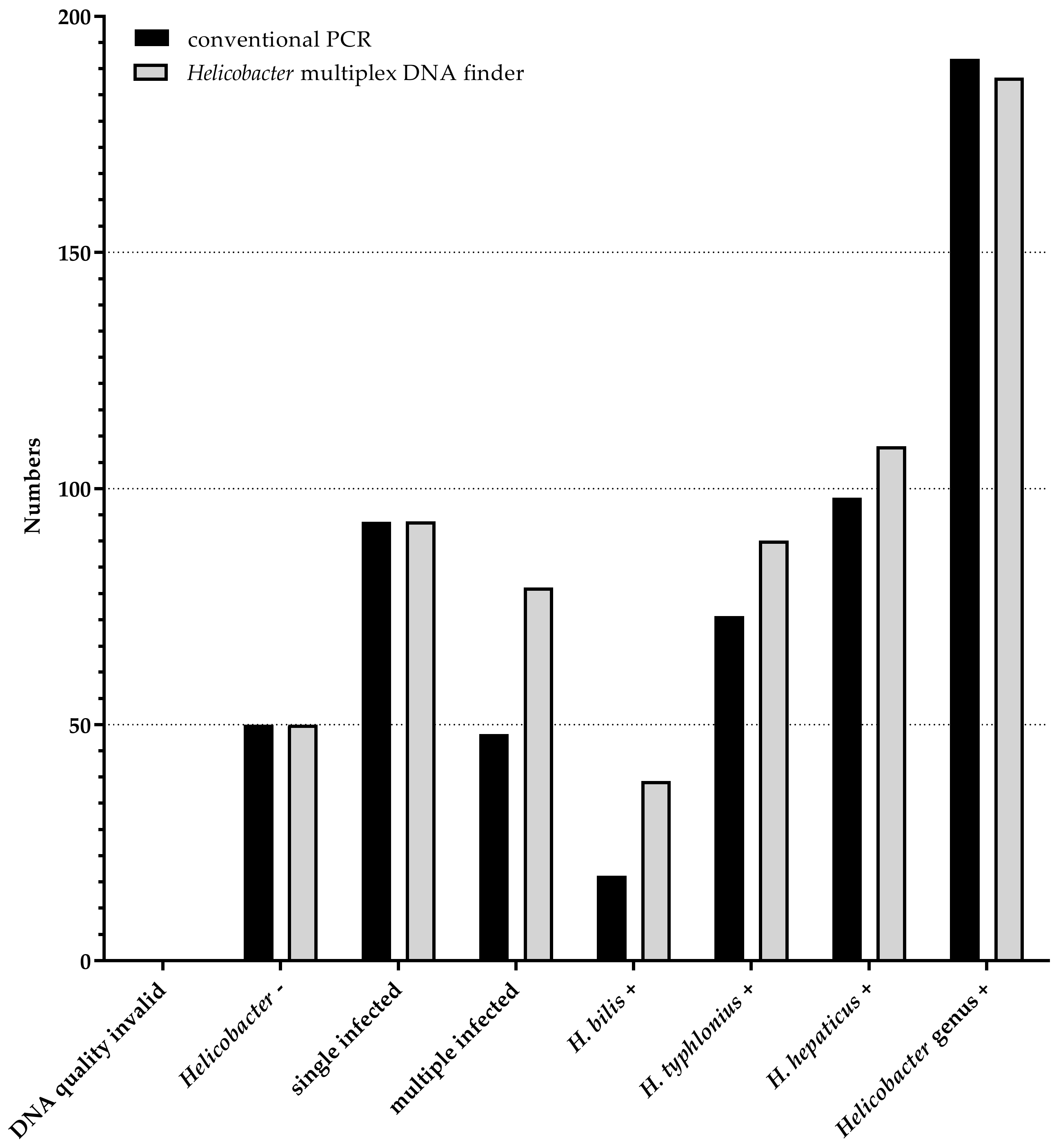
3.3. Comparison of Helicobacter Multiplex DNA Finder to Conventional PCR

For a direct comparison of the conventional PCR and the novel Helicobacter multiplex DNA finder, the detection of H. hepaticus, H. typhlonius, H. bilis and of the Helicobacter genus were analyzed. Since H. typhlonius showed weak cross-reactivities with polA, H. typhlonius was defined as positive where the polA and H. typhlonius netMFI ratio was below 100.
DNA was extracted from mouse samples (n = 241) including liver (n = 16), duodenum (n = 16) and pooled fecal samples (n = 209) of colony and sentinel mice from routine health monitoring at the DKFZ animal facility and applied to both assays. Of all included negative PCR and hybridization controls (n = 208), one was positive for H. typhlonius and H. cinaedi (0.5%) with a low netMFI value.
All 241 mouse samples had a good DNA quality control, meaning either polA (n = 237) and/or any Helicobacter/Campylobacter (n = 191) was detected. The Helicobacter multiplex DNA finder detected Helicobacter spp. in 191 samples (79%) with 93 single infections with either H. hepaticus as the most prevalent type, followed by H. typhlonius and H. bilis (Figure 1). The Helicobacter genus was detected in all but two of the 191 samples, which were species-positive only.
Fifty samples were concordantly Helicobacter spp. and/or genus-negative by the conventional PCR and the Helicobacter multiplex DNA finder and 189 were concordantly positive for any Helicobacter spp. and/or genus, resulting in a kappa of 0.98 (95%CI 0.94–1.00).
Of all 189 concordantly Helicobacter-positive samples, 48 samples were excluded from further analyses since conventional PCR detected Helicobacter genus only without subsequent species-specific analysis.
Among these 141 samples, the overall species concordance was substantial (kappa = 0.614 (95%CI 0.51–0.718)) with 185 concordantly and 41 discordantly H. bilis, H. hepaticus and H. typhlonius identified infections (Figure 1). In total, 66 samples were identified as single infections by both assays. In one sample the conventional PCR detected H. hepaticus, whereas the multiplex DNA finder detected H. typhlonius. This one sample was negative for the genus Helicobacter in the multiplex DNA finder, indicating low DNA quality. Twenty-seven samples were defined as multiple-infected by the Helicobacter multiplex DNA finder, with 11 H. typhlonius, eight H. bilis, and 12 H. hepaticus identified as co-infections that were missed by conventional PCR. Three samples were identified as multiple-infected by conventional PCR only, where the additional H. typhlonius co-infection was missed by the Helicobacter multiplex DNA in all three samples. The high number of multiple-infected samples detected by the Helicobacter multiplex DNA finder can be explained by a higher analytical sensitivity in comparison to conventional PCR.

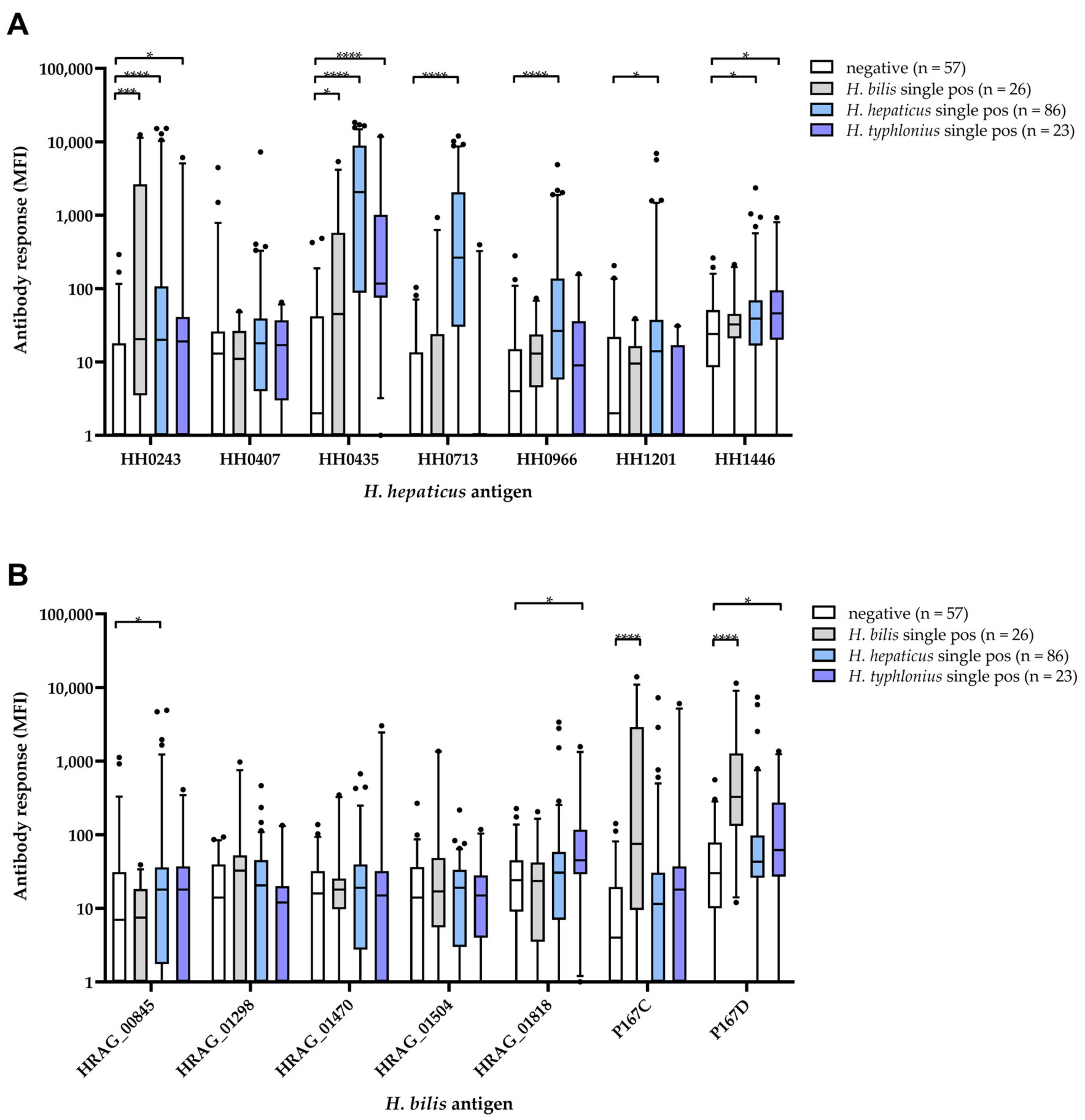
3.4. Comparison of H. hepaticus and H. bilis Multiplex Serology to Helicobacter Multiplex DNA Finder Results

In total, sera of n = 338 mice from the health monitoring of animals housed at DKFZ were analyzed for antibody responses to each of the seven antigens of H. hepaticus and H. bilis, as well as 13 H. pylori proteins. Four serum samples showed invalid serology results and were excluded from further analysis (Supplementary Figure S1). Among the remaining 334 sera, we compared continuous as well as binary sero-responses between Helicobacter-negative mice (n = 57) as well as H. bilis (n = 26), H. hepaticus (n = 86), and H. typhlonius (n = 23) single DNA-positive mice to assess sensitivity and species-specificity of Helicobacter multiplex serology.
Continuous antibody responses (in MFI) to all H. hepaticus proteins except HH0407 were significantly higher in the H. hepaticus single DNA-positive group compared to the Helicobacter genus DNA-negative group. However, for proteins HH0713, HH0966, and HH1201 only, these antibody responses did not show cross-reactive responses in H. bilis or H. typhlonius DNA-positive mice (Figure 2A).
In the case of H. bilis proteins, only antibody responses to P167C and D were significantly higher in the H. bilis DNA-positive mice compared to DNA-negatives. Antibody responses to P167D were also found to be significantly higher in the H. typhlonius DNA-positive group (Figure 2B). Of note, we found elevated antibody responses to H. pylori proteins HP0875, HP0887_2 and HP1098 in H. typhlonius single DNA-positive mice compared to DNA-negative mice (Supplementary Figure S2).
We applied a cut-off for sero-positivity to H. hepaticus and H. bilis antigens that allowed for a maximum of 5% sero-positive mice in the DNA-negative group (Table 5). The highest sero-prevalence among H. hepaticus DNA single-positive mice was given by H. hepaticus proteins HH0435 (71%) and HH0713 (64%). However, 44% of H. typhlonius and 35% of H. bilis single-positive mice were also sero-positive to HH0435 (Table 5). HH0713, in contrast, appeared more specific, with only 4% sero-positives in H. typhlonius and H. bilis DNA-positive mice. The highest sero-prevalence among H. bilis DNA single-positive mice was achieved with H. bilis proteins P167C and P167D (46% and 62%, respectively). Sero-prevalence to these two proteins was low in H. hepaticus (P167C: 11%, P167D: 13%) and H. typhlonius (P167C: 17%, P167D: 22%) single-positive mice (Table 5).

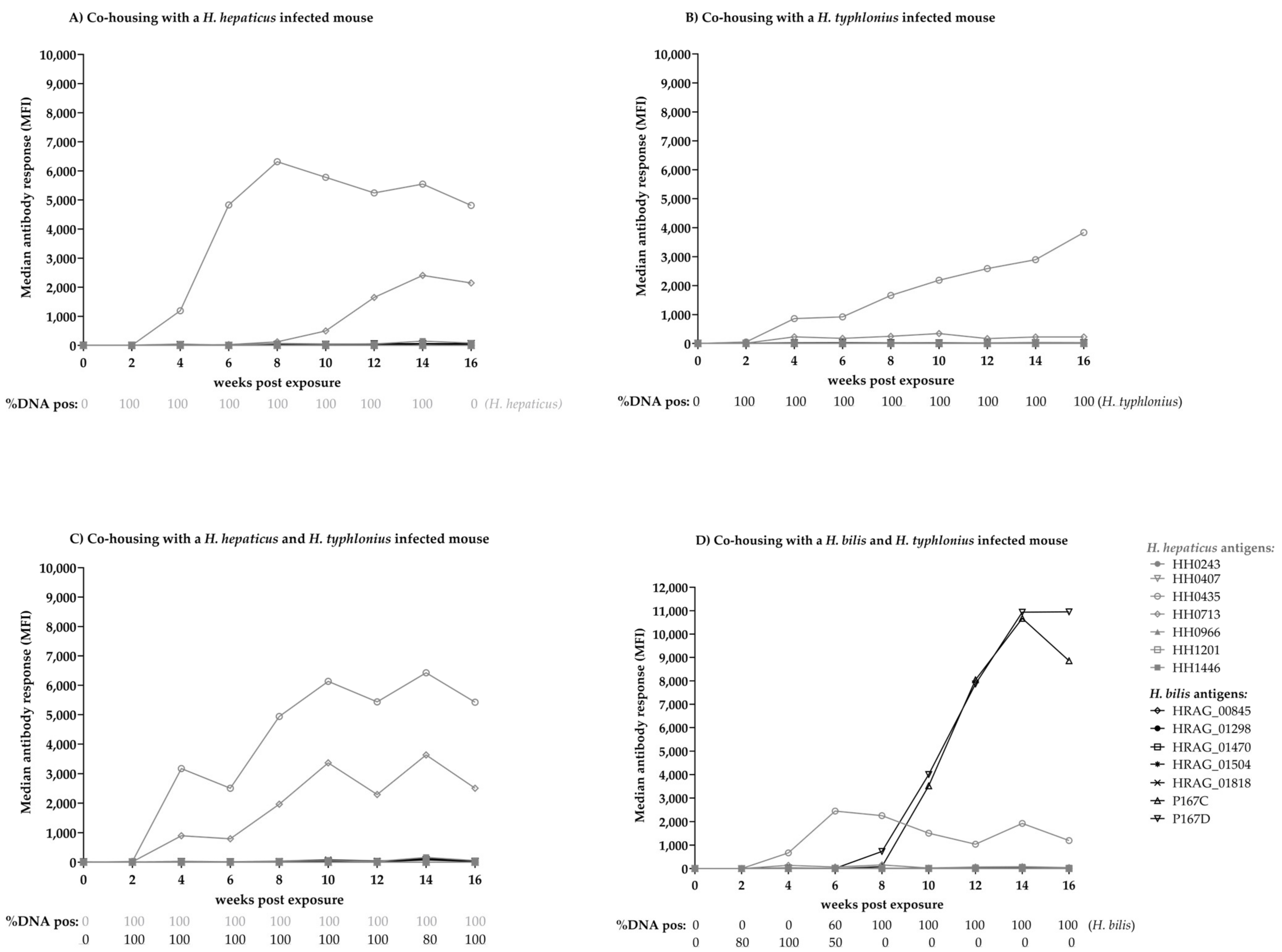
3.5. Experimental Infection of Mice with Helicobacter spp.

Each group of five mice was co-housed with a donor mouse with known Helicobacter infection status (H. hepaticus single-infected, H. typhlonius single-infected, dual H. hepaticus/H. typhlonius infected, and dual H. bilis/H. typhlonius infection). The expected Helicobacter spp. infection status of all donor mice was confirmed by the Helicobacter multiplex DNA finder and remained positive at wpe 0, 2 and 16. In total, 178 of 180 (98.9%) bi-weekly collected fecal samples of exposed mice had a valid DNA quality. H. typhlonius DNA was identified two wpe in all five single-infected mice and in nine out of ten mice with dual infection. H. typhlonius DNA-positivity was detected until 16 wpe in single-infected mice and in mice dual-infected with H. hepaticus. However, in mice dual-infected with H. bilis, H. typhlonius DNA was not detectable after six wpe (Figure 3). H. hepaticus DNA was also detected two wpe and remained positive until 16 wpe in nine out of ten mice, regardless of single or dual infection. Time between exposure and DNA detection was longer for H. bilis, at six and eight wpe for all five mice, and was not cleared until 16 wpe.
Three negative control mice co-housed with a Helicobacter-negative mouse were sampled bi-weekly (n = 27 samples), and had a good DNA quality and were negative for Helicobacter genus but positive for H. hepaticus (n = 3) and H. typhlonius (n = 1).
The experimentally infected mice were followed-up bi-weekly for the development of antibody responses to H. bilis and H. hepaticus proteins with multiplex serology. In the case of H. hepaticus and H. typhlonius single- and dual-infection, the animals developed an antibody response to proteins HH0435 and HH0713 at the earliest four wpe and two weeks after DNA detection in feces. H. hepaticus single-infected mice reached a median plateau of ~8000 and ~6000 MFI to antigens HH0435 and HH0713 eight and 14 wpe, respectively (Figure 3A). The H. typhlonius single-infected mice developed cross-reactive antibody responses to these two proteins, albeit with an overall lower maximum median antibody response (~3500 MFI to HH0435 and ~350 MFI to HH0713) (Figure 3B). H. hepaticus and H. typhlonius dual-infected mice reached a median antibody response of ~6000 MFI and ~3500 MFI to HH0435 and HH0713, respectively (Figure 3C). None of these mice developed antibody responses to H. bilis proteins P167C and D.
Mice in the H. bilis and H. typhlonius dual-infected group developed antibody responses to HH0435 four wpe and two weeks after the first H. typhlonius DNA detection in feces (Figure 3D). At the earliest, six wpe but also two weeks after H. bilis DNA detection in feces, mice also developed antibody responses to H. bilis proteins P167C and P167D with up to 10,000 MFI at wpe 14 (Figure 3D).
A fifth group of mice was exposed to Helicobacter-negative mice as a negative control. None of these mice sero-converted to any H. hepaticus or H. bilis protein during the course of follow-up.

4. Discussion

Experimental and natural infections of mice with rodent Helicobacter spp. such as H. bilis and H. hepaticus are associated with inflammatory and cancerous diseases of the enterohepatic tract [10,12,13,35,36]. Since the infection can alter experimental results, health monitoring of laboratory animals detecting Helicobacter is recommended by the FELASA [15,37].
So far, Helicobacter detection in routine health monitoring is mostly accomplished by PCR covering single or a small groups of Helicobacter spp. and subsequent gel electrophoresis or qPCR, both being time-, labor- and/or cost-intensive [2,17,18,19,38,39,40]. Moreover, these PCR assays most often target 16S rRNA, which is known to have homologies between Helicobacter spp. >94%, hindering a species-specific detection [16,41]. Hence, we developed a fluorescent bead-based species-specific multiplex PCR as well as multiplex serology assay with the potential to detect up to 100 agents per reaction (according to Luminex, Austin, TX, USA). The 96-well format allows fast, simple and highly reproducible analyses of up to 500 samples in less than five days, excluding DNA extraction and data output, and offers objective identification of agents [29,42].
The Helicobacter multiplex DNA finder includes not only the FELASA-recommended species H. hepaticus, H. bilis, and H. typhlonius, but also closely related rodent- and/or human-infecting H. pylori, H. heilmannii, H. cinaedi, H. pullorum, and H. muridarum. We furthermore enclosed one distantly related C. jejuni as a specificity control. Additionally, the detection of the Helicobacter genus is included in the Helicobacter multiplex DNA finder, giving the opportunity to identify any Helicobacter besides the above-mentioned species. Additional species-specific primers and probes targeting other Helicobacter spp. can be integrated into the Helicobacter multiplex DNA finder whenever needed. Moreover, the detection of other agents such as DNA and RNA viruses, bacteria and parasites relevant for health monitoring of laboratory animals can be assimilated [29]. The integration of a DNA quality control into the Helicobacter multiplex DNA finder gives information about the validity of samples and reduces the number of false-negative results. Analytical sensitivity of the Helicobacter DNA multiplex finder ranged from 10 to 1000 copies which is comparable to published qPCR assays and to other multiplex Helicobacter PCR assays, assuming that one bacterial cell contains at least one 16S rRNA copy [38,39,40]. We determined the sensitivity in detecting Helicobacter infections in comparison to published conventional singleplex PCR assays routinely applied for monitoring laboratory animals at DKFZ, and found the novel Helicobacter multiplex DNA finder to be highly comparable (kappa = 0.95 (95%CI 0.901–0.999)), with a more sensitive detection of H. hepaticus, H. bilis, and H. typhlonius, analyzing 241 samples with both assays.
So far, serological assays for the detection of Helicobacter spp. are not routinely applied in the health monitoring of laboratory animals. Previous attempts to detect antibodies to different Helicobacter spp. often applied whole or membrane protein extracts that may result in the detection of cross-reacting antibodies and consequently low specificity. The application of individual proteins might increase specificity and a multiplex approach could thereby allow high sensitivity to be maintained [25,26,43]. Based on our experience from a highly sensitive and specific H. pylori multiplex serology, we attempted here to re-assess the performance of serology in the routine health monitoring of laboratory animals [27]. To do so, we selected a set of potential immunogenic proteins for each H. hepaticus and H. bilis and applied these in a multiplex serology assay. Comparing antibody responses to results from the Helicobacter multiplex DNA finder in murine samples from routine health monitoring showed, however, a low sensitivity (maximum 71% for H. hepaticus and 62% for H. bilis) of the newly developed serological assay. Additionally, we detected a substantial cross-reactivity with H. typhlonius-infected mice, resulting also in low species-specificity.
The low sensitivity of the antibody assay in comparison to DNA detection in fecal samples likely results from the differential ability of the infected mice to build an antibody immune response due to their genetic background and genetic modification and/or the timing of sampling in relation to time-point of infection. Using experimentally infected CD1 outbred mice, we were able to show that Helicobacter antibodies are indeed detectable two weeks after Helicobacter DNA is identified, and that a plateau of maximum antibody response was observable six weeks after DNA detection. However, this infection experiment of immune-competent mice and sampling at defined time-points after infection does not reflect real-life scenarios in routine health monitoring.
In summary, the serological assay developed here is inferior to the Helicobacter multiplex DNA finder when it comes to sensitivity and species-specificity. However, due to low-costs and high-throughput application in combination with screening for other serologically assessed infectious agents [44] the multiplex Helicobacter serology could give the opportunity to monitor more frequently than the FELASA-recommended quarterly diagnostics. A possible screening scenario could also be to run the Helicobacter multiplex DNA finder as triage test for serologically positive mice [45].
An additional application of interest, for both the Helicobacter multiplex DNA finder as well as multiplex serology, could be in human samples. In humans, H. pylori was classified as a carcinogen for the development of gastric cancer in 1994, and since then multiple biomarker studies have been conducted to identify individuals at risk for developing cancer [46]. A causal involvement of Helicobacter spp. other than H. pylori in human disease is less well described. H. hepaticus and H. bilis have also been isolated from the human enterohepatic tract, but their prevalence and specific disease associations in humans remain unclear [47,48,49,50,51,52,53]. The Helicobacter multiplex serology described here has already been applied in a study assessing the association of different Helicobacter spp. with non-alcoholic fatty liver disease, a precursor of liver cancer, and indeed sero-positivity to H. hepaticus protein HH0713 was associated with disease [54]. Both the established novel Helicobacter multiplex DNA finder and the Helicobacter multiplex serology can in future be applied to large epidemiological studies with human samples to obtain more knowledge about the prevalence of H. hepaticus and H. bilis and their potential association with cancer. However, DNA extraction methods might be different in human samples and could require the establishment of new protocols.

5. Conclusions

We have developed two multiplex assays identifying infection with different Helicobacter spp. by the detection of DNA in the Helicobacter multiplex DNA finder and antibodies in the Helicobacter multiplex serology. While the Helicobacter multiplex serology lacks species-specificity and sensitivity, the Helicobacter multiplex DNA finder can be readily implemented in the routine health monitoring of laboratory animals. Due to the high-throughput applicability, however, both assays are promising tools to be used in large epidemiological studies investigating Helicobacter infection and disease correlation in humans.

Supplementary Materials

The following supporting information can be downloaded at: https://www.mdpi.com/article/10.3390/microorganisms11020249/s1, Figure S1: Murine samples to assess sensitivity and specificity of the newly developed Helicobacter multiplex DNA finder and Helicobacter multiplex serology; Figure S2: Antibody responses [MFI] to H. pylori antigens by Helicobacter multiplex DNA finder result in murine samples taken at routine diagnostics; Table S1: H. hepaticus and H. bilis proteins included in multiplex serology and their amino acid identities with proteins of related Helicobacter species

Author Contributions

Conceptualization, J.B., K.S. and D.H.; Methodology/resources/software/investigation/validation, J.B., M.S., B.B., K.S. and D.H.; data curation: J.B., K.S. and D.H.; visualization: J.B. and D.H.; writing—original draft preparation, J.B. and D.H.; writing—review and editing, K.S., M.S. and B.B.; All authors have read and agreed to the published version of the manuscript.

Funding

This research received no external funding.

Data Availability Statement

The data presented in this study are available on request from the corresponding author.

Acknowledgments

We thank M. Pawlita for discussion and André Leischwitz and Indra Gierse for experimental help.

Conflicts of Interest

The authors declare no conflict of interest.

References

  1. Mannion, A.; Shen, Z.; Fox, J.G. Comparative genomics analysis to differentiate metabolic and virulence gene potential in gastric versus enterohepatic Helicobacter species. BMC Genom. 2018, 19, 830. [Google Scholar] [CrossRef] [PubMed]
  2. Shames, B.; Fox, J.G.; Dewhirst, F.; Yan, L.; Shen, Z.; Taylor, N.S. Identification of widespread Helicobacter hepaticus infection in feces in commercial mouse colonies by culture and PCR assay. J. Clin. Microbiol. 1995, 33, 2968–2972. [Google Scholar] [CrossRef] [PubMed] [Green Version]
  3. Fox, J.G.; Dewhirst, F.E.; Tully, J.G.; Paster, B.J.; Yan, L.; Taylor, N.S.; Collins, M.J.; Gorelick, P.L.; Ward, J.M. Helicobacter hepaticus Sp-Nov, a Microaerophilic Bacterium Isolated from Livers and Intestinal Mucosal Scrapings from Mice. J. Clin. Microbiol. 1994, 32, 1238–1245. [Google Scholar] [CrossRef] [PubMed] [Green Version]
  4. Liu, Z.; Ramer-Tait, A.E.; Henderson, A.L.; Demirkale, C.Y.; Nettleton, D.; Wang, C.; Hostetter, J.M.; Jergens, A.E.; Wannemuehler, M.J. Helicobacter bilis colonization enhances susceptibility to Typhlocolitis following an inflammatory trigger. Dig. Dis. Sci. 2011, 56, 2838–2848. [Google Scholar] [CrossRef]
  5. Ge, Z.; Sterzenbach, T.; Whary, M.T.; Rickman, B.H.; Rogers, A.B.; Shen, Z.; Taylor, N.S.; Schauer, D.B.; Josenhans, C.; Suerbaum, S.; et al. Helicobacter hepaticus HHGI1 is a pathogenicity island associated with typhlocolitis in B6.129-IL10 tm1Cgn mice. Microbes Infect. Inst. Pasteur 2008, 10, 726–733. [Google Scholar] [CrossRef] [Green Version]
  6. Ge, Z.; Lee, A.; Whary, M.T.; Rogers, A.B.; Maurer, K.J.; Taylor, N.S.; Schauer, D.B.; Fox, J.G. Helicobacter hepaticus urease is not required for intestinal colonization but promotes hepatic inflammation in male A/JCr mice. Microb. Pathog. 2008, 45, 18–24. [Google Scholar] [CrossRef] [Green Version]
  7. Whary, M.T.; Fox, J.G. Natural and experimental Helicobacter infections. Comp. Med. 2004, 54, 128–158. [Google Scholar]
  8. Fox, J.G.; Rogers, A.B.; Whary, M.T.; Taylor, N.S.; Xu, S.; Feng, Y.; Keys, S. Helicobacter bilis-associated hepatitis in outbred mice. Comp. Med. 2004, 54, 571–577. [Google Scholar]
  9. Li, X.; Fox, J.G.; Whary, M.T.; Yan, L.; Shames, B.; Zhao, Z. SCID/NCr mice naturally infected with Helicobacter hepaticus develop progressive hepatitis, proliferative typhlitis, and colitis. Infect. Immun. 1998, 66, 5477–5484. [Google Scholar] [CrossRef] [Green Version]
  10. Franklin, C.L.; Riley, L.K.; Livingston, R.S.; Beckwith, C.S.; Besch-Williford, C.L.; Hook, R.R., Jr. Enterohepatic lesions in SCID mice infected with Helicobacter bilis. Lab. Anim. Sci. 1998, 48, 334–339. [Google Scholar]
  11. Ward, J.M.; Anver, M.R.; Haines, D.C.; Melhorn, J.M.; Gorelick, P.; Yan, L.; Fox, J.G. Inflammatory large bowel disease in immunodeficient mice naturally infected with Helicobacter hepaticus. Lab. Anim. Sci. 1996, 46, 15–20. [Google Scholar]
  12. Fox, J.G.; Yan, L.; Shames, B.; Campbell, J.; Murphy, J.C.; Li, X. Persistent hepatitis and enterocolitis in germfree mice infected with Helicobacter hepaticus. Infect. Immun. 1996, 64, 3673–3681. [Google Scholar] [CrossRef] [Green Version]
  13. Fox, J.G.; Li, X.; Yan, L.; Cahill, R.J.; Hurley, R.; Lewis, R.; Murphy, J.C. Chronic proliferative hepatitis in A/JCr mice associated with persistent Helicobacter hepaticus infection: A model of helicobacter-induced carcinogenesis. Infect. Immun. 1996, 64, 1548–1558. [Google Scholar] [CrossRef] [Green Version]
  14. Ward, J.M.; Fox, J.G.; Anver, M.R.; Haines, D.C.; George, C.V.; Collins, M.J., Jr.; Gorelick, P.L.; Nagashima, K.; Gonda, M.A.; Gilden, R.V.; et al. Chronic active hepatitis and associated liver tumors in mice caused by a persistent bacterial infection with a novel Helicobacter species. J. Natl. Cancer Inst. 1994, 86, 1222–1227. [Google Scholar] [CrossRef]
  15. FELASAWorking Group on Revision of Guidelines for Health Monitoring of Rodents Rabbits; Mähler, M.; Berard, M.; Feinstein, R.; Gallagher, A.; Illgen-Wilcke, B.; Pritchett-Corning, K.; Raspa, M. FELASA recommendations for the health monitoring of mouse, rat, hamster, guinea pig and rabbit colonies in breeding and experimental units. Lab. Anim. 2014, 48, 178–192. [Google Scholar]
  16. Poynter, S.; Phipps, J.D.; Naranjo-Pino, A.; Sanchez-Morgado, J.M. Difficulties in the molecular diagnosis of Helicobacter rodent infections. Vet. Microbiol. 2009, 134, 272–278. [Google Scholar] [CrossRef] [Green Version]
  17. Riley, L.K.; Franklin, C.L.; Hook, R.R., Jr.; Besch-Williford, C. Identification of murine helicobacters by PCR and restriction enzyme analyses. J. Clin. Microbiol. 1996, 34, 942–946. [Google Scholar] [CrossRef] [Green Version]
  18. Fox, J.G.; Yan, L.L.; Dewhirst, F.E.; Paster, B.J.; Shames, B.; Murphy, J.C.; Hayward, A.; Belcher, J.C.; Mendes, E.N. Helicobacter bilis sp. nov., a novel Helicobacter species isolated from bile, livers, and intestines of aged, inbred mice. J. Clin. Microbiol. 1995, 33, 445–454. [Google Scholar] [CrossRef] [Green Version]
  19. Franklin, C.L.; Riley, L.K.; Livingston, R.S.; Beckwith, C.S.; Hook, R.R., Jr.; Besch-Williford, C.L.; Hunziker, R.; Gorelick, P.L. Enteric lesions in SCID mice infected with “Helicobacter typhlonicus”, a novel urease-negative Helicobacter species. Lab. Anim. Sci. 1999, 49, 496–505. [Google Scholar]
  20. Boutin, S.R.; Shen, Z.; Roesch, P.L.; Stiefel, S.M.; Sanderson, A.E.; Multari, H.M.; Pridhoko, E.A.; Smith, J.C.; Taylor, N.S.; Lohmiller, J.J.; et al. Helicobacter pullorum outbreak in C57BL/6NTac and C3H/HeNTac barrier-maintained mice. J. Clin. Microbiol. 2010, 48, 1908–1910. [Google Scholar] [CrossRef] [Green Version]
  21. Pisani, P.; Whary, M.T.; Nilsson, I.; Sriamporn, S.; Wadstrom, T.; Fox, J.G.; Ljungh, A.; Forman, D. Cross-reactivity between immune responses to Helicobacter bilis and Helicobacter pylori in a population in Thailand at high risk of developing cholangiocarcinoma. Clin. Vaccine Immunol. 2008, 15, 1363–1368. [Google Scholar] [CrossRef] [PubMed] [Green Version]
  22. Ananieva, O.; Nilsson, I.; Vorobjova, T.; Uibo, R.; Wadstrom, T. Immune Responses to Bile-Tolerant Helicobacter Species in Patients with Chronic Liver Diseases, a Randomized Population Group, and Healthy Blood Donors. Clin. Vaccine Immunol. 2002, 9, 1160–1164. [Google Scholar] [CrossRef] [PubMed] [Green Version]
  23. Nilsson, I.; Lindgren, S.; Eriksson, S.; Wadstrom, T. Serum antibodies to Helicobacter hepaticus and Helicobacter pylori in patients with chronic liver disease. Gut 2000, 46, 410–414. [Google Scholar] [CrossRef] [PubMed] [Green Version]
  24. Feng, S.; Kendall, L.V.; Hodzic, E.; Wong, S.; Lorenzana, E.; Freet, K.; Ku, K.S.; Luciw, P.A.; Barthold, S.W.; Khan, I.H. Recombinant Helicobacter bilis protein P167 for mouse serodiagnosis in a multiplex microbead assay. Clin. Diagn. Lab. Immunol. 2004, 11, 1094–1099. [Google Scholar] [CrossRef] [Green Version]
  25. Feng, S.; Hodzic, E.; Kendall, L.V.; Smith, A.; Freet, K.; Barthold, S.W. Cloning and expression of a Helicobacter bilis immunoreactive protein. Clin. Diagn. Lab. Immunol. 2002, 9, 627–632. [Google Scholar] [CrossRef] [Green Version]
  26. Livingston, R.S.; Riley, L.K.; Hook, R.R., Jr.; Besch-Williford, C.L.; Franklin, C.L. Cloning and expression of an immunogenic membrane-associated protein of Helicobacter hepaticus for use in an enzyme-linked immunosorbent assay. Clin. Diagn. Lab. Immunol. 1999, 6, 745–750. [Google Scholar] [CrossRef] [Green Version]
  27. Michel, A.; Waterboer, T.; Kist, M.; Pawlita, M. Helicobacter pylori multiplex serology. Helicobacter 2009, 14, 525–535. [Google Scholar] [CrossRef]
  28. Available online: https://eur-lex.europa.eu/LexUriServ/LexUriServ.do?uri=OJ:L:2010:276:0033:0079:de:PDF (accessed on 1 December 2022).
  29. Hofler, D.; Nicklas, W.; Mauter, P.; Pawlita, M.; Schmitt, M. A bead-based multiplex assay for the detection of DNA viruses infecting laboratory rodents. PLoS ONE 2014, 9, e97525. [Google Scholar] [CrossRef]
  30. Schmitt, M.; Bravo, I.G.; Snijders, P.J.; Gissmann, L.; Pawlita, M.; Waterboer, T. Bead-based multiplex genotyping of human papillomaviruses. J. Clin. Microbiol. 2006, 44, 504–512. [Google Scholar] [CrossRef] [Green Version]
  31. Chien, C.C.; Taylor, N.S.; Ge, Z.; Schauer, D.B.; Young, V.B.; Fox, J.G. Identification of cdtB homologues and cytolethal distending toxin activity in enterohepatic Helicobacter spp. J. Med. Microbiol. 2000, 49, 525–534. [Google Scholar] [CrossRef] [Green Version]
  32. Suerbaum, S.; Josenhans, C.; Sterzenbach, T.; Drescher, B.; Brandt, P.; Bell, M.; Droge, M.; Fartmann, B.; Fischer, H.P.; Ge, Z.; et al. The complete genome sequence of the carcinogenic bacterium Helicobacter hepaticus. Proc. Natl. Acad. Sci. USA 2003, 100, 7901–7906. [Google Scholar] [CrossRef] [Green Version]
  33. Available online: https://blast.ncbi.nlm.nih.gov/Blast.cgi (accessed on 3 December 2020).
  34. Waterboer, T.; Sehr, P.; Michael, K.M.; Franceschi, S.; Nieland, J.D.; Joos, T.O.; Templin, M.F.; Pawlita, M. Multiplex human papillomavirus serology based on in situ-purified glutathione s-transferase fusion proteins. Clin. Chem. 2005, 51, 1845–1853. [Google Scholar] [CrossRef] [Green Version]
  35. Fox, J.G.; Gorelick, P.L.; Kullberg, M.C.; Ge, Z.; Dewhirst, F.E.; Ward, J.M. A novel urease-negative Helicobacter species associated with colitis and typhlitis in IL-10-deficient mice. Infect. Immun. 1999, 67, 1757–1762. [Google Scholar] [CrossRef]
  36. Donovan, J.C.; Mayo, J.G.; Rice, J.M.; Ward, J.M.; Fox, J.G. Helicobacter-associated hepatitis of mice. Lab. Anim. Sci. 1993, 43, 403. [Google Scholar]
  37. Whary, M.T.; Fox, J.G. Detection, eradication, and research implications of Helicobacter infections in laboratory rodents. Lab. Anim. 2006, 35, 25–36. [Google Scholar] [CrossRef]
  38. Neubert, V.; Sadek, A.; Burell, T.; Ralser, A.; Erhard, M.; Gerhard, M.; Seidel, K.; Kalali, B. Validation and improvement of a multiplex PCR method to detect murine Helicobacter species in feces samples of mice. Helicobacter 2022, 27, e12888. [Google Scholar] [CrossRef]
  39. Ge, Z.; White, D.A.; Whary, M.T.; Fox, J.G. Fluorogenic PCR-based quantitative detection of a murine pathogen, Helicobacter hepaticus. J. Clin. Microbiol. 2001, 39, 2598–2602. [Google Scholar] [CrossRef] [Green Version]
  40. Drazenovich, N.L.; Franklin, C.L.; Livingston, R.S.; Besselsen, D.G. Detection of rodent Helicobacter spp. by use of fluorogenic nuclease polymerase chain reaction assays. Comp. Med. 2002, 52, 347–353. [Google Scholar]
  41. Dewhirst, F.E.; Shen, Z.; Scimeca, M.S.; Stokes, L.N.; Boumenna, T.; Chen, T.; Paster, B.J.; Fox, J.G. Discordant 16S and 23S rRNA gene phylogenies for the genus Helicobacter: Implications for phylogenetic inference and systematics. J. Bacteriol. 2005, 187, 6106–6118. [Google Scholar] [CrossRef] [Green Version]
  42. Schmitt, M.; Pawlita, M. High-throughput detection and multiplex identification of cell contaminations. Nucleic Acids Res. 2009, 37, e119. [Google Scholar] [CrossRef]
  43. Kendall, L.V.; Feng, S.; Hodzic, E.; Freet, K.; Barthold, S.W. Use of the P167 recombinant antigen for serodiagnosis of Helicobacter bilis. Comp. Med. 2004, 54, 44–48. [Google Scholar] [PubMed]
  44. Schmidt, K.; Butt, J.; Mauter, P.; Vogel, K.; Erles-Kemna, A.; Pawlita, M.; Nicklas, W. Development of a multiplex serological assay reveals a worldwide distribution of murine astrovirus infections in laboratory mice. PLoS ONE 2017, 12, e0187174. [Google Scholar] [CrossRef] [PubMed] [Green Version]
  45. Whary, M.T.; Cline, J.H.; King, A.E.; Hewes, K.M.; Chojnacky, D.; Salvarrey, A.; Fox, J.G. Monitoring sentinel mice for Helicobacter hepaticus, H. rodentium, and H. bilis infection by use of polymerase chain reaction analysis and serologic testing. Comp. Med. 2000, 50, 436–443. [Google Scholar] [PubMed]
  46. IARC; World Health Organization. Schistosomes, Liver Flukes and Helicobacter pylori. In Monographs on the Evaluation of Carcinogenic Risks to Humans; Iarc Working Group on the Evaluation of Carcinogenic Risks to Humans; International Agency for Research on Cancer: Lyon, France, 1994; Volume 61, pp. 1–241. [Google Scholar]
  47. Kosaka, T.; Tajima, Y.; Kuroki, T.; Mishima, T.; Adachi, T.; Tsuneoka, N.; Fukuda, K.; Kanematsu, T. Helicobacter bilis colonization of the biliary system in patients with pancreaticobiliary maljunction. Br. J. Surg. 2010, 97, 544–549. [Google Scholar] [CrossRef] [PubMed]
  48. Hamada, T.; Yokota, K.; Ayada, K.; Hirai, K.; Kamada, T.; Haruma, K.; Chayama, K.; Oguma, K. Detection of Helicobacter hepaticus in human bile samples of patients with biliary disease. Helicobacter 2009, 14, 545–551. [Google Scholar] [CrossRef] [Green Version]
  49. Rocha, M.; Avenaud, P.; Menard, A.; Le Bail, B.; Balabaud, C.; Bioulac-Sage, P.; de Magalhaes Queiroz, D.M.; Megraud, F. Association of Helicobacter species with hepatitis C cirrhosis with or without hepatocellular carcinoma. Gut 2005, 54, 396–401. [Google Scholar] [CrossRef] [Green Version]
  50. Pellicano, R.; Mazzaferro, V.; Grigioni, W.F.; Cutufia, M.A.; Fagoonee, S.; Silengo, L.; Rizzetto, M.; Ponzetto, A. Helicobacter species sequences in liver samples from patients with and without hepatocellular carcinoma. World J. Gastroenterol. 2004, 10, 598–601. [Google Scholar] [CrossRef]
  51. Fox, J.G.; Dewhirst, F.E.; Shen, Z.; Feng, Y.; Taylor, N.S.; Paster, B.J.; Ericson, R.L.; Lau, C.N.; Correa, P.; Araya, J.C.; et al. Hepatic Helicobacter species identified in bile and gallbladder tissue from Chileans with chronic cholecystitis. Gastroenterology 1998, 114, 755–763. [Google Scholar] [CrossRef]
  52. Segura-Lopez, F.K.; Aviles-Jimenez, F.; Guitron-Cantu, A.; Valdez-Salazar, H.A.; Leon-Carballo, S.; Guerrero-Perez, L.; Fox, J.G.; Torres, J. Infection with Helicobacter bilis but not Helicobacter hepaticus was Associated with Extrahepatic Cholangiocarcinoma. Helicobacter 2015, 20, 223–230. [Google Scholar] [CrossRef]
  53. Zhou, D.; Wang, J.D.; Weng, M.Z.; Zhang, Y.; Wang, X.F.; Gong, W.; Quan, Z.W. Infections of Helicobacter spp. in the biliary system are associated with biliary tract cancer: A meta-analysis. Eur. J. Gastroenterol. Hepatol. 2013, 25, 447–454. [Google Scholar] [CrossRef]
  54. Alvarez, C.S.; Florio, A.A.; Butt, J.; Rivera-Andrade, A.; Kroker-Lobos, M.F.; Waterboer, T.; Camargo, M.C.; Freedman, N.D.; Graubard, B.I.; Lazo, M.; et al. Associations between Helicobacter pylori with nonalcoholic fatty liver disease and other metabolic conditions in Guatemala. Helicobacter 2020, 25, e12756. [Google Scholar] [CrossRef]
Figure 1. Identification of Helicobacter by Helicobacter multiplex DNA finder (grey bars) and conventional PCR (black bars). Numbers of samples defined as Helicobacter-negative, single- (i.e., only one Helicobacter spp. identified in one sample) and multiple-infected. H. bilis, H. typhlonius, H. hepaticus, Helicobacter genus positive are shown on the y-axis. The chi-squared test of independence indicated strong correlation between conventional PCR and the Helicobacter multiplex DNA finder (p < 0.0001) for any parameter compared (Helicobacter-, single-infected, multiple-infected, H. bilis, H. typhlonius, H. hepaticus and Helicobacter genus).
Microorganisms 11 00249 g001
Figure 2. Antibody responses [MFI] to (A) H. hepaticus and (B) H. bilis antigens by Helicobacter multiplex DNA finder result in murine samples taken in the course of routine health monitoring. Boxes represent the 25th to 75th and whiskers the 5th to 95th percentile, solid lines show the median. Dots represent data points lying outside the 5th and 95th percentiles, respectively. Wilcoxon–Mann–Whitney test was applied to compare continuous antibody responses [MFI] in the individual DNA-positive groups to the DNA-negative group: * p-value < 0.05, *** p-value < 0.001, **** p-value < 0.0001.
Microorganisms 11 00249 g002
Figure 3. Median antibody response [MFI] to H. hepaticus and H. bilis antigens and % DNA-positivity in feces after co-housing of each group of five mice with (A) a H. hepaticus-infected mouse, (B) a H. typhlonius-infected mouse, (C) a H. hepaticus/H. typhlonius dual-infected mouse and (D) a H. bilis/H. typhlonius dual-infected mouse over a course of 16 weeks follow-up. Each group of five mice was co-housed with Helicobacter-infected donor mice as indicated and tested bi-weekly for DNA-shedding of Helicobacter in feces by Helicobacter multiplex DNA finder as well as for development of an antibody response to H. hepaticus and H. bilis antigens by multiplex serology. The antibody response is given as the median among the five mice per group at each time point assessed. DNA-positivity in % is given below the plot for each week post exposure.
Microorganisms 11 00249 g003
Table 1. Helicobacter genus- and species-specific primers in conventional PCR and in the Helicobacter multiplex DNA finder.
PCR TargetGene Target Primer/Probe NameSequence (5′-3′)Fragment SizeReference
Helicobacter genus16S rRNAh276 forwardCTATGACGGGTATCCGGC400 bpRiley et al. [17]
h676 reverseATTCCACCTACCTCTCCCA
H. hepaticus16S rRNAB38 forwardGCATTTGAAACTGTTACTCTG417 bpShames et al. [2]
B39 reverseCTGTTTTCAAGCTCCCC
H. typhlonius16S rRNAHt 184 forwardTTAAAGATATTCTAGGGGTATAT474 bpFranklin et al. [19]
Ht 640 reverseTCTCCCATACTCTAGAGTGA
H. bilis16S rRNAC62 forwardAGAACTGCATTTGAAACTACTTT638 bpFox et al. [18]
C12 reverseGGTATTGCATCTCTTTGTATGT
Table 2. Helicobacter genus- and species-specific primers in the Helicobacter multiplex DNA finder.
PCR TargetGene Target Primer/Probe NameSequence (5′-3′)Fragment Size
H. muridarumgyAafw2_muridCCAAATGCCAGAGATGGAT122 bp
bw1_muridCCGATTACATCACCAACTAT
p_muridTATGAATGAGCTAAACCTTACACA
ggtbfw2_murGCCACTAGAGATATGTATCTTG112 bp
bw2_murGCATAGCACTCATTCCTT
p1_murAGATGTCCAATAATAGAA
H. pylorigyAfw1_pyloriGATCGCYGTRGGGATGGC144 bp
bw1_pyloriAGTGGGAAARTCAGGCCCTT
p_pyloriCTTTAGYGCATGTCTTAGAA
ggtfw1_pyloriTTAGACAAGCAAGGCAATGT94 bp
bw2_pyloriACATCGCTTCCATGCCCGC
p1_pyloriAAATAGCCATCTTCRCTG
H. bilisgyAfw1_bilisTGCCTAATGCAAGAGATGG126 bp
bw1_bilisCCATTACTTCCCCCACAAT
p_bilisCGATGAAGCATAATCTAGGG
ggtfw2_bilisGGATAATAAAGGTAATGTGATTCC138 bp
bw2_bilisGCAGGTTGCATGAGTTC
p3_bilisGCTAAATATCCAAGTGTTGAAGCA
H. heilmanniigyAFw2_HheilmCTTGCAAATAGGCGATCT129 bp
bw1_HheilmCGCATGATCTAAGTGAAG
p2_HheilmTTCTCCTGCTCTAGCCC
H. typhloniusgyAfw2_typhloniusATTGTAGGTAGGGCGTTA149 bp
bw2_typhloniusTGGTATTTACCAATCACATC
p_typhloniusGATGAACGAGCTAAGCCTTTCACC
H. cinaedigyAfw1_cinaediTACCAGATGCTAAAGATGG122 bp
bw1_cinaediAATCACATCGCCAACAAT
p_cinaediAATGAATGAGCTAAGCCTCTCT
H. hepaticusgyAfw1_hepaticusCCTGACGCAAAAGATGG122 bp
bw1_hepaticusATTTACCAATTACATCGCCTAC
p_hepaticusAATGAATGAGCTTAATCTCTCACC
H. pullorumgyAfw1_HpullAATGGAATAAGAGAGGCTTA128 bp
bw_HpullGCTTTATTGACCTGATAGGGA
p1_HpullTTAATGCGCCCTCTCCCTG
C. jejunigyAfw2_CjejuniATGAAACTTGGTCGTTTAACA178 bp
bw2_CjejuniGAGTAATACGTGGCACA
p_CjejuniCTTGCTTGAAAATTTAATTCG
ggtfw2_CjejuniTGTATCTTGATAGCAAAGGAGAA105 bp
bw1_ CjejuniGATCAAGCATAGCACTCATACC
p1_CjejuCAGCTAGATAACCTATAGT
Helicobacter genus16S rRNAfw2.3_heli_uniGAGTATGGGAGAGGTAGGTGGAATTC110 bp
bw2.2_heli_uniTAATCCTGTTTGCTCCCCACGC
p2.2_heli_uniCAATGAGTATTCCTCTTGA
a gyrase A, b gamma-glutamyltransferase.
Table 3. Analytical sensitivity of Helicobacter multiplex DNA finder.
SpeciesGeneAnalytical Sensitivity [# of Copies/PCR] a
H. muridarumgyA10
ggt100
H. pylorigA10
ggt10
H. bilisgyA100
ggt10
H. heilmanniigyA1000
H. typhloniusgyA1000
H. cinaedigyA10
H. hepaticusgyA10
H. pullorumgyA10
C. jejunigyA10,000
ggt10
Helicobacter genus16SrRNA10
Mus musculuspolA10
Homo sapienspolA10
a determined in duplicates.
Table 4. Analytical specificity of Helicobacter multiplex DNA finder.
Bacteria-Specific ProbeQC Probe
H. muridarumH. muridarumH. pyloriH. pyloriH. bilisH. bilisH. heilmanniiH. typhloniusH. cinaediH. hepaticusH. pullorumC. jejuniC. jejuniHeli
PCR-Template GenegAggtgyAggtgAggtgyAgAgyAgyAgyAgyAggt16SrRNApolA
H. muridarumgyA340a1 b1111111111111
ggt11241111111111111
H. pylorigyA1130111111111111
ggt11113611114 c111111
H. bilisgA11112591111111111
ggt11111644111111111
H. heilmanniigyA1111118711111111
H. typhloniusgA11111111231111111
H. cinaedigyA11111115226111111
H. hepaticusgyA11111111163911111
H. pullorumgyA11111111115121111
C. jejunigyA111111111117111
ggt1111111111219211
Helicobacter genus 16SrRNA11111111111114421
M. musculuspolA11111111211621111152
H. caniswhole genome11111111111116101
C. lariwhole genome111111111116111862
C. coliwhole genome111111111111111
murine faeceswhole genome11111111011501111095
a Signals above cut-off (value given in bold), b netMFI values of PCR products hybrized to a mixture of 15 distinct bead sets, background values were subtracted and negative values set to 1 MFI, c cross-reactivity of PCR product to non-specific probes, e.g., H. cinaedi PCR template with H. pylori probe (underlined value).
Table 5. Sero-positivity to H. hepaticus and H. bilis antigens by Helicobacter multiplex DNA finder result.
N (%) by DNA Result
AntigenCut-Off
[MFI] a
Negative
(n = 57)
H. bilis
(n = 26)
H. hepaticus
(n = 86)
H. typhlonius
(n = 23)
H. hepaticus
  HH02431282 (4)8 (31)20 (23)3 (13)
  HH04079562 (4)0 (0)1 (1)0 (0)
  HH04352472 (4)9 (35)61 (71)10 (44)
  HH0713731 (2)1 (4)55 (64)1 (4)
  HH09661152 (4)0 (0)25 (29)2 (9)
  HH12011362 (4)0 (0)11 (13)0 (0)
  HH14461682 (4)1 (4)11 (13)4 (17)
H. bilis
  HRAG_008454752 (4)0 (0)4 (5)0 (0)
  HRAG_01298842 (4)4 (15)6 (7)2 (9)
  HRAG_01470952 (4)2 (8)8 (9)2 (9)
  HRAG_01504892 (4)4 (15)1 (1)1 (4)
  HRAG_018181462 (4)2 (8)6 (7)5 (22)
  P167C892 (4)12 (46)11 (13)4 (17)
  P167D2882 (4)16 (62)9 (11)5 (22)
a cut-off applied to allow for a maximum of 5% sero-positive mice in the DNA-negative group.
Disclaimer/Publisher’s Note: The statements, opinions and data contained in all publications are solely those of the individual author(s) and contributor(s) and not of MDPI and/or the editor(s). MDPI and/or the editor(s) disclaim responsibility for any injury to people or property resulting from any ideas, methods, instructions or products referred to in the content.

