Article

Co-Infection of Potential Tick-Borne Pathogens of the Order Rickettsiales and Borrelia burgdorferi s. l. and Their Link to Season and Area in Germany

1
Institute for Bioanalysis, Department of Applied Sciences, Coburg University of Applied Sciences and Arts, 96450 Coburg, Germany
2
Synlab Medical Care Unit, Department of Molecular biology, Tick Laboratory, 92637 Weiden in der Oberpfalz, Germany
3
Bavarian Health and Food Safety Authority (LGL), National Reference Center for Borrelia, 85764 Oberschleißheim, Germany
4
Institute of Infectology, Friedrich-Loeffler-Institute, Federal Research Institute of Animal Health, 17493 Greifswald, Germany
5
Bayreuth Center of Ecology and Environmental Research (BayCEER), University of Bayreuth, 95440 Bayreuth, Germany
*
Author to whom correspondence should be addressed.
Microorganisms 2023, 11(1), 157; https://doi.org/10.3390/microorganisms11010157
Submission received: 17 November 2022 / Revised: 2 January 2023 / Accepted: 3 January 2023 / Published: 7 January 2023
(This article belongs to the Special Issue Advances in Tick-Borne Diseases Research)

Abstract

:
The prevalence of potential human pathogenic members of the order Rickettsiales differs between Borrelia burgdorferi sensu lato-positive and -negative tick microbiomes. Here, co-infection of members of the order Rickettsiales, such as Rickettsia spp., Anaplasma phagocytophilum, Wolbachia pipientis, and Neoehrlichia mikurensis as well as B. burgdorferi s.l. in the tick microbiome was addressed. This study used conventional PCRs to investigate the diversity and prevalence of the before-mentioned bacteria in 760 nucleic acid extracts of I. ricinus ticks detached from humans, which were previously tested for B. burgdorferi s.l.. A gltA gene-based amplicon sequencing approach was performed to identify Rickettsia species. The prevalence of Rickettsia spp. (16.7%, n = 127) and W. pipientis (15.9%, n = 121) were similar, while A. phagocytophilum was found in 2.8% (n = 21) and N. mikurensis in 0.1% (n = 1) of all ticks. Co-infection of B. burgdorferi s. l. with Rickettsia spp. was most frequent. The gltA gene sequencing indicated that Rickettsia helvetica was the dominant Rickettsia species in tick microbiomes. Moreover, R, monacensis and R. raoultii were correlated with autumn and area south, respectively, and a negative B. burgdorferi s. l. finding. Almost every fifth tick carried DNA of at least two of the human pathogenic bacteria studied here.

1. Introduction

In Germany, up to 24 tick species, which belong to seven different genera, are present. Nevertheless, Ixodes ricinus remains the most widespread tick species in Germany [1,2,3] and represents the most important vector for tick-borne pathogens [4,5]. Bacterial tick-borne pathogens cause infectious diseases such as Lyme borreliosis or rickettsioses and threaten human and animal health worldwide. The order Rickettsiales contain a variety of tick-borne pathogens, which can cause neoehrlichiosis (Neoehrlichia mikurensis), anaplasmosis (e.g., Anaplasma phagocytophilum), or rickettsioses (e.g., Rickettsia aeschlimanniii) [6,7,8]. Therefore, knowledge of their distribution in ticks is essential for risk assessment and disease prevention [9].
Rickettsia spp. are transmitted both transstadially and transovarially in a vector population, and ticks, fleas, lice, and mites, are their main reservoir [10,11]. The genus Rickettsia comprises at least 30 obligate intracellular Gram-negative species, more than half being recognized or potential human pathogens [12,13]. These species are classified into four groups: spotted fever group, epidemic typhus group, the Rickettsia bellii group, and the Rickettsia canadensis group [13,14]. In addition to endosymbiotic Rickettsia species, the human pathogenic species R. raoultii, R. massiliae, R. slovaca, R. aeschlimannii, R. monacensis, and R. helvetica can be assigned to the spotted fever group. The most common Rickettsia species in I. ricinus is R. helvetica, and its potential pathogenicity is increasingly discussed [15]. For some Rickettsia species vertebrates such as birds (R. aeschlimannii and R. helvetica), reptiles (R. helvetica and R. monacensis), and mammals (R. helvetica and R. monacensis) have also been described as potential hosts [13,15,16].
Another tick-borne pathogen of the order Rickettsiales is the obligate intracellular species A. phagocytophilum [17], which in Europe uses I. ricinus as a vector and, among others, deer as reservoir hosts [18]. As A. phagocytophilum is exclusively transstadially transmitted, the presence of a competent vertebrate host is necessary to maintain the natural cycle [18]. Strains of A. phagocytophilum are classified as either pathogenic, less-pathogenic or non-pathogenic [5,17]. Human granulocytic anaplasmosis in Europe has been occasionally reported [19].
Recently, the first successful isolation of N. mikurensis from clinical specimens was reported [20,21]. It is now also known that mainly small mammals serve as reservoir hosts and most likely I. ricinus as a vector for Germany [21,22].
W. pipientis are symbionts localized either as a commensal or mutualist in the Malpighian tubules and/or ovaries of ticks [23]. No human infection with W. pipientis has been reported, although it is consistently found in I. ricinus.
The risk of simultaneous transmission of different tick-borne pathogens, as well as the risk of co-infection in humans from a tick bite, has already been addressed [5,24]. Co-infection of A. phagocytophilum with Rickettsia spp. (2.2% [17]), B. burgdorferi s. l. (12.3% [25]), or Babesia microti (1.8% [26]) in I. ricinus was documented.
To investigate the diversity and the quantity of tick-borne pathogens in the tick microbiome, various molecular methods, including next-generation sequencing (NGS), have been used [27]. Microbiome members may interact within the tick and with each other either commensally or mutualistically [27]. Sometimes 16S rRNA gene-based surveys cannot provide a sufficient phylogenetic resolution below genus level [28]. Therefore, for example, protein-coding genes have also been employed to identify members of the genus Rickettsia (e.g., gltA or ompA/C) [29,30]. The gltA gene codes for citrate synthase, which catalyzes the synthesis of isocitrate from oxaloacetate and acetyl coA, the first step in the Krebs cycle [31]. Heterogeneity of gltA gene sequences from tick-associated and human pathogenic Rickettsia species has a divergence of 1% to 4%. In addition, the ompA and ompC genes have been used to assess variability within Rickettsia species [32]. Both genes encode for a major outer-membrane protein and are relevant genes for immunogenicity.
The goal of this study was to determine (I) the (co-)prevalence of Rickettsia spp., A. phagocytophilum, W. pipientis, and/or N. mikurensis in I. ricinus tick nucleic acid extracts, and (II) whether the variables such as B. burgdorferi s. l. finding, season/month, or area of the collection significantly affected the bacteria’s (co-)prevalence.

2. Materials and Methods

2.1. Ethics Statements

No formal approval was required for this study because we used existing tick nucleic acid extracts.

2.2. Nucleic Acid Extracts of Ticks and Pool Composition

In total, 2029 I. ricinus ticks without the differentiation of developmental stages were sent by physicians or directly by clients (human host) to the accredited tick laboratory of Synlab Medical Care Centre (MVZ) Weiden from January to December of 2018 (Supplementary Figure S1). Subsequently, nucleic acids of each tick were extracted by Synlab MVZ, as described [33], to analyze the tick-borne pathogens (e.g., species of B. burgdorferi s. l., Rickettsia spp., and tick-borne encephalitis) requested by the physicians or client. After nucleic acid extraction, the presence of different B. burgdorferi s. l. strains were tested by a real-time TaqMan PCR targeting the ospA gene with the primer pair (5′-AATATTTATTGGGAATAGGTCTAA-3’ and 5′-CACCAGGCAAATCTACTGA-3’) and the probe (tm-FA TTAATAGCATGYAAGCAAAATGTTAGCA) as outlined earlier [34].
Based on the 2029 ticks sent to Synlab MVZ Weiden, a map of Germany was created using the associated postal code as described earlier [34] to illustrate the origin of the ticks (Supplementary Figure S2). A total of 760 tick extracts, consisting of 50 µL each, were selected arbitrarily based on the variables of area, season, month, and B. burgdorferi s. l findings and were used for a more detailed analyses (Table 1 and Table S1) and stored at −80°C until further use.
Subsequently, 2 µL of nucleic acid extract each from 10 ticks were pooled, sorted in each case according to B. burgdorferi s. l. finding (negative or positive), area, and month of submission date. However, since we already received extracted DNA from ticks, no morphological information could be considered in this study. Thus, 76 individual tick nucleic acid extract pools (760 ticks) were created, of which 38 pools were B. burgdorferi s. l.-positive, and 38 pools were B. burgdorferi s. l.-negative (Supplementary Table S1).

2.3. Protein-Coding Gene-Based PCR to Screen for Bacteria of the Order Rickettsiales

The pools of the nucleic acid extracts of ticks were screened for A. phagocytophilum, N. mikurensis, Rickettsia spp., and W. pipientis by conventional PCR. For a 50 µL reaction, 10 µL 5× HF buffer (Thermo Fisher Scientific, Waltham, MA, USA), 1 µL dNTP Mix [10 mM] (Thermo Fisher Scientific), 0.5 µL Phusion high-fidelity polymerase (Thermo Fisher Scientific), 10 µL 5× PCR-Enhancer (Biozym Scientific GmbH, Hessisch Oldendorf, Germany), 5 µL respective primer pairs (details see Table 2), 16.5 µL UltraPure™ DNase/RNase-free distilled water (Thermo Fisher Scientific) and 2 µL (5 to 75 ng genomic DNA) for each tick nucleic acid extract were applied per PCR reaction. UltraPure™ water (Thermo Fisher Scientific) was used as negative control. Nucleic acid extracts of A. phagocytophilum (extracted from I. ricinus), N. mikurensis (extracted from Apodemus agrarius), R. helvetica (extracted from I. ricinus), or W. pipientis (extracted from Culex pipiens laboratory colony) were used as positive controls. The PCR thermal profiles of W. pipientis, Rickettsia spp., or A. phagocytophilum specific PCR reactions were an initial denaturation at 98 °C for 2 min followed by 35 cycles of denaturation at 98 °C for 10 s, annealing at 56.2 °C (wsp-81F/wsp-691R), 54.3 °C (RH314/RH654) or 64.3 °C (ApMSP2f/ApMSP2r) for 30 s, extension at 72 °C for 30 s, and the final elongation at 72 °C for 10 min in a T100™ Thermal Cycler (Bio-Rad Laboratories GmbH, Feldkirchen, Germany). For N. mikurensis, the thermal profile of PCR had an initial denaturation at 98 °C for 2 min followed by 55 cycles of denaturation at 98 °C for 15 s, annealing at 59 °C (NM-128s/NM-1152as) for 30 s, extension at 72 °C for 45 s, and the final elongation at 72 °C for 10 min in a T100™ Thermal Cycler (Bio-Rad Laboratories GmbH). PCR amplicons were checked on a 3% agarose gel stained with ethidium bromide and visualized by UV light (Genoplex, VWR International GmbH, Darmstadt, Germany). Whenever a nucleic acid extract pool was positive for one of the respective species, the individual tick extracts were tested for the respective pathogen(s). Tick extracts that tested positive for Rickettsia spp. were sent to LGC Genomics GmbH (Berlin, Germany) for Illumina MiSeq paired-end sequencing using the same PCR primer pairs (RH314/RH654, Table 2) as sequencing primers. Raw bioinformatic data pre-processing was carried out as recently reported [33].

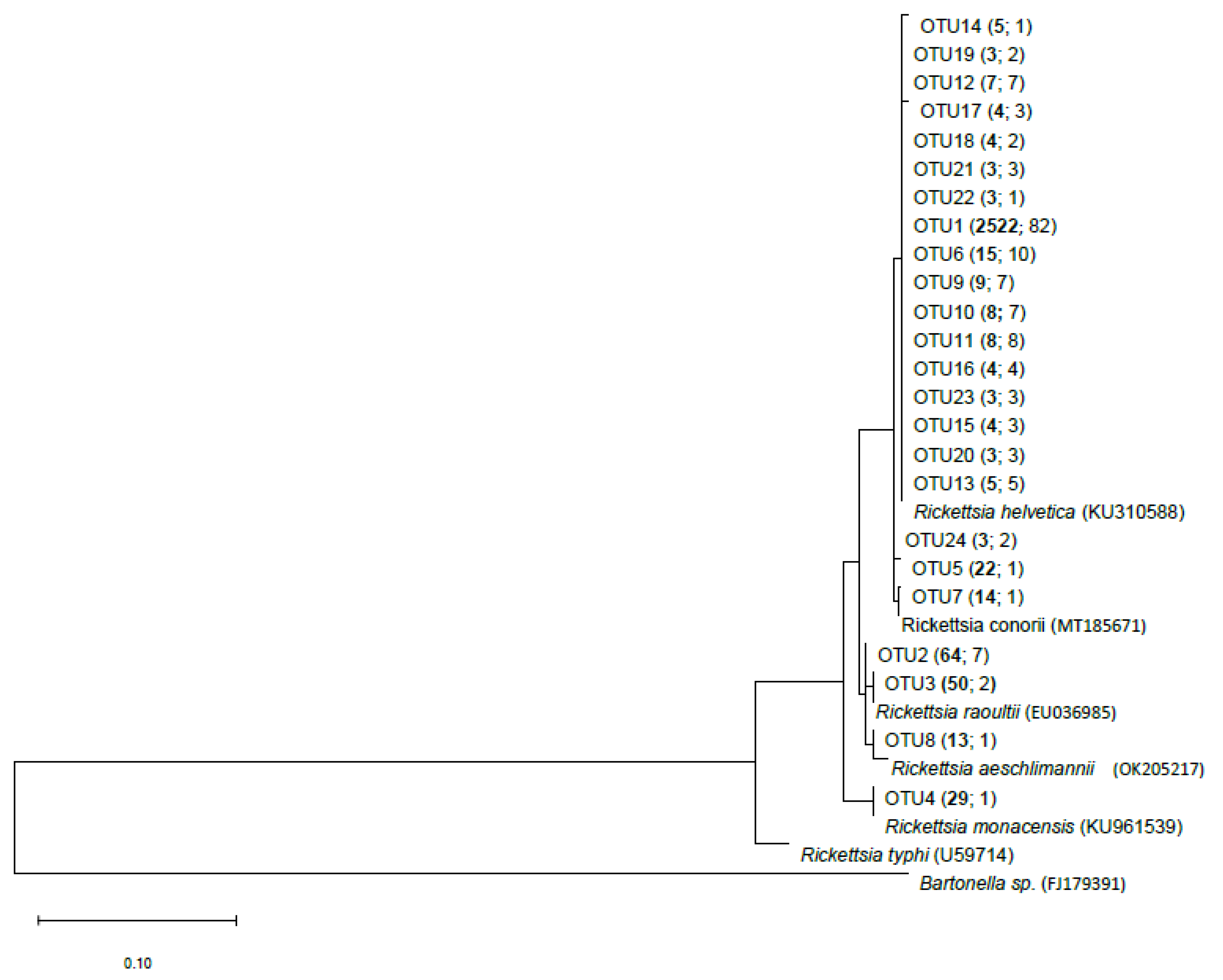
2.4. gltA Gene-Based Sequence Analyses

For the gltA gene-based sequencing data analysis, a reference database was created by downloading 13 gltA gene sequences described within the genus Rickettsia and transmitted by ticks, fleas, or mites from the National Center for Biotechnology Information (NCBI) web-based database (https://www.ncbi.nlm.nih.gov/; last data update for this study was carried out 27 December 2022) [14,39]. The obtained gltA gene sequences were trimmed at the gltA primer pair sequences (Table 2) and were used for an alignment by using the MEGA X software version 10.2.3 [40] and the MUSCLE algorithm [41].
The tick-borne gltA gene sequences were assembled and aligned to the gltA sequence database on the Galaxy training platform [42,43]. As Galaxy is used so far as the 16S rRNA gene pipeline, a few steps were modified to analyze gltA gene sequences. Data cleanup was conducted with a maximum sequence length of 325 bp. Sequences that were poorly aligned between the start- and endpoint of the gltA gene sequence, including homopolymers with a length greater than or equal to six bases, were removed from the dataset. Thereafter sequence alignment was pre-clustered to assemble sequences that were nearly identical to each other. Sequences with a difference of one in 100 bases potentially represent sequencing errors and not biological variability (here, three out of 300 bp). According to the Galaxy training platform, after removing chimeras, OTU clustering was performed on the obtained sequences [43].
OTUs were sorted in decreasing order according to the number of gltA gene-based amplicon sequence reads. Subsequently, all OTUs with a read number less than three reads (singletons and doubletons) were removed from the dataset. An OTU count table with 24 OTUs was obtained (Supplementary Table S2. Based on the final alignment a DNA-based phylogenetic tree was calculated by the neighbor-joining method using the Tamura–Nei model in MEGA X [40]. In total, 1397 positions in the final alignment were used for tree construction.

2.5. Statistics

The PCR results of each tick nucleic acid extract were analysed using the Fisher exact test with Bonferroni correction for each individual comparison of prevalence with other variables (B. burgdorferi s. l. finding, area, month, season, (Table 1)) to determine, which variable affected the tick-borne pathogen prevalence significantly. A Fisher exact test with Bonferroni correction was also performed for examining the prevalence of Rickettsia associated OTUs in nucleic acid extracts as revealed by the gltA gene-based amplicon sequencing data. All tests were performed with the R software version 3.5.2. The significance level for the Fisher exact test was set as α = 0.05. All statistical analyses were performed by RStudio, and Fisher exact test data were illustrated using Origin 2017 (OriginLab Corporation, Northampton, MA, USA).

3. Results

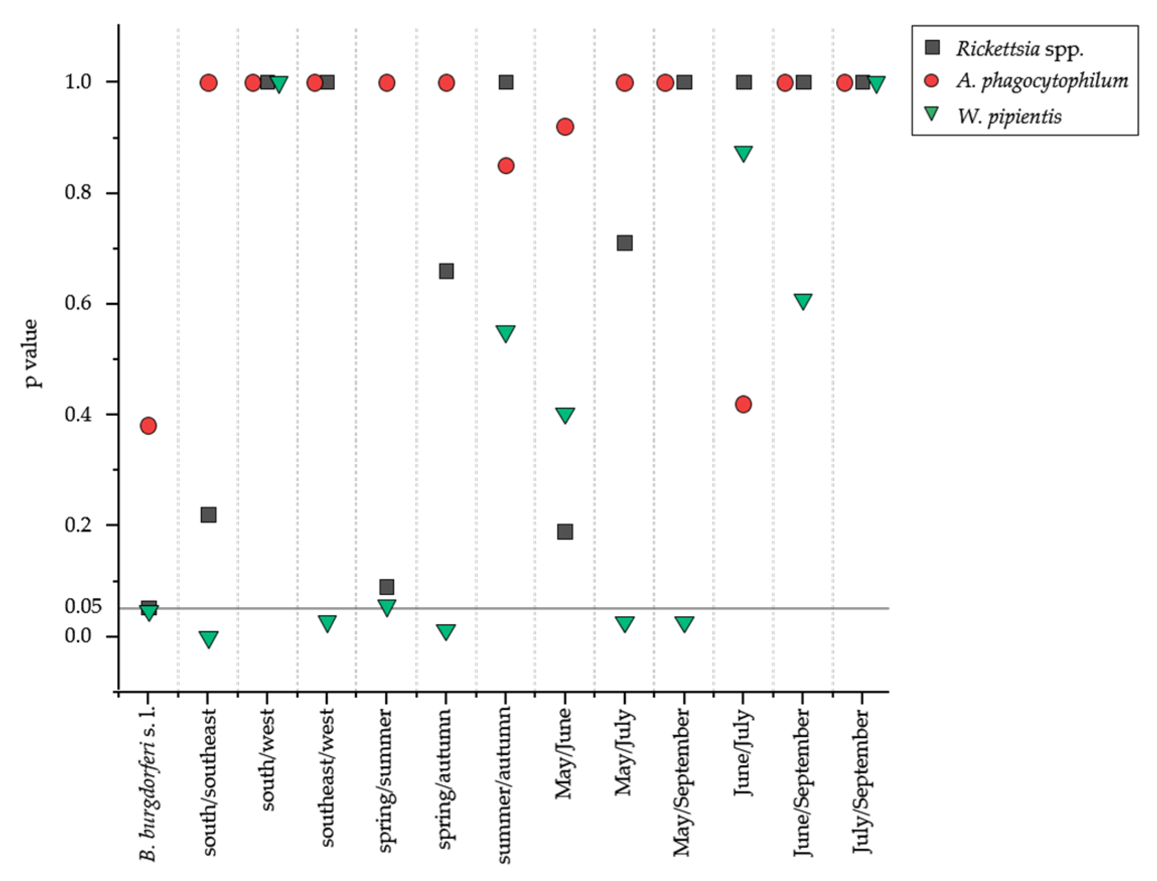
3.1. Rickettsia spp. and W. pipientis Showed Highest Prevalence in Tick Extracts

Among the 760 tick nucleic acid extracts tested, 16.7% were Rickettsia spp. positive, and 15.9% were W. pipientis positive. In addition, 2.8% of the 760 ticks were positive for A. phagocytophilum and 0.1% for N. mikurensis. The prevalence of Rickettsia spp. and W. pipientis was similar comparing B. burgdorferi s. l.-positive (n = 380) and B. burgdorferi s. l.-negative (n = 380) tick nucleic acid extracts (Table 3).
Furthermore, 62 (8.1%) tick nucleic acid extracts were characterized with more than one member of the order Rickettsiales or with additional co-infection of B. burgdorferi s. l., of which two (n = 60/7.9%) or three (n = 2/0.3%) tick-borne pathogens were found simultaneously (Table 4).
In particular, a high ratio of co-infection of B. burgdorferi s. l. and Rickettsia spp. or W. pipientis was identified, and a significant effect between the co-infection of B. burgdorferi s. l. and Rickettsia spp. (p = 4.15 × 10⁻²) as well as W. pipientis (p = 4.71 × 10⁻²) was observed (Figure 1 and Table 4). B. burgdorferi s. l. with Rickettsia spp. (64.9%) and B. burgdorferi s. l. with W. pipientis (36.4%) co-occurred particularly frequently in the area of Weiden i. d. Oberpfalz (n = 242) and Neustadt a.d. Waldnaab (n = 229), respectively, as part of the southeast area (Supplementary Table S1).
The variables of area, month, and season affected the prevalence of respective potential tick-borne pathogens of the order Rickettsiales in the tick nucleic acid extracts. There was a significantly higher prevalence of W. pipientis in the area southeast compared to the areas south and west. Further, the prevalence of W. pipientis was affected by season (highest in spring) or month (highest in May), while A. phagocytophilum and Rickettsia spp. showed no significance against any of the variables investigated (Figure 1).

3.2. Rickettsia-Positive Ticks were Dominated by R. helvetica

The gltA gene was sequenced from 127 Rickettsia-positive nucleic acid extracts by Illumina MiSeq paired-end sequencing, and 24 different OTUs remained after filtering procedures. As a result of blasting the sequences of the OTUs, five different Rickettsia species were identified (Figure 2 and Supplementary Table S2), and R. helvetica dominated 20 out of the 24 OTUs (94.4% of 2805 total sequence reads). Subsequently prevalent were R. aeschlimannii and R. yenbekshikazakhensis, respectively, each with two OTUs (OTU 2 and OTU 8, both 2.7%), R. raoultii with one OTU (OTU 3, 1.8%), and R. monacensis with one OTU (OTU 4, 1.0%) (Figure 2 and Supplementary Table S2).

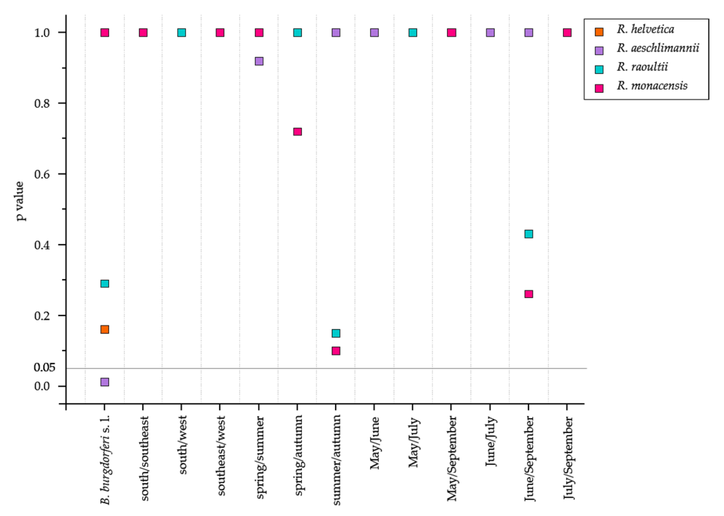
3.3. Prevalence of Rickettsia Species was Affected by B. burgdorferi s. l. Co-Infection

The identified Rickettsia species were then tested for significance to the presence of B. burgdorferi s. l., the variables area, month and season (Figure 3). However, no association with any environmental variable was detected for R. aeschlimannii, but a significant effect was identified with B. burgdorferi s. l. (p = 0.011). For R. helvetica, on the other hand, no significant effect was found for any of the environmental variables tested.

4. Discussion

4.1. Co-Infection of Potential Tick-Borne Pathogens Are Season Dependent

This study used conventional PCRs to investigate the diversity and prevalence of Rickettsia spp., A. phagocytophilum, W. pipientis, and N. mikurensis in 760 nucleic acid extracts of I. ricinus ticks detached from humans, which were previously tested for the presence of B. burgdorferi s.l.. Results were correlated to the variables season, area, as well as B. burgdorferi s. l. prevalence of each tick extract via Fisher exact test. The prevalence of Rickettsia spp. in ticks varies widely within Europe (0.5–66%) and also within Germany (1–30.4%), depending on the infected host species [4,8]. Thus, the prevalence of Rickettsia spp. determined here is 16.7%, which is within the expected range [44,45,46]. In contrast, as described here in the study, a similarly high prevalence of W. pipientis has not yet been described in literature. Evidence suggests that these endosymbionts significantly impact their vectors’ fitness, reproduction, immunity, and other characteristics [24,47]. Thus, the viability of the tick itself and tick-borne pathogens appears to be dependent on Wolbachia [24,38]. In addition, using the Fisher exact test with a Bonferroni correction, a significant difference was found in the prevalence of W. pipientis in the variables of season (spring (14.0%) and autumn (12.4%)), and month (May (14.0%) and July (24.8%) or rather September (12.4%)) (Figure 1). The reason can be explained by the tick infection with W. pipientis as the ticks are usually parasitized by the wasp Ixodiphagus hookeri, which carries Wolbachia [38,48]. Since the wasps lay their eggs in I. ricinus in summer and the development of I. hookeri continues only with the blood meal of the nymph in summer, an increased prevalence of W. pipientis in autumn and a flattening of the Wolbachia infection over the winter months is plausible [48].
The prevalence of A. phagocytophilum, on the other hand, has been adequately described and is reported to have a prevalence of 1–17.4% in I. ricinus ticks in Germany [8]. The 2.8% prevalence detected here is thus low. According to Svitalkova et al., the developmental stage of the tick itself influences the prevalence of A. phagocytophilum [49]. They observed a high prevalence of A. phagocytophilum, especially in adult I. ricinus, whereas the prevalence in nymphs was low [49]. However, since no morphological data on the ticks sent in were prepared by the physicians or the diagnostic laboratory, it can only be assumed that the ticks sent in were nymphs rather than adults.
The simultaneous prevalence of two or three potential tick-borne pathogens in 7.9% (n = 760) of the examined ticks indicates a potential contact of the ticks to a broad spectrum of pathogens and/or hosts (Table 4) [44]. Klitgaard et al. summarized that double infection occurs in 1–22% of the examined I. ricinus ticks in Europe, and Borrelia spp. is involved in most co-infections [50]. A high co-infection of B. burgdorferi s. l. and Rickettsia spp. or W. pipientis were also shown here (Table 4), supported by a significant effect (Figure. 1). Concerning Borrelia spp. and Rickettsia spp., a similarly high co-infection [45], as well as a positive association [4], have already been described in the literature.
Our prevalence of 0.4% for B. burgdorferi s. l. and A. phagocytophilum co-infection is in line with other studies from Germany [45]. However, a higher rate could have been expected, as it is known that A. phagocytophilum increases the colonization ability of B. burgdorferi s. l. and leads to more severe disease courses with diverse disease manifestations through immunosuppressive effects [16,50]. However, since a relatively low A. phagocytophilum prevalence was obtained here, the detected co-infection seems logical.
It should be mentioned that the observed co-infections here should also be considered as purely incidental. First, nucleic acid extracts from ticks infected with B. burgdorferi s. l. were directly detected, and second, the prevalence of each pathogen was significantly higher as a co-infection, therefore we consider an interaction between the pathogens likely.

4.2. Prevalence of R. helvetica to Environmental Variables

I. ricinus is dominated by R. helvetica in Europe and Germany [4,15] compared to other continents such as North America, where R. rickettsii is the predominant species of the genus Rickettsia [13]. In addition, 94.4% of the gltA gene sequencing reads of Rickettsia-positive ticks were identical to sequences of R. helvetica (Figure 2; Supplementary Table S3). Since I. ricinus simultaneously serves as a vector of R. helvetica, and additional highly efficient transovarial transmission is very likely here, R. helvetica can be permanently maintained in the tick life cycle [8,17]. Additionally, Rickettsia spp. can be passed on to the eggs during fertilization by male ticks [17]. The high reservoir competence of diverse vertebrate hosts, such as deer, mice, or hedgehogs ensures geographic distribution and availability in diverse habitats [15]. Assignment of OTU 2 and OTU 8 is ambiguous as both OTUs matched 100% to R. aeschlimannii and Ca. R. yenbekshikazakhensis (Supplementary Tables S2 and S3). Turebekov et al. described how R. aeschlimannii and Ca. R. yenbekshikazakhensis carry identical gltA gene sequences. Therefore, further investigations on these species in the future should be aimed at completely excluding the prevalence of Ca. R. yenbekshikazakhensis in Germany. Nevertheless, the presence of Ca. R. yenbekshikazakhensis in Germany is considered very improbable as it has so far only been found in Kazakhstan [51]. Moreover, the associated tick species Haemaphysalis punctata was identified as a probable vector for Ca. R. yenbekshikazakhensis, and this tick species is not part of the natural German tick diversity today [51,52]. However, R. aeschlimannii has occasionally been detected before in the microbiome of I. ricinus but was found to be transmitted by ticks of the genus Hyalomma in the African region [53,54]. An prevalence of these Rickettsia species in Germany is nevertheless possible, as R. aeschlimannii infected Hyalomma ticks have already been collected from several migratory birds, which occur in Germany and elsewhere [15]. R. monacensis in 1.0% of the reads seems realistic, as the species is distributed on one side throughout Europe and associated with I. ricinus [8,44,54]. However, the host’s risk of R. monacensis manifesting infection can be considered low, as only four clinical cases have been described recently [54].
Thus, while I. ricinus mainly harbours the Rickettsia species R. helvetica, R. aeschlimannii, and R. monacensis [8], the presence of R. raoultii in the dataset (Figure 2, Supplementary Table S2) is novel. R. raoultii has already been described in Germany [15] but only connected with D. reticulatus and D. marginatum [15,17]. R. raoultii was present in 2.2% of the examined ticks. As R. helvetica and R. monacensis are mainly associated with I. ricinus, R. raoultii is strongly connected to Dermacentor ticks. As species identification was not carried out, the identified R. raoultii must likely have been harboured by an individual belonging to the genus Dermacentor. Therefore, molecular biological confirmation should be performed in the future to confirm the morphological identification of the tick. Yet, a general seasonality of Rickettsia spp. has been observed in several studies [17,46,55,56], which we also found (Figure 1 and Section 3.1). The results of the Fisher exact test for the Rickettsia species support our assumption that Rickettsia species are adapted to the environmental variables as they revealed no dependencies with respect to the variables tested in this study (Figure 3). Moreover, the problem of PCR inhibition is crucial for both PCR and sequencing approaches, which have already been observed in the tick microbiome studies [57]. Therefore, upcoming studies should be aware of such PCR drawbacks.

5. Conclusions

The risk of co-infection of several potential tick-borne pathogens in tick microbiomes was high in the studied areas of Germany. Almost every fifth tick collected from humans carried at least two of the (potential) pathogens or genus (Rickettsia spp.) containing human pathogenic species investigated here. In addition, a few rarer rickettsial species (R. monacensis, R. raoultii) were identified as not established in the vector I. ricinus, suggesting that they use other vectors and live in smaller ecological niches. Furthermore, Ca. R. yenbekshikazakhensis was found, which is not considered to be established in Germany, or the vector I. ricinus, which requires further investigation. In contrast to the other human pathogenic species of the genus, the predominant species in Germany, R. helvetica, seems to have found better mechanisms to establish itself in I. ricinus and its hosts, thus ensuring its survival.
Nevertheless, the lack of morphological determination of the tick in the diagnostic laboratory must be considered here. In the future, this should be integrated into the analysis process. Only based on the data, such as species, sex, or stage, exclusion criteria of (potential) tick-borne pathogens can be created, which subsequently simplifies the evaluation of critical test results with very high probability.

Supplementary Materials

The following supporting information can be downloaded at: https://www.mdpi.com/article/10.3390/microorganisms11010157/s1, Table S1. Composition of the pools with associated PCR results from member of the order Rickettsiales, including information on co-infection; Table S2. OTU count table of the gltA gene-based amplicon sequencing results; Table S3. Overview of the Rickettsia species included in the gltA gene-based database and their distribution; Figure S1. Flowchart of pool assembly from tick nucleic acid extracts to assess tick-associated pathogens of the order Rickettsiales, and subsequent gltA gene-based sequencing; Figure S2. Origin of I. ricinus ticks on a map of Germany with identification of the districts and district-free cities for the year 2018. Colors indicate the available tick nucleic acid extracts per post code area in the figure.

Author Contributions

Conceptualization, M.N., V.F.; methodology, A.H., T.M.; validation, A.H.; formal analysis, A.H.; investigation, M.N.; resources, M.N.; writing—original draft preparation, A.H. and M.N.; writing—review and editing, A.H., V.F., C.S., T.M. and M.N.; visualization, A.H.; supervision, M.N.; project administration, M.N.; funding acquisition, M.N. All authors have read and agreed to the published version of the manuscript.

Funding

This research was funded by the Bavarian State Ministry of Science and Art.

Institutional Review Board Statement

Not applicable.

Informed Consent Statement

Not applicable.

Data Availability Statement

The gltA gene sequences for tick samples were deposited in the NCBI nucleotide sequence databases under accession PRJNA839573.

Acknowledgments

We thank Rebekka Pohl and Christian Klos of SYNLAB Holding Germany GmbH in Weiden for allowing us to use their equipment and reagents for nucleic acid extraction of ticks. We thank Julia Braun for her support in molecular biological analyses of nucleic acid extracts and statistical analysis. We gratefully acknowledge the financial support of the Bavarian State Ministry of Science and Art.

Conflicts of Interest

The authors declare that they have no competing interests.

References

  1. Boehnke, D.; Gebhardt, R.; Petney, T.; Norra, S. On the complexity of measuring forests microclimate and interpreting its relevance in habitat ecology: The example of Ixodes ricinus ticks. Parasites Vectors 2017, 10, 549. [Google Scholar] [CrossRef] [PubMed] [Green Version]
  2. Dobler, G.; Fingerle, V.; Hagedorn, P.; Pfeffer, M.; Silaghi, C.; Tomaso, H.; Henning, K.; Niedrig, M. Gefahren der Übertragung von Krankheitserregern durch Schildzecken in Deutschland. Bundesgesundheitsblatt Gesundh. Gesundh. 2014, 57, 541–548. [Google Scholar] [CrossRef] [Green Version]
  3. Rubel, F.; Brugger, K.; Chitimia-Dobler, L.; Dautel, H.; Meyer-Kayser, E.; Kahl, O. Atlas of ticks (Acari: Argasidae, Ixodidae) in Germany. Exp. Appl. Acarol. 2021, 84, 183–214. [Google Scholar] [CrossRef] [PubMed]
  4. Kowalec, M.; Szewczyk, T.; Welc-Falęciak, R.; Siński, E.; Karbowiak, G.; Bajer, A. Rickettsiales occurrence and co-occurrence in Ixodes ricinus ticks in natural and urban areas. Microb. Ecol. 2019, 77, 890–904. [Google Scholar] [CrossRef] [PubMed] [Green Version]
  5. Welc-Falęciak, R.; Kowalec, M.; Karbowiak, G.; Bajer, A.; Behnke, J.M.; Siński, E. Rickettsiaceae and Anaplasmataceae infections in Ixodes ricinus ticks from urban and natural forested areas of Poland. Parasites Vectors 2014, 7, 121. [Google Scholar] [CrossRef] [PubMed] [Green Version]
  6. Karbowiak, G.; Biernat, B.; Stańczak, J.; Szewczyk, T.; Werszko, J. The role of particular tick developmental stages in the circulation of tick-borne pathogens affecting humans in Central Europe. 3. Rickettsiae. Ann. Parasitol. 2016, 62, 89–100. [Google Scholar] [CrossRef]
  7. Karbowiak, G.; Biernat, B.; Stańczak, J.; Werszko, J.; Wróblewski, P.; Szewczyk, T.; Sytykiewicz, H. The role of particular ticks developmental stages in the circulation of tick-borne pathogens in Central Europe. 4. Anaplasmataceae. Ann. Parasitol. 2016, 62, 267–284. [Google Scholar] [CrossRef]
  8. Rizzoli, A.; Silaghi, C.; Obiegala, A.; Rudolf, I.; Hubálek, Z.; Földvári, G.; Plantard, O.; Vayssier-Taussat, M.; Bonnet, S.; Spitalská, E.; et al. Ixodes ricinus and its transmitted pathogens in urban and peri-urban areas in Europe: New hazards and relevance for public health. Front. Public Health 2014, 2, 251. [Google Scholar] [CrossRef]
  9. Knoll, S.; Springer, A.; Hauck, D.; Schunack, B.; Pachnicke, S.; Strube, C. Regional, seasonal, biennial and landscape-associated distribution of Anaplasma phagocytophilum and Rickettsia spp. infections in Ixodes ticks in northern Germany and implications for risk assessment at larger spatial scales. Ticks Tick Borne Dis. 2021, 12, 101657. [Google Scholar] [CrossRef]
  10. Parola, P.; Paddock, C.D.; Raoult, D. Tick-borne rickettsioses around the world: Emerging diseases challenging old concepts. Clin. Microbiol. Rev. 2005, 18, 719–756. [Google Scholar] [CrossRef]
  11. Sprong, H.; Wielinga, P.R.; Fonville, M.; Reusken, C.; Brandenburg, A.H.; Borgsteede, F.; Gaasenbeek, C.; van der Giessen, J.W. Ixodes ricinus ticks are reservoir hosts for Rickettsia helvetica and potentially carry flea-borne Rickettsia species. Parasites Vectors 2009, 2, 41. [Google Scholar] [CrossRef] [Green Version]
  12. Yang, J.; Tian, Z.; Liu, Z.; Niu, Q.; Han, R.; Li, Y.; Guan, G.; Liu, J.; Liu, G.; Luo, J.; et al. Novel spotted fever group rickettsiae in Haemaphysalis qinghaiensis ticks from Gansu, northwest China. Parasites Vectors 2016, 9, 146. [Google Scholar] [CrossRef] [PubMed] [Green Version]
  13. Piotrowski, M.; Rymaszewska, A. Expansion of tick-borne rickettsioses in the world. Microorganisms 2020, 8, 1906. [Google Scholar] [CrossRef]
  14. Dobler, G.; Wölfel, R. Typhus and other rickettsioses: Emerging infections in Germany. Dtsch. Arztebl. Int. 2009, 106, 348–354. [Google Scholar] [CrossRef]
  15. Parola, P.; Paddock, C.D.; Socolovschi, C.; Labruna, M.B.; Mediannikov, O.; Kernif, T.; Abdad, M.Y.; Stenos, J.; Bitam, I.; Fournier, P.-E.; et al. Update on tick-borne rickettsioses around the world: A geographic approach. Clin. Microbiol. Rev. 2013, 26, 657–702. [Google Scholar] [CrossRef] [PubMed] [Green Version]
  16. Borşan, S.-D.; Ionică, A.M.; Galon, C.; Toma-Naic, A.; Peştean, C.; Sándor, A.D.; Moutailler, S.; Mihalca, A.D. High diversity, prevalence, and co-infection rates of tick-borne pathogens in ticks and wildlife hosts in an urban area in Romania. Front. Microbiol. 2021, 12, 645002. [Google Scholar] [CrossRef]
  17. Blazejak, K.; Janecek, E.; Strube, C. A 10-year surveillance of Rickettsiales (Rickettsia spp. and Anaplasma phagocytophilum) in the city of Hanover, Germany, reveals Rickettsia spp. as emerging pathogens in ticks. Parasites Vectors 2017, 10, 588. [Google Scholar] [CrossRef] [PubMed] [Green Version]
  18. André, M.R. Diversity of Anaplasma and Ehrlichia/Neoehrlichia agents in terrestrial wild carnivores worldwide: Implications for human and domestic animal health and wildlife conservation. Front. Vet. Sci. 2018, 5, 293. [Google Scholar] [CrossRef]
  19. Matei, I.A.; Estrada-Peña, A.; Cutler, S.J.; Vayssier-Taussat, M.; Varela-Castro, L.; Potkonjak, A.; Zeller, H.; Mihalca, A.D. A review on the eco-epidemiology and clinical management of human granulocytic anaplasmosis and its agent in Europe. Parasites Vectors 2019, 12, 599. [Google Scholar] [CrossRef]
  20. Wass, L.; Grankvist, A.; Bell-Sakyi, L.; Bergström, M.; Ulfhammer, E.; Lingblom, C.; Wennerås, C. Cultivation of the causative agent of human neoehrlichiosis from clinical isolates identifies vascular endothelium as a target of infection. Emerg. Microbes Infect. 2019, 8, 413–425. [Google Scholar] [CrossRef]
  21. Pedersen, B.N.; Jenkins, A.; Paulsen, K.M.; Okbaldet, Y.B.; Edgar, K.S.; Lamsal, A.; Soleng, A.; Andreassen, Å.K. Distribution of Neoehrlichia mikurensis in Ixodes ricinus ticks along the coast of Norway: The western seaboard is a low-prevalence region. Zoonoses Public Health 2020, 67, 130–137. [Google Scholar] [CrossRef] [Green Version]
  22. Silaghi, C.; Woll, D.; Mahling, M.; Pfister, K.; Pfeffer, M. Candidatus Neoehrlichia mikurensis in rodents in an area with sympatric existence of the hard ticks Ixodes ricinus and Dermacentor reticulatus, Germany. Parasites Vectors 2012, 5, 285. [Google Scholar] [CrossRef] [Green Version]
  23. Subramanian, G.; Sekeyova, Z.; Raoult, D.; Mediannikov, O. Multiple tick-associated bacteria in Ixodes ricinus from Slovakia. Ticks Tick Borne Dis. 2012, 3, 406–410. [Google Scholar] [CrossRef]
  24. Portillo, A.; Palomar, A.M.; de Toro, M.; Santibáñez, S.; Santibáñez, P.; Oteo, J.A. Exploring the bacteriome in anthropophilic ticks: To investigate the vectors for diagnosis. PLoS ONE 2019, 14, e0213384. [Google Scholar] [CrossRef] [PubMed] [Green Version]
  25. Ben, I.; Lozynskyi, I. Prevalence of Anaplasma phagocytophilum in Ixodes ricinus and Dermacentor reticulatus and coinfection with Borrelia burgdorferi and tick-borne encephalitis virus in western Ukraine. Vector Borne Zoonotic Dis. 2019, 19, 793–801. [Google Scholar] [CrossRef] [PubMed] [Green Version]
  26. Sytykiewicz, H.; Karbowiak, G.; Hapunik, J.; Szpechciński, A.; Supergan-Marwicz, M.; Goławska, S.; Sprawka, I.; Czerniewicz, P. Molecular evidence of Anaplasma phagocytophilum and Babesia microti co-infections in Ixodes ricinus ticks in central-eastern region of Poland. Ann. Agric. Environ. Med. 2012, 19, 45–49. [Google Scholar]
  27. Greay, T.L.; Gofton, A.W.; Paparini, A.; Ryan, U.M.; Oskam, C.L.; Irwin, P.J. Recent insights into the tick microbiome gained through next-generation sequencing. Parasites Vectors 2018, 11, 12. [Google Scholar] [CrossRef]
  28. Klindworth, A.; Pruesse, E.; Schweer, T.; Peplies, J.; Quast, C.; Horn, M.; Glöckner, F.O. Evaluation of general 16S ribosomal RNA gene PCR primers for classical and next-generation sequencing-based diversity studies. Nucleic Acids Res. 2013, 41, e1. [Google Scholar] [CrossRef] [PubMed]
  29. Janecek, E.; Streichan, S.; Strube, C. SNP-based real-time pyrosequencing as a sensitive and specific tool for identification and differentiation of Rickettsia species in Ixodes ricinus ticks. BMC Infect. Dis. 2012, 12, 261. [Google Scholar] [CrossRef] [Green Version]
  30. Bunikis, J.; Garpmo, U.; Tsao, J.; Berglund, J.; Fish, D.; Barbour, A.G. Sequence typing reveals extensive strain diversity of the Lyme borreliosis agents Borrelia burgdorferi in North America and Borrelia afzelii in Europe. Microbiology 2004, 150, 1741–1755. [Google Scholar] [CrossRef] [Green Version]
  31. Krebs, H.A.; Johnson, W.A. Metabolism of ketonic acids in animal tissues. Biochem. J. 1937, 31, 645–660. [Google Scholar] [CrossRef] [PubMed]
  32. Blanda, V.; D’Agostino, R.; Giudice, E.; Randazzo, K.; La Russa, F.; Villari, S.; Vullo, S.; Torina, A. New real-time PCRs to differentiate Rickettsia spp. and Rickettsia conorii. Molecules 2020, 25, 4431. [Google Scholar] [CrossRef] [PubMed]
  33. Hoffmann, A.; Fingerle, V.; Noll, M. Analysis of tick surface decontamination methods. Microorganisms 2020, 8, 987. [Google Scholar] [CrossRef] [PubMed]
  34. Hoffmann, A.; Müller, T.; Fingerle, V.; Noll, M. Presence of human pathogens of the Borrelia burgdorferi sensu lato complex shifts the sequence read abundances of tick microbiomes in two german locations. Microorganisms 2021, 9, 1814. [Google Scholar] [CrossRef] [PubMed]
  35. Courtney, J.W.; Kostelnik, L.M.; Zeidner, N.S.; Massung, R.F. Multiplex real-time PCR for detection of Anaplasma phagocytophilum and Borrelia burgdorferi. J. Clin. Microbiol. 2004, 42, 3164–3168. [Google Scholar] [CrossRef] [PubMed] [Green Version]
  36. Capelli, G.; Ravagnan, S.; Montarsi, F.; Ciocchetta, S.; Cazzin, S.; Porcellato, E.; Babiker, A.M.; Cassini, R.; Salviato, A.; Cattoli, G.; et al. Occurrence and identification of risk areas of Ixodes ricinus-borne pathogens: A cost-effectiveness analysis in north-eastern Italy. Parasites Vectors 2012, 5, 61. [Google Scholar] [CrossRef] [Green Version]
  37. Hildebrandt, A.; Krämer, A.; Sachse, S.; Straube, E. Detection of Rickettsia spp. and Anaplasma phagocytophilum in Ixodes ricinus ticks in a region of Middle Germany (Thuringia). Ticks Tick-Borne Dis. 2010, 1, 52–56. [Google Scholar] [CrossRef]
  38. Plantard, O.; Bouju-Albert, A.; Malard, M.-A.; Hermouet, A.; Capron, G.; Verheyden, H. Detection of Wolbachia in the tick Ixodes ricinus is due to the presence of the hymenoptera endoparasitoid Ixodiphagus hookeri. PLoS ONE 2012, 7, e30692. [Google Scholar] [CrossRef] [Green Version]
  39. National Center for Biotechnology Information. National Center for biotechnology information. Available online: https://www.ncbi.nlm.nih.gov/ (accessed on 27 December 2022).
  40. Kumar, S.; Stecher, G.; Li, M.; Knyaz, C.; Tamura, K. MEGA X: Molecular Evolutionary Genetics Analysis across computing platforms. Mol. Biol. Evol. 2018, 35, 1547–1549. [Google Scholar] [CrossRef]
  41. Edgar, R.C. MUSCLE: Multiple sequence alignment with high accuracy and high throughput. Nucleic Acids Res. 2004, 32, 1792–1797. [Google Scholar] [CrossRef] [Green Version]
  42. Hiltemann, S.; Batut, B.; Clements, D. 16S Microbial analysis with Mothur (extended) (galaxy training materials). Available online: https://training.galaxyproject.org/training-material/topics/metagenomics/tutorials/mothur-miseq-sop/tutorial.html (accessed on 13 April 2021).
  43. Batut, B.; Hiltemann, S.; Bagnacani, A.; Baker, D.; Bhardwaj, V.; Blank, C.; Bretaudeau, A.; Brillet-Guéguen, L.; Čech, M.; Chilton, J.; et al. Community-driven data analysis training for biology. Cell Syst. 2018, 6, 752–758.e1. [Google Scholar] [CrossRef]
  44. Schicht, S.; Schnieder, T.; Strube, C. Rickettsia spp. and coinfections with other pathogenic microorganisms in hard ticks from northern Germany. J. Med. Entomol. 2012, 49, 766–771. [Google Scholar] [CrossRef] [Green Version]
  45. Tappe, J.; Jordan, D.; Janecek, E.; Fingerle, V.; Strube, C. Revisited: Borrelia burgdorferi sensu lato infections in hard ticks (Ixodes ricinus) in the city of Hanover (Germany). Parasites Vectors 2014, 7, 441. [Google Scholar] [CrossRef] [PubMed] [Green Version]
  46. Tappe, J.; Strube, C. Anaplasma phagocytophilum and Rickettsia spp. infections in hard ticks (Ixodes ricinus) in the city of Hanover (Germany): Revisited. Ticks Tick Borne Dis. 2013, 4, 432–438. [Google Scholar] [CrossRef] [PubMed]
  47. Zindel, R.; Gottlieb, Y.; Aebi, A. Arthropod symbioses: A neglected parameter in pest- and disease-control programmes. J. Appl. Ecol. 2011, 48, 864–872. [Google Scholar] [CrossRef] [Green Version]
  48. Collatz, J.; Selzer, P.; Fuhrmann, A.; Oehme, R.M.; Mackenstedt, U.; Kahl, O.; Steidle, J.L.M. A hidden beneficial: Biology of the tick-wasp Ixodiphagus hookeri in Germany. J. Appl. Entomol. 2011, 135, 351–358. [Google Scholar] [CrossRef]
  49. Svitálková, Z.; Haruštiaková, D.; Mahríková, L.; Berthová, L.; Slovák, M.; Kocianová, E.; Kazimírová, M. Anaplasma phagocytophilum prevalence in ticks and rodents in an urban and natural habitat in south-western Slovakia. Parasites Vectors 2015, 8, 276. [Google Scholar] [CrossRef] [Green Version]
  50. Klitgaard, K.; Kjær, L.J.; Isbrand, A.; Hansen, M.F.; Bødker, R. Multiple infections in questing nymphs and adult female Ixodes ricinus ticks collected in a recreational forest in Denmark. Ticks Tick-Borne Dis. 2019, 10, 1060–1065. [Google Scholar] [CrossRef]
  51. Turebekov, N.; Abdiyeva, K.; Yegemberdiyeva, R.; Dmitrovsky, A.; Yeraliyeva, L.; Shapiyeva, Z.; Amirbekov, A.; Oradova, A.; Kachiyeva, Z.; Ziyadina, L.; et al. Prevalence of Rickettsia species in ticks including identification of unknown species in two regions in Kazakhstan. Parasites Vectors 2019, 12, 197. [Google Scholar] [CrossRef]
  52. Akimov, I.; Nebogatkin, I. Distribution of the tick Haemaphysalis punctata (Acari, Ixodidae) in Ukraine. Vestn. Zool. 2012, 46, e-46–e-51. [Google Scholar] [CrossRef] [Green Version]
  53. Guccione, C.; Colomba, C.; Tolomeo, M.; Trizzino, M.; Iaria, C.; Cascio, A. Rickettsiales in Italy. Pathogens 2021, 10, 181. [Google Scholar] [CrossRef] [PubMed]
  54. Portillo, A.; Santibáñez, S.; García-Álvarez, L.; Palomar, A.M.; Oteo, J.A. Rickettsioses in Europe. Microbes Infect. 2015, 17, 834–838. [Google Scholar] [CrossRef] [PubMed]
  55. May, K.; Strube, C. Prevalence of Rickettsiales (Anaplasma phagocytophilum and Rickettsia spp.) in hard ticks (Ixodes ricinus) in the city of Hamburg, Germany. Parasitol. Res. 2014, 113, 2169–2175. [Google Scholar] [CrossRef] [PubMed]
  56. Biernat, B.; Stańczak, J.; Michalik, J.; Sikora, B.; Cieniuch, S. Rickettsia helvetica and R. monacensis infections in immature Ixodes ricinus ticks derived from sylvatic passerine birds in west-central Poland. Parasitol. Res. 2016, 115, 3469–3477. [Google Scholar] [CrossRef] [Green Version]
  57. Pedersen, B.N.; Jenkins, A.; Kjelland, V. Tick-borne pathogens in Ixodes ricinus ticks collected from migratory birds in southern Norway. PLoS ONE 2020, 15, e0230579. [Google Scholar] [CrossRef] [PubMed]
Figure 1. Effect of environmental variables on the prevalence of Rickettsia spp., A. phagocytophilum, N. mikurensis, and W. pipientis as revealed by the Fisher exact test with Bonferroni correction. Symbol legend for each species is included in the figure. Details of the characteristics of the variables can be found in Table 1. The significance level of 0.05 is indicated.
Microorganisms 11 00157 g001
Figure 2. DNA-based neighbor-joining tree of partial gltA gene sequences. Environmental sequences retrieved from this study are indicated as OTU with their total sequence read numbers in bold and frequency of OTUs both in brackets. The gltA gene sequences from reference organisms were retrieved from GenBank and their accession numbers are indicated in brackets. The scale bar represents 0.1 nucleotide substitutions per site. For a sample-specific overview, see Supplementary Table S2.
Microorganisms 11 00157 g002
Figure 3. Effect of environmental variables on the prevalence of R. helvetica, R. aeschlimannii, R. raoultii, and R. monacensis as revealed by the Fisher exact test with Bonferroni correction. Symbol legend for each species is included in the figure. Details of the characteristics of the variables can be found in Table 1. The significance level of 0.05 is indicated.
Microorganisms 11 00157 g003
Table 1. Characteristics of the variables of each tick nucleic acid extract for subsequent Fisher exact test. Seasons were categorized according to the meteorological calendar (spring: 1 March, summer: 1 June, autumn: 1 September); n indicates how many tick nucleic acid extracts were available per characteristic.
VariableCharacteristic
B. burgdorferi s. l.Positive (n = 380), negative (n = 380)
AreaSouth (n = 120), southeast (n = 560), west (n = 80)
MonthMay (n = 180), June (n = 380), July (n = 140), September (n = 60)
SeasonSpring (n = 180), summer (n = 520), autumn (n = 60)
Table 2. Primer pairs used in this study to detect species of the order Rickettsiales in tick nucleic acid extracts.
Bacterial Species/ GenusTarget GeneAmplicon Length [bp]Final Primer Concentration (nM)Primer Set *References
A. phagocytophilummsp277400ApMSP2f:
5′-ATGGAAGGTAGTGTTGGTTATGGTATT-3′
ApMSP2r:
5′-TTGGTCTTGAAGCGCTCGTA-3′
[35]
N. mikurensisgroEL1024500NM-128f:
5′-AACAGGTGAAACACTAGATAAGTCCAT-3′
NM-1152f:
5′-TTCTACTTTGAACATTTGAAGAATTACTAT-3′
[36]
Rickettsia spp.gltA340400RH314f:
5′-AAACAGGTTGCTCATCATTC-3′
RH654r:
5′-AGAGCATTTTTTATTATTGG-3′
[37]
W. pipientiswsp591500wsp-81f:
5′-TGGTCCAATAAGTGATGAAGAAAC-3′
wsp-691r:
5′-AAAAATTAAACGCTACTCCA-3′
[38]
* f, forward and r, reverse. Primers were named as introduced in the respective reference.
Table 3. Presence of A. phagocytophilum, N. mikurensis, Rickettsia spp., and W. pipientis in individual tick nucleic acid extracts with B. burgdorferi s. l.-negative (n = 380) and B. burgdorferi s. l.-positive (n = 380) findings.
Bacterial Species or GenusB. burgdorferi s. l. FindingPrevalence of
Positive Findings
Total Prevalence of
Positive Findings
A. phagocytophilumnegative13 (1.7%)21 (2.8%)
positive8 (1.0%)
N. mikurensisnegative1 (0.1%)1 (0.1%)
positive0 (0.0%)
Rickettsia spp.negative53 (7.0%)127 (16.7%)
positive74 (9.7%)
W. pipientisnegative50 (6.6%)121 (15.9%)
positive71 (9.3%)
Table 4. Co-infection of B. burgdorferi s. l., A. phagocytophilum, N. mikurensis and/or Rickettsia spp. of individual nucleic acid extracts from ticks. Prevalence is indicated by numbers and their relative prevalence in percentage (n = 760).
Frequence of co-InfectionGenus/ SpeciesPrevalenceTotal
Prevalence
DoubleB. burgdorferi s. l. + A. phagocytophilum3/0.4%60 (7.9%)
B. burgdorferi s. l. + Rickettsia spp.57/7.5%
TripleB. burgdorferi s. l. + A. Phagocytophilum + Rickettsia spp.2/0.3%2/0.3%
62/8.1%
Disclaimer/Publisher’s Note: The statements, opinions and data contained in all publications are solely those of the individual author(s) and contributor(s) and not of MDPI and/or the editor(s). MDPI and/or the editor(s) disclaim responsibility for any injury to people or property resulting from any ideas, methods, instructions or products referred to in the content.

