Case Report

Detection of Helicobacter pylori from Extracted Teeth of a Patient with Idiopathic Thrombocytopenic Purpura

1
Department of Oral and Maxillofacial Surgery II, Osaka University Graduate School of Dentistry, Osaka 565-0871, Japan
2
Department of Pediatric Dentistry, Osaka University Graduate School of Dentistry, Osaka 565-0871, Japan
3
Department of Pediatric Dentistry, Hiroshima University Graduate School of Biomedical and Health Sciences, Hiroshima 734-8553, Japan
*
Author to whom correspondence should be addressed.
Microorganisms 2022, 10(11), 2285; https://doi.org/10.3390/microorganisms10112285
Submission received: 3 August 2022 / Revised: 8 November 2022 / Accepted: 9 November 2022 / Published: 17 November 2022
(This article belongs to the Special Issue Advances in the Diagnosis and Treatment of Helicobacter pylori)

Abstract

:
Immune thrombocytopenic purpura (ITP) is an autoimmune disease characterized by isolated cryptogenic thrombocytopenia due to a transient or persistent reduction in platelet count. Many patients with ITP have shown improved platelet count after Helicobacter pylori eradication therapy. However, there have been no studies regarding H. pylori in the oral cavity of patients with ITP. Here, we describe a patient with ITP whose oral samples exhibited H. pylori. A 64-year-old woman with ITP came to our hospital with chief complaints that required oral surgery, including tooth extraction and cystectomy. Bacterial DNA from H. pylori was confirmed on the extracted tooth, but was not detected in the saliva taken at the time. Bacterial DNA from H. pylori was detected on the suture around the extraction socket, which was removed at 10 days post-operation. However, H. pylori DNA was not detected in other oral samples at 10 or 30 days post-operation. A urea breath test was carried out in the gastrointestinal clinic at 60 days post-operation, which revealed no presence of H. pylori in the gastrointestinal tract. These results suggest that teeth with severe bacterial infections may be a potential reservoir of H. pylori for patients with ITP.

1. Introduction

Immune thrombocytopenic purpura (also known as idiopathic thrombocytopenic purpura; ITP) is an autoimmune disease characterized by isolated thrombocytopenia with fewer than 100,000/µL platelets due to a transient or persistent reduction in platelet count [1]. Although ITP is presumably caused by the autoimmune destruction of platelets in the spleen, the detailed mechanism underlying this phenomenon remains unknown. When the platelet count falls below 50,000/µL due to ITP, bleeding may be evident [2]. Furthermore, when the platelet count drops to less than 10,000/µL, severe bleeding symptoms such as oral bleeding, hematuria, and intracranial hemorrhage may occur [3,4,5,6,7]. When these symptoms develop, corticosteroids, high-dose gamma-globulin therapy, platelet infusion, and splenectomy should be considered [3,8].
Helicobacter pylori is a Gram-negative microaerophilic human gastric pathogen with strong urease activity and a helical structure [9]. H. pylori infection, which causes chronic gastritis, peptic ulcer disease and gastric cancer [9], can be diagnosed via the histological detection of bacteria in gastric biopsies, urea breath test and stool antigen test [10].
Associations between H. pylori and extragastric diseases, such as neurological diseases, respiratory diseases, and hematologic diseases, have been previously reported [11,12]. In 1998, it was reported that the number of platelets increased in patients with ITP after the eradication of H. pylori [13]. In addition, studies focusing on the relationship between ITP and H. pylori infection have been reported in recent years [14,15], and the eradication of H. pylori is regarded as an effective treatment for ITP [14,15]. Examination of H. pylori in gastric infections in patients with ITP is typically performed by non-invasive tests such as the urea breath test and stool antigen test; invasive tests (e.g., endoscopic biopsy) are not recommended due to the difficulty involved in maintaining hemostasis [16].
H. pylori was previously classified as a Campylobacter species, which are known to be major periodontopathic bacteria [9]. Therefore, the oral cavity is a possible route of H. pylori infection, and a potential reservoir for the bacteria [17]. There are approximately 700 bacterial species in the oral cavity, and it is not possible to specifically detect H. pylori from the oral cavity by the same methods used to detect H. pylori from gastric tissue. To detect H. pylori in the oral cavity, molecular microbiological analyses such as polymerase chain reaction (PCR) have been widely used [18].
The detection rate of H. pylori by PCR from the oral cavity has been reported to be 0–100% [19], and it is not easy to identify the actual infection rate. To overcome the difficulty of detecting H. pylori from oral specimens, we designed a novel primer set based on the whole-genome information of about 50 H. pylori strains in our database [20,21]. Among the entire genome of H. pylori, 16S rRNA, vacA, cagA, glmM (ureC), and ureA have been used frequently for H. pylori detection by PCR [22,23,24,25,26]. We searched for homology between all H. pylori in each of these genes. As a result, only ureA had six consecutive sequences of 20 or more bases conserved in the approximately 50 H. pylori strains [20], and we selected some of these regions as primer sets for H. pylori detection.
Although molecular biological analysis is used as a tool for diagnosis of H. pylori infection in the oral cavity, there have been no published studies regarding H. pylori detection in oral samples taken from patients with ITP. In the present report, we describe the detection of H. pylori in the oral cavity of a patient with ITP who underwent oral surgery comprising tooth extraction and cystectomy.

2. Materials and Methods

2.1. Ethics Statement

This study was conducted in full adherence to the Declaration of Helsinki. This study protocol was approved by the Ethics Committee of Osaka University Graduate School of Dentistry (approval: H27-E17-1). Written consent to participate in this study was obtained from the subject patient. The patient first came to our clinic on 19 October 2017, underwent surgery on 13 December 2017, and the consultation at our clinic was completed on 12 January 2018.

2.2. DNA Extraction

Bacterial DNA was extracted using previously described methods [20]. Briefly, oral specimens were resuspended in 250 μL of 10 mM Tris-HCl (pH 8.0) containing 100 mM NaCl and 1 mM EDTA. The cells were collected using centrifugation and lysed in 600 µL of Cell Lysis Solution (Qiagen, Düsseldorf, Germany) and incubated at 80 °C for 5 min, followed by the addition of 3 μL of RNase A (10 mg/mL; Qiagen) and incubation at 37 °C for 30 min. Protein Precipitation Solution (Qiagen) was added, vigorously vortexed for 20 s, and then centrifuged at 10,000× g for 3 min. The supernatant was combined with 600 μL of isopropanol (Wako Pure Chemical Industries, Tokyo, Japan) and centrifuged. The precipitate was then resuspended in 70% ethanol (Wako Pure Chemical Industries), centrifuged, combined with 100 μL of TE buffer (10 mM Tris-HCl, 1 mM EDTA [pH 8.0]), and used as bacterial DNA for the PCR method.

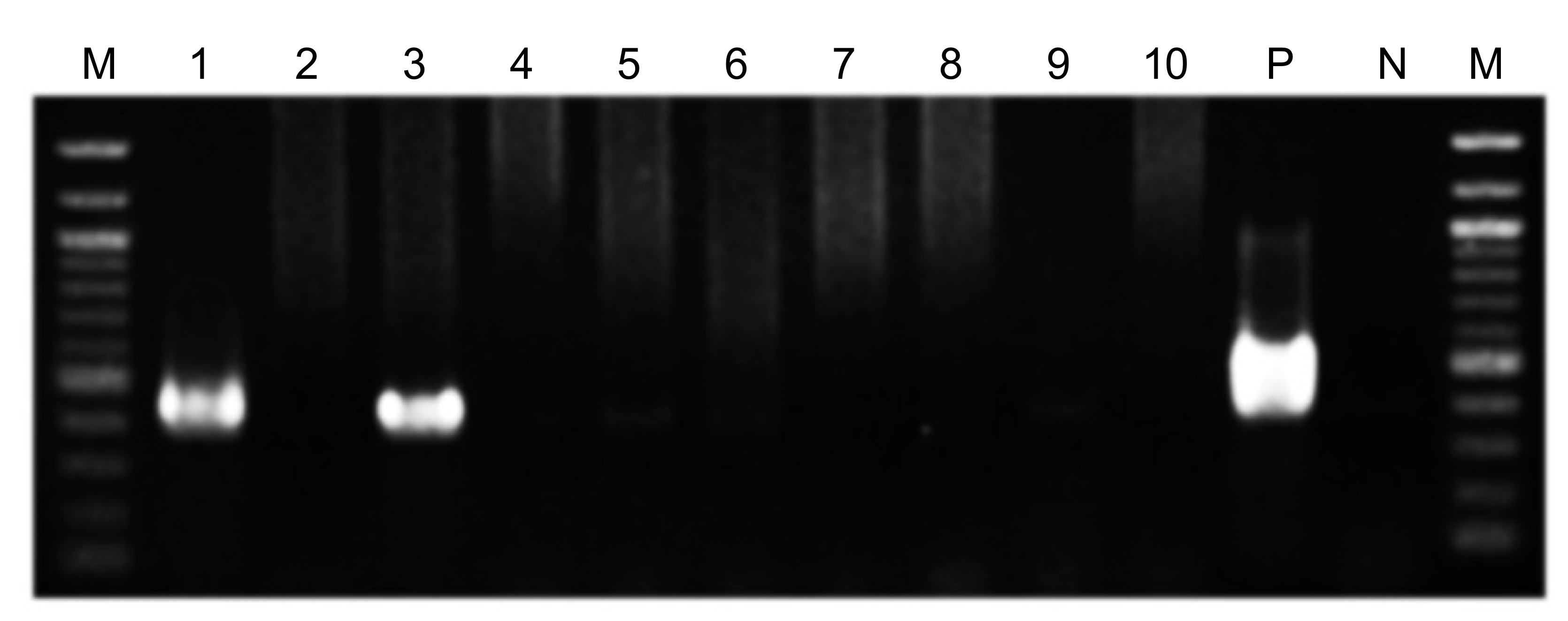
2.3. PCR Detection of Bacteria
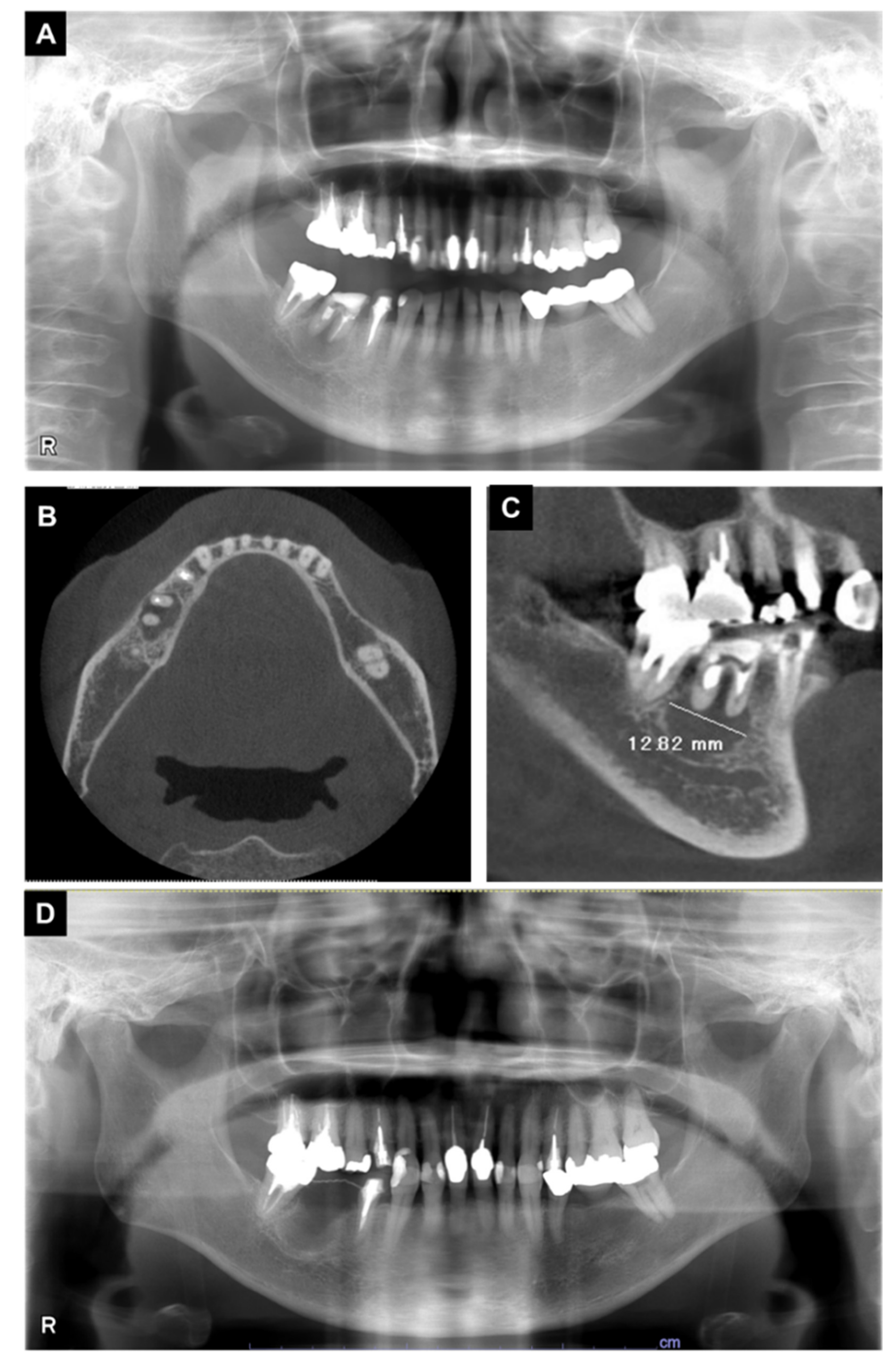
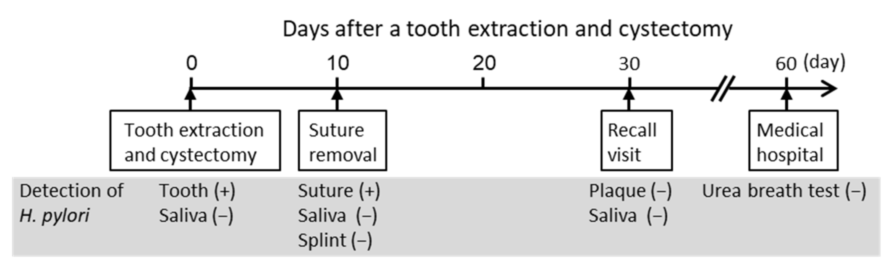
We previously searched the whole-genome sequences of about 50 H. pylori strains in a database to construct a PCR method to detect H. pylori from oral specimens with high sensitivity and specificity [20]. We found that more than 20 nucleotides of sequences were consecutively conserved in all H. pylori strains at multiple locations in the ureA gene. We designed primer sets for H. pylori detection using some of these sequences in the ureA gene (Table 1).
PCR with either of these forward and reverse primer combinations detected 102 – 103 colony forming units (CFU) of H. pylori [21]. To further improve the detection sensitivity, we developed a nested PCR method. In the nested PCR, the first-step PCR for H. pylori detection was performed using primers ureA-aF and ureA-bR, and the second-step PCR (nested PCR) using primers ureA-bF and ureA-aR (Figure 1).
For the first-step PCR, 2 μL of bacterial DNA extracted from oral samples was used in a 20 μL reaction. For the second-step PCR, 1 μL of the first-step PCR amplification product was used as a template for the reaction in a total volume of 20 μL. Both the first and second steps of nested PCR were performed as described previously, using TaKaRa Ex Taq polymerase (Takara Bio. Inc., Otsu, Japan) [21]. During each nested PCR reaction, sterile distilled water was used in place of bacterial DNA as a negative control to ensure that no false positive reactions occurred. PCR amplification was performed with the following cycling parameters: initial denaturation at 95 °C for 4 min, followed by 30 amplification cycles (95 °C for 30 s, 55 °C for 30 s, and 72 °C for 30 s), and a final extension at 72 °C for 7 min. The amplicon size of the first-step PCR product was 488 bp and the amplicon size of the second-step PCR product was 383 bp [21].

3. Case Report

A 64-year-old woman with ITP was presented to the Department of Oral and Maxillofacial Surgery at Osaka University Dental Hospital for tooth extraction and cystectomy due to a radicular cyst. She had been diagnosed with ITP in the Department of Hematology three years prior. The patient’s platelet count was stable at approximately 30–50 × 103/μL (Figure 2); thus, the hematologist considered her to be at low risk of hemorrhagic diathesis, and she received a routine dental check-up.
In an intraoral examination, temporary sealing with cement was observed on the right mandibular first molar (Figure 3). Although intraoral examination showed no obvious gingival swelling around the roots of the right mandibular molar region, panoramic radiography showed a cystic lesion around the root apices of the right mandibular first molar (Figure 4A–C). Therefore, we selected tooth extraction and cystectomy as treatment. Although the patient’s preoperative platelet count was 36 × 103/μL (Table 2), which is below the normal range, we performed the tooth extraction and cystectomy under intravenous sedation without platelet transfusion on the basis of the hematologist’s opinion that the patient was at low risk of hemorrhagic diathesis (Figure 4D). No abnormal bleeding was observed in the socket, and the operative site was packed with Surgicel® (Ethicon Inc., New Brunswick, NJ, USA) and suture; a splint was then placed for hemostasis. The patient received 1 g of cefazolin intravenously during tooth extraction and 100 mg of cefditoren pivoxil, three times a day after each meal, for three days after tooth extraction. The patient did not use an antimicrobial mouthwash such as chlorhexidine after tooth extraction.
To analyze whether H. pylori was present in the patient’s oral cavity, samples of the extracted tooth were taken intra-operatively with the patient’s consent (Figure 5). In addition, 1 mL of unstimulated saliva was collected in sterile plastic tubes intra-operatively and post-operatively (after 10 and 30 days). A suture and splint were included in the analysis; these were removed at 10 days post-operation. Finally, dental plaque samples were taken from four teeth (upper left second premolar, lower left second premolar, lower right second premolar, and lower right second molar) during a follow-up visit at 30 days post-operation. The patient was not given any restrictions such as brushing their teeth or eating before the collection of oral samples.
The first-step PCR did not detect H. pylori DNA in the oral specimens. the second-step PCR detected H. pylori DNA on the extracted teeth, but not in the saliva taken intra-operatively (Figure 6). In addition, the second-step PCR detected H. pylori on the suture used at the extracted tooth site, which was removed from the site 10 days post-operation, whereas H. pylori was not detected in saliva or on the splint taken on the same day. Moreover, H. pylori was not detected in dental plaque samples from four teeth or in saliva samples taken 30 days post-operation. The bands obtained by the nested PCR method were sequenced using previously described methods [21,27], and were confirmed to correspond to H. pylori genes.
Based on these results, we referred the patient to a gastroenterologist to examine the presence of H. pylori in the gastrointestinal tract. On day 60 post-operation, the gastroenterologist performed a urea breath test using urea labeled with 13C, which returned a negative result for H. pylori.
During the follow-up at our hospital, one-month post-operative panoramic radiography showed no problems (Figure 7). A family dentist performed subsequent prosthetic treatment and follow-up. More than four years have passed since the tooth extraction, and there have been no visits to our clinic due to post-operative problems.

4. Discussion

An association between ITP and H. pylori infection was first reported in 1998 [13]. Within the past 20 years, a large number of studies have demonstrated that H. pylori is an etiological factor in the onset of ITP [14,15]. Platelet count was reported to be significantly increased by 3.5–4.2-fold after H. pylori eradication therapy in patients with ITP [14,15]. However, all prior studies were focused on the relationship between ITP and H. pylori infection in gastric tissues. Therefore, we examined whether H. pylori was present in the oral cavity of a patient with ITP.
We investigated the distribution of H. pylori in oral samples from the patient, such as saliva and the extracted tooth. Detection of H. pylori in these oral samples was performed by nested PCR using H. pylori detection primer sets targeting the ureA gene, as in our previous study [20]. Nested PCR showed H. pylori DNA on the extracted tooth, but not in saliva taken intra-operatively. In addition, saliva samples taken at 10 and 30 days post-operation were negative for H. pylori, as were dental plaque samples taken from four different teeth at 30 days post-operation. However, H. pylori DNA was detected on a suture used in the extraction socket, which was removed at 10 days post-operation. These results indicate that H. pylori was localized to the extracted tooth but was not present in saliva or on most other oral surfaces.
The diagnosis of H. pylori infection includes bacterial culture, urea breath test, urease test, histology, and serology results [28]. However, H. pylori detection methods are generally designed to detect H. pylori from gastric tissues, and cannot detect H. pylori in the oral cavity, where about 700 bacterial species exist [20]. Therefore, molecular biological analyses such as PCR, real-time PCR, and nested PCR methods are widely used to detect H. pylori in the oral cavity [20]. Even using these molecular biological methods, it is difficult to detect H. pylori accurately and sensitively from the oral cavity [20,21]. We previously examined various detection methods that were less likely to produce false-positive results [20,21]. As a result, we confirmed that the nested PCR method specifically detected only H. pylori, and did not detect closely related species including H. felis or H. pullorum, demonstrating that the incidence of false positives was extremely low. The nested PCR method also has high sensitivity for the detection of H. pylori; as few as 1–10 CFU were detected, even in the presence of other oral bacteria and in oral tissues from oral specimens [21]. Therefore, more than 1–10 CFU of H. pylori were present in the extracted tooth and sutures of the ITP patient. The nested PCR method used in the present study detected H. pylori in oral specimens including the saliva, dental plaque, extracted teeth, and inflamed dental pulp [29,30,31].
Recently, it was reported that a unique oral microbiome forms in subjects under special systemic conditions including disease or congenital anomalies [32]. Furthermore, the oral conditions of individuals in many developing countries in Asia are worse than those in Japan [33,34]. The unique oral environment greatly influences H. pylori colonization in the oral cavity. For example, the association between periodontal disease and oral H. pylori infection has attracted attention. H. pylori colonize in periodontal pockets formed by periodontal disease [35], and periodontal treatment reduces H. pylori reinfection into the stomach [36]. H. pylori and Porphyromonas gingivalis, a major periodontopathic bacteria, are often detected together in the oral cavity [37]. H. pylori is also more likely to be detected in the presence of a specific periodontopathic bacterial group called the red complex (Porphyromonas gingivalis, Treponema denticola, and Tannerella forsythia) and less likely to be detected in the presence of the orange complex (Prevotella intermedia, Prevotella nigrescens, and Campylobacter rectus) or green complex (Capnocytophaga ochracea, Capnocytophaga sputigena, and Eikenella corrodens) [37]. In the present case report, H. pylori colonized the cystic lesion around the root apices of an ITP patient, and the specific environment or oral bacterial species present in the lesion may be involved in the colonization of H. pylori. These results suggest that the prevention of dental disease is essential in controlling H. pylori colonization.
Table 3 summarizes all of the studies that have used the same nested PCR method for H. pylori detection as used in this study. Among those papers, the oral specimen with the highest H. pylori detection rate was the inflamed dental pulp, with a positivity rate of 38.9% [21]. In contrast, in the other papers, the H. pylori detection rate from dental pulp ranged from 2.6% to 12.0% [31,37], and that from extracted teeth ranged from 9.2% to 17.9% [29,37]. The lower detection rate of H. pylori in these pulps and extracted teeth than in the inflamed dental pulp may be due to the inclusion of teeth with periodontal diseases that did not develop pulpal infection. In our case report, a panoramic X-ray finding showed no alveolar bone resorption, and the cause of the radicular cyst may be reinfection of the pulp that had undergone root canal treatment. Therefore, preventing the development of dental caries-induced inflamed pulp is crucial, as it could serve as a reservoir for H. pylori in ITP.
In the nested PCR used in this study, H. pylori DNA was not detected in all oral specimens in the first-step PCR. In the second-step PCR, H. pylori DNA was detected in the extracted tooth and in the sutures used at the tooth extraction site on the 10th post-operative day. In a study using inflamed pulp from 131 subjects, the detection rate of H. pylori in first-step PCR was 3.1% [21]. In contrast, the detection rate of H. pylori in second-step PCR using identical specimens was 38.9% [21]—significantly higher than that in first-step PCR. These results suggest that oral lesions mainly contain enough H. pylori to be detected by second-step PCR but not by first-step PCR.
The oral cavity is a possible reservoir for H. pylori, and thus plays a crucial role in both H. pylori transmission and gastric infection [38]. In the present case, the patient completed a urea breath test after tooth extraction, which revealed that no H. pylori was present in the gastrointestinal tract. H. pylori may not have been detected in the gastrointestinal tract because it was present only in a limited portion of the extracted tooth, and was not present in the gastrointestinal tract before the tooth extraction. Conversely, H. pylori may have been present in both the extracted tooth and the gastrointestinal tract; thus, H. pylori was eliminated by extraction of the tooth that constituted a reservoir for bacterial colonization. If possible, a urea breath test should have been performed before tooth extraction to demonstrate the presence of H. pylori in the gastric tissue. In future studies, a larger number of patients with ITP should be analyzed, focusing on the presence of H. pylori in both the oral cavity and the gastrointestinal tract, to confirm whether eradication of H. pylori from the oral cavity leads to its eradication from the gastrointestinal tract.
We prescribed 1 g of cefazolin intravenously during tooth extraction and 100 mg of cefditoren pivoxil, three times a day after each meal, for three days after tooth extraction. The main purpose of antibiotic administration after tooth extraction is to prevent infection. However, β-lactam antibiotics such as cefazolin and cefditoren pivoxil are also used to eradicate H. pylori [30], and they may be effective at eradicating H. pylori remaining in the tooth extraction sockets.
This study had some limitations. First, our data have only highlighted a simple casual relationship between oral H. pylori and ITP. To clarify the many unknown factors involved, a longer follow-up of a large number of patients is required to determine the association between H. pylori in the oral cavity and a clinical improvement in ITP. We should monitor changes in patient condition after the removal of H. pylori, including improved clinical findings of ITP and reinfection of H. pylori. In addition, the relationship between H. pylori in the oral cavity and that in the gastric tissues needs to be clarified. Second, since the subject had no subjective symptoms in the digestive system and her platelets were decreased, the subject requested a urea breath test, which is a non-invasive and simple test. However, if possible, a definitive diagnosis should be performed by culture tests for H. pylori using tissue samples from gastroscopy. In addition, antibody testing and histopathologic analysis of H. pylori should be performed. Third, since our treatment was completed after tooth extraction, we only had information on platelet count immediately after tooth extraction. Previous reports have shown that platelet count significantly increases in ITP patients 6 months after eradication treatment [14,15]. Therefore, data on platelet count should be collected for at least 6 months, even after removal of H. pylori in the oral cavity.

5. Conclusions

H. pylori DNA was detected in the oral cavity, localized around the extracted tooth of a patient with ITP. Although the causal relationship between the oral detection of H. pylori and ITP is still unknown, teeth with severe bacterial infections, such as radicular cysts, are potential reservoirs of H. pylori. Based on these findings, dental treatment or oral care that leads to the eradication or reduction of H. pylori from the oral cavity is recommend in patients with diseases such as ITP.

Author Contributions

M.H. and R.N. designed the study and wrote the initial draft of the manuscript under the supervision of N.U.; K.N., M.H., R.N., S.M., Y.O. and H.K. contributed to the analysis and interpretation of data and assisted in the preparation of the manuscript. All authors have read and agreed to the published version of the manuscript.

Funding

This work was supported by the Japan Society for the Promotion of Science (JSPS) KAKENHI Grant Number 18K17252 and 21K10184.

Data Availability Statement

Not applicable.

Acknowledgments

We thank Rewa Yanagisawa, Department of Pediatric Dentistry, Osaka University Graduate School of Dentistry, for technical support with molecular analyses.

Conflicts of Interest

The authors declare no conflict of interest.

References

  1. Rodeghiero, F.; Stasi, R.; Gernsheimer, T.; Michel, M.; Provan, D.; Arnold, D.M.; Bussel, J.B.; Cines, D.B.; Chong, B.H.; Cooper, N.; et al. Standardization of terminology, definitions and outcome criteria in immune thrombocytopenic purpura of adults and children: Report from an international working group. Blood 2009, 113, 2386–2393. [Google Scholar] [CrossRef] [PubMed]
  2. Swinkels, M.; Rijkers, M.; Voorberg, J.; Vidarsson, G.; Leebeek, F.W.G.; Jansen, A.J.G. Emerging concepts in immune thrombocytopenia. Front. Immunol. 2018, 9, 880. [Google Scholar] [CrossRef] [PubMed]
  3. Kayal, L.; Jayachandran, S.; Singh, K. Idiopathic thrombocytopenic purpura. Contemp. Clin. Dent. 2014, 5, 410–414. [Google Scholar] [CrossRef] [PubMed]
  4. Lilleyman, J.S. Intracranial haemorrhage in idiopathic thrombocytopenic purpura. Paediatric Haematology Forum of the British Society for Haematology. Arch. Dis. Child. 1994, 71, 251–253. [Google Scholar] [CrossRef] [PubMed] [Green Version]
  5. Butros, L.J.; Bussel, J.B. Intracranial hemorrhage in immune thrombocytopenic purpura: A retrospective analysis. J. Pediatr. Hematol. Oncol. 2003, 25, 660–664. [Google Scholar] [CrossRef]
  6. Psaila, B.; Petrovic, A.; Page, L.K.; Menell, J.; Schonholz, M.; Bussel, J.B. Intracranial hemorrhage (ICH) in children with immune thrombocytopenia (ITP): Study of 40 cases. Blood 2009, 114, 4777–4783. [Google Scholar] [CrossRef] [Green Version]
  7. Bal, M.V.; Koyuncuoglu, C.Z.; Saygun, I. Immune thrombocytopenic purpura presenting as unprovoked gingival hemorrhage: A case report. Open Dent. J. 2014, 8, 164–167. [Google Scholar] [CrossRef]
  8. Bussel, J.B. The use of intravenous gamma-globulin in idiopathic thrombocytopenic purpura. Clin. Immunol. Immunopathol. 1989, 53, S147–S155. [Google Scholar] [CrossRef]
  9. Fennerty, M.B. Helicobacter pylori. Arch. Intern. Med. 1994, 154, 721–727. [Google Scholar] [CrossRef]
  10. Wang, Y.K.; Kuo, F.C.; Liu, C.J.; Wu, M.C.; Shih, H.Y.; Wang, S.S.; Wu, J.Y.; Kuo, C.H.; Huang, Y.K.; Wu, D.C. Diagnosis of Helicobacter pylori infection: Current options and developments. World J. Gastroenterol. 2015, 21, 11221–11235. [Google Scholar] [CrossRef]
  11. De Korwin, J.D.; Ianiro, G.; Gibiino, G.; Gasbarrini, A. Helicobacter pylori infection and extragastric diseases in 2017. Helicobacter 2017, 22 (Suppl. 1), e12411. [Google Scholar] [CrossRef]
  12. Goni, E.; Franceschi, F. Helicobacter pylori and extragastric diseases. Helicobacter 2016, 21 (Suppl 1), 45–48. [Google Scholar] [CrossRef]
  13. Gasbarrini, A.; Franceschi, F.; Tartaglione, R.; Landolfi, R.; Pola, P.; Gasbarrini, G. Regression of autoimmune thrombocytopenia after eradication of Helicobacter pylori. Lancet 1998, 352, 878. [Google Scholar] [CrossRef]
  14. Hwang, J.J.; Lee, D.H.; Yoon, H.; Shin, C.M.; Park, Y.S.; Kim, N. The effects of Helicobacter pylori eradication therapy for chronic idiopathic thrombocytopenic purpura. Gut. Liver 2016, 10, 356–361. [Google Scholar] [CrossRef] [PubMed]
  15. Amiri, M. Impact of Helicobacter pylori eradication therapy on platelet counts in patients with chronic idiopathic thrombocytopenic purpura. Glob. J. Health Sci. 2015, 8, 35–40. [Google Scholar] [CrossRef] [Green Version]
  16. Stasi, R.; Provan, D. Helicobacter pylori and chronic ITP. Hematol. Am. Soc. Hematol. Educ. Program 2008, 2008, 206–211. [Google Scholar] [CrossRef] [PubMed] [Green Version]
  17. Krasteva, A.; Panov, V.; Krasteva, A.; Kisselova, A. Oral cavity and systemic diseases—Helicobacter pylori and dentistry. Biotechnol. Biotechnol. Equip. 2011, 25, 2447–2451. [Google Scholar] [CrossRef] [Green Version]
  18. Westblom, T.U.; Bhatt, B.D. Diagnosis of Helicobacter pylori infection. Curr. Top. Microbiol. Immunol. 1999, 241, 215–235. [Google Scholar] [CrossRef]
  19. Silva, D.G.; Tinoco, E.M.; Rocha, G.A.; Rocha, A.M.; Guerra, J.B.; Saraiva, I.E.; Queiroz, D.M. Helicobacter pylori transiently in the mouth may participate in the transmission of infection. Mem. Inst. Oswaldo. Cruz. 2010, 105, 657–660. [Google Scholar] [CrossRef] [Green Version]
  20. Ogaya, Y.; Nomura, R.; Watanabe, Y.; Nakano, K. Detection of Helicobacter pylori DNA in inflamed dental pulp specimens from Japanese children and adolescents. J. Med. Microbiol. 2015, 64, 117–123. [Google Scholar] [CrossRef]
  21. Nomura, R.; Ogaya, Y.; Matayoshi, S.; Morita, Y.; Nakano, K. Molecular and clinical analyses of Helicobacter pylori colonization in inflamed dental pulp. BMC Oral Health 2018, 18, 64. [Google Scholar] [CrossRef] [PubMed] [Green Version]
  22. Mapstone, N.P.; Lynch, D.A.; Lewis, F.A.; Axon, A.T.; Tompkins, D.S.; Dixon, M.F.; Quirke, P. Identification of Helicobacter pylori DNA in the mouths and stomachs of patients with gastritis using PCR. J. Clin. Pathol. 1993, 46, 540–543. [Google Scholar] [CrossRef] [PubMed]
  23. Miyabayashi, H.; Furihata, K.; Shimizu, T.; Ueno, I.; Akamatsu, T. Influence of oral Helicobacter pylori on the success of eradication therapy against gastric Helicobacter pylori. Helicobacter 2000, 5, 30–37. [Google Scholar] [CrossRef] [PubMed]
  24. Wang, J.; Chi, D.S.; Laffan, J.J.; Li, C.; Ferguson, D.A., Jr.; Litchfield, P.; Thomas, E. Comparison of cytotoxin genotypes of Helicobacter pylori in stomach and saliva. Dig. Dis. Sci. 2002, 47, 1850–1856. [Google Scholar] [CrossRef]
  25. Park, C.Y.; Kwak, M.; Gutierrez, O.; Graham, D.Y.; Yamaoka, Y. Comparison of genotyping Helicobacter pylori directly from biopsy specimens and genotyping from bacterial cultures. J. Clin. Microbiol. 2003, 41, 3336–3338. [Google Scholar] [CrossRef] [Green Version]
  26. Smith, S.I.; Oyedeji, K.S.; Arigbabu, A.O.; Cantet, F.; Megraud, F.; Ojo, O.O.; Uwaifo, A.O.; Otegbayo, J.A.; Ola, S.O.; Coker, A.O. Comparison of three PCR methods for detection of Helicobacter pylori DNA and detection of cagA gene in gastric biopsy specimens. World J. Gastroenterol. 2004, 10, 1958–1960. [Google Scholar] [CrossRef]
  27. Nakano, K.; Inaba, H.; Nomura, R.; Nemoto, H.; Takeda, M.; Yoshioka, H.; Matsue, H.; Takahashi, T.; Taniguchi, K.; Amano, A.; et al. Detection of cariogenic Streptococcus mutans in extirpated heart valve and atheromatous plaque specimens. J. Clin. Microbiol. 2006, 44, 3313–3317. [Google Scholar] [CrossRef] [Green Version]
  28. Goodwin, C.S.; Mendall, M.M.; Northfield, T.C. Helicobacter pylori infection. Lancet 1997, 349, 265–269. [Google Scholar] [CrossRef]
  29. Hamada, M.; Nomura, R.; Ogaya, Y.; Matayoshi, S.; Kadota, T.; Morita, Y.; Uzawa, N.; Nakano, K. Potential involvement of Helicobacter pylori from oral specimens in overweight body-mass index. Sci. Rep. 2019, 9, 4845. [Google Scholar] [CrossRef] [Green Version]
  30. Kadota, T.; Ogaya, Y.; Hatakeyama, R.; Nomura, R.; Nakano, K. Comparison of oral flora before and after triple therapy for Helicobacter pylori eradication in patient with gastric disease. Odontology 2019, 107, 261–267. [Google Scholar] [CrossRef]
  31. Iwai, K.; Watanabe, I.; Yamamoto, T.; Kuriyama, N.; Matsui, D.; Nomura, R.; Ogaya, Y.; Oseko, F.; Adachi, K.; Takizawa, S.; et al. Association between Helicobacter pylori infection and dental pulp reservoirs in Japanese adults. BMC Oral. Health 2019, 19, 267. [Google Scholar] [CrossRef] [PubMed] [Green Version]
  32. Mitsuhata, C.; Kado, N.; Hamada, M.; Nomura, R.; Kozai, K. Characterization of the unique oral microbiome of children with Down syndrome. Sci. Rep. 2022, 12, 14150. [Google Scholar] [CrossRef] [PubMed]
  33. Asao, Y.; Iwamoto, Y.; Mitsuhata, C.; Naito, M.; Kozai, K. Three-year survey of oral hygiene conditions of Cambodian public primary school children. J. Oral Sci. 2022, 64, 208–211. [Google Scholar] [CrossRef] [PubMed]
  34. Asao, Y.; Iwamoto, Y.; Chea, C.; Chher, T.; Mitsuhata, C.; Naito, M.; Kozai, K. The effect of improving oral health literacy among teachers on the oral health condition of primary schoolchildren in Cambodia. Eur. J. Paediatr. Dent. 2022, in press. [Google Scholar]
  35. Lauritano, D.; Cura, F.; Candotto, V.; Gaudio, R.M.; Mucchi, D.; Carinci, F. Periodontal pockets as a reservoir of Helicobacter pylori causing relapse of gastric ulcer: A review of the literature. J. Biol. Regul. Homeost. Agents 2015, 29, 123–126. [Google Scholar]
  36. Tongtawee, T.; Wattanawongdon, W.; Simawaranon, T. Effects of periodontal therapy on eradication and recurrence of Helicobacter pylori infection after successful treatment. J. Int. Med. Res. 2019, 47, 875–883. [Google Scholar] [CrossRef] [Green Version]
  37. Kadota, T.; Hamada, M.; Nomura, R.; Ogaya, Y.; Okawa, R.; Uzawa, N.; Nakano, K. Distribution of Helicobacter pylori and periodontopathic bacterial species in the oral cavity. Biomedicines 2020, 8, 161. [Google Scholar] [CrossRef]
  38. Payão, S.L.; Rasmussen, L.T. Helicobacter pylori and its reservoirs: A correlation with the gastric infection. World. J. Gastrointest. Pharmacol. Ther. 2016, 7, 126–132. [Google Scholar] [CrossRef]
Figure 1. Schematic diagram of positions of the designed primers for Helicobacter pylori detection.
Microorganisms 10 02285 g001
Figure 2. Platelet counts in the year leading up to the surgery. The gray color indicates the standard value for healthy subjects.
Microorganisms 10 02285 g002
Figure 3. Intraoral photographs taken at the first visit to our hospital. The first molar was temporarily sealed with cement.
Microorganisms 10 02285 g003
Figure 4. X-ray examination findings during the initial visit. (A) Panoramic radiography; (B,C) computed tomography images. Radiolucent findings were observed at the root apex of the first molar. (D) Panoramic radiography images after surgery.
Microorganisms 10 02285 g004
Figure 5. Timeline of the present case after tooth extraction and cystectomy.
Microorganisms 10 02285 g005
Figure 6. Detection of Helicobacter pylori in oral samples. Polymerase chain reaction results showing the second-step PCR assay using ureA-bF and ureA-aR primer sets for detection of H. pylori in oral samples. Lanes: 1, extracted tooth; 2, saliva taken immediately pre-operation; 3, suture taken 10 days post-operation; 4, saliva taken 10 days post-operation; 5, splint taken 10 days post-operation; 6, dental plaque (upper left second premolar) taken 30 days post-operation; 7, dental plaque (lower left second premolar) taken 30 days post-operation; 8, dental plaque (lower right second premolar) taken 30 days post-operation; 9, dental plaque (lower right second molar) taken 30 days post-operation; 10, saliva taken 30 days post-operation; P (positive control), H. pylori ATCC 43504; N (negative control), sterile water; M, 100-bp DNA ladder.
Microorganisms 10 02285 g006
Figure 7. A panoramic radiography image taken during the one-month follow-up period after surgery.
Microorganisms 10 02285 g007
Table 1. H. pylori detection primers.
PrimerSequence (5′–3′)Size (bp)References
First step PCR
ureA-aFATG AAA CTC ACC CCA AAA GA488[21]
ureA-bRCCG AAA GTT TTT TCT CTG TCA AAG TCT A
Second step PCR
ureA-bFAAA CGC AAA GAA AAA GGC ATT AA383[21]
ureA-aRTTC ACT TCA AAG AAA TGG AAG TGT GA
Table 2. Results of the preoperative blood analysis.
ItemAbbreviationValueStandard Value
Red blood cell countRBC4.5 × 106/μL3.8–4.8 × 106/μL
White blood cell countWBC4.1 × 103/μL4.0–9.0 × 103/μL
Platelet countPlt36 × 103/μL130–400 × 103/μL
HemoglobinHb13.1 g/dL12.0–16.0 g/dL
Activated partial thromboplastin timeAPTT28.6 s22.0–39.0 s
Prothrombin timePT (s)12.7 s10.0–13.0 s
Prothrombin time activityPT (%)89.10%80.0–120.0%
Prothrombin time-international normalized ratioPT-INR1.11.0 ± 0.15
Table 3. Previous studies of H. pylori detected in the oral cavity, by the same nested PCR method as used in this study.
Study No.Oral SpecimenNumber of Subjects (Age)History of Gastrointestinal DiseaseDetection Rates of H. pyloriReferences
1Inflamed dental pulpn = 131 (1–19 years)Unknown38.90%[21]
2Dental plaque (Extracted tooth)n = 87Digestive diseases9.20%[29]
Saliva(20–83 years;(n = 16)14.90%
mean 45.6 years)
3Dental pulpn = 192Urine antibody test positive (n = 25)12.00%[31]
Dental plaque(mean 58.6 years)1.00%
Saliva 0%
4Dental plaquen = 1 (29 years)Stomachache and Urine antibody test positive100%[30]
Saliva100%
5Dental pulpn = 39 (16–70 years;Previous H. pylori infection in gastric tissue (n = 4) 2.60%[37]
Dental plaque (Extracted tooth)mean 35.3 years)17.90%
Saliva 5.10%
Publisher’s Note: MDPI stays neutral with regard to jurisdictional claims in published maps and institutional affiliations.

