

Green Chemistry Based Synthesis of Zinc Oxide Nanoparticles Using Plant Derivatives of Calotropis gigantea (Giant Milkweed) and Its Biological Applications against Various Bacterial and Fungal Pathogens
 , , , , , , ,
Abstract
:1. Introduction
2. Materials and Methods
2.1. Plant Material
2.2. Collection of Culture Media of Various Fungi and Bacteria
2.3. Preparation of Plant Extracts
2.4. Green Synthesis of Nanoparticles
2.5. Characterization of Zinc Nanoparticles
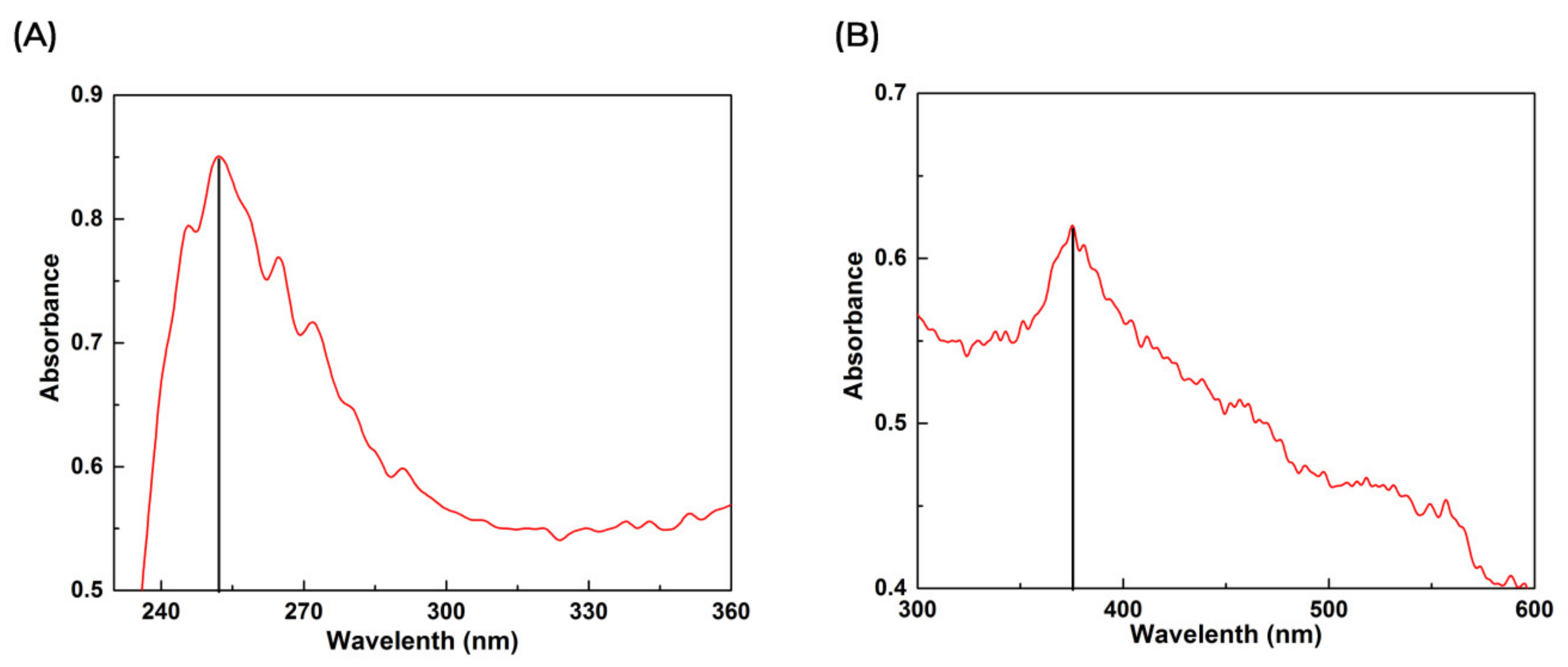
2.5.1. Ultraviolet Spectroscopy (UV-Vis)
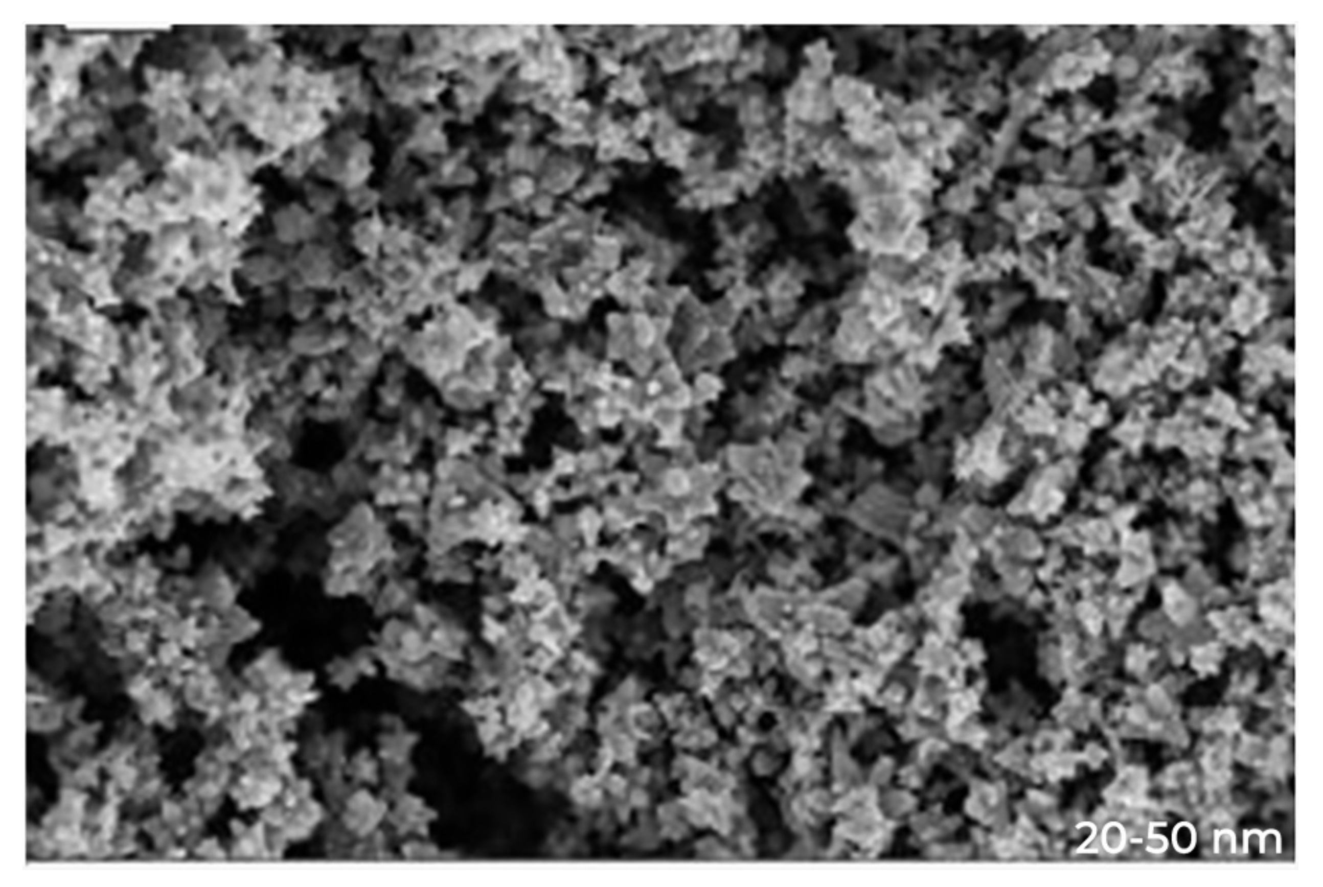
2.5.2. SEM Analysis
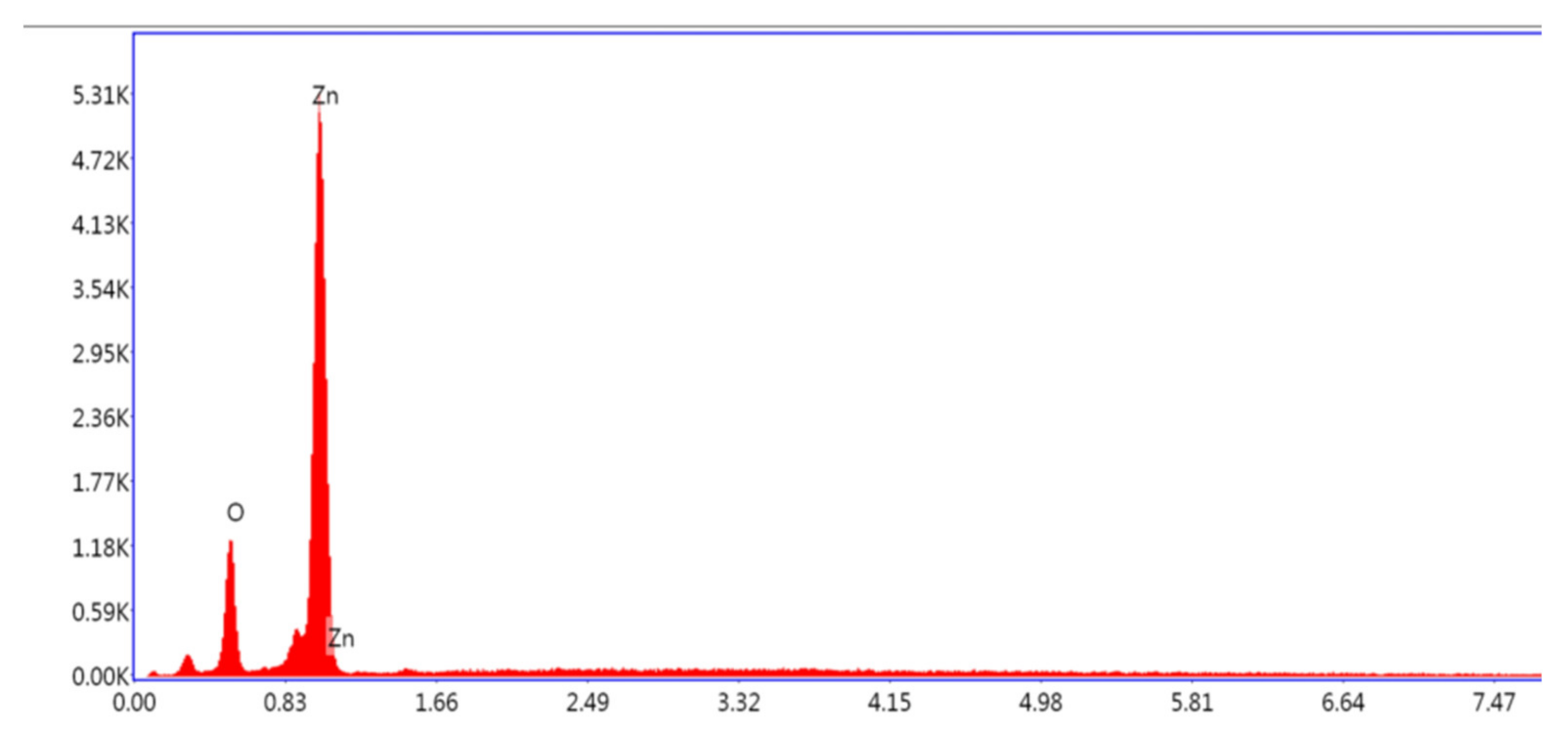
2.5.3. EDX Analysis
2.5.4. FTIR Analysis
2.6. Enzyme Inhibition Assays
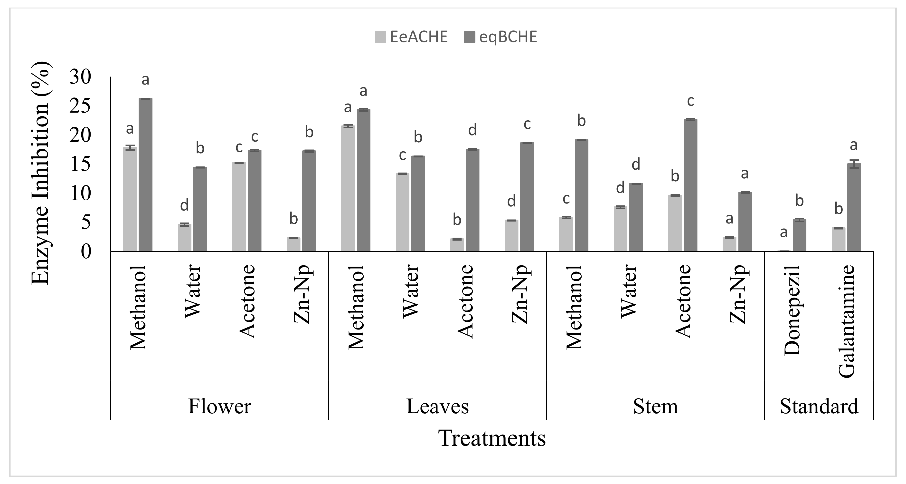
2.6.1. Inhibition of ACHE and BCHE
2.6.2. Inhibition of Urease
2.7. Antimicrobial Activity of Plant Extracts and Zn Nanoparticles
2.7.1. Disc Diffusion Method
2.7.2. Determining Minimum Inhibitory Concentration (MIC)
2.8. Statistical Analysis
3. Results
3.1. Analysis of Green synthesis of ZnO NPs
3.1.1. Visual and Ultraviolet Spectroscopy (UV-Vis)
3.1.2. Scanning Electron Microscopy (SEM)
3.1.3. Energy Dispersive X-ray Analysis (EDX)
3.1.4. FT-IR Spectroscopy
3.2. Comparison of Plant Extracts and Nanoparticles
3.3. Determination of AChE and BChE Inhibitory Activity
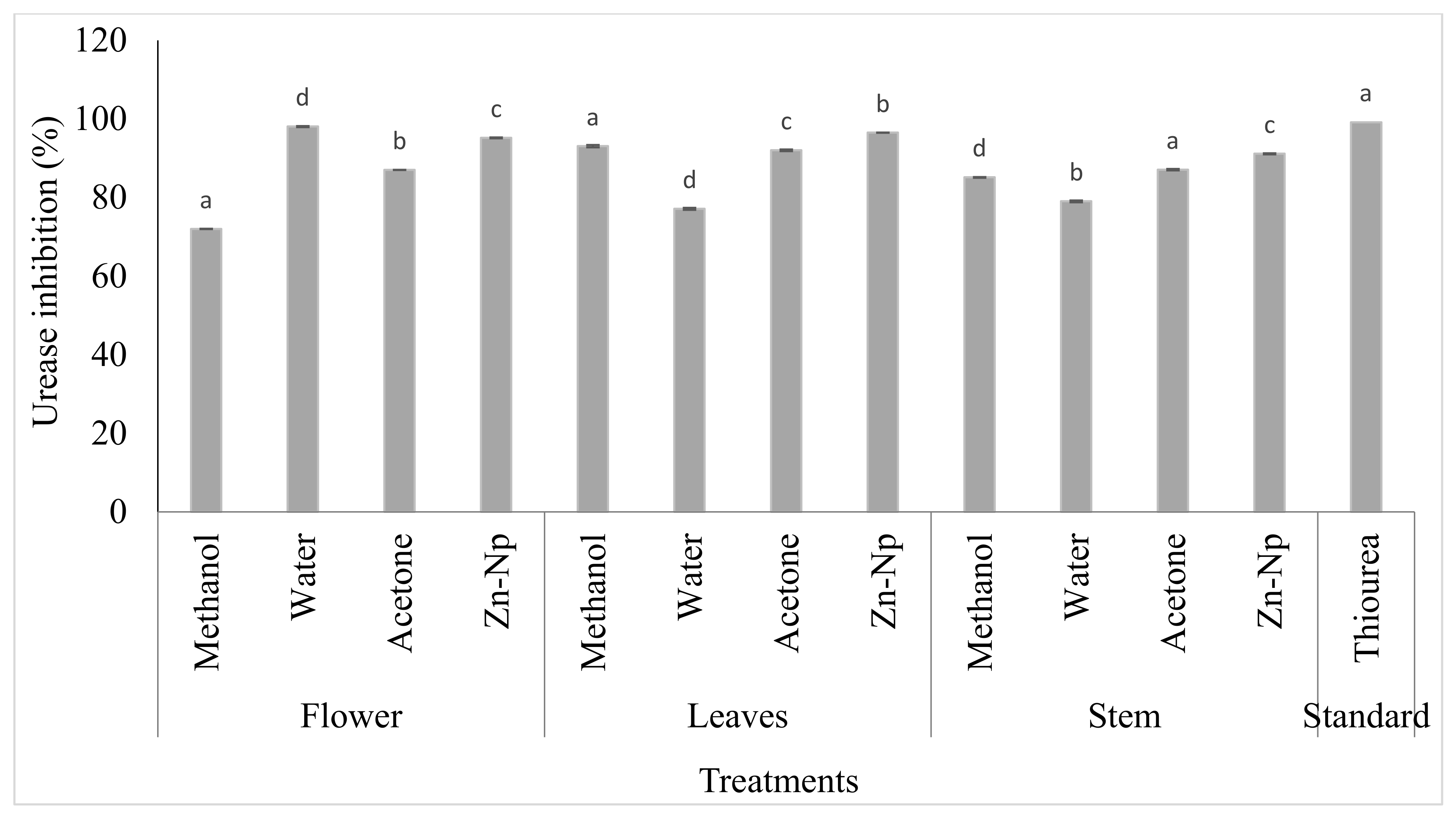
3.4. Urease Assay
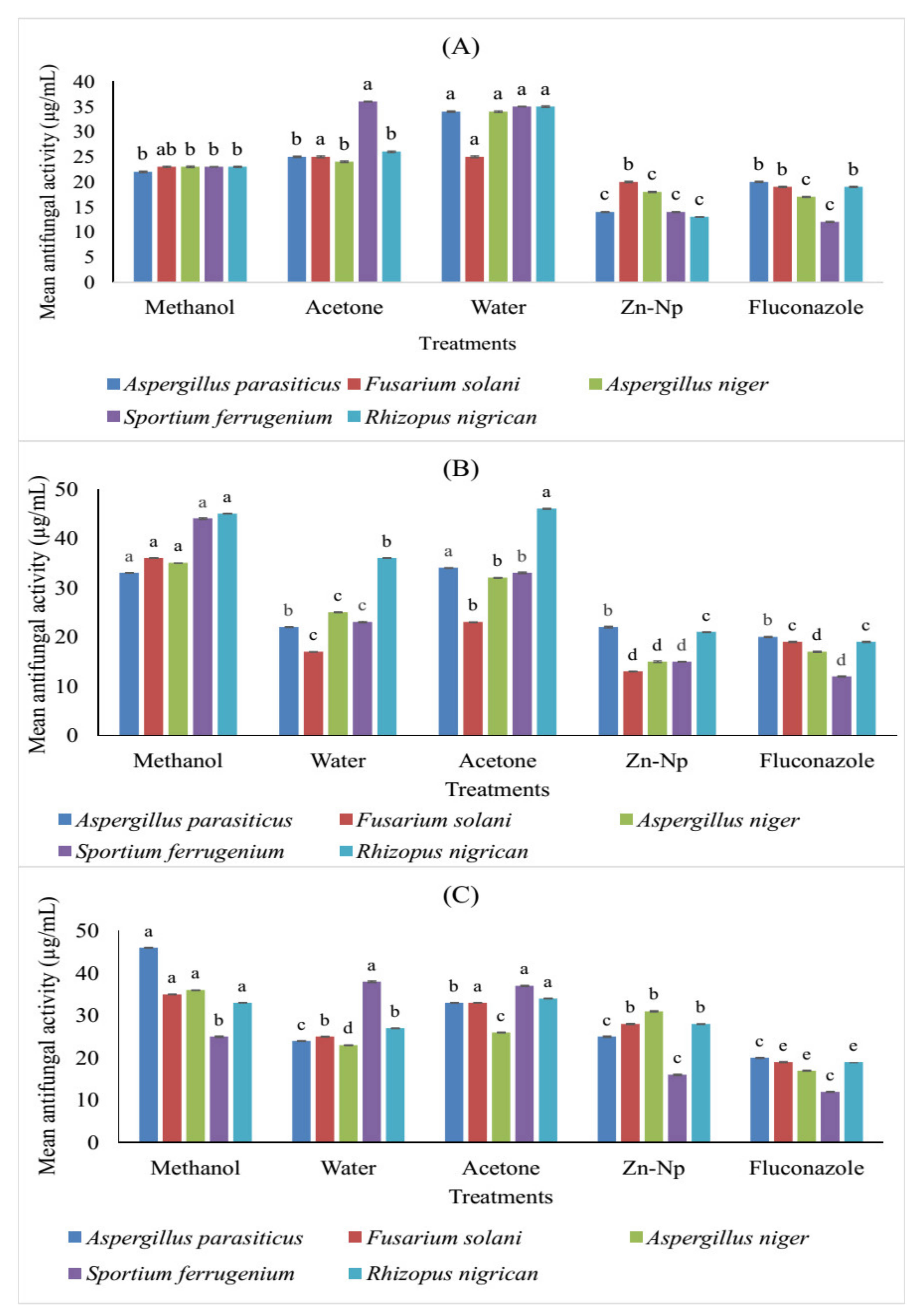
3.5. Antifungal Activity of Zinc Nanoparticles and Plant Extracts
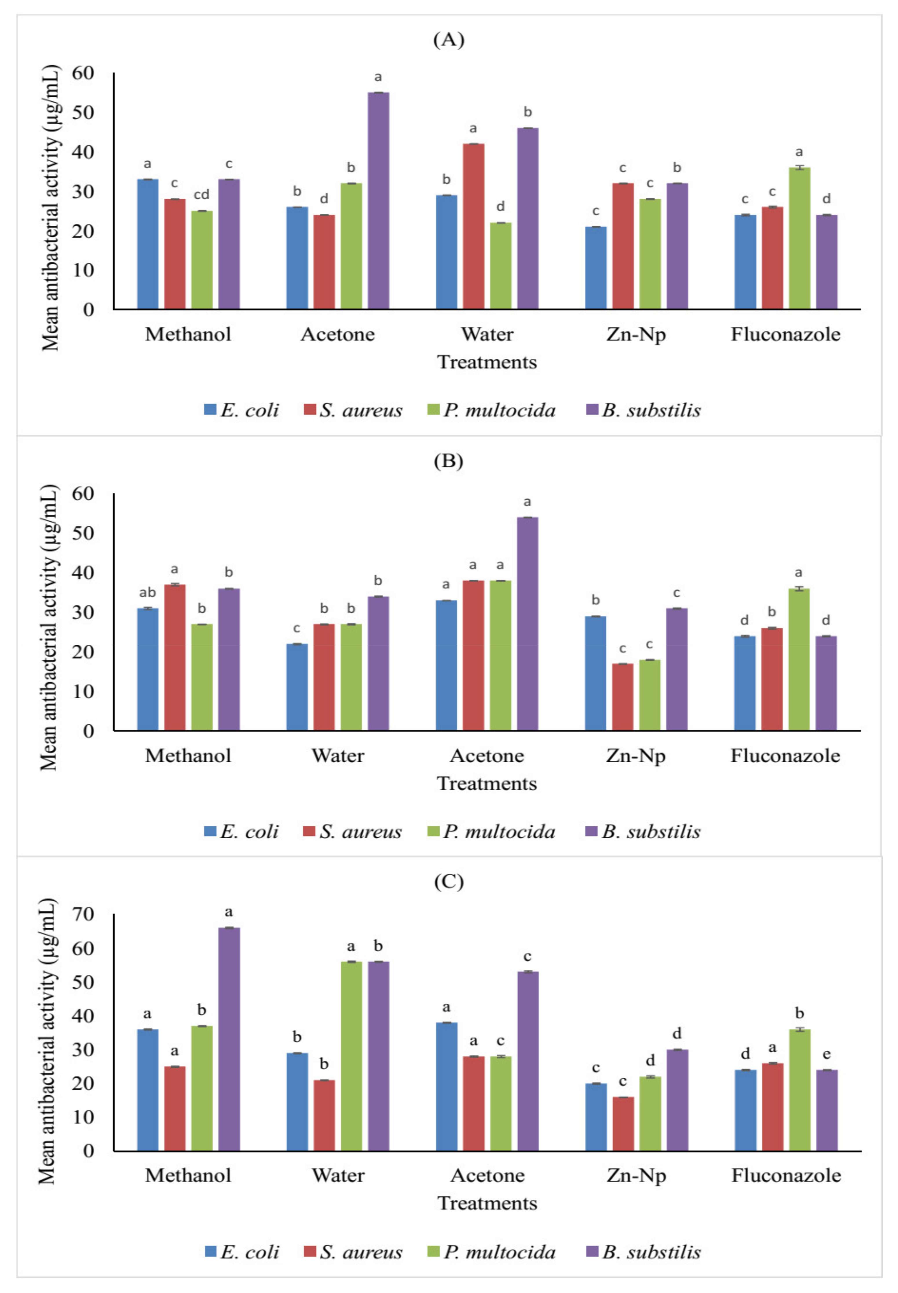
3.6. Antimicrobial Activity of Zinc Nanoparticles and Plant Extracts
4. Discussion
5. Conclusions
Author Contributions
Funding
Informed Consent Statement
Data Availability Statement
Acknowledgments
Conflicts of Interest
References
- Hu, Y.; Niemeyer, C.M. From DNA nanotechnology to material systems engineering. Adv. Mater. 2019, 31, 1806294. [Google Scholar] [CrossRef] [PubMed] [Green Version]
- Subramani, K.; Ahmed, W. Nanotechnology and its applications in dentistry—An introduction. In Emerging Nanotechnologies in Dentistry; William Andrew Publishing: Norwich, NY, USA, 2018; pp. 1–15. [Google Scholar]
- Kaplan, S.; Radin, J. Bounding an emerging technology: Para-scientific media and the Drexler-Smalley debate about nanotechnology. Soc. Stud. Sci. 2011, 41, 457–485. [Google Scholar] [CrossRef]
- Auffan, M.; Rose, J.; Bottero, J.Y.; Lowry, G.V.; Jolivet, J.P.; Wiesner, M.R. Towards a definition of inorganic nanoparticles from environmental, health and safety perspective. Nat. Nanotechnol. 2009, 4, 634–641. [Google Scholar] [PubMed]
- Christian, P.; Von der Kammer, F.; Baalousha, M.; Hofmann, T. Nanoparticles: Structure, properties, preparation and behaviour in environmental media. Ecotoxicology 2008, 17, 326–343. [Google Scholar]
- Suci, P.A.; Berglund, D.L.; Liepold, L.; Brumfield, S.; Pitts, B.; Davison, W.; Oltrogge, L.; Hoyt, K.O.; Codd, S.; Stewart, P.S.; et al. High-density targeting of a viral multifunctional nanoplatform to a pathogenic, biofilm-forming bacterium. Chem. Biol. 2007, 14, 387–398. [Google Scholar] [CrossRef] [Green Version]
- Cañas-Carrell, J.E.; Li, S.; Parra, A.M.; Shrestha, B. Metal oxide nanomaterials: Health and environmental effects. In Health and Environmental Safety of Nanomaterials; Woodhead Publishing: Sawston, UK, 2014; pp. 200–221. [Google Scholar]
- Dreaden, E.C.; Alkilany, A.M.; Huang, X.; Murphy, C.J.; El-Sayed, M.A. The golden age: Gold nanoparticles for biomedicine. Chem. Soc. Rev. 2012, 41, 2740–2779. [Google Scholar] [CrossRef] [Green Version]
- Konishi, Y.; Ohno, K.; Saitoh, N.; Nomura, T.; Nagamine, S.; Hishida, H.; Takahashi, Y.; Uruga, T. Bioreductive deposition of platinum nanoparticles on the bacterium Shewanella algae. J. Biotechnol. 2007, 128, 648–653. [Google Scholar] [CrossRef]
- Willner, I.; Baron, R.; Willner, B. Growing metal nanoparticles by enzymes. Adv. Mater. 2006, 18, 1109–1120. [Google Scholar] [CrossRef]
- Ahmad, N.; Sharma, S.; Singh, V.N.; Shamsi, S.F.; Fatma, A.; Mehta, B.R. Biosynthesis of silver nanoparticles from Desmodiumtriflorum: A novel approach towards weed utilization. Biotechnol. Res. Int. 2011, 11, 1–10. [Google Scholar] [CrossRef] [Green Version]
- Nethavhanani, T. Synthesis of Zinc Oxide Nanoparticles by a Green Process and the Investigation of Their Physical Properties. Master’s Thesis, University of Western Cape, Cape Town, South Africa, 2017. [Google Scholar]
- Duan, H.; Wang, D.; Li, Y. Green chemistry for nanoparticle synthesis. Chem. Soc. Rev. 2015, 44, 5778–5792. [Google Scholar] [CrossRef]
- Ambika, S.; Sundrarajan, M. Green biosynthesis of ZnO nanoparticles using Vitexnegundo L. extract: Spectroscopic investigation of interaction between ZnO nanoparticles and human serum albumin. J. Photochem. Photobiol. B: Biol. 2015, 149, 143–148. [Google Scholar] [CrossRef]
- Malik, P.; Shankar, R.; Malik, V.; Sharma, N.; Mukherjee, T.K. Green chemistry based benign routes for nanoparticle synthesis. J. Nanoparticles 2014, 2014, 302429. [Google Scholar] [CrossRef] [Green Version]
- Rai, M.; Yadav, A.; Gade, A. CRC 675—Current trends in phytosynthesis of metal nanoparticles. Crit. Rev. Biotechnol. 2008, 28, 277–284. [Google Scholar] [CrossRef]
- Zhao, F.; Yao, D.; Guo, R.; Deng, L.; Dong, A.; Zhang, J. Composites of polymer hydrogels and nanoparticulate systems for biomedical and pharm. Nanomaterials 2015, 5, 2054–2130. [Google Scholar] [CrossRef] [PubMed] [Green Version]
- Taylor, E.; Webster, T.J. Reducing infections through nanotechnology and nanoparticles. Int. J. Nanomed. 2011, 6, 1463. [Google Scholar]
- Liu, Y.P.; Zhao, Y.L.; Feng, T.; Cheng, G.G.; Zhang, B.H.; Li, Y.; Cai, X.H.; Luo, X.D. Melosuavines A–H, cytotoxic bisindole alkaloid derivatives from Melodinus suaveolens. J. Nat. Prod. 2013, 76, 2322–2329. [Google Scholar] [CrossRef]
- Choi, B.J.; Jeong, D.S.; Kim, S.K.; Rohde, C.; Choi, S.; Oh, J.H.; Kim, H.J.; Hwang, C.S.; Szot, K.; Waser, R.; et al. Resistive switching mechanism of TiO2 thin films grown by atomic-layer deposition. J. Appl. Phys. 2005, 98, 033715. [Google Scholar] [CrossRef] [Green Version]
- Chen, S.F.; Li, J.P.; Qian, K.; Xu, W.P.; Lu, Y.; Huang, W.X.; Yu, S.H. Large scale photochemical synthesis of M@ TiO2 nanocomposites (M= Ag, Pd, Au, Pt) and their optical properties, CO oxidation performance, and antibacterial effect. Nano Res. 2010, 3, 244–255. [Google Scholar] [CrossRef] [Green Version]
- Yang, J.; Li, Q.; Sun, D.; Lu, Y.; Su, Y.; Yang, X.; Hong, J. Biosynthesis of silver and gold nanoparticles by novel sundried Cinnamomumcamphora leaf. Nanotechnology 2007, 18, 105104. [Google Scholar]
- Scheffer, R.P. Role of toxinsin evolution and ecology of plantpathogenic fungi. Experientia 1991, 47, 804–811. [Google Scholar] [CrossRef]
- Zhang, F.; Wu, X.; Chen, Y.; Lin, H. Application of silver nanoparticles to cotton fabric as an antibacterial textile finish. Fibers Polym. 2009, 10, 496–501. [Google Scholar] [CrossRef] [Green Version]
- Sani, A.; Hassan, D.; Khalil, A.T.; Mughal, A.; El-Mallul, A.; Ayaz, M.; Maaza, M. Floral extracts-mediated green synthesis of NiO nanoparticles and their diverse pharmacological evaluations. J. Biomol. Struct. Dyn. 2020, 39, 4133–4147. [Google Scholar] [CrossRef]
- Sahu, P.K.; Giri, D.D.; Singh, R.; Pandey, P.; Gupta, S.; Shrivastava, A.K.; Pandey, K.D. Therapeutic and medicinal uses of Aloe vera: A review. Pharmacol. Pharm. 2013, 4, 599. [Google Scholar] [CrossRef]
- Bingtao, A.; Peddimounika, R.; Rohitha, S.R.M.; Premkumar, P. Evaluation of antioxidant and antibacterial efficacy of flowers of Calotropisgigantea. Asian J. Pharm. Pharmacol. 2015, 4, 821–824. [Google Scholar]
- Meena, A.K.; Bansal, P.; Kumar, S. Plants-herbal wealth as a potential source of ayurvedic drugs. Asian J. Tradit. Med. 2009, 4, 152–170. [Google Scholar]
- Kareem, S.O.; Akpan, I.; Osho, M.B. Calotropisprocera (Sodom apple)––A potential material for enzyme purification. Bioresour. Technol. 2003, 87, 133–135. [Google Scholar] [CrossRef]
- Joseph, B.; George, J.; Jeevitha, M.V.; Charles, S. Pharmacological and biological overview on Calotropis gigantean: A comprehensive review. Int. Res. J. Pharm. Appl. Sci. 2013, 3, 219–223. [Google Scholar]
- Kumar, G.; Karthik, L.; Rao, K.V.B. A review on pharmacological and phytochemical profile of Calotropisgigantea Linn. Pharmacologyonline 2011, 1, 1–8. [Google Scholar]
- Creno, R.J.; Wenk, R.E.; Bohlig, P. Automated micro measurement of urea using urease and the Berthelot reaction. Am. J. Clin. Pathol. 1970, 54, 828–832. [Google Scholar] [CrossRef]
- El-Mohamedy, R.S.; Abdalla, A.M. Evaluation of antifungal activity of Moringa oleifera extracts as natural fungicide against some plant pathogenic fungi in vitro. J. Agric. Technol. 2014, 10, 963–982. [Google Scholar]
- Dhingra, O.D.; Sinclair, J.B. Basic Plant Pathology Methods; CRC Press Inc.: Boca Raton, FL, USA, 1985; pp. 132–163. [Google Scholar]
- Steel, R.G.; Torrie, J.H.; Dickey, D.A. Principles and Procedures of Statistics: A Biometrical Approach, 3rd ed.; McGraw Hill Book International Co.: Singapore, 1997. [Google Scholar]
- Ali, K.; Dwivedi, S.; Azam, A.; Saquib, Q.; Al-Said, M.S.; Alkhedhairy, A.A.; Musarrat, J. Aloe vera extract functionalized zinc oxide nanoparticles as nanoantibiotics against multi-drug resistant clinical bacterial isolates. J. Colloid Interface Sci. 2016, 472, 145–156. [Google Scholar] [CrossRef]
- Elumalai, K.; Velmurugan, S. Green synthesis, characterization and antimicrobial activities of zinc oxide nanoparticles from the leaf extract of Azadirachta indica (L.). Appl. Surf. Sci. 2015, 345, 329–336. [Google Scholar] [CrossRef]
- Benelli, G. Plant-mediated biosynthesis of nanoparticles as an emerging tool against mosquitoes of medical and veterinary importance: A review. Parasitol. Res. 2016, 115, 23–34. [Google Scholar] [CrossRef]
- Benelli, G. Plant-borne ovicides in the fight against mosquito vectors of medical and veterinary importance: A systematic review. Parasitol. Res. 2015, 114, 3201–3212. [Google Scholar] [CrossRef]
- Banumathi, B.; Malaikozhundan, B.; Vaseeharan, B. Invitro acaricidal activity of ethnoveterinary plants and green synthesis of zinc oxide nanoparticles against Rhipicephalus (Boophilus) microplus. Vet. Parasitol. 2016, 216, 93–100. [Google Scholar] [CrossRef]
- Patil, B.N.; Taranath, T.C. Limoniaacidissima L. leaf mediated synthesis of zinc oxide nanoparticles: A potent tool against Mycobacterium tuberculosis. Int. J. Mycobacteriology 2016, 5, 197–204. [Google Scholar] [CrossRef] [Green Version]
- Nagajyothi, P.C.; Sreekanth, T.V.M. Green synthesis of metallic and metal oxide nanoparticles and their antibacterial activities. In Green Processes for Nanotechnology; Springer: Cham, Germany, 2015; pp. 99–117. [Google Scholar]
- Kwon, Y.J.; Kim, K.H.; Lim, C.S.; Shim, K.B. Characterization of ZnO nanopowders by the polymerized complex method via an organochemical route. J. Ceram. Process. Res. 2002, 3, 146–149. [Google Scholar]
- Murugan, K.; Benelli, G.; Ayyappan, S.; Dinesh, D.; Panneerselvam, C.; Nicoletti, M.; Hwang, J.S.; Kumar, P.M.; Subramaniam, J.; Suresh, U. Toxicity of seaweed-synthesized silver nanoparticles against the filariasis vector Culex quinquefasciatus and its impact on predation efficiency of the cyclopoid crustacean Mesocyclopslongisetus. Parasitol. Res. 2015, 114, 2243–2253. [Google Scholar] [CrossRef]
- Murugan, K.; Benelli, G.; Panneerselvam, C.; Subramaniam, J.; Jeyalalitha, T.; Dinesh, D.; Nicoletti, M.; Hwang, J.S.; Suresh, U.; Madhiyazhagan, P. Cymbopogon citratus-synthesized gold nanoparticles boost the predation efficiency of copepod Mesocyclopsaspericornis against malaria and dengue mosquitoes. Exp. Parasitol. 2015, 153, 129–138. [Google Scholar] [CrossRef]
- Sathyavathi, R.; Krishna, M.B.; Rao, S.V.; Saritha, R.; Rao, D.N. Biosynthesis of silver nanoparticles using Coriandrum sativum leaf extract and their application in nonlinear optics. Adv. Sci. Lett. 2010, 3, 138–143. [Google Scholar] [CrossRef] [Green Version]
- Sportelli, M.C.; Izzi, M.; Loconsole, D.; Sallustio, A.; Picca, R.A.; Felici, R.; Chironna, M.; Cioffi, N.; 2022. On the efficacy of ZnO nanostructures against SARS-CoV-2. Int. J. Mol. Sci. 2022, 23, 3040. [Google Scholar] [CrossRef]
- Reynisson, E.; Lauzon, H.L.; Magnússon, H.; Jónsdóttir, R.; Ólafsdóttir, G.; Marteinsson, V.; Hreggviðsson, G.Ó. Bacterial composition and succession during storage of North-Atlantic cod (Gadusmorhua) at superchilled temperatures. BMC Microbiol. 2009, 9, 250. [Google Scholar] [CrossRef] [Green Version]
- Mittal, A.K.; Chisti, Y.; Banerjee, U.C. Synthesis of metallic nanoparticles using plant extracts. Biotechnol. Adv. 2013, 31, 346–356. [Google Scholar] [CrossRef]
- Eloff, J.N. Avoiding pitfalls in determining antimicrobial activity of plant extracts and publishing the results. BMC Complement. Altern. Med. 2019, 19, 1–8. [Google Scholar] [CrossRef] [Green Version]
- Türkan, F.; Atalar, M.N.; Aras, A.; Gülçin, İ.; Bursal, E. ICP–MS and HPLC analyses, enzyme inhibition and antioxidant potential of Achillea schischkinii Sosn. Bioorganic Chem. 2020, 94, 103333. [Google Scholar] [CrossRef]
- Mukherjee, P.K.; Kumar, V.; Mal, M.; Houghton, P.J. Acetylcholinesterase inhibitors from plants. Phytomedicine 2007, 14, 289–300. [Google Scholar] [CrossRef]
- Khalil, A.T.; Ovais, M.; Ullah, I.; Ali, M.; Shinwari, Z.K.; Hassan, D.; Maaza, M. Sageretiathea (Osbeck.) modulated biosynthesis of NiO nanoparticles and their in vitro. pharmacognostic, antioxidant and cytotoxic potential. Artif. Cells Nanomed. Biotechnol. 2018, 46, 838–852. [Google Scholar] [CrossRef] [Green Version]
- Kwak, J.I.; Yoon, S.J.; An, Y.J. Long-term effects of ZnO nanoparticles on exoenzyme activities in planted soils. Environ. Eng. Res. 2017, 22, 224–229. [Google Scholar] [CrossRef] [Green Version]
- Mason, C.; Vivekanandhan, S.; Misra, M.; Mohanty, A.K. Switchgrass (Panicumvirgatum) extract mediated green synthesis of silver nanoparticles. World J. Nano Sci. Eng. 2012, 2, 47. [Google Scholar] [CrossRef]
- AJ Awllia, J.; Al-Ghamdi, M.; Huwait, E.; Javaid, S.; Rasheed, S.; IqbalChoudhary, M. Flavonoids as natural inhibitors of jack bean urease Enzyme. Lett. Drug Des. Discov. 2016, 13, 243–249. [Google Scholar] [CrossRef]
- Ellman, G.L.; Courtney, K.D.; Andres Jr, V.; Featherstone, R.M. A new and rapid colorimetric determination of acetylcholinesteraseactivity. Biochem. Pharmacol. 1961, 7, 88–95. [Google Scholar] [CrossRef]
- Samoisy, A.K.; Mahomoodally, M.F. Ethnopharmacological analysis of medicinal plants used against noncommunicable diseases in Rodrigues Island, Indian Ocean. J. Ethnopharmacol. 2015, 173, 20–38. [Google Scholar] [CrossRef]
- Kumar, G.; Karthik, L.; Rao, K.B.; Kirthi, A.V.; Rahuman, A.A. Larvicidal, repellent and ovicidal activity of Calotropisgigantea against Culexgelidus, Culextritaeniorhynchus (Diptera: Culicidae). Int. J. Agric. Technol. 2012, 8, 869–880. [Google Scholar]
- Bharathi, P.; Thomas, A.; Thomas, A.; Krishnan, S.; Ravi, T.K. Anti bacterial activity of leaf extracts of Calotropisgigantea Linn. against certain gram-negative and gram-positive bacteria. Int. J. Chem. Sci. 2011, 9, 919–923. [Google Scholar]
- Alam, M.A.; Habib, M.R.; Nikkon, R.; Rahman, M.; Karim, M.R. Antimicrobial Activity of Akanda (Calotropisgigantea L.) on Some Pathogenic Bacteria. Bangladesh J. Sci. Ind. Res. 2008, 43, 397–404. [Google Scholar] [CrossRef]


| Plant Parts | Solvents | EeAChE and eqBChE SI * | IC50 µM |
|---|---|---|---|
| Flower | Methanol | 1.3 | 49.03 ± 0.08 |
| Acetone | 1.2 | b 19.03 ± 0.06 | |
| ZnNPs | 1.3 | 16.1 ± 0.15 | |
| Water | 3.9 | 30.07 ± 0.18 | |
| Leaves | Methanol | 1.2 | 43.06 ± 0.16 |
| Acetone | 8.6 | 25.03 ± 0.09 | |
| ZnNPs | 1.4 | 22.1 ± 0.03 | |
| Water | 1.3 | 23.14 ± 0.26 | |
| Stem | Methanol | 3.3 | b 17.15 ± 0.07 |
| Acetone | 3.8 | 23.08 ± 0.11 | |
| ZnNPs | 1.7 | 30.1 ± 0.13 | |
| Water | 3.8 | 31.04 ± 0.17 | |
| A** | Donepezil | 180 | - |
| Galantamine | 3.7 | - | |
| Thiourea | - | 21.25 ± 0.17 |
Publisher’s Note: MDPI stays neutral with regard to jurisdictional claims in published maps and institutional affiliations. |
© 2022 by the authors. Licensee MDPI, Basel, Switzerland. This article is an open access article distributed under the terms and conditions of the Creative Commons Attribution (CC BY) license (https://creativecommons.org/licenses/by/4.0/).
Share and Cite
Farooq, A.; Khan, U.A.; Ali, H.; Sathish, M.; Naqvi, S.A.H.; Iqbal, S.; Ali, H.; Mubeen, I.; Amir, M.B.; Mosa, W.F.A.; et al. Green Chemistry Based Synthesis of Zinc Oxide Nanoparticles Using Plant Derivatives of Calotropis gigantea (Giant Milkweed) and Its Biological Applications against Various Bacterial and Fungal Pathogens. Microorganisms 2022, 10, 2195. https://doi.org/10.3390/microorganisms10112195
Farooq A, Khan UA, Ali H, Sathish M, Naqvi SAH, Iqbal S, Ali H, Mubeen I, Amir MB, Mosa WFA, et al. Green Chemistry Based Synthesis of Zinc Oxide Nanoparticles Using Plant Derivatives of Calotropis gigantea (Giant Milkweed) and Its Biological Applications against Various Bacterial and Fungal Pathogens. Microorganisms. 2022; 10(11):2195. https://doi.org/10.3390/microorganisms10112195
Chicago/Turabian StyleFarooq, Ammara, Umair A. Khan, Haider Ali, Manda Sathish, Syed Atif Hasan Naqvi, Shehzad Iqbal, Haider Ali, Iqra Mubeen, Muhammad Bilal Amir, Walid F. A. Mosa, and et al. 2022. "Green Chemistry Based Synthesis of Zinc Oxide Nanoparticles Using Plant Derivatives of Calotropis gigantea (Giant Milkweed) and Its Biological Applications against Various Bacterial and Fungal Pathogens" Microorganisms 10, no. 11: 2195. https://doi.org/10.3390/microorganisms10112195