Article

One Health Approach to Rickettsiosis: A Five-Year Study on Spotted Fever Group Rickettsiae in Ticks Collected from Humans, Animals and Environment

1
Istituto Zooprofilattico Sperimentale dell’Umbria e delle Marche “Togo Rosati”, Via Gaetano Salvemini, 06126 Perugia, Italy
2
Department of Veterinary Medicine, University of Perugia, Via San Costanzo 4, 06126 Perugia, Italy
*
Authors to whom correspondence should be addressed.
Microorganisms 2022, 10(1), 35; https://doi.org/10.3390/microorganisms10010035
Submission received: 19 November 2021 / Revised: 17 December 2021 / Accepted: 23 December 2021 / Published: 25 December 2021
(This article belongs to the Special Issue Advances in Tick-Borne Diseases Research)

Abstract

:
The spotted fever group of Rickettsiae is a heterogeneous group of Rickettsiae transmitted by ticks, causing similar diseases in humans (spotted fever). Until recently, it was supposed that a single pathogenic tick-borne SFG Rickettsia circulated in each different geographic area and that R. conorii subsp. conorii was the SFG Rickettsiae circulating in Italy, but in the last decade, thanks to molecular diagnostic, several different Rickettsia species, previously not considered pathogenic for decades, have been isolated from ticks and definitively associated to human disease, also in Italy. The present survey was carried out with the aim of investigating the presence of different SFG Rickettsia species in a geographic area where no information was available. Ticks collected from animals submitted to necropsy, removed from humans in local hospitals and collected from the environment were identified and tested by PCR for Rickettsia spp. based on the gltA gene, and positive PCR products were sequenced. A total of 3286 ticks were collected. Fifteen tick species were recognized, the most represented (79.52%) species in the collection was Ixodes ricinus, followed by Rhipicephalus sanguineus (9.13%). The overall prevalence of Rickettsia infection was 7.58%. Eight species of Rickettsia were identified, the most frequent was R. monacensis (56%), followed by R. helvetica (25.50%). Noteworthy, is the detection in the present study of Rrhipicephali, detected only twice in Italy. These are the first data available on SFG Rickettsiae circulation in the study area and they can be considered as starting point to assess the possible risk for humans.

1. Introduction

Rickettsioses are worldwide vector borne zoonoses caused by obligate intracellular Gram-native bacteria (order, Rickettsiales; family, Rickettsiaceae; genera Rickettsia and Orientia) infesting eukaryotic cells. They are among the oldest known vector-borne diseases [1]. Phylogenetics based on whole-genome analysis data determined that the genus Rickettsia comprises 27 recognized species and several dozen as-yet uncharacterized strains. According to Szokoli and collaborators [2], the Rickettsia genus could be divided into four groups: the spotted fever group (R. rickettsii, R. conorii, R. parkeri, and several others), typhus group (R. prowazekii and R. typhi), ancestral group (R. bellii and R. canadensis, not known to be pathogenic), and transitional group (R. akari, R. australis, and R. felis) [3]. The spotted fever group (SFG) is a heterogeneous group of Rickettsiae transmitted by ticks causing a similar disease in humans (spotted fever). Until recently, it was supposed that a single pathogenic tick-borne SFG Rickettsia circulated in each different geographic area, but in the last decade, to make puzzling the picture of SFG Rickettsiae in the world, several different Rickettsia species isolated from ticks, previously not considered pathogenic for decades, were definitively associated to human disease, as happened to R. massiliae [4]. Ticks are obligate hematophagous acarines that parasitize all vertebrates in almost every region of the world [5], they are second to mosquitoes as major vectors of pathogens to humans and play a primary role in disease transmission in animals [6]. However, given the transovarial transmission (TOT) with a high level of filial infection rate (FIR) described in ticks for the associated Rickettsia species, they are not only the vectors of SFG Rickettsiae, but they can be considered their main reservoir, even if for some SFG Rickettsia species, an amplifier role is suspected for some vertebrate species, such as rodents [4].
For this reason, SFG Rickettsia species and their respective vectors are very closely associated and, as consequence, the circulation of a SFG Rickettsia species in a territory strictly depends on the presence of its vectors and, consequently, is affected by the hosts’ population dynamics [7]. For many years, it was assumed that R. conorii subsp. conorii was the only agent of spotted fever circulating in Italy and that the pathognomonic clinical manifestation of Mediterranean spotted fever (MSF) was the only one developed after the SFG Rickettsia infection in humans. MSF, in fact, is constantly reported in warmer areas of the country during summer according to the biology of the tick Rhipicepahlus sanguineus, which, at those latitudes, behaves as an exophilic tick [1,4].
Thanks to the improved diagnostic skills and, mainly, to the use of molecular tools, in the last decade different SFG Rickettsiae have been identified in the Italian territory, including Rickettsia slovaca, Rickettsia aeschlimanni, Rickettsia massilliae, Rickettsia monacensis, Rickettsia conorii subsp. Israelensis, Rickettsia conorii subsp. Indica, Rickettsia raoultii, Rickettsia helvetica, Rickettsia hoogstraalii, R. peacockii, R. rhiphicephali, and R. felis [1,4,8,9,10,11,12,13].
Recently, in a retrospective study, 5989 cases of non-typhus rickettsioses were identified from 2001 to 2015 in humans in Italy, giving an average annual incidence of 0.88 (95%CI: 0.80; 0.96) per 100,000 [14]. In the Marche region, 72 human cases were reported in the same study and clinical cases are continuously reported in the region. As for other tick-borne diseases, a large part of the diagnosis of rickettsiosis in humans is made by clinical symptoms and serology, lacking the identification of the pathogen involved, and thus, the true incidence is likely to be underestimated, leading to the hypothesis that there are seven times more cases than officially reported [15].
The present study intended to investigate the circulation of SFG Rickettsiae in ticks collected in the Marche region and the bordering Republic of San Marino by specific molecular analysis and sequencing data, providing knowledge useful to better define the complex puzzle of SFG Rickettsiae circulation

2. Materials and Methods
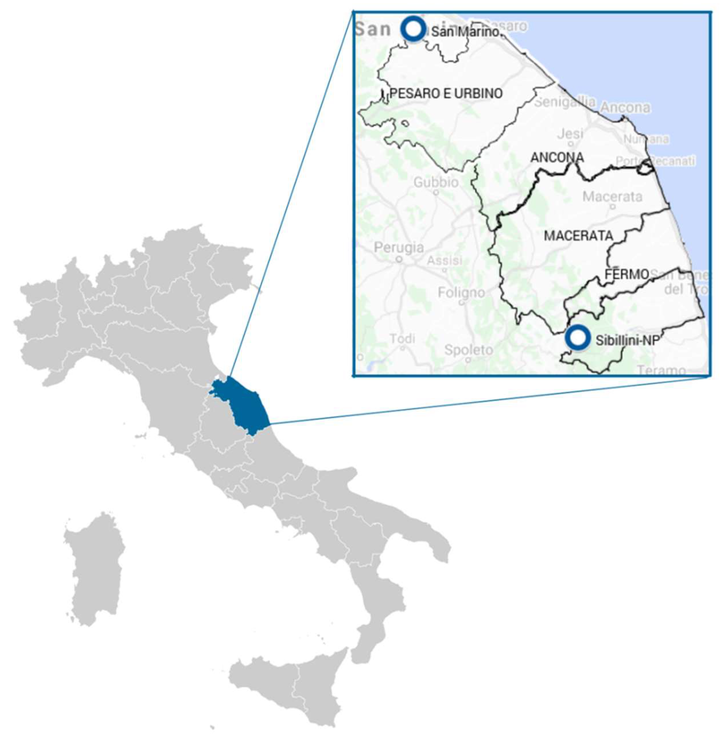
2.1. Study Areas

The Marche region is located in central Italy and extends over an area of 9694 square kilometers of the central Adriatic slope. Most of the region is mountainous or hilly, the main features being the Apennine chain along the internal boundary and an extensive system of hills rapidly descending towards the Adriatic. With the only exception of Monte Vettore (2476 m a.s.l.), the mountains do not exceed 2400 m a.s.l. The territory covers different environments characterized by high biodiversity in terms of wildlife, with several natural parks, such as Monti Sibillini National Park (Monti Sibillini NP) (Figure 1), a natural protected area of 71,437 square kilometers located in the Apenninic area of the region. Meanwhile, rural farming is common in the area, especially for small ruminants and beef cattle. Republic San Marino is an independent state landlocked by Italy, on the border between the regions of Emilia Romagna and Marche and about 10 km from the Adriatic coast (Figure 1). It is part of the Apennine mountain range and shares the same environmental features with the northern part of Marche region.

2.2. Sampling and Tick Identification

The Istituto Zooprofilattico Sperimentale dell’Umbria e delle Marche (IZSUM) is a veterinary public health laboratory the operates in the Umbria and Marche regions.
It receives carcasses of domestic and wild animals for diagnostic purposes and in the framework of specific animal health surveillance and control programs. Regarding zoonoses transmitted by ticks, in the Marche region, the Center for Entomology and Vector-borne Diseases (CERVe), located at IZSUM, has activated a service for tick identification and pathogen testing in ticks removed from humans.
In the period 2011–2015, ticks were collected from animal carcasses, humans, and the environment.
Ticks were collected from animals during the inspection before necropsy; carcasses were systematically examined for the presence of ticks around the soft parts of their bodies (ears, the inner sides of the hind and forelegs, perineal area), neck, and thorax.
Ticks recovered from humans were removed from patients by hospital medical staff and sent to IZSUM laboratory for species identification and pathogen detection. No clinical data of patients were available to the laboratory for privacy reasons.
Free-living ticks were collected in 22 sampling sites placed in two different geographical areas: 18 sites in the Republic of San Marino (RSM), and 4 sites in Monti Sibillini National Park (Figure 1). Dragging was performed during spring and fall using a 1 m2 white blanket, stopping every 2.5 m to prevent tick detachment. All ticks were immediately placed in flacons with 70% ethanol, properly labeled, and sent to IZSUM laboratory. Tick identification was performed using taxonomic keys available in the literature [16]. Before DNA extraction, immature free-living ticks were pooled according to species, dragging session details, and developmental stage, with a maximum of 100 larvae and of 50 nymphs in each pool, while adult ticks were tested singularly.

2.3. DNA Extraction and PCR

The single adult ticks and pooled samples were homogenized by TissueLyser (Qiagen) and DNA was extracted using the QIAamp® cador® Pathogen Mini Kit (Qiagen, Hilden, Germany), according to manufacturer’s protocol. Quality and quantity were tested with an Eppendorf biophotometer.
The extracted DNA was examined for the presence of Rickettsia spp. citrate synthase-encoding gene (gltA) by using RpCs877 F and RpCs1258 R primers (5′-GGGGGCCTGCTCACGGCGG-3′; 5′-AATTGCAAAAAGTACAGTGAACA-3′) and amplifying 381 base pair (bp) fragments as described by Roux and collaborators [17].
The reaction was performed in a total volume of 50 μL containing: 1× PCR buffer, 2.0 mM of MgCl2, 0.2 mM of dNTP, 0.5 μM of each primer, 0.05 U/μL of Taq DNA polymerase, 5.0 μL of template DNA, and distilled water. Positive and negative controls were included in each run. Cycling conditions for gltA amplification were: 5 min of denaturation at 94 °C, 35 cycles at 94 °C for 30 s, annealing for 45 s at 50 °C and 72 °C for 45 s, with a final extension step at 72 °C for 7 min. The amplifications were followed by horizontal electrophoresis on a 2% agarose gel in TAE 1× buffer (w/v), and were visualized with GelRed (10,000× Biotium) under UV transillumination.

2.4. DNA Sequence Analysis

PCR-positive products were purified with a Roche® purification kit and the obtained amplicons were sequenced using Big Dye Terminator-Applied Biosystem. The nucleotide sequences were aligned with the CLUSTALW program using software BioEdit v7.2.5 (Tom Hall Ibis Biosciences, Carlsbad, CA, USA). The nucleotide sequence identity was compared with reference strains in GenBank database using BLAST (Basic Local Alignment Search Tool) provided by the National Center for Biotechnology Information.

3. Results

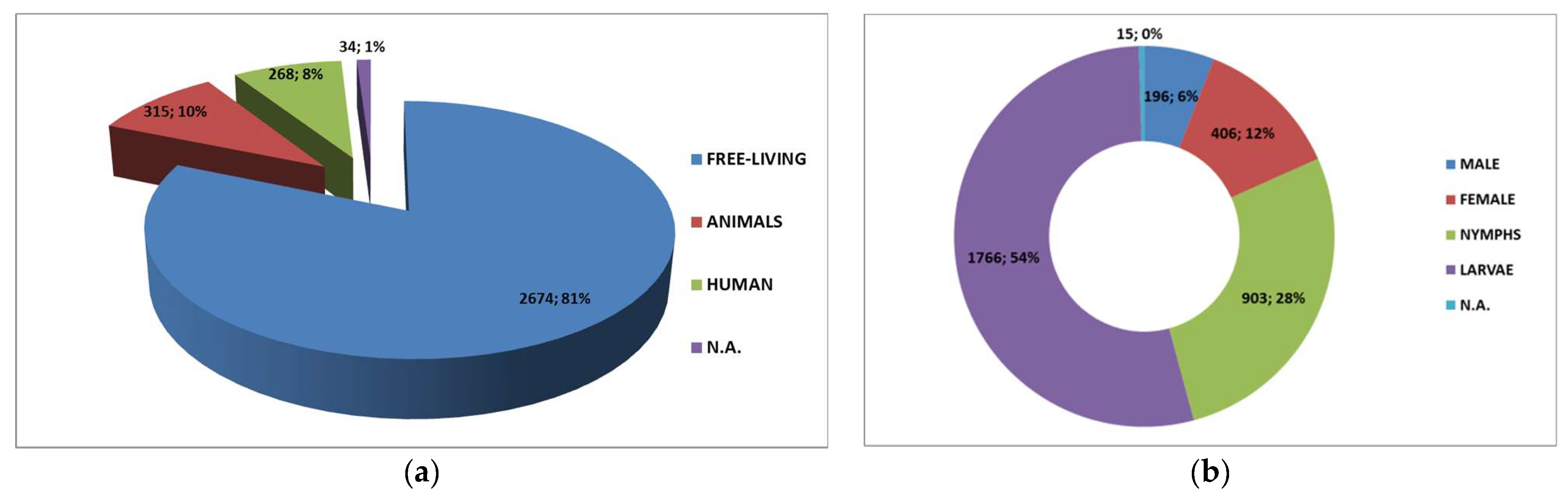
3.1. Tick Collection and Identification

A total of 3286 tick samples were collected. The most frequent source of ticks was the environment, with 2674 (81%) of free-living ticks out of 3286 total.
Ticks were collected from the following group of animals: fam. Cervidae, fam. Bovidae, fam. Canidae, fam. Felidae, ord. Rodents, and fam. Suidae. Detailed results of the collection are shown in the Figure 2.
Out of 3286 collected tick samples, 3282 were identified at the species level, while only one was identified at the genus level and three ticks were not determined at the species nor the genus level. Overall, 15 tick species were recognized, the most represented (79.52%) species in the collection was Ixodes ricinus, followed by Rhipicephalus sanguineus (9.13%), with 11.35% made up of the remaining species. Table 1 summarizes tick ID results.

3.2. Rickettsia DNA Detection

All tick samples were screened by PCR for a specific Rickettsia DNA target. The Rickettsia gltA gene was detected in 250 samples.
The overall prevalence of Rickettsia infection was 7.58% (249/3286) of the total samples (Table 2), with a positivity percentage of 6.06%, 20% and 9.33% in ticks collected from environment, animals, and humans, respectively. Table 2 summarizes total results of PCR for Rickettsia in each tick species.

3.3. Rickettsia Identification by Sequencing

To confirm results and identify Rickettsia species, all positive PCR products detected in ticks were sequenced. Identification of Rickettsia species based on DNA sequencing was successfully obtained in 200 out of 249 positive samples. R. monacesis was the most frequent (56%). Table 3 summarizes data on Rickettsia sequencing in relation to tick species and to the different sources.

4. Discussion

The study provides novel insights on the SFG Rickettsiae circulation in the environment in the study area and even if significantly affected by biases, such as the features of the passive monitoring (mainly wild hosts and according seasonality and hunting season) and the low territory coverage of the environmental sampling, it has some strengthening points, such as the diversity of the source of ticks, the length of interval time of the survey, and the consequent high number of samples tested (3286), including 244 from humans. The overall positivity for SFG Rickettsiae was 7.58% (249/3286), which was significantly lower than what was reported in a similar study carried out on ticks collected from wild animals in the Abruzzi region by Pascucci and collaborators [13], who found 52.25% on 178 pools of ixodid ticks, and the results produced by Scarpulla and collaborators [8] on 113 ticks collected from hosts and environment in Latium and Tuscany regions (12.4%), whereas, if we consider only the positivity in ticks collected from animals (19.81%), our results are comparable to the findings of Ebani and collaborators [18] in Tuscany (20.78%). Positivity percentage in our study in free-living ticks (6.06%) was lower than what has been recently reported in a study carried out testing free-living ticks in a suburban area in Latium 25.8% where, however, 255 ticks had been selected randomly out of 518 ticks, and singularly tested [19]. Different ways of pooling ticks in the studies may have affected Rickettsia testing results, making them not easily comparable. Ticks belonging to five genera (Dermacentor, Haemaphysalis, Hyalomma, Ixodes, and Rhipicephalus) were collected during the study, with the most frequent species being Ixodes ricinus (79.52%), followed by Rhipicephalus sanguineus (9.13%). Both species are documented as the most frequent in central Italy, but even if in our study the proportion is likely affected by the bias represented by the source: 81% of the collection, in fact, was represented by free-living ticks collected by dragging carried out during “spring time”. These sampling techniques and the collection season are, in fact, recognized as the most suitable for Ixodes ricinus because of its hunting behavior (ambushing) and for its seasonal dynamic. Similarly, the high prevalence of immaturity in the collection may likely be a consequence of the predominance in free-living ticks and of Ixodes ricinus among them. Ixodes ricinus was confirmed as the most frequent tick species biting humans in central Italy (244/270), this finding is in accordance to the results of Pascucci and Cammà [20] for the northern part of Marche region. Considering that 9.26% of ticks collected from humans tested positive for Rickettsia spp., the human population may be considered at risk for Rickettsiosis in the study area, even though the more detailed epidemiological investigation that comprises spatial analysis needs to be continued. Six different species of Rickettsia belonging to the SFG were identified by sequencing—R. monacensis was by far the most frequent species in our collection, since more than half of the sequenced samples (56%) belonged to this species. This result is in accordance with the situation previously described in Tuscany [8], and in Emilia Romagna [21]. Similarly, Pascucci and collaborators [13] identified R. monacensis in 40.9% out of 93 Rickettsia SFG-positive pools. Remarkably, the frequency of R. helvetica in our samples reached 25.37%. Similarly to what was described by Pascucci and collaborators [13], R. helvetica was identified exclusively in ticks (51 out of 2613) belonging to I. ricinus species, the main vector and natural reservoir [4], while R. monacensis, even though found mainly in I. ricinus (107 out of 2613), was identified also in I. acuminatus and Rh. sanguineus, thus confirming what has been suggested by Madeddu and collaborators [22] regarding the possible implication of species other than I. ricinus in the transmission of R. monacensis. Furthermore, R. monacensis and R. helvetica had just been detected in I. ricinus collected from dogs in the bordering region (Umbria) [23]. Both Rickettsia species are recognized as occasional agents of spotted fever in Italy [1,3,22]. The finding of a Rickettsia identified as Rickettsia rhipicephali is in line with what was described by Otranto and collaborator [10] for the first time in Sicily
R. rhipicephali, isolated for the first time from Rh. sanguineus ticks collected from dogs in Mississippi in 1975 and sporadically reported in Europe [4], was detected in our study, surprisingly not only in Rhipicephalus ticks (GenBank Accession Number OL906195) but also in Ixodes ricinus (GenBank Accession Numbers OL906194 and OL906196), leading us to hypothesize a possible role in the transmission. This Rickettsia has been identified in ticks from France, Portugal, Greece (including the Island of Cephalonia), Croatia [4], and in 2018 again in Italy using gltA sequencing by Guerden and collaborators [11]. As for other recently identified species, it is not yet confirmed as human pathogen [4]
Conversely, R. massiliae, recognized as an agent of human diseases also in Italy, was detected in our study in Rhipicephalus ticks, confirming literature data that describe R. massiliae presence all over the Rh. turanicus and Rh. sanguineus geographic distribution [4,13]. Though less common than that found in a similar study carried on in central Italy [13], R. slovaca was revealed in our study. This Rickettsia is associated with a syndrome in humans characterized by scalp eschars and neck lymphadenopathy following tick bite. Initially, this syndrome was named TIBOLA (tick-borne lymphadenopathy) or DEBONEL (Dermacentor-borne necrotic erythema and lymphadenopathy), and, after 2010, SENLAT (scalp eschar and neck lymphadenopathy after a tick bite) [4] because of its strict but not exclusive association with Dermacentor ticks, also described in our study, where R. slovaca was also detected in Hyalomma and Ixodes ticks. The reason for a low frequency of R. slovaca may be linked to the low representativeness of Dermacentor marginatus ticks in our collection (0.40%). Moreover, in the present study R. hoogstraalii, a SFG Rickettsia with unknown pathogenicity that is closely related to R. felis, an emerging pathogen known to be transmitted by several arthropods [4], was detected in Ixodes ricinus. R. hoogstraalii had been detected for the first time in Italy by Chisu and collaborators in Sardinia [9]. Tt was originally isolated in 2006, from H. sulcata ticks collected from sheep and goats in Croatia [24], in H. punctata ticks collected in Cyprus [25], and in H. punctata and in H. sulcata ticks from Spain [26]. It has spread worldwide and been identified in other tick species [9], and, to our knowledge, this is the second report of R. hoogstraalii in Italy and the first in the peninsular territory. Further confirmation of Rickettsia species identification should be carried out using different Rickettsia molecular targets, such as OmpA and OmpB. Moreover, the complexity of SFG Rickettsiae biological cycles with different tick species acting as vectors /reservoirs and, consequently, with several vertebrate hosts involved, makes the system extremely sensitive to the influence of climate changes that can be carried out both directly on the vectors modifying its survival and activities, or indirectly on host availability [27]. Thus, additional studies on the relationship between SFG Rickettsiae and climate change should be performed in the study area in order to identify possible scenarios.

5. Conclusions

Our findings are the first comprehensive data available on SFG Rickettsiae circulation in the study area and, even if to be supported by further molecular investigation and biases affected, they can be considered as starting point to assess spotted fever risk for humans and to set up surveillance and public awareness activities that are essential prevention tools. A factual assessment of zoonoses risk for humans in contexts placed in natural environments rich in biodiversity with high levels of interaction between humans, domestic and wild animals, vectors, and transmitted pathogens, such as the study area, necessitates a One Health approach that considers human and animal health as strictly interconnected with ecosystem health. This concept is even more essential in the risk assessment for vector-borne diseases.

Author Contributions

Conceptualization, I.P., E.A. and S.G.; methodology, E.A., I.P., C.C. and S.G. software, E.A.; validation, E.A., C.C. and A.d.D.; formal analysis, I.P. and M.G.M.; investigation, E.A., C.C., A.N., M.M. and B.M.; resources, S.G.; data curation, E.A.; I.P. and S.G. writing—original draft preparation, I.P. and S.G.; writing—review and editing, I.P., S.G. and E.A.; visualization, I.P., S.C. and G.M.; supervision, S.G. and S.C.; project administration, S.G. and G.M.; funding acquisition, S.G. and S.C. All authors have read and agreed to the published version of the manuscript.

Funding

This research received no external funding.

Institutional Review Board Statement

Not applicable.

Informed Consent Statement

Not applicable.

Data Availability Statement

The data presented in this study are available in the present article Pascucci, I.; Antognini, E.; Canonico, C.; Montalbano, M.G.; Necci, A.; Di Donato, A.d.; Moriconi, M.; Morandi, B.; Morganti, G.; Crotti, S.; and Gavaudan S. One Health Approach to Rickettsiosis: A Five Years Study on Spotted Fever Group Rickettsiae in Ticks Collected from Humans, Animals and Environment.

Conflicts of Interest

The authors declare no conflict of interest.

References

  1. Parola, P.; Paddock, C.D.; Raoult, D. Tick-borne rickettsioses around the world: Emerging diseases challenging old concepts. Clin. Microbiol. Rev. 2005, 18, 719–756. [Google Scholar] [CrossRef] [PubMed] [Green Version]
  2. Szokoli, F.; Castelli, M.; Sabaneyeva, E.; Schrallhammer, M.; Krenek, S.; Doak, T.G.; Berendonk, T.U.; Petroni, G. Disentangling the taxonomy of Rickettsiales and description of two novel symbionts (“Candidatus Bealeia Paramacronuclearis” and “Candidatus Fokinia Cryptica”) Sharing the Cytoplasm of the Ciliate Protist Paramecium biaurelia. Appl. Environ. Microbiol. 2016, 82, 7236–7247. [Google Scholar] [CrossRef] [PubMed] [Green Version]
  3. Guccione, C.; Colomba, C.; Tolomeo, M.; Trizzino, M.; Iaria, C.; Cascio, A. Rickettsiales in Italy. Pathogens 2021, 10, 181. [Google Scholar] [CrossRef]
  4. Parola, P.; Paddock, C.D.; Socolovschi, C.; Labruna, M.B.; Mediannikov, O.; Kernif, T.; Abdad, M.Y.; Stenos, J.; Bitam, I.; Fournier, P.-E.; et al. Update on tick borne rickettsioses around the world: A geographic approach. Clin. Microbiol. Rev. 2013, 26, 657–702. [Google Scholar] [CrossRef] [PubMed] [Green Version]
  5. Parola, P.; Raoult, D. Ticks and tick borne bacterial diseases in humans: An emerging infectious threat. Clin. Infect. Dis. 2001, 32, 897–928. [Google Scholar] [CrossRef]
  6. Brites-Neto, J.; Duarte, K.M.R.; Martins, T.F. Tick-borne infections in human and animal population worldwide. Vet. World 2015, 8, 301–315. [Google Scholar] [CrossRef]
  7. Socolovschi, C.; Mediannikov, O.; Raoult, D.; Parola, P. The relationship between spotted fever group rickettsiae and ixodid ticks. Vet. Res. 2009, 40, 34. [Google Scholar] [CrossRef] [Green Version]
  8. Scarpulla, M.; Barlozzari, G.; Marcario, A.; Salvato, L.; Blanda, V.; De Liberato, C.; D’Agostini, C.; Torina, A.; Macrì, G. Molecular detection and characterization of spotted fever group rickettsiae in ticks from Central Italy. Ticks Tick Borne Dis. 2016, 7, 1052–1056. [Google Scholar] [CrossRef] [Green Version]
  9. Chisu, V.; Leulmi, H.; Masala, G.; Piredda, M.; Foxi, C.; Parola, P. Detection of Rickettsia hoogstraalii, Rickettsia helvetica, Rickettsia massiliae, Rickettsia slovaca and Rickettsia aeschlimannii in ticks from Sardinia, Italy. Ticks Tick Borne Dis. 2017, 8, 347–352. [Google Scholar] [CrossRef]
  10. Otranto, D.; Dantas-Torres, F.; Giannelli, A.; Latrofa, M.S.; Cascio, A.; Cazzin, S.; Ravagnan, S.; Montarsi, F.; Zanzani, S.A.; Manfredi, M.T.; et al. Ticks infesting humans in Italy and associated pathogens. Parasit. Vectors 2014, 7, 328. [Google Scholar] [CrossRef] [Green Version]
  11. Geurden, T.; Becskei, C.; Six, R.H.; Maeder, S.; Latrofa, M.S.; Otranto, D.; Farkas, R. Detection of Tick-Borne Pathogens in Ticks from Dogs and Cats in Different European Countries. Ticks Tick-Borne Dis. 2018, 9, 1431–1436. [Google Scholar] [CrossRef] [PubMed]
  12. Raele, D.A.; Galante, D.; Pugliese, N.; Salandra, G.L.; Cafiero, M.A. Spotted Fever Group Rickettsiae Associated with Ixodid Ticks in Wild Environment in Southern Italy. Microbiologyopen 2018, 7, e00527. [Google Scholar] [CrossRef] [Green Version]
  13. Pascucci, I.; Di Domenico, M.; Curini, V.; Cocco, A.; Averaimo, D.; D’Alterio, N.; Cammà, C. Diversity of Rickettsia in Ticks Collected in Abruzzi and Molise Regions (Central Italy). Microorganisms 2019, 7, 696. [Google Scholar] [CrossRef] [Green Version]
  14. Gomez-Barroso, D.; Vescio, M.F.; Bella, A.; Ciervo, A.; Busani, L.; Rizzo, C.; Rezza, G.; Pezzotti, P. Mediterranean spotted fever rickettsiosis in Italy, 2001–2015: Spatio-temporal distribution based on hospitalization records. Ticks Tick Borne Dis. 2019, 10, 43–50. [Google Scholar] [CrossRef]
  15. de Sousa, R.; Nobrega, S.D.; Bacellar, F.; Torgal, J. Mediterranean spotted fever in Portugal: Risk factors for fatal outcome in 105 hospitalized patients. Ann. N. Y. Acad. Sci. 2003, 990, 285–294. [Google Scholar] [CrossRef]
  16. Manilla, G. Acari Ixodida. In Fauna d’Italia, 1st ed.; Edizioni Calderini: Bologna, Italy, 1998. [Google Scholar]
  17. Roux, V.; Rydkina, E.; Eremeeva, M.; Raoult, D. Citrate synthase gene comparison, a new tool for phylogenetic analysis, and its application for the rickettsiae. Int. J. Syst. Bacteriol. 1997, 47, 252–261. [Google Scholar] [CrossRef] [PubMed] [Green Version]
  18. Ebani, V.V.; Bertelloni, F.; Turchi, B.; Filogari, D.; Cerri, D. Molecular survey of tick-borne pathogens in ixodid ticks collected from hunted wild animals in Tuscany, Italy. Asian Pac. J. Trop. Med. 2015, 8, 714–717. [Google Scholar] [CrossRef] [Green Version]
  19. Mancini, F.; Vescio, M.F.; Toma, L.; Di Luca, M.; Severini, F.; Cacciò, S.M.; Mariano, C.; Nicolai, G.; Laghezza Masci, V.; Fausto, A.M.; et al. Detection of tick-borne pathogens in ticks collected in the suburban area of Monte Romano, Lazio Region, Central Italy. Ann. Dell’istituto Super. Sanità 2019, 55, 143–150. [Google Scholar] [CrossRef]
  20. Pascucci, I.; Cammà, C. Lyme disease and the detection of Borrelia burgdorferi genospecies in Ixodes ricinus ticks from central Italy. Vet. Ital. 2010, 46, 173–188. [Google Scholar] [PubMed]
  21. Maioli, G.; Pistone, D.; Bonilauri, P.; Pajoro, M.; Barbieri, I.; Mulatti, P.; Vicari, N.; Dottori, M. Etiological agents of rickettsiosis and anaplasmosis in ticks collected in Emilia-Romagna region (Italy) during 2008 and 2009. Exp. Appl. Acarol. 2012, 57, 199–208. [Google Scholar] [CrossRef]
  22. Madeddu, G.; Mancini, F.; Caddeo, A.; Ciervo, A.; Babudieri, S.; Maida, I.; Fiori, M.L.; Rezza, G.; Mura, M.S. Rickettsia monacensis as cause of Mediterranean spotted fever-like illness, Italy. Emerg. Infect. Dis. 2012, 18, 702–704. [Google Scholar] [CrossRef]
  23. Morganti, G.; Gavaudan, S.; Canonico, C.; Ravagnan, S.; Olivieri, E.; Diaferia, M.; Marenzoni, M.L.; Antognoni, M.T.; Capelli, G.; Silaghi, C.; et al. Molecular survey on Rickettsia spp., Anaplasma phagocytophilum, Borrelia burgdorferi Sensu Lato, and Babesia spp. in Ixodes ricinus Ticks infesting dogs in Central Italy. Vector Borne Zoonotic Dis. 2017, 17, 743–748. [Google Scholar] [CrossRef] [PubMed]
  24. Duh, D.; Punda-Polic, V.; Avsic-Zupanc, T.; Bouyer, D.; Walker, D.H.; Popov, V.L.; Jelovsek, M.; Gracner, M.; Trilar, T.; Bradaric, N.; et al. Rickettsia hoogstraalii sp. nov., isolated from hard- and soft-bodied ticks. Int. J. Syst. Evol. Microbiol. 2010, 60, 977–984. [Google Scholar] [CrossRef] [PubMed] [Green Version]
  25. Chochlakis, D.; Ioannou, I.; Sandalakis, V.; Dimitriou, T.; Kassinis, N.; Papadopoulos, B.; Tselentis, Y.; Psaroulaki, A. Spotted fever group rickettsiae in ticks in Cyprus. Microb. Ecol. 2012, 63, 314–323. [Google Scholar] [CrossRef]
  26. Marquez, F.J. Spotted fever group Rickettsia in ticks from southeastern Spain natural parks. Exp. Appl. Acarol. 2008, 45, 185–194. [Google Scholar] [CrossRef] [PubMed]
  27. Dumic, I.; Severnini, E. “Ticking Bomb”: The Impact of Climate Change on the Incidence of Lyme Disease. Can. J. Infect. Dis. Med. Microbiol. 2018, 2018. [Google Scholar] [CrossRef] [Green Version]
Figure 1. Geographical localization of the Marche region in Italy and of the dragging sites.
Microorganisms 10 00035 g001
Figure 2. Distribution of source (a) and development stages (b) of tick samples.
Microorganisms 10 00035 g002
Table 1. Tick ID results.
AdultsNymphsLarvaeN.A.Total%
MaleFemale
Ixodes ricinus11633584413171261379.52%
Rhipicephalus sanguineus182212237113009.13%
Haemaphysalis punctata41242210601935.87%
Rhipicephalus bursa1209801013.07%
Haemaphysalis parva25710150.46%
Dermacentor marginatus76000130.40%
Hyalomma marginatum48000120.37%
Ixodes acuminatus20460120.37%
Rhipicephalus pusillus001000100.30%
Ixodes gibbosus0230050.15%
Haemaphysalis sulcata2010030.09%
Not determined0000330.09%
Hyalomma lusitanicum0200020.06%
Ixodes frontalis1100020.06%
Ixodes spp.0001010.03%
Ixodes ventalloi1000010.03%
1954079031766153286100.00%
Table 2. Results of PCR for Rickettsia spp.
Tick SpeciesFree-LivingAnimalsHumanN.A.Total Ticks for SpeciesPositives for Species% Positive for Species% Positive for Total
TotPos%TotPos%TotPos%TotPos%
Ixodes ricinus21761587.26%1923920%244239.43%10 26132208.42%6.70%
Rhipicephalus sanguineus26041.54%281968%1119.09%11100%300258.33%0.76%
Haemaphysalis punctata1230 390 10 300 19300.00%0.00%
Rhipicephalus bursa990 20 00 00 10100.00%0.00%
Haemaphysalis parva40 10110%10 00 1516.67%0.03%
Dermacentor marginatus10 9111%30 00 1317.69%0.03%
Hyalomma marginatum00 110 00 10 1200.00%0.00%
Ixodes acuminatus90 2150%10 00 1218.33%0.03%
Rhipicephalus pusillus00 100 00 00 1000.00%0.00%
Ixodes gibbosus00 00 50 00 500.00%0.00%
Haemaphysalis sulcata00 30 00 00 300.00%0.00%
Not determined00 30 00 00 300.00%0.00%
Hyalomma lusitanicum00 00 2150%00 2150.00%0.03%
Ixodes frontalis20 00 00 00 200.00%0.00%
Ixodes spp.00 00 00 10 100.00%0.00%
Ixodes ventalloi00 10 00 00 100.00%0.00%
TOTAL26741626.06%3106220.00%268259.33%3412.94%3286250 7.58%
Table 3. Rickettsia species identification by sequencing.
Free-LivingAnimalsHumanN.ATotal
SFG Rickettsia Species
Identification in Tick Specimens
Tick SpeciesTotPosTotPosTotPosTotPosTotPos% PositiveFrequence
R.monacensis
Ixodes acuminatus902110001218.33%
Ixodes ricinus21767919217244111026131074.09%
Rhipicephalus sanguineus26042801101030041.33%
Total R. monacensis 1123.40%56.00%
R.helveticaIxodes ricinus21764519222444102613511.95%
Total R. helvetica 511.55%25.50%
R.hoogstraaliiIxodes ricinus217601922244010261320.08%
Total R. hoogstralii 20.06%1.00%
R. massiliaeRhipicephalus sanguineus26002891101 30093.0%
Total R.massiliae 90.27%4.50%
R.rhipicephaliRhipicephalus sanguineus2601028011010300103.3%
Ixodes ricinus217691920244010261390.34%
Total R. rhipicephali 190.6%9.50%
R.slovacaHyalomma lusitanicum000021002150%
Ixodes ricinus217601922244010261320.08%
Dermacentor marginatus109130001317.69%
Total R. slovaca 40.12%2.00%
Rickettsia SFGIxodes ricinus217611920244010261310.04%
Rhipicepahlus sanguineus26002801101130010.33%
Total Rickettsia SFG 20.06%1.00%
Rickettsia spp.Rhipicephalus sanguineus26002801111030010.33%
Total Rickettsia spp. 10.03%0.50%
Total Positive 200
Publisher’s Note: MDPI stays neutral with regard to jurisdictional claims in published maps and institutional affiliations.

