Next Issue
Volume 9, January
Previous Issue
Volume 8, September


Pathogens, Volume 8, Issue 4 (December 2019) – 144 articles

Cover Story (view full-size image): 
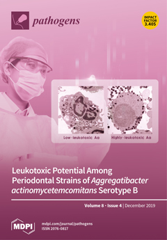
Leukotoxin is an important and powerful tool expressed by the periodontal pathogen, Aggregatibacter actinomycetemcomitans. The high leukotoxic potential of the virulent JP2 genotype of A. actinomycetemcomitans has been studied extensively. In order to obtain a comprehensive understanding of the leukotoxic potential of different genotypes of A. actinomycetemcomitans serotype b, a geographically widespread collection of strains has been analyzed by several methods. The present study of the leukotoxic potential of both the JP2 and non-JP2 genotype strains of A.actinomycetemcomitans emphasizes the importance of using more than one method when assessing the leukotoxin-related virulence capacity of A. actinomycetemcomitans. View this paper
  • Issues are regarded as officially published after their release is announced to the table of contents alert mailing list.
  • You may sign up for e-mail alerts to receive table of contents of newly released issues.
  • PDF is the official format for papers published in both, html and pdf forms. To view the papers in pdf format, click on the "PDF Full-text" link, and use the free Adobe Reader to open them.
Order results
Result details
Section
Select all
Export citation of selected articles as:
12 pages, 1269 KiB  
Article
Development of Monoclonal Antibody Specific to Foot-and-Mouth Disease Virus Type A for Serodiagnosis
by Quyen Thi Nguyen, Jihyun Yang, Jae-Won Byun, Hyun Mi Pyo, Mi-Young Park, Bok Kyung Ku, Jinju Nah, Soyoon Ryoo, Sung-Hwan Wee, Kang-Seuk Choi and Haryoung Poo
Pathogens 2019, 8(4), 301; https://doi.org/10.3390/pathogens8040301 - 17 Dec 2019
Cited by 7 | Viewed by 3702
Abstract
Foot-and-mouth disease (FMD) is a highly contagious and economically devastating disease affecting cloven-hoofed livestock worldwide. FMD virus (FMDV) type A is one of the most common causes of FMD outbreaks among the seven FMDV serotypes, and its serological diagnosis is therefore important to [...] Read more.
Foot-and-mouth disease (FMD) is a highly contagious and economically devastating disease affecting cloven-hoofed livestock worldwide. FMD virus (FMDV) type A is one of the most common causes of FMD outbreaks among the seven FMDV serotypes, and its serological diagnosis is therefore important to confirm FMDV type A infection and to determine FMD vaccine efficacy. Here, we generated monoclonal antibodies (mAbs) specific to FMDV type A via hybridoma systems using an inactivated FMDV type A (A22/Iraq/1964) and found 4 monoclones (#29, #106, #108, and #109) with high binding reactivity to FMDV type A among 594 primary clones. In particular, the #106 mAb had a higher binding reactivity to the inactivated FMDV type A than the other mAbs and a commercial mAb. Moreover, the #106 mAb showed no cross-reactivity to inactivated FMDV type South African territories 1, 2, and 3, and low reactivity to inactivated FMDV type O (O1 Manisa). Importantly, the solid-phase competitive ELISA (SPCE) using horseradish peroxidase (HRP)-conjugated #106 mAb detected FMDV type A-specific Abs in sera from FMD type A-vaccinated cattle more effectively than a commercial SPCE. These results suggest that the newly developed FMDV type A-specific mAb might be useful for diagnostic approaches for detecting Abs against FMDV type A. Full article
Show Figures

Figure 1

13 pages, 4673 KiB  
Article
Endocytic Pathway of Feline Coronavirus for Cell Entry: Differences in Serotype-Dependent Viral Entry Pathway
by Tomomi Takano, Yumeho Wakayama and Tomoyoshi Doki
Pathogens 2019, 8(4), 300; https://doi.org/10.3390/pathogens8040300 - 16 Dec 2019
Cited by 14 | Viewed by 7219
Abstract
Feline coronavirus (FCoV) is a pathogen causing a lethal infectious disease in cats, feline infectious peritonitis. It has two serotypes (type I FCoV and type II FCoV). According to our previous study, type I FCoV infection is inhibited by compounds inducing intracellular cholesterol [...] Read more.
Feline coronavirus (FCoV) is a pathogen causing a lethal infectious disease in cats, feline infectious peritonitis. It has two serotypes (type I FCoV and type II FCoV). According to our previous study, type I FCoV infection is inhibited by compounds inducing intracellular cholesterol accumulation, whereas type II FCoV infection is not inhibited. Intracellular cholesterol accumulation was reported to disrupt late endosome function. Based on these findings, types I and II FCoV are considered to enter the cytosol through late and early endosomes, respectively. We investigated whether the antiviral activities of a late endosome trafficking inhibitor and cholesterol-accumulating agents are different between the FCoV serotypes. The late endosome trafficking inhibitor did not inhibit type II FCoV infection, but it inhibited type I FCoV infection. Type I FCoV infection was inhibited by cholesterol-accumulating triazoles, but not by non-cholesterol-accumulating triazoles. These phenomena were observed in both feline cell lines and feline primary macrophages. This study provides additional information on the differences in intracellular reproductive cycle between type I and type II FCoV. Full article
(This article belongs to the Special Issue Feline Infectious Peritonitis)
Show Figures

Graphical abstract

59 pages, 2178 KiB  
Review
Lyme Disease Frontiers: Reconciling Borrelia Biology and Clinical Conundrums
by Vladimir V. Bamm, Jordan T. Ko, Iain L. Mainprize, Victoria P. Sanderson and Melanie K. B. Wills
Pathogens 2019, 8(4), 299; https://doi.org/10.3390/pathogens8040299 - 16 Dec 2019
Cited by 27 | Viewed by 16637
Abstract
Lyme disease is a complex tick-borne zoonosis that poses an escalating public health threat in several parts of the world, despite sophisticated healthcare infrastructure and decades of effort to address the problem. Concepts like the true burden of the illness, from incidence rates [...] Read more.
Lyme disease is a complex tick-borne zoonosis that poses an escalating public health threat in several parts of the world, despite sophisticated healthcare infrastructure and decades of effort to address the problem. Concepts like the true burden of the illness, from incidence rates to longstanding consequences of infection, and optimal case management, also remain shrouded in controversy. At the heart of this multidisciplinary issue are the causative spirochetal pathogens belonging to the Borrelia Lyme complex. Their unusual physiology and versatile lifestyle have challenged microbiologists, and may also hold the key to unlocking mysteries of the disease. The goal of this review is therefore to integrate established and emerging concepts of Borrelia biology and pathogenesis, and position them in the broader context of biomedical research and clinical practice. We begin by considering the conventions around diagnosing and characterizing Lyme disease that have served as a conceptual framework for the discipline. We then explore virulence from the perspective of both host (genetic and environmental predispositions) and pathogen (serotypes, dissemination, and immune modulation), as well as considering antimicrobial strategies (lab methodology, resistance, persistence, and clinical application), and borrelial adaptations of hypothesized medical significance (phenotypic plasticity or pleomorphy). Full article
(This article belongs to the Special Issue Pathogenesis of Fungal and Bacterial Microbes)
Show Figures

Figure 1

21 pages, 2879 KiB  
Article
Repeat-Induced Point Mutations Drive Divergence between Fusarium circinatum and Its Close Relatives
by Stephanie van Wyk, Brenda D. Wingfield, Lieschen De Vos, Nicolaas A. van der Merwe, Quentin C. Santana and Emma T. Steenkamp
Pathogens 2019, 8(4), 298; https://doi.org/10.3390/pathogens8040298 - 14 Dec 2019
Cited by 9 | Viewed by 3577
Abstract
The Repeat-Induced Point (RIP) mutation pathway is a fungal-specific genome defense mechanism that counteracts the deleterious effects of transposable elements. This pathway permanently mutates its target sequences by introducing cytosine to thymine transitions. We investigated the genome-wide occurrence of RIP in the pitch [...] Read more.
The Repeat-Induced Point (RIP) mutation pathway is a fungal-specific genome defense mechanism that counteracts the deleterious effects of transposable elements. This pathway permanently mutates its target sequences by introducing cytosine to thymine transitions. We investigated the genome-wide occurrence of RIP in the pitch canker pathogen, Fusarium circinatum, and its close relatives in the Fusarium fujikuroi species complex (FFSC). Our results showed that the examined fungi all exhibited hallmarks of RIP, but that they differed in terms of the extent to which their genomes were affected by this pathway. RIP mutations constituted a large proportion of all the FFSC genomes, including both core and dispensable chromosomes, although the latter were generally more extensively affected by RIP. Large RIP-affected genomic regions were also much more gene sparse than the rest of the genome. Our data further showed that RIP-directed sequence diversification increased the variability between homologous regions of related species, and that RIP-affected regions can interfere with homologous recombination during meiosis, thereby contributing to post-mating segregation distortion. Taken together, these findings suggest that RIP can drive the independent divergence of chromosomes, alter chromosome architecture, and contribute to the divergence among F. circinatum and other members of this economically important group of fungi. Full article
(This article belongs to the Section Fungal Pathogens)
Show Figures

Figure 1

15 pages, 1829 KiB  
Article
Effects of Pseudomonas aeruginosa on Microglial-Derived Extracellular Vesicle Biogenesis and Composition
by Leandra B. Jones, Sanjay Kumar, Courtnee’ R. Bell, Veolonda A. Peoples, Brennetta J. Crenshaw, Mamie T. Coats, Jessica A. Scoffield, Glenn C. Rowe, Brian Sims and Qiana L. Matthews
Pathogens 2019, 8(4), 297; https://doi.org/10.3390/pathogens8040297 - 14 Dec 2019
Cited by 6 | Viewed by 3195
Abstract
The packaging of molecular constituents inside extracellular vesicles (EVs) allows them to participate in intercellular communication and the transfer of biological molecules, however the role of EVs during bacterial infection is poorly understood. The goal of this study was to examine the effects [...] Read more.
The packaging of molecular constituents inside extracellular vesicles (EVs) allows them to participate in intercellular communication and the transfer of biological molecules, however the role of EVs during bacterial infection is poorly understood. The goal of this study was to examine the effects of Pseudomonas aeruginosa (P. aeruginosa) infection on the biogenesis and composition of EVs derived from the mouse microglia cell line, BV-2. BV-2 cells were cultured in exosome-free media and infected with 0, 1.3 × 10⁴, or 2.6 × 10⁴ colony forming units per milliliter P. aeruginosa for 72 h. The results indicated that compared with the control group, BV-2 cell viability significantly decreased after P. aeruginosa infection and BV-2-derived EVs concentration decreased significantly in the P. aeruginosa-infected group. P. aeruginosa infection significantly decreased chemokine ligand 4 messenger RNA in BV-2-derived infected EVs, compared with the control group (p ≤ 0.05). This study also revealed that heat shock protein 70 (p ≤ 0.05) and heat shock protein 90β (p ≤ 0.001) levels of expression within EVs increased after P. aeruginosa infection. EV treatment with EVs derived from P. aeruginosa infection reduced cell viability of BV-2 cells. P. aeruginosa infection alters the expression of specific proteins and mRNA in EVs. Our study suggests that P. aeruginosa infection modulates EV biogenesis and composition, which may influence bacterial pathogenesis and infection. Full article
Show Figures

Graphical abstract

5 pages, 214 KiB  
Brief Report
No Confirmed Cases of Taenia solium Taeniasis in a Group of Recently Arrived Sub-Saharan Migrants to Italy
by Lorenzo Zammarchi, Marta Tilli, Antonia Mantella, Annarita Botta, Alessandra Nicoletti, Héctor Hugo García, Yesenia Castillo, Donatella Aquilini, Sara Boccalini and Alessandro Bartoloni
Pathogens 2019, 8(4), 296; https://doi.org/10.3390/pathogens8040296 - 14 Dec 2019
Cited by 3 | Viewed by 2099
Abstract
One-hundred and sixty-four migrants from Sub-Saharan Africa to Italy were screened with the Taenia solium specific enzyme-linked immunosorbent assay coproantigen (ELISA CoAg) and four (2.4%) were recorded as positive, but with optical density values near to the cut-off. No ELISA CoAg positive samples [...] Read more.
One-hundred and sixty-four migrants from Sub-Saharan Africa to Italy were screened with the Taenia solium specific enzyme-linked immunosorbent assay coproantigen (ELISA CoAg) and four (2.4%) were recorded as positive, but with optical density values near to the cut-off. No ELISA CoAg positive samples were confirmed by parasitological methods. Low positivity could be attributed to false positive result or cross-reaction with other Taenia species. Further studies are needed to assess the role of migration on sporadic autochthonous transmission of T. solium taeniasis/cysticercosis in Europe. Full article
(This article belongs to the Special Issue Tackling Foodborne Parasitic Infections)
11 pages, 1253 KiB  
Article
Water Quality as a Predictor of Legionella Positivity of Building Water Systems
by David Pierre, Julianne L. Baron, Xiao Ma, Frank P. Sidari III, Marilyn M. Wagener and Janet E. Stout
Pathogens 2019, 8(4), 295; https://doi.org/10.3390/pathogens8040295 - 13 Dec 2019
Cited by 29 | Viewed by 4634
Abstract
Testing drinking water systems for the presence of Legionella colonization is a proactive approach to assess and reduce the risk of Legionnaires’ disease. Previous studies suggest that there may be a link between Legionella positivity in the hot water return line or certain [...] Read more.
Testing drinking water systems for the presence of Legionella colonization is a proactive approach to assess and reduce the risk of Legionnaires’ disease. Previous studies suggest that there may be a link between Legionella positivity in the hot water return line or certain water quality parameters (temperature, free chlorine residual, etc.) with distal site Legionella positivity. It has been suggested that these measurements could be used as a surrogate for testing for Legionella in building water systems. We evaluated the relationship between hot water return line Legionella positivity and other water quality parameters and Legionella colonization in premise plumbing systems by testing 269 samples from domestic cold and hot water samples in 28 buildings. The hot water return line Legionella positivity and distal site positivity only demonstrated a 77.8% concordance rate. Hot water return line Legionella positivity compared to distal site positivity had a sensitivity of 55% and a specificity of 96%. There was poor correlation and a low positive predictive value between the hot water return line and distal outlet positivity. There was no correlation between Legionella distal site positivity and total bacteria (heterotrophic plate count), pH, free chlorine, calcium, magnesium, zinc, manganese, copper, temperature, total organic carbon, or incoming cold-water chlorine concentration. These findings suggest that hot water return line Legionella positivity and other water quality parameters are not predictive of distal site positivity and should not be used alone to determine the building’s Legionella colonization rate and effectiveness of water management programs. Full article
(This article belongs to the Special Issue Legionella Contamination in Water Environment)
Show Figures

Figure 1

10 pages, 226 KiB  
Article
Serological and Molecular Investigation of Brucella Species in Dogs in Pakistan
by Tariq Jamil, Falk Melzer, Iahtasham Khan, Mudassar Iqbal, Muhammad Saqib, Muhammad Hammad Hussain, Stefan Schwarz and Heinrich Neubauer
Pathogens 2019, 8(4), 294; https://doi.org/10.3390/pathogens8040294 - 13 Dec 2019
Cited by 21 | Viewed by 3731
Abstract
Brucellosis is an important bacterial zoonosis caused by B. abortus and B. melitensis in Pakistan. The status of canine brucellosis caused by B. canis remains obscure. In total, 181 serum samples were collected from stray and working dogs in two different prefectures viz. [...] Read more.
Brucellosis is an important bacterial zoonosis caused by B. abortus and B. melitensis in Pakistan. The status of canine brucellosis caused by B. canis remains obscure. In total, 181 serum samples were collected from stray and working dogs in two different prefectures viz. Faisalabad (n = 87) and Bahawalpur (n = 94). Presence of antibodies against B. canis and B. abortus/B. melitensis was determined using the slow agglutination test (SAT) and ELISA, respectively. Real-time PCR was performed to detect and differentiate Brucella DNA at the species level. In Faisalabad, the serological prevalence was found to be 9.2% (8/87) and 10.3% (9/87) by SAT and ELISA, respectively. Only one of the ELISA positive samples (1.15%) yielded amplification for B. abortus DNA. In Bahawalpur, 63.8% (60/94) samples were found positive by SAT; however, none of the samples was positive by ELISA or by real-time PCR. Location, age (≥1 year) and body condition (weak) were found to be associated with B. canis infection, whereas presence of wounds was found to be associated with B. abortus infection only. These findings point towards a risk of transmission from dog to livestock and humans and vice versa. The study expects to draw the attention of concerned authorities towards infection prevention and animal welfare. This study warrants further epidemiological investigation on brucellosis in pet dogs and their owners. To the best of our knowledge, this is first ever report on B. canis and B. abortus in dogs in Pakistan. Full article
(This article belongs to the Section Animal Pathogens)
10 pages, 1240 KiB  
Article
Klebsiella pneumoniae—A Useful Pathogenic Strain for Biotechnological Purposes: Diols Biosynthesis under Controlled and Uncontrolled pH Levels
by Laura Mitrea and Dan Cristian Vodnar
Pathogens 2019, 8(4), 293; https://doi.org/10.3390/pathogens8040293 - 11 Dec 2019
Cited by 26 | Viewed by 4360
Abstract
Despite being a well-known human pathogen, Klebsiella pneumoniae plays a significant role in the biotechnology field, being considered as a microbial cell factory in terms of valuable chemical biosynthesis. In this work, Klebsiella pneumoniae DSMZ 2026 was investigated for its potential to biosynthesize [...] Read more.
Despite being a well-known human pathogen, Klebsiella pneumoniae plays a significant role in the biotechnology field, being considered as a microbial cell factory in terms of valuable chemical biosynthesis. In this work, Klebsiella pneumoniae DSMZ 2026 was investigated for its potential to biosynthesize 1,3-propanediol (PDO) and 2,3-butanediol (BDO) during batch fermentation under controlled and uncontrolled pH levels. The bacterial strain was cultivated at a bioreactor level, and it was inoculated in 2 L of specific mineral broth containing 50 g/L of glycerol as the main carbon source. The process was conducted under anaerobic conditions at 37 °C and 180 RPM (rotations per minute) for 24 h. The effect of pH oscillation on the biosynthesis of PDO and BDO was investigated. Samples were taken every 3 h and specific tests were performed: pH measurement, main substrate consumption, PDO and BDO production. The cell morphology was analyzed on both solid and liquid media. After 24 h of cultivation, the maximum concentrations of PDO and BDO were 28.63 ± 2.20 g/L and 18.10 ± 1.10 g/L when the pH value was maintained at 7. Decreased concentrations of PDO and BDO were achieved (11.08 ± 0.14 g/L and 7.35 ± 0.00 g/L, respectively) when the pH level was not maintained at constant values. Moreover, it was identified the presence of other metabolites (lactic, citric, and succinic acids) in the cultivation media at the beginning of the process, after 12 h and 24 h of cultivation. Full article
Show Figures

Graphical abstract

18 pages, 3128 KiB  
Article
Tuberculosis Epidemiology and Badger (Meles meles) Spatial Ecology in a Hot-Spot Area in Atlantic Spain
by Pelayo Acevedo, Miguel Prieto, Pablo Quirós, Isabel Merediz, Lucía de Juan, José Antonio Infantes-Lorenzo, Roxana Triguero-Ocaña and Ana Balseiro
Pathogens 2019, 8(4), 292; https://doi.org/10.3390/pathogens8040292 - 10 Dec 2019
Cited by 12 | Viewed by 3785
Abstract
We provide a temporal overview (from 2012 to 2018) of the outcomes of tuberculosis (TB) in the cattle and badger populations in a hot-spot in Asturias (Atlantic Spain). We also study the badger’s spatial ecology from an epidemiological perspective in order to describe [...] Read more.
We provide a temporal overview (from 2012 to 2018) of the outcomes of tuberculosis (TB) in the cattle and badger populations in a hot-spot in Asturias (Atlantic Spain). We also study the badger’s spatial ecology from an epidemiological perspective in order to describe hazardous behavior in relation to TB transmission between cattle and badgers. Culture and single intradermal tuberculin test (SITT) were available for cattle as part of the National Program for the Eradication of TB. A field survey was also carried out in order to determine the paddocks and buildings used by each farm, and the information obtained was stored by using geographic information systems. Moreover, eighty-three badgers were submitted for necropsy and subsequent bacteriological studies. Ten badgers were also tracked, using global positioning system (GPS) collars. The prevalence of TB in cattle herds in the hot-spot increased from 2.2% in 2012 to 20% in 2016; it then declined to 0.0% in 2018. In contrast, the TB prevalence in badgers increased notably (from 5.55% in 2012–2015 to 10.64% in 2016–2018). Both cattle and badgers shared the same strain of Mycobacterium bovis. The collared badgers preferred paddocks used by TB-positive herds in spring and summer (when they were more active). The males occupied larger home ranges than the females (Khr95: males 149.78 ± 25.84 ha and females 73.37 ± 22.91 ha; Kcr50: males 29.83 ± 5.69 ha and females 13.59 ± 5.00 ha), and the home ranges were smaller in autumn and winter than in summer. The averages of the index of daily and maximum distances traveled by badgers were 1.88 ± (SD) 1.20 km and 1.99 ± 0.71 km, respectively. One of them presented a dispersive behavior with a maximum range of 18.3 km. The most preferred habitat was apple orchards in all seasons, with the exception of winter, in which they preferred pastures. Land uses and landscape structure, which have been linked with certain livestock-management practices, provide a scenario of great potential for badger–cattle interactions, thus enhancing the importance of the badgers’ ecology, which could potentially transmit TB back to cattle in the future. Full article
(This article belongs to the Special Issue Tuberculosis Epidemiology and Control in Multi-Host Systems)
Show Figures

Figure 1

13 pages, 1596 KiB  
Article
Genetic Basis and Physiological Effects of Lipid A Hydroxylation in Pseudomonas aeruginosa PAO1
by Alessandra Lo Sciuto, Matteo Cervoni, Roberta Stefanelli, Maria Concetta Spinnato, Alessandra Di Giamberardino, Carmine Mancone and Francesco Imperi
Pathogens 2019, 8(4), 291; https://doi.org/10.3390/pathogens8040291 - 10 Dec 2019
Cited by 16 | Viewed by 3511
Abstract
Modifications of the lipid A moiety of lipopolysaccharide influence the physicochemical properties of the outer membrane of Gram-negative bacteria. Some bacteria produce lipid A with a single hydroxylated secondary acyl chain. This hydroxylation is catalyzed by the dioxygenase LpxO, and is important for [...] Read more.
Modifications of the lipid A moiety of lipopolysaccharide influence the physicochemical properties of the outer membrane of Gram-negative bacteria. Some bacteria produce lipid A with a single hydroxylated secondary acyl chain. This hydroxylation is catalyzed by the dioxygenase LpxO, and is important for resistance to cationic antimicrobial peptides (e.g., polymyxins), survival in human blood, and pathogenicity in animal models. The lipid A of the human pathogen Pseudomonas aeruginosa can be hydroxylated in both secondary acyl chains, but the genetic basis and physiological role of these hydroxylations are still unknown. Through the generation of single and double deletion mutants in the lpxO1 and lpxO2 homologs of P. aeruginosa PAO1 and lipid A analysis by mass spectrometry, we demonstrate that both LpxO1 and LpxO2 are responsible for lipid A hydroxylation, likely acting on different secondary acyl chains. Lipid A hydroxylation does not appear to affect in vitro growth, cell wall stability, and resistance to human blood or antibiotics in P. aeruginosa. In contrast, it is required for infectivity in the Galleria mellonella infection model, without relevantly affecting in vivo persistence. Overall, these findings suggest a role for lipid A hydroxylation in P. aeruginosa virulence that could not be directly related to outer membrane integrity. Full article
(This article belongs to the Section Human Pathogens)
Show Figures

Figure 1

18 pages, 1256 KiB  
Review
NF-κB and MicroRNA Deregulation Mediated by HTLV-1 Tax and HBZ
by Stefania Fochi, Vincenzo Ciminale, Elisabetta Trabetti, Umberto Bertazzoni, Donna M. D’Agostino, Donato Zipeto and Maria Grazia Romanelli
Pathogens 2019, 8(4), 290; https://doi.org/10.3390/pathogens8040290 - 10 Dec 2019
Cited by 19 | Viewed by 4356
Abstract
The risk of developing adult T-cell leukemia/lymphoma (ATLL) in individuals infected with human T-cell lymphotropic virus 1 (HTLV-1) is about 3–5%. The mechanisms by which the virus triggers this aggressive cancer are still an area of intensive investigation. The viral protein Tax-1, together [...] Read more.
The risk of developing adult T-cell leukemia/lymphoma (ATLL) in individuals infected with human T-cell lymphotropic virus 1 (HTLV-1) is about 3–5%. The mechanisms by which the virus triggers this aggressive cancer are still an area of intensive investigation. The viral protein Tax-1, together with additional regulatory proteins, in particular HTLV-1 basic leucine zipper factor (HBZ), are recognized as relevant viral factors required for both viral replication and transformation of infected cells. Tax-1 deregulates several cellular pathways affecting the cell cycle, survival, and proliferation. The effects of Tax-1 on the NF-κB pathway have been thoroughly studied. Recent studies also revealed the impact of Tax-1 and HBZ on microRNA expression. In this review, we summarize the recent progress in understanding the contribution of HTLV-1 Tax- and HBZ-mediated deregulation of NF-κB and the microRNA regulatory network to HTLV-1 pathogenesis. Full article
(This article belongs to the Special Issue HTLV-1 Disease)
Show Figures

Figure 1

10 pages, 2165 KiB  
Article
Molecular Detection and Characterization of Borrelia garinii (Spirochaetales: Borreliaceae) in Ixodes nipponensis (Ixodida: Ixodidae) Parasitizing a Dog in Korea
by Seung-Hun Lee, Youn-Kyoung Goo, Paul John L. Geraldino, Oh-Deog Kwon and Dongmi Kwak
Pathogens 2019, 8(4), 289; https://doi.org/10.3390/pathogens8040289 - 06 Dec 2019
Cited by 9 | Viewed by 3047
Abstract
The present study aimed to detect and characterize Borrelia spp. in ticks attached to dogs in Korea. Overall, 562 ticks (276 pools) attached to dogs were collected and tested for Borrelia infection by PCR targeting the 5S-23S rRNA intergenic spacer region (rrf-rrl). One [...] Read more.
The present study aimed to detect and characterize Borrelia spp. in ticks attached to dogs in Korea. Overall, 562 ticks (276 pools) attached to dogs were collected and tested for Borrelia infection by PCR targeting the 5S-23S rRNA intergenic spacer region (rrf-rrl). One tick larva (pool level, 0.4%; individual level, 0.2%) was confirmed by sequencing Borrelia garinii, a zoonotic pathogen. For molecular characterization, the outer surface protein A (ospA) and flagellin genes were analyzed. Phylogenetic ospA analysis distinguished B. garinii from B. bavariensis, which has been recently identified as a novel Borrelia species. On the other hand, phylogenetic analysis showed that single gene analysis involving rrf-rrl or flagellin was not sufficient to differentiate B. garinii from B. bavariensis. In addition, the B. garinii-infected tick was identified as Ixodes nipponensis by sequencing according to mitochondrial 16S rRNA and the second transcribed spacer region. To our knowledge, this is the first study to report the molecular detection of B. garinii in I. nipponensis parasitizing a dog in Korea. Continuous monitoring of tick-borne pathogens in ticks attached to animals is required to avoid disease distribution and possible transmission to humans. Full article
(This article belongs to the Section Animal Pathogens)
Show Figures

Figure 1

18 pages, 3242 KiB  
Article
Surfactant Protein A Impairs Genital HPV16 Pseudovirus Infection by Innate Immune Cell Activation in A Murine Model
by Sylvia Ujma, Sinead Carse, Alisha Chetty, William Horsnell, Howard Clark, Jens Madsen, Rose-Marie Mackay, Alastair Watson, Mark Griffiths, Arieh A. Katz and Georgia Schäfer
Pathogens 2019, 8(4), 288; https://doi.org/10.3390/pathogens8040288 - 06 Dec 2019
Cited by 11 | Viewed by 3505
Abstract
Infection by oncogenic human papillomavirus (HPV) is the principle cause of cervical cancer and other anogenital cancers. The majority of cervical cancer cases occur in low- and middle-income countries (LMIC). Prophylactic vaccines exist to combat HPV infection but accessibility to these in LMIC [...] Read more.
Infection by oncogenic human papillomavirus (HPV) is the principle cause of cervical cancer and other anogenital cancers. The majority of cervical cancer cases occur in low- and middle-income countries (LMIC). Prophylactic vaccines exist to combat HPV infection but accessibility to these in LMIC is limited. Alternative preventative measures against HPV infection are therefore also needed to control cervical cancer risk. HPV employs multiple mechanisms to evade the host immune response. Therefore, an approach to promote HPV recognition by the immune system can reduce infection. Surfactant proteins A and D (SP-A and SP-D) are highly effective innate opsonins of pathogens. Their function is primarily understood in the lung, but they are also expressed at other sites of the body, including the female reproductive tract (FRT). We hypothesized that raised levels of SP-A and/or SP-D may enhance immune recognition of HPV and reduce infection. Co-immunoprecipitation and flow cytometry experiments showed that purified human SP-A protein directly bound HPV16 pseudovirions (HPV16-PsVs), and the resulting HPV16-PsVs/SP-A complex enhanced uptake of HPV16-PsVs by RAW264.7 murine macrophages. In contrast, a recombinant fragment of human SP-D bound HPV16-PsVs weakly and had no effect on viral uptake. To assess if SP-A modulates HPV16-PsVs infection in vivo, a murine cervicovaginal challenge model was applied. Surprisingly, neither naïve nor C57BL/6 mice challenged with HPV16-PsVs expressed SP-A in the FRT. However, pre-incubation of HPV16-PsVs with purified human SP-A at a 1:10 (w/w) ratio significantly reduced the level of HPV16-PsV infection. When isolated cells from FRTs of naïve C57BL/6 mice were incubated with HPV16-PsVs and stained for selected innate immune cell populations by flow cytometry, significant increases in HPV16-PsVs uptake by eosinophils, neutrophils, monocytes, and macrophages were observed over time using SP-A-pre-adsorbed virions compared to control particles. This study is the first to describe a biochemical and functional association of HPV16 virions with the innate immune molecule SP-A. We show that SP-A impairs HPV16-PsVs infection and propose that SP-A is a potential candidate for use in topical microbicides which provide protection against new HPV infections. Full article
Show Figures

Figure 1

9 pages, 1863 KiB  
Article
The Impact of SsPI-1 Deletion on Streptococcus suis Virulence
by Yan Zhao, Gang Li, Xin-Yue Yao, Shu-Guang Lu, Jing Wang, Xiao-Dong Shen and Ming Li
Pathogens 2019, 8(4), 287; https://doi.org/10.3390/pathogens8040287 - 06 Dec 2019
Cited by 3 | Viewed by 2549
Abstract
(1) Background: Streptococcus suis is an important zoonotic pathogen that infects pigs and can occasionally cause life-threatening systemic infections in humans. Two large-scale outbreaks of streptococcal toxic shock-like syndrome in China suggest that the pathogenicity of S. suis has been changing in recent [...] Read more.
(1) Background: Streptococcus suis is an important zoonotic pathogen that infects pigs and can occasionally cause life-threatening systemic infections in humans. Two large-scale outbreaks of streptococcal toxic shock-like syndrome in China suggest that the pathogenicity of S. suis has been changing in recent years. Genetic analysis revealed the presence of a chromosomal pathogenicity island (PAI) designated SsPI-1 in Chinese epidemic S. suis strains. The purpose of this study is to define the role of SsPI-1 in the virulence of S. suis. (2) Methods: A SsPI-1 deletion mutant was compared to the wild-type strain regarding the ability to attach to epithelial cells, to cause host disease and mortality, and to stimulate host immune response in experimental infection of piglets. (3) Results: Deletion of SsPI-1 significantly reduces adherence of S. suis to epithelial cells and abolishes the lethality of the wild-type strain in piglets. The SsPI-1 mutant causes no significant pathological lesions and exhibits an impaired ability to induce proinflammatory cytokine production. (4) Conclusions: Deletion of the SsPI-1 PAI attenuates the virulence of this pathogen. We conclude that SsPI-1 is a critical contributor to the evolution of virulence in epidemic S. suis. Full article
Show Figures

Figure 1

13 pages, 2756 KiB  
Article
Inactivation of Classical Swine Fever Virus in Porcine Serum Samples Intended for Antibody Detection
by Denise Meyer, Anja Petrov and Paul Becher
Pathogens 2019, 8(4), 286; https://doi.org/10.3390/pathogens8040286 - 05 Dec 2019
Cited by 6 | Viewed by 3630
Abstract
Shipping of serum samples that were taken from pigs infected with classical swine fever (CSF) virus is frequently requested with the objective of serological analyses, not only for diagnostic purposes but also for exchange of reference materials that are used as control material [...] Read more.
Shipping of serum samples that were taken from pigs infected with classical swine fever (CSF) virus is frequently requested with the objective of serological analyses, not only for diagnostic purposes but also for exchange of reference materials that are used as control material of diagnostic assays. On the basis of the fact that an outbreak with CSF is associated with enormous economic losses, biological safety during the exchange of reference material is of great importance. The present study aimed to establish a pragmatic approach for reliable CSF virus (CSFV) inactivation in serum without impairing antibody detection. Considering the fact that complement inactivation through heating is routinely applied, the basic idea was to combine heat treatment with the dilution of serum in a detergent containing buffer in order to facilitate the inactivation process. The results show that treatment of serum samples with phosphate buffered saline-Tween20 (final concentration = 0.15%) along with incubation at 56 °C for 30 min inactivated CSFV and such treatment with ≤ 0.25% PBS-Tween20 does not impair subsequent antibody detection by ELISA or virus neutralization test. This minimizes the risk of virus contamination and represents a valuable contribution to a safer CSF diagnosis on a national and international level. Full article
(This article belongs to the Special Issue Classical Swine Fever)
Show Figures

Figure 1

18 pages, 4408 KiB  
Article
Morphology and Multi-Gene Phylogeny Reveal Pestalotiopsis pinicola sp. nov. and a New Host Record of Cladosporium anthropophilum from Edible Pine (Pinus armandii) Seeds in Yunnan Province, China
by Saowaluck Tibpromma, Peter E. Mortimer, Samantha C. Karunarathna, Fangdong Zhan, Jianchu Xu, Itthayakorn Promputtha and Kai Yan
Pathogens 2019, 8(4), 285; https://doi.org/10.3390/pathogens8040285 - 04 Dec 2019
Cited by 20 | Viewed by 4394
Abstract
This study contributes new knowledge on the diversity of conidial fungi in edible pine (Pinus armandii) seeds found in Yunnan Province, China and emphasizes the importance of edible seed products to ensure food safety standards. We isolated two fungal species, one [...] Read more.
This study contributes new knowledge on the diversity of conidial fungi in edible pine (Pinus armandii) seeds found in Yunnan Province, China and emphasizes the importance of edible seed products to ensure food safety standards. We isolated two fungal species, one on the pine seed coat and the other on the endosperm of the pine seed. The two fungal species were identified as Pestalotiopsis pinicola sp. nov. and a new host record Cladosporium anthropophilum. Characteristic morphological features of Pestalotiopsis pinicola were used alongside results from multi-gene phylogenetic analysis to distinguish it from currently known species within the genus. Cladosporium anthropophilum was identified as a new host record based on morphological features and phylogenetic analysis. In addition, detailed descriptions, scanned electron microscopy morphology, illustrations, and phylogenetic trees are provided to show the placement of these species. Full article
(This article belongs to the Section Plant Pathogens)
Show Figures

Figure 1

18 pages, 3669 KiB  
Article
Phenotypic Characterization of Rhodococcus equi Biofilm Grown In Vitro and Inhibiting and Dissolving Activity of Azithromycin/Rifampicin Treatment
by Elisa Rampacci, Maria Luisa Marenzoni, Stefano Giovagnoli, Fabrizio Passamonti, Mauro Coletti and Donatella Pietrella
Pathogens 2019, 8(4), 284; https://doi.org/10.3390/pathogens8040284 - 04 Dec 2019
Cited by 6 | Viewed by 3190
Abstract
Microbial biofilm has been implicated in a wide range of chronic infections. In spite of the fact that Rhodococcus equi is a recognized cause of chronic disease in animals and humans, few studies have focused on the sessile phenotype of R. equi. [...] Read more.
Microbial biofilm has been implicated in a wide range of chronic infections. In spite of the fact that Rhodococcus equi is a recognized cause of chronic disease in animals and humans, few studies have focused on the sessile phenotype of R. equi. The aim of this research was to phenotypically characterize the biofilm development of R. equi and its answerability for hypo-responsiveness to macrolides and rifampicin. Biofilm formation is initiated by bacterial adhesion to the surface. In this work, the ability of R. equi to adhere to the surface of human lung epithelial cells was detected by a fluorometric adhesion test performed on 40 clinical isolates. Subsequently, the capability of R. equi to produce biofilm was investigated by colorimetric, fluorescence and scanning electron microscopy analysis, revealing a general slow growth of rhodococcal biofilm and different sessile phenotypes among field isolates, some also including filamented bacteria. Azithromycin treatment produced a higher long-term inhibition and dissolution of R. equi biofilms than rifampicin, while the two antibiotics combined boosted the anti-biofilm effect in a statistically significant manner, although this was not equally effective for all R. equi isolates. Increasing the MIC concentrations of drugs tenfold alone and in combination did not completely eradicate pre-formed R. equi biofilms, while a rifampicin-resistant isolate produced an exceptionally abundant extracellular matrix. These results have strengthened the hypothesis that biofilm production may occur as an antibiotic tolerance system in R. equi, potentially determining persistence and, eventually, chronic infection. Full article
(This article belongs to the Section Animal Pathogens)
Show Figures

Figure 1

10 pages, 769 KiB  
Review
Tasked with a Challenging Objective: Why Do Neutrophils Fail to Battle Pseudomonas aeruginosa Biofilms
by Jennifer Geddes-McAlister, Abirami Kugadas and Mihaela Gadjeva
Pathogens 2019, 8(4), 283; https://doi.org/10.3390/pathogens8040283 - 04 Dec 2019
Cited by 14 | Viewed by 4394
Abstract
Multidrug-resistant (MDR) bacterial infections are a leading cause of mortality, affecting approximately 250,000 people in Canada and over 2 million people in the United States, annually. The lack of efficacy of antibiotic-based treatments is often caused by inability of the drug to penetrate [...] Read more.
Multidrug-resistant (MDR) bacterial infections are a leading cause of mortality, affecting approximately 250,000 people in Canada and over 2 million people in the United States, annually. The lack of efficacy of antibiotic-based treatments is often caused by inability of the drug to penetrate bacterial biofilms in sufficient concentrations, posing a major therapeutic challenge. Here, we review the most recent information about the architecture of Pseudomonas aeruginosa biofilms in vivo and describe how advances in imaging and mass spectroscopy analysis bring about novel therapeutic options and challenge existing dogmas. Full article
(This article belongs to the Special Issue Pathogenesis of Fungal and Bacterial Microbes)
Show Figures

Figure 1

13 pages, 1679 KiB  
Article
Transcriptomic Analysis of Aggregatibacter actinomycetemcomitans Core and Accessory Genes in Different Growth Conditions
by Natalia O. Tjokro, Weerayuth Kittichotirat, Annamari Torittu, Riikka Ihalin, Roger E. Bumgarner and Casey Chen
Pathogens 2019, 8(4), 282; https://doi.org/10.3390/pathogens8040282 - 03 Dec 2019
Cited by 4 | Viewed by 2705
Abstract
Aggregatibacter actinomycetemcomitans genome can be divided into an accessory gene pool (found in some but not all strains) and a core gene pool (found in all strains). The functions of the accessory genes (genomic islands and non-island accessory genes) are largely unknown. We [...] Read more.
Aggregatibacter actinomycetemcomitans genome can be divided into an accessory gene pool (found in some but not all strains) and a core gene pool (found in all strains). The functions of the accessory genes (genomic islands and non-island accessory genes) are largely unknown. We hypothesize that accessory genes confer critical functions for A. actinomycetemcomitans in vivo. This study examined the expression patterns of accessory and core genes of A. actinomycetemcomitans in distinct growth conditions. We found similar expression patterns of island and non-island accessory genes, which were generally lower than the core genes in all growth conditions. The median expression levels of genomic islands were 29%–37% of the core genes in enriched medium but elevated to as high as 63% of the core genes in nutrient-limited media. Several putative virulence genes, including the cytolethal distending toxin operon, were found to be activated in nutrient-limited conditions. In conclusion, genomic islands and non-island accessory genes exhibited distinct patterns of expression from the core genes and may play a role in the survival of A. actinomycetemcomitans in nutrient-limited environments. Full article
Show Figures

Figure 1

11 pages, 1997 KiB  
Article
A Recombinant Influenza A/H1N1 Carrying A Short Immunogenic Peptide of MERS-CoV as Bivalent Vaccine in BALB/c Mice
by Mahmoud M. Shehata, Ahmed Kandeil, Ahmed Mostafa, Sara H. Mahmoud, Mokhtar R. Gomaa, Rabeh El-Shesheny, Richard Webby, Ghazi Kayali and Mohamed A. Ali
Pathogens 2019, 8(4), 281; https://doi.org/10.3390/pathogens8040281 - 02 Dec 2019
Cited by 4 | Viewed by 5377
Abstract
Middle East Respiratory Syndrome Coronavirus (MERS-CoV) became a global human health threat since its first documentation in humans in 2012. An efficient vaccine for the prophylaxis of humans in hotspots of the infection (e.g., Saudi Arabia) is necessary but no commercial vaccines are [...] Read more.
Middle East Respiratory Syndrome Coronavirus (MERS-CoV) became a global human health threat since its first documentation in humans in 2012. An efficient vaccine for the prophylaxis of humans in hotspots of the infection (e.g., Saudi Arabia) is necessary but no commercial vaccines are yet approved. In this study, a chimeric DNA construct was designed to encode an influenza A/H1N1 NA protein which is flanking immunogenic amino acids (aa) 736–761 of MERS-CoV spike protein. Using the generated chimeric construct, a novel recombinant vaccine strain against pandemic influenza A virus (H1N1pdm09) and MERS-CoV was generated (chimeric bivalent 5 + 3). The chimeric bivalent 5 + 3 vaccine strain comprises a recombinant PR8-based vaccine, expressing the PB1, HA, and chimeric NA of pandemic 2009 H1N1. Interestingly, an increase in replication efficiency of the generated vaccine strain was observed when compared to the PR8-based 5 + 3 H1N1pdm09 vaccine strain that lacks the MERS-CoV spike peptide insert. In BALB/c mice, the inactivated chimeric bivalent vaccine induced potent and specific neutralizing antibodies against MERS-CoV and H1N1pdm09. This novel approach succeeded in developing a recombinant influenza virus with potential use as a bivalent vaccine against H1N1pdm09 and MERS-CoV. This approach provides a basis for the future development of chimeric influenza-based vaccines against MERS-CoV and other viruses. Full article
Show Figures

Figure 1

25 pages, 9675 KiB  
Article
A Dual Role for Macrophages in Modulating Lung Tissue Damage/Repair during L2 Toxocara canis Infection
by Berenice Faz-López, Héctor Mayoral-Reyes, Rogelio Hernández-Pando, Pablo Martínez-Labat, Derek M. McKay, Itzel Medina-Andrade, Jonadab E. Olguín and Luis I. Terrazas
Pathogens 2019, 8(4), 280; https://doi.org/10.3390/pathogens8040280 - 02 Dec 2019
Cited by 14 | Viewed by 3110
Abstract
Macrophages that are classically activated (M1) through the IFN-γ/STAT1 signaling pathway have a major role in mediating inflammation during microbial and parasitic infections. In some cases, unregulated inflammation induces tissue damage. In helminth infections, alternatively activated macrophages (M2), whose activation occurs mainly via [...] Read more.
Macrophages that are classically activated (M1) through the IFN-γ/STAT1 signaling pathway have a major role in mediating inflammation during microbial and parasitic infections. In some cases, unregulated inflammation induces tissue damage. In helminth infections, alternatively activated macrophages (M2), whose activation occurs mainly via the IL-4/STAT6 pathway, have a major role in mediating protection against excessive inflammation, and has been associated with both tissue repair and parasite clearance. During the lung migratory stage of Toxocara canis, the roles of M1 and M2 macrophages in tissue repair remain unknown. To assess this, we orally infected wild-type (WT) and STAT1 and STAT6-deficient mice (STAT1−/− and STAT6−/−) with L2 T. canis, and evaluated the role of M1 or M2 macrophages in lung pathology. The absence of STAT1 favored an M2 activation pattern with Arg1, FIZZ1, and Ym1 expression, which resulted in parasite resistance and lung tissue repair. In contrast, the absence of STAT6 induced M1 activation and iNOS expression, which helped control parasitic infection but generated increased inflammation and lung pathology. Next, macrophages were depleted by intratracheally inoculating mice with clodronate-loaded liposomes. We found a significant reduction in alveolar macrophages that was associated with higher lung pathology in both WT and STAT1−/− mice; in contrast, STAT6−/− mice receiving clodronate-liposomes displayed less tissue damage, indicating critical roles of both macrophage phenotypes in lung pathology and tissue repair. Therefore, a proper balance between inflammatory and anti-inflammatory responses during T. canis infection is necessary to limit lung pathology and favor lung healing. Full article
(This article belongs to the Section Immunological Responses and Immune Defense Mechanisms)
Show Figures

Figure 1

18 pages, 4308 KiB  
Article
Generation and Immunogenicity of a Recombinant Pseudorabies Virus Co-Expressing Classical Swine Fever Virus E2 Protein and Porcine Circovirus Type 2 Capsid Protein Based on Fosmid Library Platform
by Muhammad Abid, Teshale Teklue, Yongfeng Li, Hongxia Wu, Tao Wang, Hua-Ji Qiu and Yuan Sun
Pathogens 2019, 8(4), 279; https://doi.org/10.3390/pathogens8040279 - 01 Dec 2019
Cited by 14 | Viewed by 3890
Abstract
Pseudorabies (PR), classical swine fever (CSF), and porcine circovirus type 2 (PCV2)-associated disease (PCVAD) are economically important infectious diseases of pigs. Co-infections of these diseases often occur in the field, posing significant threat to the swine industry worldwide. gE/gI/TK-gene-deleted vaccines are safe and [...] Read more.
Pseudorabies (PR), classical swine fever (CSF), and porcine circovirus type 2 (PCV2)-associated disease (PCVAD) are economically important infectious diseases of pigs. Co-infections of these diseases often occur in the field, posing significant threat to the swine industry worldwide. gE/gI/TK-gene-deleted vaccines are safe and capable of providing full protection against PR. Classical swine fever virus (CSFV) E2 glycoprotein is mainly used in the development of CSF vaccines. PCV2 capsid (Cap) protein is the major antigen targeted for developing PCV2 subunit vaccines. Multivalent vaccines, and especially virus-vectored vaccines expressing foreign proteins, are attractive strategies to fight co-infections for various swine diseases. The gene-deleted pseudorabies virus (PRV) can be used to develop promising and economical multivalent live virus-vectored vaccines. Herein, we constructed a gE/gI/TK-gene-deleted PRV co-expressing E2 of CSFV and Cap of PCV2 by fosmid library platform established for PRV, and the expression of E2 and Cap proteins was confirmed using immunofluorescence assay and western blotting. The recombinant virus propagated in porcine kidney 15 (PK-15) cells for 20 passages was genetically stable. The evaluation results in rabbits and pigs demonstrate that rPRVTJ-delgE/gI/TK-E2-Cap elicited detectable anti-PRV antibodies, but not anti-PCV2 or anti-CSFV antibodies. These findings provide insights that rPRVTJ-delgE/gI/TK-E2-Cap needs to be optimally engineered as a promising trivalent vaccine candidate against PRV, PCV2 and CSFV co-infections in future. Full article
(This article belongs to the Section Vaccines and Therapeutic Developments)
Show Figures

Figure 1

9 pages, 4127 KiB  
Article
Aggregatibacter actinomycetemcomitans Biofilm Reduces Gingival Epithelial Cell Keratin Expression in an Organotypic Gingival Tissue Culture Model
by Arzu Beklen, Annamari Torittu, Riikka Ihalin and Marja Pöllänen
Pathogens 2019, 8(4), 278; https://doi.org/10.3390/pathogens8040278 - 01 Dec 2019
Cited by 6 | Viewed by 2350
Abstract
Epithelial cells express keratins, which are essential for the structural integrity and mechanical strength of the cells. In the junctional epithelium (JE) of the tooth, keratins such as K16, K18, and K19, are expressed, which is typical for non-differentiated and rapidly dividing cells. [...] Read more.
Epithelial cells express keratins, which are essential for the structural integrity and mechanical strength of the cells. In the junctional epithelium (JE) of the tooth, keratins such as K16, K18, and K19, are expressed, which is typical for non-differentiated and rapidly dividing cells. The expression of K17, K4, and K13 keratins can be induced by injury, bacterial irritation, smoking, and inflammation. In addition, these keratins can be found in the sulcular epithelium and in the JE. Our aim was to estimate the changes in K4, K13, K17, and K19 expression in gingival epithelial cells exposed to Aggregatibacter actinomycetemcomitans. An organotypic gingival mucosa and biofilm co-culture was used as a model system. The effect of the biofilm after 24 h was assessed using immunohistochemistry. The structure of the epithelium was also studied with transmission electron microscopy (TEM). The expression of K17 and K19, as well as total keratin expression, decreased in the suprabasal layers of epithelium, which were in close contact with the A. actinomycetemcomitans biofilm. The effect on keratin expression was biofilm specific. The expression of K4 and K13 was low in all of the tested conditions. When stimulated with the A. actinomycetemcomitans biofilm, the epithelial contact site displayed a thick necrotic layer on the top of the epithelium. The A. actinomycetemcomitans biofilm released vesicles, which were found in close contact with the epithelium. After A. actinomycetemcomitans irritation, gingival epithelial cells may lose their resistance and become more vulnerable to bacterial infection. Full article
Show Figures

Figure 1

16 pages, 3600 KiB  
Article
High Adhesion and Increased Cell Death Contribute to Strong Biofilm Formation in Klebsiella pneumoniae
by Siddhi Desai, Kinjal Sanghrajka and Devarshi Gajjar
Pathogens 2019, 8(4), 277; https://doi.org/10.3390/pathogens8040277 - 01 Dec 2019
Cited by 33 | Viewed by 7051
Abstract
Klebsiella pneumoniae (Kp), is a frequent cause of hospital and community-acquired infections and WHO had declared it as a “priority pathogen”. Biofilm is a major virulence factor of Kp and yet the mechanism of strong biofilm formation in Kp is unclear. [...] Read more.
Klebsiella pneumoniae (Kp), is a frequent cause of hospital and community-acquired infections and WHO had declared it as a “priority pathogen”. Biofilm is a major virulence factor of Kp and yet the mechanism of strong biofilm formation in Kp is unclear. A key objective of the present study is to investigate the differences between strong and weak biofilms formed by clinical isolates of Kp on various catheters and in different media conditions and to identify constituents contributing to strong biofilm formation. Quantification of matrix components (extracellular DNA (eDNA), protein, exopolysaccharides (EPS), and bacterial cells), confocal laser scanning microscopy (CLSM), field emission gun scanning electron microscopy (FEG-SEM) and flow-cytometry analysis were performed to compare strong and weak biofilm matrix. Our results suggest increased biofilm formation on latex catheters compared to silicone and silicone-coated latex catheters. Higher amounts of eDNA, protein, EPS, and dead cells were observed in the strong biofilm of Kp. High adhesion capacity and cell death seem to play a major role in formation of strong Kp biofilms. The enhanced eDNA, EPS, and protein in the biofilm matrix appear as a consequence of increased cell death. Full article
(This article belongs to the Section Human Pathogens)
Show Figures

Graphical abstract

9 pages, 267 KiB  
Article
The Prevalence of HSV, HHV-6, HPV and Mycoplasma genitalium in Chlamydia trachomatis positive and Chlamydia trachomatis Negative Urogenital Samples among Young Women in Finland
by Suvi Korhonen, Kati Hokynar, Tiina Eriksson, Kari Natunen, Jorma Paavonen, Matti Lehtinen and Mirja Puolakkainen
Pathogens 2019, 8(4), 276; https://doi.org/10.3390/pathogens8040276 - 01 Dec 2019
Cited by 5 | Viewed by 3578
Abstract
Chlamydia trachomatis, Mycoplasma genitalium, herpes simplex virus (HSV) and human papillomavirus (HPV) cause sexually transmitted infections. In addition, human herpesvirus 6 (HHV-6) may be a genital co-pathogen. The prevalence rates of HSV, HHV-6, HPV, M. genitalium, and the C. trachomatis [...] Read more.
Chlamydia trachomatis, Mycoplasma genitalium, herpes simplex virus (HSV) and human papillomavirus (HPV) cause sexually transmitted infections. In addition, human herpesvirus 6 (HHV-6) may be a genital co-pathogen. The prevalence rates of HSV, HHV-6, HPV, M. genitalium, and the C. trachomatis ompA genotypes were investigated by PCR in urogenital samples of the C. trachomatis nucleic acid amplification test positive (n = 157) and age-, community- and time-matched negative (n = 157) women. The prevalence of HPV DNA was significantly higher among the C. trachomatis positives than the C. trachomatis negatives (66% vs. 25%, p < 0.001). The prevalence of HSV (1.9% vs. 0%), HHV-6 (11% vs. 14%), and M. genitalium DNA (4.5% vs. 1.9%) was not significantly different between the C. trachomatis-positive and -negative women. Thirteen per cent of test-of-cure specimens tested positive for C. trachomatis. The prevalence of HSV, HHV-6, HPV, M. genitalium, and the C. trachomatis ompA genotypes did not significantly differ between those who cleared the C. trachomatis infection (n = 105) and those who did not (n = 16). The higher prevalence of HPV DNA among the C. trachomatis positives suggests greater sexual activity and increased risk for sexually transmitted pathogens. Full article
(This article belongs to the Special Issue Chlamydia trachomatis Infections)
16 pages, 1669 KiB  
Article
Characterization of the Barley Net Blotch Pathosystem at the Center of Origin of Host and Pathogen
by Moshe Ronen, Hanan Sela, Eyal Fridman, Rafael Perl-Treves, Doris Kopahnke, Alexandre Moreau, Roi Ben-David and Arye Harel
Pathogens 2019, 8(4), 275; https://doi.org/10.3390/pathogens8040275 - 29 Nov 2019
Cited by 9 | Viewed by 3703
Abstract
Net blotch (NB) is a major disease of barley caused by the fungus Pyrenophora teres f. teres (Ptt), and P. teres f. maculata (Ptm). Ptt and Ptm infect the cultivated crop (Hordeum vulgare) and its wild relatives [...] Read more.
Net blotch (NB) is a major disease of barley caused by the fungus Pyrenophora teres f. teres (Ptt), and P. teres f. maculata (Ptm). Ptt and Ptm infect the cultivated crop (Hordeum vulgare) and its wild relatives (H. vulgare ssp. spontaneum and H. murinum ssp. glaucum). The main goal of this research was to study the NB-causing pathogen in the crop center of origin. To address this, we have constructed a Ptt (n = 15) and Ptm (n = 12) collection isolated from three barley species across Israel. Isolates were characterized genetically and phenotypically. Aggressiveness of the isolates was determined based on necrotrophic growth rate on detached leaves of barley. In addition, isolates were genetically characterized by the mating type, followed by phylogenetic analysis, clustering them into seven groups. The analysis showed no significant differentiation of isolates based on either geographic origin, host of origin or form (Ptt vs. Ptm). Nevertheless, there was a significant difference in aggressiveness among the isolates regardless of host species, geographic location or sampling site. Moreover, it was apparent that the isolates derived from wild hosts were more variable in their necrotrophic growth rate, compared to isolates sampled from cultivated hosts, thereby suggesting that NB plays a major role in epidemiology at the center of barley origin where most of the diversity lies. Ptm has significantly higher necrotrophic and saprotrophic growth rates than Ptt, and for both a significant negative correlation was found between light intensity exposure and growth rates. Full article
Show Figures

Figure 1

18 pages, 806 KiB  
Article
The Transcriptional Profile of Trichophyton rubrum Co-Cultured with Human Keratinocytes Shows New Insights about Gene Modulation by Terbinafine
by Monise Fazolin Petrucelli, Josie Budag Matsuda, Kamila Peroni, Pablo Rodrigo Sanches, Wilson Araújo Silva, Jr., Rene Oliveira Beleboni, Nilce Maria Martinez-Rossi, Mozart Marins and Ana Lúcia Fachin
Pathogens 2019, 8(4), 274; https://doi.org/10.3390/pathogens8040274 - 29 Nov 2019
Cited by 11 | Viewed by 3129
Abstract
The dermatophyte Trichophyton rubrum is the main causative agent of dermatophytoses worldwide. Although a superficial mycosis, its incidence has been increasing especially among diabetic and immunocompromised patients. Terbinafine is commonly used for the treatment of infections caused by dermatophytes. However, cases of resistance [...] Read more.
The dermatophyte Trichophyton rubrum is the main causative agent of dermatophytoses worldwide. Although a superficial mycosis, its incidence has been increasing especially among diabetic and immunocompromised patients. Terbinafine is commonly used for the treatment of infections caused by dermatophytes. However, cases of resistance of T. rubrum to this allylamine were reported even with the efficacy of this drug. The present study is the first to evaluate the effect of terbinafine using a co-culture model of T. rubrum and human keratinocytes, mimicking a fungus-host interaction, in conjunction with RNA-seq technique. Our data showed the repression of several genes involved in the ergosterol biosynthesis cascade and the induction of genes encoding major facilitator superfamily (MFS)- and ATP-binding cassette superfamily (ABC)-type membrane transporter which may be involved in T. rubrum mechanisms of resistance to this drug. We observed that some genes reported in the scientific literature as candidates of new antifungal targets were also modulated. In addition, we found the modulation of several genes that are hypothetical in T. rubrum but that possess known orthologs in other dermatophytes. Taken together, the results indicate that terbinafine can act on various targets related to the physiology of T. rubrum other than its main target of ergosterol biosynthetic pathway. Full article
Show Figures

Figure 1

12 pages, 6108 KiB  
Article
Immunosuppressive Compounds Affect the Fungal Growth and Viability of Defined Aspergillus Species
by Stanislaw Schmidt, Michael Hogardt, Asuman Demir, Frauke Röger and Thomas Lehrnbecher
Pathogens 2019, 8(4), 273; https://doi.org/10.3390/pathogens8040273 - 29 Nov 2019
Cited by 5 | Viewed by 2782
Abstract
Immunosuppressive drugs are administered to a number of patients; e.g., to allogeneic hematopoietic stem cell transplant recipients. Immunosuppressive drugs impair the immune system and thus increase the risk of invasive fungal disease, but may exhibit antifungal activity at the same time. We investigated [...] Read more.
Immunosuppressive drugs are administered to a number of patients; e.g., to allogeneic hematopoietic stem cell transplant recipients. Immunosuppressive drugs impair the immune system and thus increase the risk of invasive fungal disease, but may exhibit antifungal activity at the same time. We investigated the impact of various concentrations of three commonly used immunosuppressive compounds—cyclosporin A (CsA), methylprednisolone (mPRED), and mycophenolic acid (MPA)—on the growth and viability of five clinically important Aspergillus species. Methods included disc diffusion, optical density of mycelium, and viability assays such as XTT. MPA and CsA had a species-specific and dose-dependent inhibitory effect on the growth of all Aspergillus spp. tested, although growth inhibition by MPA was highest in A. niger, A. flavus and A. brasiliensis. Both agents exhibited species-specific hyphal damage, which was higher when the immunosuppressants were added to growing conidia than to mycelium. In contrast, mPRED increased the growth of A. niger, but had no major impact on the growth and viability of any of the other Aspergillus species tested. Our findings may help to better understand the interaction of drugs with Aspergillus species and ultimately may have an impact on individualizing immunosuppressive therapy. Full article
(This article belongs to the Section Human Pathogens)
Show Figures

Figure 1

18 pages, 3216 KiB  
Article
Ionomic Differences between Susceptible and Resistant Olive Cultivars Infected by Xylella fastidiosa in the Outbreak Area of Salento, Italy
by Giusy D’Attoma, Massimiliano Morelli, Pasquale Saldarelli, Maria Saponari, Annalisa Giampetruzzi, Donato Boscia, Vito Nicola Savino, Leonardo De La Fuente and Paul A. Cobine
Pathogens 2019, 8(4), 272; https://doi.org/10.3390/pathogens8040272 - 28 Nov 2019
Cited by 37 | Viewed by 5841
Abstract
Olive quick decline syndrome (OQDS) is a devastating disease of olive trees in the Salento region, Italy. This disease is caused by the bacterium Xylella fastidiosa, which is widespread in the outbreak area; however, the “Leccino” variety of olives has proven to [...] Read more.
Olive quick decline syndrome (OQDS) is a devastating disease of olive trees in the Salento region, Italy. This disease is caused by the bacterium Xylella fastidiosa, which is widespread in the outbreak area; however, the “Leccino” variety of olives has proven to be resistant with fewer symptoms and lower bacterial populations than the “Ogliarola salentina” variety. We completed an empirical study to determine the mineral and trace element contents (viz; ionome) of leaves from infected trees comparing the two varieties, to develop hypotheses related to the resistance of Leccino trees to X. fastidiosa infection. All samples from both cultivars tested were infected by X. fastidiosa, even if leaves were asymptomatic at the time of collection, due to the high disease pressure in the outbreak area and the long incubation period of this disease. Leaves were binned for the analysis by variety, field location, and infected symptomatic and infected asymptomatic status by visual inspection. The ionome of leaf samples was determined using inductively coupled plasma optical emission spectroscopy (ICP-OES) and compared with each other. These analyses showed that Leccino variety consistently contained higher manganese (Mn) levels compared with Ogliarola salentina, and these levels were higher in both infected asymptomatic and infected symptomatic leaves. Infected asymptomatic and infected symptomatic leaves within a host genotype also showed differences in the ionome, particularly a higher concentration of calcium (Ca) and Mn levels in the Leccino cultivar, and sodium (Na) in both varieties. We hypothesize that the ionome differences in the two varieties contribute to protection against disease caused by X. fastidiosa infection. Full article
Show Figures

Figure 1

Previous Issue
Next Issue
Back to TopTop