Article

A U.S. Isolate of Theileria orientalis Ikeda Is Not Transstadially Transmitted to Cattle by Rhipicephalus microplus

1
Department of Veterinary Microbiology and Pathology, Washington State University, Pullman, WA 99164, USA
2
Animal Disease Research Unit, USDA-ARS, Pullman, WA 99164, USA
3
Department of Animal Medicine, The Faculty of Veterinary Medicine, The University of Alexandria, Alexandria 21944, Egypt
4
United States Department of Agriculture, Agricultural Research Service, Invasive Insect Biocontrol and Behavior Laboratory, Beltsville, MD 20705, USA
*
Author to whom correspondence should be addressed.
Pathogens 2023, 12(4), 559; https://doi.org/10.3390/pathogens12040559
Submission received: 14 March 2023 / Revised: 28 March 2023 / Accepted: 31 March 2023 / Published: 5 April 2023
(This article belongs to the Special Issue Ticks & Piroplasms: Updates and Emerging Challenges)

Abstract

:
Theileria orientalis Ikeda has caused an epidemic of bovine anemia and abortion across several U.S. states. This apicomplexan hemoparasite is transmitted by Haemaphysalis longicornis ticks; however, it is unknown if other North American ticks are competent vectors. Since the disease movement is largely determined by the host tick range(s), the prediction of the T. orientalis spread among U.S. cattle populations requires determination of additional competent tick vectors. Although Rhipicephalus microplus has mostly been eradicated from the U.S., outbreaks in populations occur frequently, and the U.S. remains at risk for reintroduction. Since R. microplus is a vector of Theileria equi and T. orientalis DNA has been detected in R. microplus, the goal of this study was to determine whether R. microplus is a competent vector of T. orientalis. Larval R. microplus were applied to a splenectomized, T. orientalis Ikeda-infected calf for parasite acquisition, removed as molted adults, and applied to two T. orientalis naïve, splenectomized calves for transmission. After 60 days, the naïve calves remained negative for T. orientalis by PCR and cytology. Additionally, T. orientalis was not detected in the salivary glands or larval progeny of acquisition-fed adults. These data suggest that R. microplus is not a competent vector of the U.S. T. orientalis Ikeda isolate.

1. Introduction

Theileria orientalis is a hemoprotozoan parasite in the phylum Apicomplexa [1]. The infection of cattle with the parasite can cause anemia, weakness, reduced milk production, reduced weight gain, abortion, and occasionally death [2,3,4,5,6,7,8]. Eleven genotypes of T. orientalis have been identified [1]. Of those, the most commonly encountered genotypes are Buffeli, Chitose, and Ikeda [1,9,10,11]. While Buffeli is generally clinically benign, Chitose and Ikeda tend to be more virulent, resulting in the increased morbidity and mortality of affected cattle [2,10]. Although small, transient outbreaks of T. orientalis Buffeli occurred sporadically in the United States (U.S.) before 2017 [12,13], T. orientalis Ikeda has since become endemic in Virginia [8,11,14,15] and has been detected in seven U.S. states to date [16]. In one recent study, up to 20% of the cattle tested at Virginia sale barns were positive for T. orientalis [11]. No vaccine is yet available to prevent disease, and there are no approved treatment options for affected cattle [2]. Since cattle that survive acute infection with T. orientalis become persistent, asymptomatic carriers [2], the continued spread of the parasite throughout the U.S. is possible as cattle are moved and the range of competent tick vectors expands.
Vector-competent ticks are the definitive hosts of T. orientalis, which spreads primarily by tick transmission [12,17]. In the U.S. and much of Asia, Australia, and New Zealand, the Asian longhorned tick, Haemaphysalis longicornis, is the primary vector of T. orientalis [2,17,18,19]. A large population of H. longicornis was detected in New Jersey in 2017 [20], and has since spread throughout eighteen states, including Virginia [15], West Virginia, North Carolina, Tennessee, Kentucky, Georgia, South Carolina, Missouri, Arkansas, Ohio, New York [21], New Jersey, Pennsylvania, Maryland, Connecticut, Delaware, Massachusetts, and Rhode Island [22,23]. Few other Haemaphysalis sp. ticks, including H. bancrofti and H. humerosa, are also known to be competent vectors of T. orientalis [2], and the parasite has also been transmitted iatrogenically and via mechanical insect vectors, including lice and biting flies [24,25].
The spread of tick-borne pathogens is largely dependent upon the geographic range of their competent tick vectors [26]. Thus, the accurate prediction of how T. orientalis will spread throughout U.S. cattle populations is dependent on determining whether other native tick species can also serve as competent vectors of the parasite. The cattle fever tick, Rhipicephalus microplus, is a major concern to U.S. cattle and equine producers due to its spread of hemoparasitic pathogens to cattle and horses, namely Babesia bovis and Theileria equi [27,28,29,30]. Although largely eradicated from the U.S. [31], this tick is still regularly encountered in buffer quarantine zones along the U.S.-Mexico border in Texas, and occasionally invades beyond the quarantine zone there [31]. With approximately 13% of all U.S. beef cattle raised in Texas [32], the determination of the vector potential of R. microplus for T. orientalis is critically important, especially considering that cattle persistently infected with T. orientalis could be transported to a region with an outbreak population of cattle fever ticks.
Since R. microplus is a competent vector of the related Apicomplexan parasites, Theileria equi, Babesia caballi, and Babesia bovis [29,30], and previous studies reported the detection of T. orientalis DNA in tissues of R. microplus ticks in T. orientalis-endemic areas [33,34,35,36,37,38], the goal of the present study was to determine whether R. microplus ticks are competent vectors of the U.S. isolate of T. orientalis Ikeda. The current study reports the outcome and implications of a controlled T. orientalis acquisition and transmission study using Holstein calves and colonized R. microplus ticks.

2. Materials and Methods

2.1. Cattle

In total, three 2–3-month-old Holstein calves obtained from local dairies were utilized in this study. Calves 1 and 2 were steers and Calf 3 was a freemartin. Prior to the study, all calves were splenectomized under the supervision of a board-certified agricultural animal internist at Washington State Veterinary Teaching Hospital and subsequently rested for 4–8 weeks to allow for full recovery. At the onset of the study, the calves were confirmed as healthy based on a physical exam, complete blood count (CBC), and serum chemistry panel, and were negative for T. orientalis via PCR for the T. orientalis Ikeda major piroplasm surface protein (MPSP, Section 2.3). Calf 1 was inoculated with T. orientalis Ikeda blood stabilate (below) and used for tick acquisition feeding. Calves 2 and 3 were subsequently utilized for tick transmission feeding. These animal experiments were approved by the University of Idaho Institutional Animal Care and Use Committees, Protocol 2021-37.

2.2. Infection of Calf 1 with Theileria orientalis Stabilate

Calf 1 was intravenously inoculated with 12 mL of cryopreserved T. orientalis Ikeda-infected erythrocyte stabilate composed of erythrocytes from 3 previously infected animals: 8.4 mL from stabilate batch 1804/3-2-22, parasitemia 2.5%; 2.4 mL from stabilate batch 1697/5-12-20, parasitemia 0.7%; and 1.2 mL from stabilate batch 1726/7-2-20, parasitemia 0.48%. All stabilates were prepared as previously described [17] and stored in liquid nitrogen until use. The stabilates were thawed rapidly, mixed with an autologous serum, and injected slowly into the jugular vein. Following inoculation, the calf was monitored for signs of anaphylaxis for 30 min.
Following inoculation, the attitude, appetite, rectal temperature, pulse, and respiratory rate were assessed daily, and the CBC and chemistry panel were assessed weekly. T. orientalis PCRs (Section 2.3) were conducted every 2–7 days. The packed cell volume (PCV) was assessed every 48 h. The percent parasitized erythrocytes (PPE) was determined every 48 h via an evaluation of the Diff-Quik-stained blood smears using the following equation: ((Total parasites in 5 fields)/(Erythrocyte count in ¼ of a field × 20)) × 100. At the end of the experiment, the calf was euthanized via an intravenous administration of sodium pentobarbital (Fatal Plus®, Vortech Pharmaceuticals, Dearborn, MI, USA).

2.3. Theileria orientalis DNA Isolation

For both cattle and ticks, the T. orientalis infection status was assessed via PCRs for T. orientalis MPSP as described in [8,17,39], with minor modifications. Briefly, the DNA was isolated from the whole bovine blood, macerated tick salivary gland tissues, or macerated larval ticks using the DNeasy Blood and Tissue kit (Qiagen, Hilden, Germany), in accordance with the manufacturer’s instructions. DNA was eluted in 100 µL of the elution buffer and the concentrations were determined using the DeNovix DS-11 series spectrophotometer/fluorometer (Denovix®, DeNovix Inc., Wilmington, DE, USA).

2.4. Theileria orientalis PCR

Following DNA isolation, a PCR master mix consisting of 22.5 µL of the AccuPrime™ Pfx SuperMix (Invitrogen™, Waltham, MA, USA), 2 µL of DNA at 20–100 ng/µL, and 1.5 µL at 10 µM each of forward (5′-CTTTGCCTAGGATACTTCCT-3′) and reverse (5′-ACGGCAAGTGGTGAGAACT-3′) primers was prepared. Conventional PCR was performed at the following thermal cycling conditions: initial denaturation at 95 °C for 5 min, followed by 35 cycles of denaturation at 95 °C for 15 s, annealing at 57 °C for 15 s, and extension at 68 °C for 1 min. DNA from a blood sample previously confirmed to be positive for T. orientalis Ikeda by microscopy and PCR was used as a positive control, and the UltraPure™ DNase/RNase-free distilled water (Thermo Fisher Scientific, Waltham, MA, USA) was used as a template in the negative control. In total, 10 µL of the PCR product was loaded onto the wells of a 1% agarose gel for visualization of the PCR product at the expected size of 776 base pairs. For assessment of bovine blood, a single aliquot of extracted DNA was assessed at each timepoint. For the tick salivary gland assessment, single aliquots of extracted DNA from the salivary glands of 15 stimulation-fed adult ticks were assessed from each group of ticks. For the assessment of larval ticks, 10–12 aliquots of extracted DNA from groups of ~20,000 macerated larvae were assessed.

2.5. Acquisition Feeding of Rhipicephalus microplus Ticks on Calf 1

The ticks utilized for the study were from a colony of the La Minita R. microplus strain, derived from an outbreak tick population in Star County, Texas in 1996 [40]. The colony has been maintained continuously (3–4 generations per year) at the Animal Disease Research Unit tick lab without the addition of new field-collected ticks since 1999, when it was acquired from Texas [40].
For acquisition, 2 groups of larval R. microplus ticks were applied to Calf 1, each comprised larvae hatched from 0.25 g of eggs (approximately 5000 ticks). The ticks were applied beneath separate, 8.5″ × 8″ cloth patches adhered to the shaved skin on the back of the calf with hip tag cement. To ensure the ticks were exposed to blood stages T. orientalis, the first group of larvae (Group 1) was applied 53 days after T. orientalis inoculation (33 days after the calf became PCR-positive for T. orientalis and 11 days after organisms were first detectable via blood smear cytology) and the second group (Group 2) was applied 13 days later (66 days post-T. orientalis inoculation).

2.6. Transmission Feeding of Rhipicephalus microplus Ticks on Calves 2 and 3

Tick feeding patches were applied to the backs of Calves 2 and 3, as described in Section 2.4. Approximately 1000 freshly molted adult R. microplus ticks from Group 1 were removed from Calf 1 16 days after application using forceps to gently extract freshly molted adults from previous exuviae (which remained attached to the host) and transferring them to a feeding patch on Calf 2. Approximately 1000 freshly molted adult R. microplus ticks from Group 2 were similarly removed 17 days after application and transferred to a feeding patch on Calf 3. These ticks were allowed to attach and feed to repletion, and replete females were subsequently collected for egg-laying. To assess whether the ticks had acquired T. orientalis from Calf 1, a subset of adult male and female ticks from each group (30 ticks total, 15 per group) were collected after 4–5 days of feeding (sufficient to allow salivary gland development and parasite replication) and the salivary glands were harvested, processed, and analyzed via T. orientalis MPSP PCR. as described in Section 2.2.
Following tick application, Calves 2 and 3 were monitored via a physical exam, CBC, chemistry panel, PCV analysis, peripheral blood PCR, and blood smear cytology for 60 days in the same manner as Calf 1, as described in Section 2.3. A sixty-day cut-off was three times the duration it took Calf 1 to become PCR-positive for T. orientalis following intravenous inoculation, which is less efficient than tick transmission. In previous studies, the calves infected via H. longicornis tick feeding became PCR-positive for T. orientalis on day 14 post-tick application [17]. Given these data, a 60-day window is likely sufficient for the development of patent infection. After 60 days, the calves were euthanized via the intravenous administration of sodium pentobarbital (Fatal Plus®, Vortech Pharmaceuticals, Dearborn, MI, USA).

2.7. Assessment of Progeny Larval Ticks for Theileria orientalis Infection

Eggs laid by replete adult female ticks from Groups 1 and 2 were incubated at 26 °C and allowed to hatch. In total, 2 g (~40,000) of progeny larvae from female ticks in each group were macerated as follows: 10 mL of sterile 1 × phosphate-buffered saline (PBS) (Thermo Fisher Scientific, Waltham, MA, USA) was added to the larvae from each group, the tick mixtures transferred to separate 15 mL Ten Broeck type tissue grinders (Thomas Scientific, Swedesboro, NJ, USA), and the ticks were macerated until the issues were completely homogenized. The homogenized tissue was then transferred to separate 25 mL Falcon® conical centrifuge tubes (Corning Life Sciences, Corning, NY, USA) and centrifuged at 1800× g for 10 min. The supernatants were collected, vortexed, distributed in 2 mL aliquots, and frozen until ready for use. DNA extraction and T. orientalis MPSP PCRs were subsequently performed, as described in Section 2.2.

3. Results

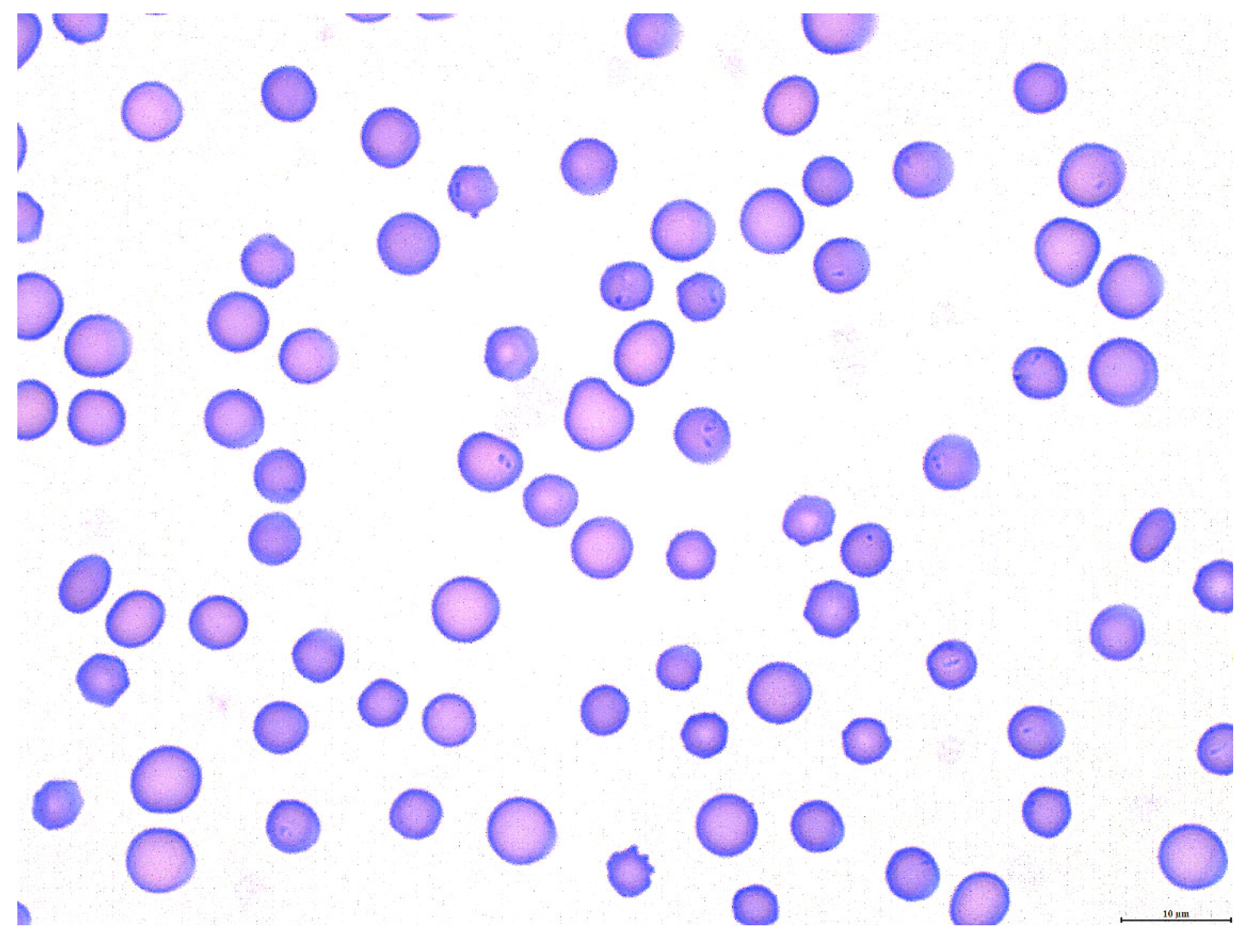
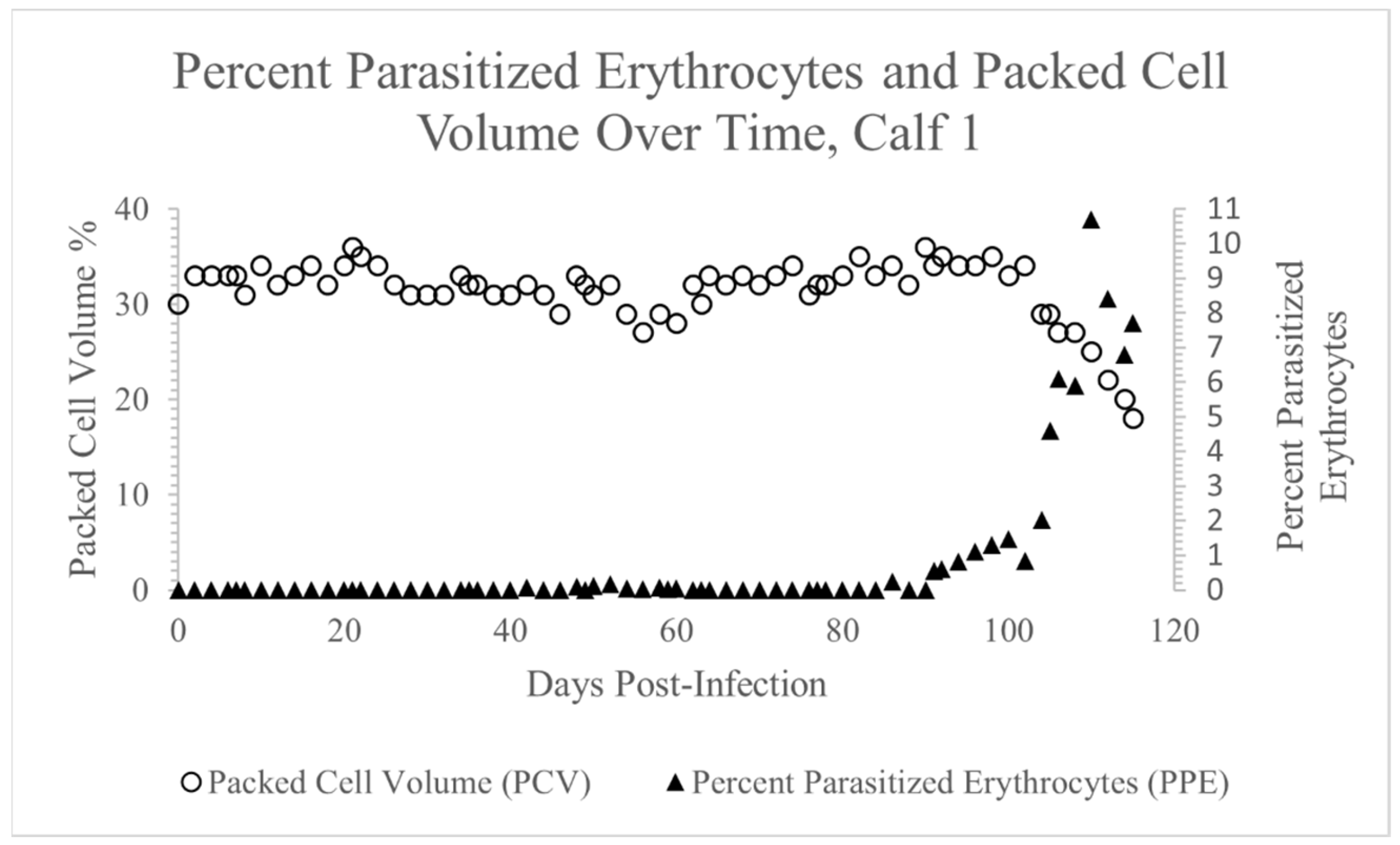
3.1. Theileria orientalis Infection of Calf 1

Theileria orientalis was first detected via PCR in the peripheral blood of Calf 1 twenty days post-inoculation (Table 1). While T. orientalis was detected in the peripheral blood of this calf via PCR for most of the remaining experimental timeline, there were scattered days during weeks 3, 5, and 11 when the parasitemia temporarily dropped below the level of PCR detection (indicated by +/−, Table 1). Piroplasms were intermittently detected via blood smear cytology (Figure 1) from day 42 post-inoculation onward, and the PPE ranged from 0.025% to 10.7% (Figure 2). The PCV remained stable until day 104, when it began to steadily decline, reaching a nadir of 18% (12% decline from pre-infection PCV) on day 115 post-inoculation (Figure 2). Apart from anemia, the complete blood counts were normal throughout the experiment. Terminally, mild hypoproteinemia and hyperbilirubinemia were noted on serum chemistry panels, and the calf exhibited mild tachypnea, depression, and lethargy secondary to anemia. As in previous experiments [17], the calf never became febrile.

3.2. Rhipicephalus microplus Acquisition Feed on Calf 1

The first group of ~5000 R. microplus larvae were applied to Calf 1 on day 53 post-inoculation and ~1000 removed sixteen days later as freshly molted adults. During this time-period, Calf 1 was PCR-positive for T. orientalis Ikeda via PCR and exhibited PPE of 0.025–0.1%. The second group of ~5000 R. microplus larvae were applied to Calf 1 on day 66 post-inoculation and ~1000 removed seventeen days later as freshly molted adults. During this time-period Calf 1 was intermittently PCR-positive for T. orientalis and exhibited a PPE of 0–0.04%.
Following removal, the ticks were applied to Calves 2 (Group 1) and 3 (Group 2) and allowed to feed. After a brief (4–5 day) period of feeding, a subset of male and female ticks from each batch were removed, dissected, and their salivary glands assessed for T. orientalis via PCR. T. orientalis was not detected in the salivary glands of any of the fifteen assayed ticks from each group (Table 2). This finding supports the conclusion that R. microplus ticks did not acquire T. orientalis while feeding on Calf 1 in this experiment.

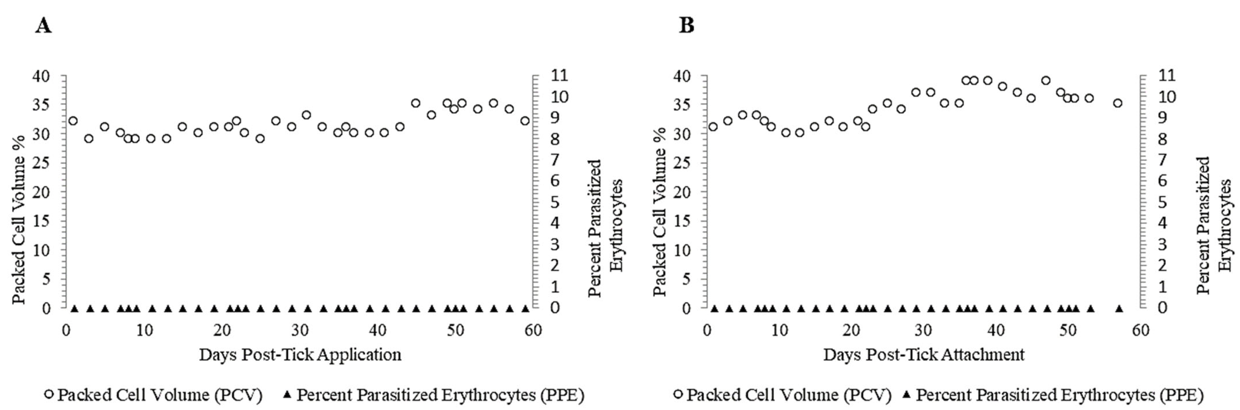
3.3. Failure of Adult R. microplus Ticks to Transmit T. orientalis to Calves 2 and 3

Following tick application, Calves 2 and 3 were monitored for evidence of T. orientalis-infection for 60 days. T. orientalis was not detected in the peripheral blood of either calf during that time-period via PCR (Table 1) or blood smear cytology (Figure 3A,B), consistent with lack of successful transmission of T. orientalis by R. microplus ticks in this experiment. Neither calf developed fever, anemia (Figure 3A,B), or any other abnormalities within the parameters assessed via physical exam, complete blood count or serum chemistry panel during this time-period.

3.4. T. orientalis Is Not Detected in Larval Progeny of R. microplus Fed on an Infected Calf

Since some Apicomplexan parasites, such as B. bovis, are transmitted transovarially by R. microplus ticks [41], ~40,000 progeny larval ticks of adults from both Groups 1 and 2 were assessed for T. orientalis via PCR. Theileria orientalis was not detected in progeny larvae from either tick group (Table 2).

4. Discussion

The results of this study suggest that R. microplus ticks are not competent vectors of the U.S. isolate of T. orientalis Ikeda via transstadial transmission. Larval stage R. microplus was applied to a T. orientalis Ikeda-infected calf and fed until adulthood. Once molted to adults, but before beginning to feed as adults, the ticks were removed and permitted to feed to repletion on two naïve, splenectomized calves to assess transstadial transmission. T. orientalis was not detected in the salivary glands of the adult R. microplus ticks after 4–5 days of transmission feeding, nor was it detected in progeny larvae of replete acquisition-fed adults. Furthermore, the two splenectomized, transmission-fed calves remained negative for T. orientalis Ikeda by both PCR and blood smear cytology at day 60 post-tick application and did not develop clinical signs consistent with infection.
Both Theileria sp. and Babesia sp. are apicomplexan hemoparasites that belong to the order Piroplasmida [42], and, although R. microplus ticks efficiently transmit B. bovis and T. equi [27,30,41,43,44], our study provides no evidence that they are vectors of T. orientalis Ikeda. This result may be related to the unique life history of R. microplus as a one-host tick. For R. microplus to be considered a competent vector of T. orientalis Ikeda, transmission must occur via at least one of two possible mechanisms: transstadial, which occurs between life stages; or transovarial, which occurs between mothers and offspring [45]. For one-host ticks such as R. microplus, transovarial transmission is one of the most efficient modes of pathogen transfer because ticks spend their entire lifecycle on a single host [46]. As mentioned, R. microplus ticks are efficient vectors of numerous pathogens, including B. bovis (bovine babesiosis), which is passed from the infected females to their offspring [41]. These offspring subsequently seek out and attach to a new host animal, perpetuating the transmission cycle [42]. In the transovarial transmission of B. bovis, the pathogen is acquired during adult feeding and moves to the midgut before being released into the hemolymph and migrating to the salivary glands and ovaries. After infecting the ovaries, kinetes are transovarially transmitted to the developing larvae, which then transmit infective sporozoites to naïve hosts via the salivary glands [45]. Successful transovarial transmission, therefore, requires several complex steps, including movement throughout the tick and evading the tick immune system [47]. Unlike Babesia sp., the Theileria sp. life cycles are not generally characterized by transovarial transmission [48]. The findings in this study are in line with this observation. We assessed ~40,000 pooled larval progeny from each tick group for T. orientalis and were unable to detect the parasite. Although the pooling of larvae allowed us to assay a larger percentage of the tick progeny for T. orientalis, it also presented a limitation as the separate assessment of larvae from individual female ticks can also provide important information. While the present data suggest a low to non-existent transovarial transmission rate (defined as the number of ticks that produce infected offspring), it is possible that rare, individual females could have a high filial infection rate (defined as the number of infected offspring produced by a single infected female). Essentially, in this case, the overall low transovarial transmission rate could have led to a dilution effect, masking rare females with a high filial infection rate. Future studies in which the offspring of individual females are tested are required to rule out this possibility.
Transstadial transmission is the most common mode of pathogen transfer for most ticks and tick-borne pathogens, including Theileria sp. [48,49] This mode of transmission works well for three-host ticks, such as Haemaphysalis longicornis, the most common vector of T. orientalis. In this system, T. orientalis Ikeda is acquired by larval or nymphal H. longicornis ticks during a blood meal. Once replete, the tick drops off the host and molts, during this period of off-host development the parasite is passed transstadially to the next stage [2,17,18]. Most tick species that transmit Theileria sp. are multi-host ticks and primarily transmit parasites transstadially [45,48]. This major life history difference may explain why R. microplus did not transmit T. orientalis in our study.
One interesting exception to this general rule is the role of R. microplus ticks in transmitting Theileria equi between infected horses. Rhipicephalus microplus can serve as a tick vector of T. equi in some cases [30,43,44]. It has been shown that T. equi can be transmitted transstadially from larval or nymphal R. microplus to adults [43,50], and that the parasite can be transmitted intrastadially between horses by R. microplus adult male ticks, which move between hosts, feeding intermittently, and mating with female ticks [44]. In these studies, the successful R. microplus acquisition of T. equi from persistently infected horses was demonstrated by the PCR-based detection of the parasite within the salivary gland tissue [43,44,50]. In contrast, we were unable to demonstrate T. orientalis within the salivary gland tissue of male or female adult ticks fed on a T. orientalis-infected calf. Similarly, while adult R. microplus transmits T. equi to naïve horses, R. microplus was unable to transmit T. orientalis to naïve calves.
The definitive reason that R. microplus is an efficient vector of T. equi but did not acquire or transmit T. orientalis in the present study is not clear. We utilized splenectomized calves in this study to maximize parasitemia and minimize the risk of false negative findings, but it is possible that the level of parasitemia to which the ticks were exposed during acquisition (0–0.1%) was insufficient for tick infection. However, in our previous T. orientalis Ikeda competency study using the same parasite strain and U.S. H. longicornis ticks, the H. longicornis ticks successfully acquired and transmitted the parasite when exposed to parasitemia of 0% in a PCR-positive calf [17], and the related parasite T. parva was readily transmitted between mammalian hosts by competent tick vectors when the parasite was below the level of detection by light microscopy [51]. Finally, R. microplus ticks successfully acquired T. equi from persistently infected horses with low-levels of parasitemia [43], suggesting that the parasite dose was not the main variable in play regarding the R. microplus competence for Theileria sp. acquisition and transmission. Regardless, it remains possible that R. microplus could acquire and/or transmit T. orientalis under field conditions in which cattle are highly tick-infested and develop extremely high parasitemia.
While this experiment demonstrated that R. microplus ticks are not likely competent vectors of T. orientalis Ikeda, T. orientalis DNA has been detected in R. microplus from other regions such as India [33,36], China [34,35], Vietnam [38], and Pakistan [37]. It should be noted that these studies tested field-collected R. microplus ticks for T. orientalis Ikeda while this experiment utilized colony ticks from Texas. Thus, there could be genetic, behavioral, or physiological differences in the lineages of R. microplus from these different regions, or differences in the T. orientalis parasite strains [52,53]. Furthermore, detection within a tick does not confirm vector competence unless additional steps to establish competency are conducted. In this controlled experiment, the lack of R. microplus vector competence is supported by three main findings: 1. T. orientalis was not detected via PCR in the salivary glands from adult ticks fed as larvae and nymphs on a known T. orientalis-positive calf; 2. T. orientalis was not detected via PCR in the larval progeny of ticks fed on a known T. orientalis-infected calf; and 3. No evidence of transmission was detected via PCR, cytology, or clinical pathology in either splenectomized, naïve calves after 60 days. To the authors’ knowledge, this is the first controlled acquisition and transmission study conducted to determine whether R. microplus is a competent vector of T. orientalis Ikeda.
As a one-host tick, R. microplus ticks live on the same host through their three major life stages. With a large percentage of its time spent on the host, it is probable that R. microplus will encounter other ticks feeding on the same host. For example, R. microplus and H. longicornis readily feed on livestock [18,19,20,21,54]. In Tengchong County, China, researchers found both species of ticks feeding on goats and both species were PCR-positive for T. orientalis [34]. While the significance of this result is unclear given the results of the present study, other tick-pathogen systems have demonstrated that pathogen spillover from a native tick species into a new tick species is possible. In Virginia, larval, nymphal, and adult stages of H. longicornis were positive for the Bourbon virus, a virus that is known to be carried by the lone star tick (Amblyomma americanum) [55]. Amblyomma americanum ticks and blood from locally harvested deer at the same site also tested positive for the Bourbon virus, suggesting that the tick-borne virus was circulating in the region and that H. longicornis could have been infected by feeding on a host that was infected with the Bourbon virus, presumably infected by A. americanum [55]. Pathogen spillover into a new tick host can lead to active pathogen circulation in the absence of the known primary tick vector. Through surveillance and experimental studies, pathogen spillover from a native tick species to a new tick species should be prioritized as a future research avenue to predict the range expansion of tick-borne pathogens. This is especially relevant to T. orientalis, which is newly endemic in the U.S.
Co-feeding next to an infected tick has also been suggested as a possible route of pathogen sharing between ticks. The primary vector for Rickettsia parkeri is the Gulf Coast tick, Amblyomma maculatum [56]. However, when A. americanum nymphs were co-feeding next to infected A. maculatum, the pathogen was successfully transstadially maintained in A. americanum adults [56]. Feeding in close spatiotemporal proximity has been illustrated as an efficient way to transmit pathogens between two ticks in the absence of systemic pathogen infection in a host [57,58]. While T. orientalis may not efficiently infect the R. microplus ticks in the absence of co-feeding H. longicornis ticks, further studies are needed to rule out Theileria sp. Transmission between ticks via the close proximity feeding of two different tick species.
T. orientalis will likely continue to cause significant losses for the U.S. cattle industry. The extent of the T. orientalis spread and the resultant development of regional endemicity is dependent both on the movement of asymptomatic carrier cattle and the range of competent tick hosts. Many tick species native to the U.S. are vectors of various Theileria sp. parasites. Thus, continued studies are needed to characterize the repertoire of tick vectors for this newly endemic pathogen of U.S. cattle. While the present study suggests that R. microplus is not likely a competent vector for T. orientalis, surveillance is recommended to monitor for interactions between H. longicornis, R. microplus, and other native U.S. ticks to ensure that pathogen spillover does not occur.

Author Contributions

Conceptualization, L.M.F., C.K.O., D.R.H., K.C.P., K.O. and G.A.S.; methodology, L.M.F., C.K.O., D.R.H. and G.A.S.; formal analysis, L.M.F., C.K.O., A.H. and D.R.H.; investigation, L.M.F., C.K.O., D.R.H. and A.H., resources, L.M.F.; data curation, L.M.F.; writing—original draft preparation, C.K.O., A.H., K.O., K.C.P. and L.M.F.; writing—review and editing, L.M.F., D.R.H., C.K.O., A.H., K.O., K.C.P. and G.A.S.; visualization, L.M.F.; supervision, L.M.F., D.R.H. and C.K.O.; project administration, L.M.F.; funding acquisition, L.M.F. All authors have read and agreed to the published version of the manuscript.

Funding

The research was funded by the USDA-ARS CRIS# 2090-32000-044-000-D.

Institutional Review Board Statement

These animal experiments were approved by the University of Idaho Institutional Animal Care and Use Committees, Protocol number 2021-37.

Informed Consent Statement

Not applicable.

Data Availability Statement

All data are provided in the manuscript.

Acknowledgments

The authors wish to acknowledge the technical expertise of Shelby Beckner and Nic Durfee. We also wish to acknowledge the animal handling expertise of Megan Jacks and her animal care staff, and tick rearing and handling expertise of Gavin Scoles.

Conflicts of Interest

The authors declare no conflict of interest.

References

  1. Sivakumar, T.; Hayashida, K.; Sugimoto, C.; Yokoyama, N. Evolution and genetic diversity of Theileria. Infect. Genet. Evol. 2014, 27, 250–263. [Google Scholar] [CrossRef] [PubMed] [Green Version]
  2. Watts, J.G.; Playford, M.C.; Hickey, K.L. Theileria orientalis: A review. N. Z. Vet. J. 2016, 64, 3–9. [Google Scholar] [CrossRef] [PubMed]
  3. Lawrence, K.; Forsyth, S.; Vaatstra, B.; McFadden, A.; Pulford, D.; Govindaraju, K.; Pomroy, W. Clinical haematology and biochemistry profiles of cattle naturally infected with Theileria orientalis Ikeda type in New Zealand. N. Z. Vet. J. 2017, 66, 21–29. [Google Scholar] [CrossRef] [PubMed]
  4. Lawrence, K.; Gedye, K.; Pomroy, W. A longitudinal study of the effect of Theileria orientalis Ikeda type infection on three New Zealand dairy farms naturally infected at pasture. Vet. Parasitol. 2019, 276, 108977. [Google Scholar] [CrossRef] [PubMed]
  5. Lawrence, K.; Gibson, M.; Hickson, R.; Gedye, K.; Hoogenboom, A.; Fermin, L.; Draganova, I.; Pomroy, W. Experimental infection of Friesian bulls with Theileria orientalis (Ikeda) and effects on the haematocrit, live weight, rectal temperature and activity. Vet. Parasitol. Reg. Stud. Rep. 2018, 14, 85–93. [Google Scholar] [CrossRef]
  6. Lawrence, K.; Lawrence, B.; Hickson, R.; Hewitt, C.; Gedye, K.; Fermin, L.; Pomroy, W. Associations between Theileria orientalis Ikeda type infection and the growth rates and haematocrit of suckled beef calves in the North Island of New Zealand. N. Z. Vet. J. 2018, 67, 66–73. [Google Scholar] [CrossRef]
  7. Perera, P.K.; Gasser, R.B.; Firestone, S.M.; Anderson, G.A.; Malmo, J.; Davis, G.; Beggs, D.S.; Jabbar, A. Oriental theileriosis in dairy cows causes a significant milk production loss. Parasites Vectors 2014, 7, 73. [Google Scholar] [CrossRef] [Green Version]
  8. Oakes, V.J.; Yabsley, M.J.; Schwartz, D.; LeRoith, T.; Bissett, C.; Broaddus, C.; Schlater, J.L.; Todd, S.M.; Boes, K.M.; Brookhart, M.; et al. Theileria orientalis Ikeda Genotype in Cattle, Virginia, USA. Emerg. Infect. Dis. 2019, 25, 1653–1659. [Google Scholar] [CrossRef] [Green Version]
  9. Eamens, G.J.; Bailey, G.; Jenkins, C.; Gonsalves, J.R. Significance of Theileria orientalis types in individual affected beef herds in New South Wales based on clinical, smear and PCR findings. Vet. Parasitol. 2013, 196, 96–105. [Google Scholar] [CrossRef]
  10. Eamens, G.J.; Gonsalves, J.R.; Jenkins, C.; Collins, D.; Bailey, G. Theileria orientalis MPSP types in Australian cattle herds associated with outbreaks of clinical disease and their association with clinical pathology findings. Vet. Parasitol. 2013, 191, 209–217. [Google Scholar] [CrossRef]
  11. Telionis, A.; Lahmers, K.; Todd, M.; Carbonello, A.; Broaddus, C.C.; Bissett, C.J.; Hungerford, L.L. Distribution of Theileria orientalis in Virginia Market Cattle, 2018–2020. Pathogens 2022, 11, 1353. [Google Scholar] [CrossRef]
  12. Uilenberg, G.; Perié, N.M.; Spanjer, A.A.; Franssen, F.F. Theileria orientalis, a cosmopolitan blood parasite of cattle: Demonstration of the schizont stage. Res. Vet. Sci. 1985, 38, 352–360. [Google Scholar] [CrossRef]
  13. Stockham, S.L.; Kjemtrup, A.M.; Conrad, P.A.; Schmidt, D.A.; Scott, M.A.; Robinson, T.W.; Tyler, J.W.; Johnson, G.C.; Carson, C.A.; Cuddihee, P. Theileriosis in a Missouri beef herd caused by Theileria buffeli: Case report, herd investigation, ultra-structure, phylogenetic analysis, and experimental transmission. Vet. Pathol. 2000, 37, 11–21. [Google Scholar] [CrossRef] [Green Version]
  14. Oakes, V.J.; Todd, S.M.; Carbonello, A.A.; Michalak, P.; Lahmers, K.K. Coinfection of cattle in Virginia with Theileria orientalis Ikeda genotype and Anaplasma marginale. J. Vet. Diagn. Investig. 2021, 34, 36–41. [Google Scholar] [CrossRef]
  15. Thompson, A.T.; White, S.; Shaw, D.; Egizi, A.; Lahmers, K.; Ruder, M.G.; Yabsley, M.J. Theileria orientalis Ikeda in host-seeking Haemaphysalis longicornis in Virginia, U.S.A. Ticks Tick-Borne Dis. 2020, 11, 101450. [Google Scholar] [CrossRef]
  16. Lokting, B. A New Tick-Borne Disease Is Killing Cattle in the US. MIT Technology Review 2022. Available online: https://www.technologyreview.com/2022/11/17/1063352/new-tick-borne-disease-killing-cattle-in-us/ (accessed on 18 February 2023).
  17. Dinkel, K.D.; Herndon, D.R.; Noh, S.M.; Lahmers, K.K.; Todd, S.M.; Ueti, M.W.; Scoles, G.A.; Mason, K.L.; Fry, L.M. A U.S. isolate of Theileria orientalis, Ikeda genotype, is transmitted to cattle by the invasive Asian longhorned tick, Haemaphysalis longicornis. Parasites Vectors 2021, 14, 157. [Google Scholar] [CrossRef]
  18. Marendy, D.; Baker, K.; Emery, D.; Rolls, P.; Stutchbury, R. Haemaphysalis longicornis: The life-cycle on dogs and cattle, with confirmation of its vector status for Theileria orientalis in Australia. Vet. Parasitol. 2019, 277, 100022. [Google Scholar] [CrossRef]
  19. Heath, A. Biology, ecology and distribution of the tick, Haemaphysalis longicornis Neumann (Acari: Ixodidae) in New Zealand. N. Z. Vet. J. 2016, 64, 10–20. [Google Scholar] [CrossRef]
  20. Rainey, T.; Occi, J.L.; Robbins, R.G.; Egizi, A. Discovery of Haemaphysalis longicornis (Ixodida: Ixodidae) Parasitizing a Sheep in New Jersey, United States. J. Med. Èntomol. 2018, 55, 757–759. [Google Scholar] [CrossRef]
  21. Tufts, D.M.; VanAcker, M.C.; Fernandez, M.P.; DeNicola, A.; Egizi, A.; Diuk-Wasser, M.A. Distribution, Host-Seeking Phenology, and Host and Habitat Associations of Haemaphysalis longicornis Ticks, Staten Island, New York, USA. Emerg. Infect. Dis. 2019, 25, 792–796. [Google Scholar] [CrossRef] [Green Version]
  22. Beard, C.B.; Occi, J.; Bonilla, D.L.; Egizi, A.M.; Fonseca, D.M.; Mertins, J.W.; Backenson, B.P.; Bajwa, W.I.; Barbarin, A.M.; Bertone, M.A.; et al. Multistate Infestation with the Exotic Disease-Vector Tick Haemaphysalis longicornis—United States, August 2017-September 2018. MMWR Morb. Mortal. Wkly. Rep. 2018, 67, 1310–1313. [Google Scholar] [CrossRef] [PubMed] [Green Version]
  23. Bonilla, D.L. National Haemaphysalis longicornis (Asian Longhorned Tick) Situation Report, USDA-APHIS, Service, Editor. 2-27-2023, USDA-APHIS. Available online: https://www.aphis.usda.gov/animal_health/animal_diseases/tick/downloads/longhorned-tick-sitrep.pdf (accessed on 28 February 2023).
  24. Hammer, J.F.; Jenkins, C.; Bogema, D.; Emery, D. Mechanical transfer of Theileria orientalis: Possible roles of biting arthropods, colostrum and husbandry practices in disease transmission. Parasites Vectors 2016, 9, 34. [Google Scholar] [CrossRef] [PubMed] [Green Version]
  25. Fujusaki, K.; Kamio, T.; Kawazu, S.; Shimizu, S.; Shimura, K. Theileria sergenti: Experimental transmission by the long-nosed cattle louse, Linognathus vituli. Ann. Trop. Med. Parasitol. 1993, 87, 217–218. [Google Scholar] [CrossRef] [PubMed]
  26. Alkishe, A.; Raghavan, R.K.; Peterson, A.T. Likely Geographic Distributional Shifts among Medically Important Tick Species and Tick-Associated Diseases under Climate Change in North America: A Review. Insects 2021, 12, 225. [Google Scholar] [CrossRef] [PubMed]
  27. Howell, J.M.; Ueti, M.W.; Palmer, G.H.; Scoles, G.A.; Knowles, D.P. Persistently Infected Calves as Reservoirs for Acquisition and Transovarial Transmission of Babesia bovis by Rhipicephalus (Boophilus) microplus. J. Clin. Microbiol. 2007, 45, 3155–3159. [Google Scholar] [CrossRef] [Green Version]
  28. Mahoney, D.F.; Wright, I.G.; Goodger, B.V.; Mirre, G.B.; Sutherst, R.W.; Utech, K.B.W. The transmission of babesia bovis in herds of european and zebu x european cattle infested with the tick, boophilus microplus. Aust. Vet. J. 1981, 57, 461–469. [Google Scholar] [CrossRef]
  29. Potgieter, F.T.; Van Vuuren, A.S. The transmission of Babesia bovis using frozen infective material obtained from Boophilus microplus larvae. Onderstepoort J. Vet. Res. 1974, 41, 79–80. [Google Scholar]
  30. Scoles, G.A.; Ueti, M.W. Vector Ecology of Equine Piroplasmosis. Annu. Rev. Èntomol. 2015, 60, 561–580. [Google Scholar] [CrossRef]
  31. Uilenberg, G. Babesia—A historical overview. Vet. Parasitol. 2006, 138, 3–10. [Google Scholar] [CrossRef]
  32. Annual Cattle Review, Texas and U.S. Inventory, USDA-NASS, Editor. 2021: Texas Field Office. Available online: https://www.nass.usda.gov/Statistics_by_State/Texas/Publications/Current_News_Release/2021_Rls/tx-cattle-review-2021.pdf (accessed on 5 February 2023).
  33. Kakati, P.; Sarmah, P.C.; Ray, D.; Bhattacharjee, K.; Sharma, R.K.; Barkalita, L.M.; Sarma, D.K.; Baishya, B.C.; Borah, P.; Stanley, B. Emergence of oriental theileriosis in cattle and its transmission through Rhipicephalus (Boophilus) microplus in Assam, India. Vet. World 2015, 8, 1099–1104. [Google Scholar] [CrossRef]
  34. Li, L.-H.; Wang, J.-Z.; Zhu, D.; Li, X.-S.; Lu, Y.; Yin, S.-Q.; Li, S.-G.; Zhang, Y.; Zhou, X.-N. Detection of novel piroplasmid species and Babesia microti and Theileria orientalis genotypes in hard ticks from Tengchong County, Southwest China. Parasitol. Res. 2020, 119, 1259–1269. [Google Scholar] [CrossRef]
  35. Hao, L.; Yuan, D.; Li, S.; Jia, T.; Guo, L.; Hou, W.; Lu, Z.; Mo, X.; Yin, J.; Yang, A.; et al. Detection of Theileria spp. in ticks, sheep keds (Melophagus ovinus), and livestock in the eastern Tibetan Plateau, China. Parasitol. Res. 2020, 119, 2641–2648. [Google Scholar] [CrossRef]
  36. Patial, V.; Gupta, T.; Angaria, S.; Bali, D.; Katoch, A.; Gautam, M.; Singh, N.K.; Sharma, M.; Chahota, R. Theileria orientalis outbreak in an organized cattle breeding farm. Vet. Parasitol. Reg. Stud. Rep. 2021, 24, 100572. [Google Scholar] [CrossRef]
  37. Rehman, A.; Conraths, F.J.; Sauter-Louis, C.; Krücken, J.; Nijhof, A.M. Epidemiology of tick-borne pathogens in the semi-arid and the arid agroecological zones of Punjab province, Pakistan. Transbound. Emerg. Dis. 2019, 66, 526–536. [Google Scholar] [CrossRef] [Green Version]
  38. Khukhuu, A.; Lan, D.T.B.; Long, P.T.; Ueno, A.; Li, Y.; Luo, Y.; de Macedo, A.C.C.; Matsumoto, K.; Inokuma, H.; Kawazu, S.-I.; et al. Molecular Epidemiological Survey of Theileria orientalis in Thua Thien Hue Province, Vietnam. J. Vet. Med. Sci. 2011, 73, 701–705. [Google Scholar] [CrossRef] [Green Version]
  39. Ota, N.; Mizuno, D.; Kuboki, N.; Igarashi, I.; Nakamura, Y.; Yamashina, H.; Hanzaike, T.; Fujii, K.; Onoe, S.; Hata, H.; et al. Epidemiological Survey of Theileria orientalis Infection in Grazing Cattle in the Eastern Part of Hokkaido, Japan. J. Vet. Med. Sci. 2009, 71, 937–944. [Google Scholar] [CrossRef] [Green Version]
  40. Scoles, G.A.; Ueti, M.W.; Noh, S.M.; Knowles, D.P.; Palmer, G.H. Conservation of Transmission Phenotype of Anaplasma marginale (Rickettsiales: Anaplasmataceae) Strains Among Dermacentor and Rhipicephalus Ticks (Acari: Ixodidae). J. Med. Èntomol. 2007, 44, 484–491. [Google Scholar] [CrossRef] [Green Version]
  41. Howell, J.M.; Ueti, M.W.; Palmer, G.H.; Scoles, G.A.; Knowles, D.P. Transovarial Transmission Efficiency of Babesia bovis Tick Stages Acquired by Rhipicephalus (Boophilus) microplus during Acute Infection. J. Clin. Microbiol. 2007, 45, 426–431. [Google Scholar] [CrossRef] [Green Version]
  42. Almazán, C.; Scimeca, R.C.; Reichard, M.V.; Mosqueda, J. Babesiosis and Theileriosis in North America. Pathogens 2022, 11, 168. [Google Scholar] [CrossRef]
  43. Ueti, M.W.; Palmer, G.H.; Kappmeyer, L.S.; Statdfield, M.; Scoles, G.A.; Knowles, D.P. Ability of the Vector Tick Boophilus microplus to Acquire and Transmit Babesia equi following Feeding on Chronically Infected Horses with Low-Level Parasitemia. J. Clin. Microbiol. 2005, 43, 3755–3759. [Google Scholar] [CrossRef] [Green Version]
  44. Ueti, M.W.; Palmer, G.H.; Scoles, G.A.; Kappmeyer, L.S.; Knowles, D.P. Persistently Infected Horses Are Reservoirs for Intrastadial Tick-Borne Transmission of the Apicomplexan Parasite Babesia equi. Infect. Immun. 2008, 76, 3525–3529. [Google Scholar] [CrossRef] [PubMed] [Green Version]
  45. Schnittger, L.; Ganzinelli, S.; Bhoora, R.; Omondi, D.; Nijhof, A.M.; Florin-Christensen, M. The Piroplasmida Babesia, Cytauxzoon, and Theileria in farm and companion animals: Species compilation, molecular phylogeny, and evolutionary insights. Parasitol. Res. 2022, 121, 1207–1245. [Google Scholar] [CrossRef] [PubMed]
  46. Bergren, N.A.; Kading, R.C. The Ecological Significance and Implications of Transovarial Transmission among the Vector-Borne Bunyaviruses: A Review. Insects 2018, 9, 173. [Google Scholar] [CrossRef] [PubMed] [Green Version]
  47. Schnittger, L.; Rodriguez, A.E.; Florin-Christensen, M.; Morrison, D.A. Babesia: A world emerging. Infect. Genet. Evol. 2012, 12, 1788–1809. [Google Scholar] [CrossRef] [PubMed]
  48. Jalovecka, M.; Hajdusek, O.; Sojka, D.; Kopacek, P.; Malandrin, L. The Complexity of Piroplasms Life Cycles. Front. Cell. Infect. Microbiol. 2018, 8, 248. [Google Scholar] [CrossRef] [Green Version]
  49. Ravindran, R.; Hembram, P.K.; Kumar, G.S.; Kumar, K.G.A.; Deepa, C.K.; Varghese, A. Transovarial transmission of pathogenic protozoa and rickettsial organisms in ticks. Parasitol. Res. 2023, 122, 691–704. [Google Scholar] [CrossRef]
  50. Peckle, M.; Santos, H.; Pires, M.; Silva, C.; Costa, R.; Vitari, G.; Camilo, T.; Meireles, N.; Paulino, P.; Massard, C. Dynamics of Theileria equi Infection in Rhipicephalus (Boophilus) microplus during the Parasitic Phase in a Chronically Infected Horse. Pathogens 2022, 11, 525. [Google Scholar] [CrossRef]
  51. Olds, C.L.; Mason, K.L.; Scoles, G.A. Rhipicephalus appendiculatus ticks transmit Theileria parva from persistently infected cattle in the absence of detectable parasitemia: Implications for East Coast fever epidemiology. Parasites Vectors 2018, 11, 126. [Google Scholar] [CrossRef] [Green Version]
  52. Low, V.L.; Tay, S.T.; Kho, K.L.; Koh, F.X.; Tan, T.K.; Lim, Y.A.L.; Ong, B.L.; Panchadcharam, C.; Norma-Rashid, Y.; Sofian-Azirun, M. Molecular characterisation of the tick Rhipicephalus microplus in Malaysia: New insights into the cryptic diversity and distinct genetic assemblages throughout the world. Parasites Vectors 2015, 8, 341. [Google Scholar] [CrossRef] [Green Version]
  53. Estrada-Peña, A.; Venzal, J.M.; Nava, S.; Mangold, A.; Guglielmone, A.A.; Labruna, M.B.; de la Fuente, J. Reinstatement of Rhipicephalus (Boophilus) australis(Acari: Ixodidae) with Redescription of the Adult and Larval Stages. J. Med. Èntomol. 2012, 49, 794–802. [Google Scholar] [CrossRef] [Green Version]
  54. Pereira, D.F.S.; Ribeiro, H.S.; Gonçalves, A.A.M.; da Silva, A.V.; Lair, D.F.; de Oliveira, D.S.; Boas, D.F.V.; Conrado, I.d.S.S.; Leite, J.C.; Barata, L.M.; et al. Rhipicephalus microplus: An overview of vaccine antigens against the cattle tick. Ticks Tick-Borne Dis. 2022, 13, 101828. [Google Scholar] [CrossRef]
  55. Cumbie, A.N.; Trimble, R.N.; Eastwood, G. Pathogen Spillover to an Invasive Tick Species: First Detection of Bourbon Virus in Haemaphysalis longicornis in the United States. Pathogens 2022, 11, 454. [Google Scholar] [CrossRef]
  56. Wright, C.L.; Sonenshine, D.E.; Gaff, H.D.; Hynes, W.L. Rickettsia parkeri Transmission to Amblyomma americanum by Cofeeding with Amblyomma maculatum (Acari: Ixodidae) and Potential for Spillover. J. Med. Entomol. 2015, 52, 1090–1095. [Google Scholar] [CrossRef]
  57. Patrican, L.A. Acquisition of Lyme disease spirochetes by cofeeding Ixodes scapularis ticks. Am. J. Trop. Med. Hyg. 1997, 57, 589–593. [Google Scholar] [CrossRef]
  58. Belli, A.; Sarr, A.; Rais, O.; Rego, R.O.M.; Voordouw, M.J. Ticks infected via co-feeding transmission can transmit Lyme borreliosis to vertebrate hosts. Sci. Rep. 2017, 7, 5006. [Google Scholar] [CrossRef] [Green Version]
Figure 1. Photomicrograph of representative blood smear from Calf 1, day 111 post-inoculation with T. orientalis. Occasional erythrocytes contain 1–2, intracytoplasmic, 1–2.5 µm × 0.5 µm piroplasms, consistent with the T. orientalis merozoite stage. Scale bar: 10 µm.
Pathogens 12 00559 g001
Figure 2. PPE and PCV over time, Calf 1. Piroplasms were first detected on day 42 post-inoculation, and from that point to the end of the experiment, PPE ranged from 0% to 10.7%. The PCV did not decline significantly until day 104, when it went into decline, reaching a nadir of 18% on day 115 post-inoculation.
Pathogens 12 00559 g002
Figure 3. PPE and PCV over time, Calves 2 (A) and 3 (B). In both calves, no parasites were detected during the 60-day monitoring period post-tick application, and the PCV remained within the normal range for the entire experiment.
Pathogens 12 00559 g003
Table 1. Weekly Theileria orientalis peripheral blood PCR results for Calves 1–3. DPI = days post-infection. During weeks 3, 5, and 11, T. orientalis was below the level of detection for at least 1 sampling day that week but was detected on other sampling points in the same week. This is indicated with +/−.
Calf1
0–7 DPI
2
8–14 DPI
3
15–21 DPI
4
22–28 DPI
5
29–35 DPI
6
36–42 DPI
7
43–49 DPI
8
50–56 DPI
9
57–63 DPI
10
64–70 DPI
11
71–77 DPI
12
78–84 DPI
13
85–91 DPI
14
92–98 DPI
15
99–105 DPI
16
106–112 DPI
17
113–119 DPI
1+/−++/−++++++/−++++++
2
3
Table 2. Results of Theileria orientalis PCR performed on adult Rhipicephalus microplus salivary glands and macerated R. microplus larvae.
Tick BatchFemales TestedMales TestedNumber Positive
Group 1 Adults690
Group 2 Adults780
Group 1 Larvae~20,000 larval ticksBatch negative
Group 2 Larvae~20,000 larval ticksBatch negative
Disclaimer/Publisher’s Note: The statements, opinions and data contained in all publications are solely those of the individual author(s) and contributor(s) and not of MDPI and/or the editor(s). MDPI and/or the editor(s) disclaim responsibility for any injury to people or property resulting from any ideas, methods, instructions or products referred to in the content.

