Article

Phytophthora Species Involved in Alnus glutinosa Decline in Portugal

1
Dipartimento Territorio e Sistemi Agro-Forestali, Università degli Studi di Padova, Viale dell’Università, 16, 35020 Legnaro, Italy
2
CESAM, Departamento de Biologia, Universidade de Aveiro, 3810-193 Aveiro, Portugal
*
Author to whom correspondence should be addressed.
Pathogens 2023, 12(2), 276; https://doi.org/10.3390/pathogens12020276
Submission received: 16 January 2023 / Revised: 2 February 2023 / Accepted: 6 February 2023 / Published: 8 February 2023

Abstract

:
Recent field surveys conducted in five common alder ecosystems in Portugal have shown the occurrence of severe canopy dieback, bleeding canker and root rot symptoms indicative of Phytophthora infections. Isolations from symptomatic tissues, rhizosphere and water samples yielded a total of 13 Phytophthora species belonging to 6 phylogenetic clades, including P. lacustris (13 isolates), P. multivora (10), P. amnicola (9), P. chlamydospora (6), P. polonica (6), P. bilorbang (4), P. plurivora (4), P. cinnamomi (3), P. asparagi (2), P. cactorum (2), P. pseudocryptogea (2), P. gonapodyides (1) and P. rosacearum (1). Results of the pathogenicity test confirmed the complex aetiology of common alder decline and the additional risk posed by Phytophthora multivora to the riparian habitats in Portugal. At the same time, the diversity of Phytophthora assemblages detected among the investigated sites suggests that different species could contribute to causing the same symptoms on this host. Two species, P. amnicola and P. rosacearum, are reported here for the first time in natural ecosystems in Europe.

1. Introduction

Alders represent an important component of European riparian and wetland vegetation. In Europe, four alder species grow spontaneously, mainly along rivers, streams and damp environments, often with a pioneer behaviour fundamental for ecological succession [1,2]. In Portugal, the only species occurring naturally is the common alder (Alnus glutinosa (L.) Gaertn.). This species is widespread in the northern and central parts of the country, mainly in the flooded plains and swamps at lower altitudes than the mountain riparian systems often associated with other broadleaved tree species such as Quercus spp. and Salix spp. [3].
Since the early 1990s, alder ecosystems have been severally impacted by an emerging disease that has contributed to their decline and regression in the European continent and some areas of North America [4,5]. Typical symptoms include general or progressive canopy dieback, stem bleeding cankers, necrotic bark lesions at the collar and root rot [6].
Many studies have investigated the causes of this disease, identifying the causal agents as some members of the Phytophthora genus [4,7,8]. The disease appears to have an extremely complex aetiology; independent surveys have ascertained the occurrence of over 30 species of Phytophthora in declining alder ecosystems between North America and Europe [5,7,9,10]. Many of these species belong to the Phytophthora clade 6 sensu [11]. This clade includes organisms closely related to aquatic environments. The ecology of several taxa is still unclear; some species are known to have a saprotrophic or opportunistic lifestyle, while a few are reported to be aggressive pathogens [12,13].
Among the other Phytophthora species associated with declining alder trees, several belong to clade 2. Phytophthora plurivora is one of the most widespread species in declining alder ecosystems in Europe, and its pathogenicity has been confirmed using different inoculation techniques [8,14,15]. In contrast, in North America, P. siskiyouensis is reported as one of the most aggressive pathogens of the Alnus species [16,17].
Despite the studies that have been conducted in Europe during the last three decades, many issues about the aetiology of alder decline remain to be clarified, as well as the distribution and impact of the different Phytophthora species among the countries. In Portugal, until now, only one study has investigated the role of Phytophthora species in alder decline [18]. The study was conducted on two alder stands along two rivers in central Portugal, confirming the involvement of two species, Phytophthora × alni and P. lacustris, in the disease aetiology.
Therefore, given the still limited information on the occurrence and impact of Phytophthora species in Portuguese riparian habitats and the recent discovery in central Portugal of five riparian ecosystems with high mortality rates of common alder trees, a study was conducted to establish the causal agents and obtain new data about the diversity and impact of Phytophthora species.

2. Materials and Methods

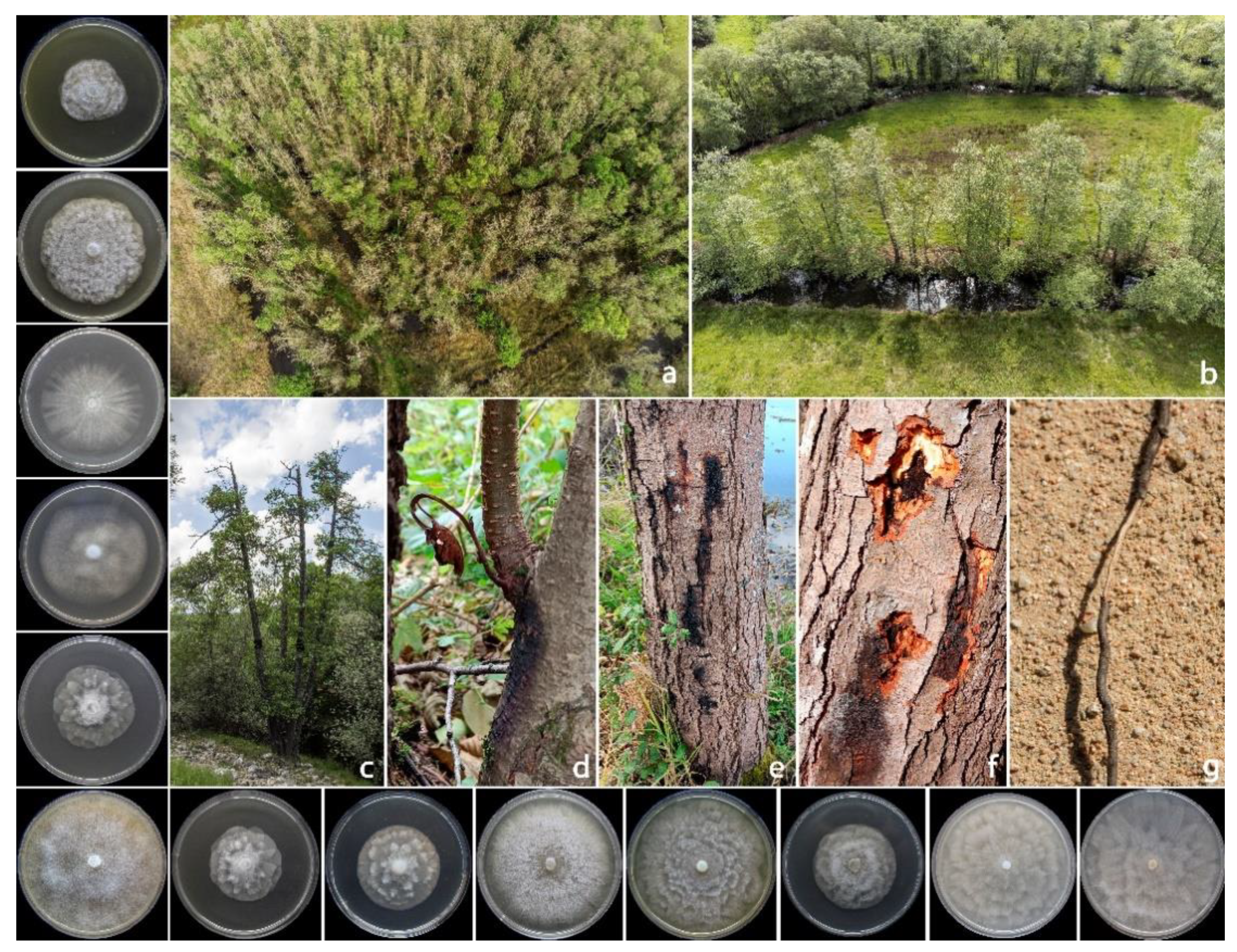
2.1. Field Surveys and Sampling Procedure

Monitoring activities were conducted during spring 2022 on five natural Alnus glutinosa stands located in the central part of Portugal, the districts of Aveiro and Guarda (Table 1). The altitude of survey sites ranged from 9 to 750 m. a.s.l.
At each site, mature alder trees were visually checked for the presence of typical Phytophthora disease symptoms, including wilting of foliage, shoot and twigs dieback, sudden death, bleeding cankers, and root and collar rot. In Sites 2 and 3, four linear transects of 50 m were randomly established to evaluate disease incidence and mortality rate, expressed as the number of symptomatic trees out of the total number of trees (DI = n/N × 100) and the number of dead trees out of the total number of trees (M = d/N × 100), respectively [19].
At each site, representative trees were randomly chosen for sampling (Table 1). Rhizosphere soil samples (about 1 L of soil and fine roots) were collected around the collar of 38 declining alder trees. Among these, eight trees were chosen for the collection of bark tissue samples, taking small fragments from the border of bleeding cankers on the stem. In Sites 2 and 3, the occurrence of Phytophthora species was also monitored in the water streams using nylon mesh bags containing 10 young cork oak (Quercus suber L.) leaves as bait [10,20]. The nylon mesh bags were positioned near the root systems of the selected alder trees.

2.2. Isolation and Identification of Phytophthora Species

In the laboratory, samples were processed to isolate the pathogens in pure culture. Rhizosphere samples were placed in plastic boxes and flooded with 2 L of distilled water. After 24 h, pittosporum (Pittosporum sp.) leaves were placed on the water surface and used as bait. Boxes were kept at 18–20 °C under natural daylight, and after 3–5 days, leaves showing dark spots were cut into small pieces (5 mm2) and placed on Petri dishes containing the selective medium PDA+ [21].
Isolation of Phytophthora species was also performed directly from the necrotic tissues, taking small inner bark fragments along the border of the bleeding cankers with a sterile scalpel in aseptic conditions and placing them in Petri dishes containing PDA+.
After ten days, the mesh bags, floating on the water surface, were collected from the stream and transferred to the laboratory. Leaves showing necrotic dark spots were cleaned in sterile distilled water for 10 s, dried on sterile papers, cut into small fragments, and used for isolation of Phytophthora, as illustrated above.
The isolates in pure culture were initially grouped in morphotypes and identified based on the colony appearance after 7 days on potato dextrose agar (PDA) and carrot agar (CA) at 20 °C in the dark, the presence/absence of chlamydospores and hyphal swelling, the biometric data of sporangia produced on CA plugs floating in unsterile water in the Petri dishes and breeding systems, as reported by Bregant et al. [10]. All isolates were preserved in glycerol at −80 °C at the Department of Biology, University of Aveiro, Portugal; some representative isolates of each species are stored on PDA and CA slants under oil in the culture collection of the Dipartimento Territorio e Sistemi Agro-Forestali, Università degli Studi di Padova, Italy.

2.3. Identification of Isolates

The identity of all isolates was confirmed by analyses of the DNA sequences. The genomic DNA of the isolates was extracted from the mycelium of 5-day-old cultures grown on PDA at 20 °C, according to the protocol reported by Möller [22]. The rDNA internal transcribed spacer region (ITS) was the locus chosen to be sequenced to identify the isolates. The primers ITS5 and ITS4 were used to amplify and sequence the entire ITS region, including the complete 5.8S gene [23]. Polymerase chain reactions (PCRs) were performed in a final volume of 25 mL reaction mixtures containing 15.75 μL of molecularly pure water, 6.25 μL of NZYTaq 2× green Master Mix (NzytechTM, Lisbon, Portugal), 1 μL of each primer at 10 pmol/μL and 1 μL of the DNA template. PCR amplification conditions were performed as described by Linaldeddu et al. [24] in a Bio-Rad C1000 touch thermal cycler (Bio-Rad Laboratories, Inc., Hercules, CA, USA). The nucleotide sequences were read and edited with FinchTV 1.4.0 (Geospiza Inc., Seattle, WA, USA). and then compared with reference sequences (ex-type culture or representative strains) retrieved in GenBank using the BLAST search function [25]. Isolates were assigned to a species when their sequences were identical (100%) to the sequence of type material or representative isolates (Table 2). Sequences from representative isolates of each species were deposited at GenBank (Table 2).

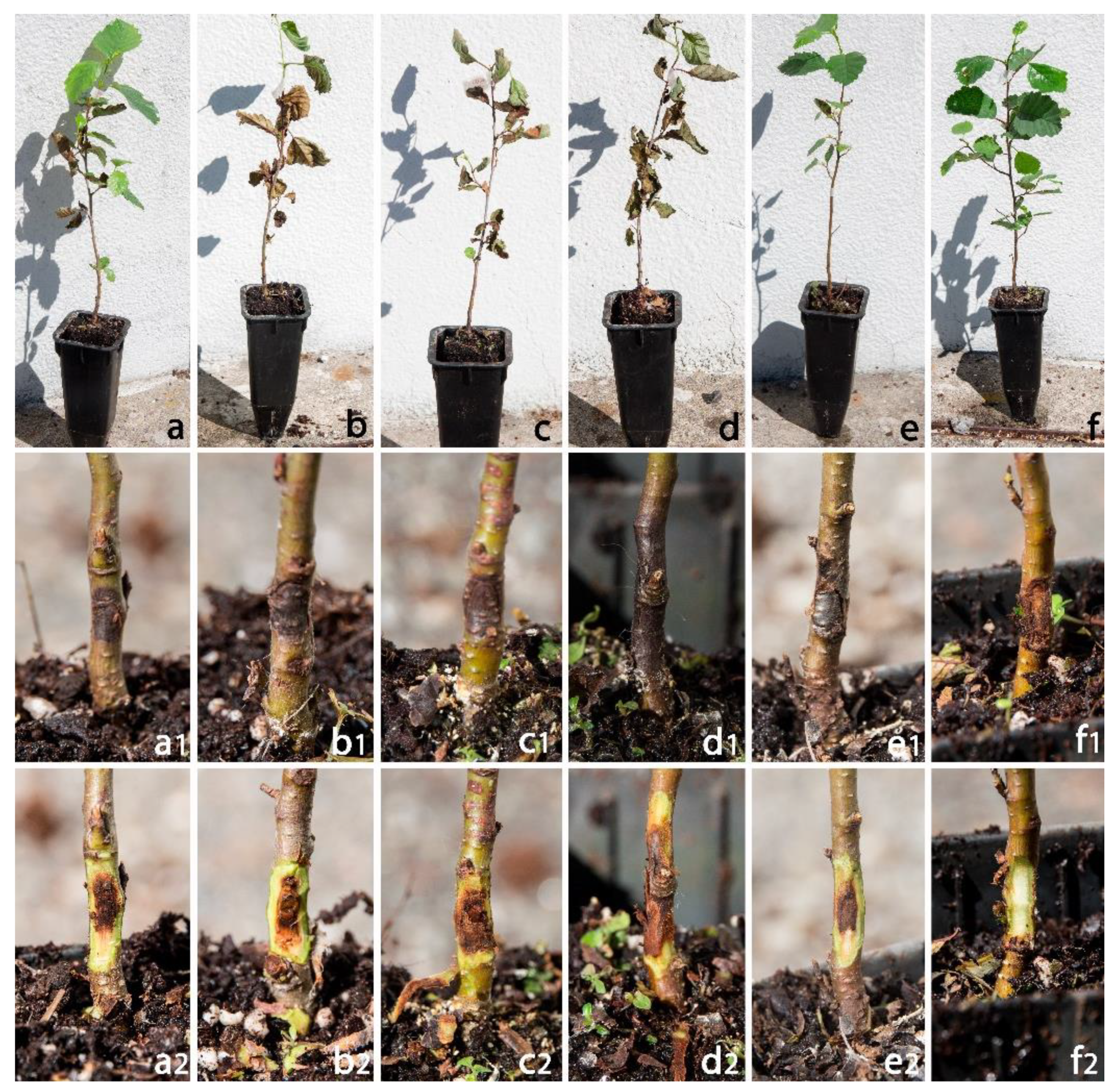
2.4. Pathogenicity Test

To confirm Koch’s postulates for new host–pathogen associations, the pathogenicity of five Phytophthora species was tested by inoculation on 1-year-old common alder seedlings grown in plastic pots (5 cm diameter, 0.5 L volume). Ten seedlings were inoculated with a representative isolate of each species, and ten were used as control. The seedlings were inoculated by wounding at the base of the stem using the protocol reported by Bregant et al. [10].
All inoculated seedlings were kept in controlled conditions at 21 °C and watered regularly for 30 days. At the end of the experimental period, seedlings were checked for the presence of internal (necrotic lesion) and external (wilting and exudates) disease symptoms. For each seedling, the outer bark was carefully removed with a scalpel, and the length of the necrotic lesion surrounding each inoculation point was measured.
The re-isolation of isolates was attempted by transferring 5 pieces of inner bark taken around the margin of the necrotic lesions onto PDA+. Growing colonies were subcultured onto CA and PDA, incubated in the dark at 20 °C and identified by morphological and molecular analyses.

2.5. Data Analysis

Pathogenicity assay data were checked for normality and then subjected to analysis of variance (ANOVA). Significant differences among mean values were determined using Fisher’s least significant differences multiple range test (p = 0.05) after one-way ANOVA using XLSTAT 2008 software (Addinsoft, Paris, France).

3. Results

3.1. Symptomatology

Field surveys conducted in five common alder ecosystems in Portugal showed the widespread occurrence of severe Phytophthora disease symptoms on young and old alder trees. Severe disease symptoms were observed mainly in the periodically flooded areas. Disease incidence, calculated at Sites 2 and 3, ranged from 75% to 83%, with an average mortality rate of 32%.
Declining trees were characterized by complex symptomatology, including extensive bleeding cankers on the lower part of the stem and, sometimes, on the branches, with irregular-shaped, inner bark reddish-brown necrosis as the result of the death of the bark tissues (Figure 1). In addition, different canopy symptoms, such as rusty shrivelled leaves, small-size leaves, shoot blight and epicormic shoots, were observed. In the late stage of the disease, infection causes a progressive or sudden decline of the whole canopy.

3.2. Aetiology

A total of sixty-three Phytophthora isolates were obtained from 60 out of 66 processed samples (positivity 90.1%). Among these, 4 isolates were obtained from bleeding cankers, 36 from rhizosphere (fine roots) and 23 from leaves used as bait along the streams. Based on morphology, colony appearance and DNA sequence data for 13 Phytophthora species, namely, P. lacustris (13 isolates), P. multivora (10), P. amnicola (9), P. chlamydospora (6), P. polonica (6), P. bilorbang (4), P. plurivora (4), P. cinnamomi (3), P. asparagi (2), P. cactorum (2), P. pseudocryptogea (2), P. gonapodyides (1) and P. rosacearum (1), were identified (Table 2).
The most common Phytophthora species isolated in this study was P. lacustris; this species was obtained mainly from water streams. Phytophthora amnicola and P. multivora were the dominant species in rhizosphere samples, whereas P. multivora was the only species occurring in all types of samples (bark tissue, rhizosphere and water). Phytophthora chlamydospora and P. plurivora were the most widespread species, occurring in three sites (Table 2). Seven out of thirteen species isolated belonged to Phytophthora ITS clade 6, the most represented clade, followed by clade 2.

3.3. Pathogenicity

At the end of the experimental period, Phytophthora-inoculated alder seedlings showed severe wilted symptoms associated with dark brown inner bark lesions that spread up and down from the inoculation point (Figure 2). All inoculated Phytophthora species proved to be pathogenic on common alder.
The average lesion length differed significantly among species (Table 3). The lesions caused by P. multivora were significantly larger than those caused by the other species. Necrotic inner bark lesions caused by P. multivora, P. chlamydospora, P. asparagi and P. amnicola progressively girdled the stem, causing wilting symptoms and the sudden death of the seedlings (Table 3). Control plants inoculated with sterile PDA plugs remained symptomless. All inoculated species were successfully re-isolated (100%) from the margin of the necrotic inner bark lesions of all seedlings. No Phytophthora isolates or other microorganisms were re-isolated from control seedlings.

4. Discussion

An extensive survey of Phytophthora-related diseases, conducted in central Portugal, showed the occurrence of 13 species, P. amnicola, P. asparagi, P. bilorbang, P. cactorum, P. chlamydospora, P. cinnamomi, P. gonapodyides, P. lacustris, P. multivora, P. plurivora, P. polonica, P. pseudocryptogea and P. rosacearum, in five riparian habitats (rivers and streams) characterized by high common alder mortality. Ten out of thirteen species were recovered from naturally declining common alder ecosystems in previous studies in Europe [7,8,9,10,26,27].
Most species found in this study are classified in the ITS clade 6 sensu [11,28]. This major clade includes a large number of saprotrophic or weak opportunistic pathogens strongly linked to aquatic environments [12]; therefore, it is not surprising that P. lacustris was found to be the dominant species. Its presence in Portugal, associated with declining alder formations, was recently documented by Kanoun-Boulè [18]. Three of the other species belonging to clade 6, P. bilorbang, P. chlamydospora and P. gonapodyides, are very common in the temperate riparian habitats of Europe and other continents, often in association with declining alders [9,10,29] whereas P. amnicola, P. asparagi and P. rosacearum are reported here for the first time on declining common alder.
Phytophthora amnicola was originally described in 2012 in Western Australia [30]. Its lifestyle appears strongly related to water, this is corroborated by the data obtained in this survey, but little is known regarding the ecology and potential impact of this species in Portugal.
From the rhizosphere of two alder trees, the colonies of P. asparagi were obtained. This species was described in the USA in 2012, but it has been known for a long time and the name is currently considered invalid [31,32]. Phytophthora asparagi has been reported in different countries as a pathogen on ornamental and crop plants [31,33]. Some recent studies have demonstrated that this species is widespread in Mediterranean environments on several species of the Mediterranean maquis [34,35]. Its diffusion in natural areas appears closely linked to the white asparagus (Asparagus albus), a preferential host that can facilitate host jumps [34].
Phytophthora rosacearum was isolated for the first time in California on Malus sp. and later from other diseased crops, such as pear in California and pomegranate in Turkey [36,37,38]. The recovery of P. rosacearum on common alder in Portugal represents the first report of this species in a natural habitat and in Europe. Based on the results obtained in the pathogenicity test, P. rosacearum can be considered a weak pathogen of common alder compared to other Phytophthora species.
The second most represented clade consists of two species, P. multivora and P. plurivora. Phytophthora multivora was the only species found in all types of samples monitored (stem bleeding cankers, necrotic fine roots and baited leaves along the stream). The discovery of this polyphagous pathogen causing the root rot and mortality of common alder in Portugal poses an additional threat to alder stands in Europe. Phytophthora multivora was previously reported to cause root and collar rot lesions on Agathis australis, Agonis flexuosa, Banksia spp., Corymbia calophylla, Eucalyptus spp., Rubus anglocandicans and Wollemia nobilis in Australia and New Zealand and on Acacia mearnsii, Alnus glutinosa, Araucaria araucana, Quercus spp., Rhododendron sp. and Salix fragilis in natural areas in Chile, Czech Republic, Germany, Hungary, New Zealand and South Africa [39,40,41,42,43,44,45,46,47,48]. The underbark inoculation test confirmed the aggressiveness of this emerging pathogen on common alder. Previous studies have ascertained its pathogenicity on Agathis australis, Agonis flexuosa, Corymbia spp., Eucalyptus spp., Banksia spp., Rubus anglocandicans and Wollemia nobilis [40,42,44,45,49,50].
The other species consistently detected from bark lesions and rhizospheres was P. plurivora, a plurivorous pathogen involved in the aetiology of several diseases of woody hosts in six continents [51,52,53,54]. The high isolation frequency of P. plurivora in this study is in accordance with the results of previous studies conducted on declining common alder trees in Italy [8,10].
Finally, the other four species were recovered less frequently. Among these, P. cactorum, P. polonica and P. pseudocryptogea are detected for the first time and are related to declining forests in Portugal; however, they are already known in common alder in other European countries [10,55]. The discovery of P. cinnamomi in riparian habitats is of particular concern due to its wide host range and the impact of this pathogen on oak forests in Portugal [56].
The fulfilment of Koch’s postulates for the five species tested in this study expands the list of pathogenic Phytophthora species for common alder to 29 (Table S1), suggesting that the disease may be caused by more than one pathogen under different environmental conditions.

5. Conclusions

While P. × alni has been getting the most attention in the last decades, the most common species isolated from declining alder trees in Europe is P. plurivora [9,10,27]. At the same time, the current trend of discovering an increasing number of pathogenic species in declining alder trees emphasizes how much more we need to learn about Phytophthora biodiversity and their impact on riparian ecosystems. In general, this work contributes to expanding knowledge on the biodiversity of Phytophthora species in the natural areas of Portugal with eight new reports (Table S2).

Supplementary Materials

The following supporting information can be downloaded at: https://www.mdpi.com/article/10.3390/pathogens12020276/s1, Table S1: Phytophthora species reported in natural ecosystems in Portugal; Table S2: Phytophthora species reported as pathogenic on Alnus glutinosa [57,58,59,60,61,62,63,64,65,66,67,68,69,70,71,72,73,74].

Author Contributions

Conceptualisation: C.B., B.T.L. and A.A.; methodology, C.B., B.T.L. and A.A.; formal analysis, C.B., E.B. and S.H.; investigation, C.B., E.B. and S.H.; resources, A.A. and B.T.L.; writing—original draft preparation, C.B.; writing—review and editing, B.T.L. and A.A.; supervision, B.T.L. and A.A.; funding acquisition, A.A. and B.T.L. All authors have read and agreed to the published version of the manuscript.

Funding

This research received no external funding.

Institutional Review Board Statement

Not applicable.

Informed Consent Statement

Not applicable.

Acknowledgments

This research has been financially supported by the Land Environment Resources and Health (L.E.R.H.) doctoral course (University of Padova). We thank the Portuguese Foundation for Science and Technology (FCT/MCTES) for the financial support to CESAM (UIDP/50017/2020 + UIDB/50017/2020 + LA/P/0094/2020) and the PhD grants of Sandra Hilário (SFRH/BD/137394/2018) and Eduardo Batista (PD/BD/135535/2018).

Conflicts of Interest

The authors declare no conflict of interest.

References

  1. Chen, Z.D.; Li, J.H. Phylogenetics and biogeography of Alnus (Betulaceae) inferred from sequences of nuclear ribosomal DNA its region. Int. J. Plant Sci. 2004, 165, 325–335. [Google Scholar]
  2. Claessens, H.; Oosterbaan, A.; Savill, P.; Rondeux, J. A review of the characteristics of black alder (Alnus glutinosa (L.) Gaertn.) and their implications for silvicultural practices. Forestry 2010, 83, 163–175. [Google Scholar]
  3. Houston Durrant, T.; de Rigo, D.; Caudullo, G. Alnus glutinosa in Europe: Distribution, habitat, usage and threats. Eur. Atlas For. Tree Species 2016, 1, 64–65. [Google Scholar]
  4. Brasier, C.; Rose, J.; Gibbs, J. An unusual Phytophthora associated with widespread alder mortality in Britain. Plant Pathol. 1995, 44, 999–1007. [Google Scholar]
  5. Sims, L.L.; Sutton, W.; Reeser, P.; Hansen, E.M. The Phytophthora species assemblage and diversity in riparian alder ecosystems of western Oregon, USA. Mycologia 2015, 107, 889–902. [Google Scholar] [CrossRef]
  6. Bjelke, U.; Boberg, J.; Oliva, J.; Tattersdill, K.; McKie, B.G. Dieback of riparian alder caused by the Phytophthora alni complex: Projected consequences for stream ecosystems. Freshw. Biol. 2016, 61, 565–579. [Google Scholar] [CrossRef]
  7. Trzewik, A.; Orlikowski, L.B.; Oszako, T.; Nowakowska, J.A.; Orlikowska, T. The characterization of Phytophthora isolates obtained from diseased Alnus glutinosa in Poland. Balt. For. 2015, 21, 44–50. [Google Scholar]
  8. Seddaiu, S.; Linaldeddu, B.T. First Report of Phytophthora acerina, P. plurivora, and P. pseudocryptogea associated with declining common alder trees in Italy. Plant Dis. 2020, 104, 1874. [Google Scholar] [CrossRef]
  9. Aday Kaya, A.G.; Lehtijärvi, A.; Şaşmaz, Y.; Nowakowska, J.A.; Oszako, T.; Doğmuş Lehtijärvi, H.T.; Woodward, S. Phytophthora species detected in the rhizosphere of Alnus glutinosa stands in the floodplain forests of Western Turkey. For. Pathol. 2018, 48, 11–14. [Google Scholar] [CrossRef]
  10. Bregant, C.; Sanna, G.P.; Bottos, A.; Maddau, L.; Montecchio, L.; Linaldeddu, B.T. Diversity and pathogenicity of Phytophthora species associated with declining alder trees in Italy and description of Phytophthora alpina sp. nov. Forests 2020, 11, 848. [Google Scholar] [CrossRef]
  11. Yang, X.; Tyler, B.M.; Hong, C. An expanded phylogeny for the genus Phytophthora. IMA Fungus 2017, 8, 355–384. [Google Scholar] [CrossRef]
  12. Brasier, C.M.; Cooke, D.E.; Duncan, J.M.; Hansen, E.M. Multiple new phenotypic taxa from trees and riparian ecosystems in Phytophthora gonapodyidesP. megasperma ITS Clade 6, which tend to be high-temperature tolerant and either inbreeding or sterile. Mycol. Res. 2003, 107, 277–290. [Google Scholar]
  13. Aghighi, S.; Hardy, G.E.S.J.; Scott, J.K.; Burgess, T.I. Phytophthora bilorbang sp. nov., a new species associated with the decline of Rubus anglocandicans (European blackberry) in Western Australia. Eur. J. Plant Pathol. 2012, 133, 841–855. [Google Scholar]
  14. Haque, M.M.; Martínez-Álvarez, P.; Lomba, J.M.; Martín-García, J.; Diez, J.J. First report of Phytophthora plurivora causing collar rot on common alder in Spain. Plant Dis. 2014, 98, 425. [Google Scholar] [CrossRef]
  15. Zamora-Ballesteros, C.; Haque, M.M.U.; Diez, J.J.; Martín-García, J. Pathogenicity of Phytophthora alni complex and P. plurivora in Alnus glutinosa seedlings. For. Pathol. 2017, 47, e12299. [Google Scholar] [CrossRef]
  16. Rooney-Latham, S.; Blomquist, C.L.; Pastalka, T.; Costello, L. Collar rot on Italian alder trees in California caused by Phytophthora siskiyouensis. Plant Health Prog. 2009, 10, 20. [Google Scholar] [CrossRef]
  17. Navarro, S.; Sims, L.; Hansen, E. Pathogenicity to alder of Phytophthora species from riparian ecosystems in western Oregon. For. Pathol. 2015, 45, 358–366. [Google Scholar] [CrossRef]
  18. Kanoun-Boulé, M.; Vasconcelos, T.; Gaspar, J.; Vieira, S.; Dias-Ferreira, C.; Husson, C. Phytophthora ×alni and Phytophthora lacustris associated with common alder decline in Central Portugal. For. Pathol. 2016, 46, 174–176. [Google Scholar] [CrossRef]
  19. Linaldeddu, B.T.; Bottecchia, F.; Bregant, C.; Maddau, L.; Montecchio, L. Diplodia fraxini and Diplodia subglobosa: The main species associated with cankers and dieback of Fraxinus excelsior in north-eastern Italy. Forests 2020, 11, 883. [Google Scholar]
  20. Huberli, D.; Hardy, G.E.S.J.; White, D.; Williams, N.; Burgess, T.I. Fishing for Phytophthora from Western Australia’s waterways: A distribution and diversity survey. Australas. Plant Pathol. 2013, 42, 251–260. [Google Scholar]
  21. Linaldeddu, B.T.; Mulas, A.A.; Bregant, C.; Piras, G.; Montecchio, L. First Report of Phytophthora pistaciae causing root and collar rot on nursery plants of Pistacia lentiscus in Italy. Plant Dis. 2020, 104, 1564. [Google Scholar]
  22. Möller, E.M.; Bahnweg, G.; Sandermann, H.; Geiger, H.H. A simple and efficient protocol for isolation of high molecular weight DNA from filamentous fungi, fruit bodies, and infected plant tissues. Nucleic Acids Res. 1992, 20, 6115–6116. [Google Scholar]
  23. White, T.J.; Bruns, T.; Lee, S.; Taylor, J. Amplification and direct sequencing of fungal ribosomal RNA genes for phylogenetics. In PCR Protocols: A Guide to Methods and Applications; Innis, M.A., Gelfand, D.H., Sninsky, J.J., White, T.J., Eds.; Academic Press: San Diego, CA, USA, 1990; pp. 315–322. [Google Scholar]
  24. Linaldeddu, B.T.; Franceschini, A.; Alves, A.; Phillips, A.J. Diplodia quercivora sp. nov.: A new species of Diplodia found on declining Quercus canariensis trees in Tunisia. Mycologia 2013, 105, 1266–1274. [Google Scholar]
  25. Altschul, S.F.; Gish, W.; Miller, W.; Myers, E.W.; Lipman, D.J. Basic local alignment search tool. J. Mol. Biol. 1990, 215, 403–410. [Google Scholar] [CrossRef]
  26. Mrazkova, M.; Černý, K.; Tomšovský, M.; Strnadova, V.; Gregorová, B.; Holub, V.; Pánek, M.; Havrdová, L.; Hejna, M. Occurrence of Phytophthora multivora and Phytophthora plurivora in the Czech Republic. Plant Prot. Sci. 2013, 49, 155–164. [Google Scholar]
  27. Matsiakh, I.; Kramarets, V.; Cleary, M. Occurrence and diversity of Phytophthora species in declining broadleaf forests in western Ukraine. For. Pathol. 2021, 51, e12662. [Google Scholar]
  28. Cooke, D.E.L.; Drenth, A.; Duncan, J.M.; Wagels, G.; Brasier, C.M. A molecular phylogeny of Phytophthora and related oomycetes. Fungal Gen. Biol. 2000, 30, 17–32. [Google Scholar]
  29. Matsiakh, I.; Oszako, T.; Kramarets, V.; Nowakowska, J.A. Phytophthora and Pythium species detected in rivers of the Polish-Ukrainian border areas. Bal. For. 2016, 22, 230–238. [Google Scholar]
  30. Burgess, T.I.; Hüberli, D.; Hardy, G.E.S.J.; Stukely, M.J.C.; Jung, T. Phytophthora amnicola T. I. Burgess & T. Jung, sp. nov. Persoonia 2012, 28, 140–141. [Google Scholar]
  31. Saude, C.; Hurtado-Gonzales, O.P.; Lamour, K.H.; Hausbeck, M.K. Occurrence and characterization of a Phytophthora sp. pathogenic to asparagus (Asparagus officinalis) in Michigan. Phytopathology 2008, 98, 1075–1083. [Google Scholar] [CrossRef]
  32. Granke, L.L.; Saude, C.; Windstam, S.T.; Webster, B.J.; Hausbeck, M.K. Phytophthora asparagi Saude & Hausbeck, sp. nov. Persoonia 2012, 28, 146–147. [Google Scholar]
  33. Cunnington, J.H.; De Alwis, S.; Pascoe, I.G.; Symes, P. The ‘asparagus’ Phytophthora infecting members of the Agavaceae at the Royal Botanic Gardens, Melbourne. Austral. Plant Pathol. 2005, 34, 413–414. [Google Scholar]
  34. Scanu, B.; Linaldeddu, B.T.; Deidda, A.; Jung, T. Diversity of Phytophthora species from declining Mediterranean maquis vegetation, including two new species, Phytophthora crassamura and P. ornamentata sp. nov. PLoS ONE 2015, 10, e0143234. [Google Scholar] [CrossRef]
  35. Riolo, M.; Aloi, F.; La Spada, F.; Sciandrello, S.; Moricca, S.; Santilli, E.; Pane, E.; Cacciola, S.O. Diversity of Phytophthora communities across different types of Mediterranean vegetation in a nature reserve area. Forests 2015, 11, 853. [Google Scholar]
  36. Hansen, E.M.; Wilcox, W.F.; Reeser, P.W.; Sutton, W. Phytophthora rosacearum and P. sansomeana, new species segregated from the Phytophthora megasperma “complex”. Mycologia 2009, 101, 129–135. [Google Scholar]
  37. Sanchez, A.D.; Sosa, M.C.; Lutz, M.C.; Carreño, G.A.; Ousset, M.J.; Lucero, G.S. Identification and pathogenicity of Phytophthora species in pear commercial orchards in Argentina. Eur. J. Plant Pathol. 2019, 154, 811–822. [Google Scholar]
  38. Kurbetli, İ.; Karaca, G.; Aydoğdu, M.; Sülü, G. Phytophthora species causing root and collar rot of pomegranate in Turkey. Eur. J. Plant Pathol. 2020, 157, 485–496. [Google Scholar]
  39. Schubert, R.; Bahnweg, G.; Nechwatal, J.; Jung, T.; Cooke, D.E.L.; Duncan, J.M.; Muller-Starck, G.; Langelbartens, C.; Sandermann, C., Jr.; Oßwald, W. Detection and quantification of Phytophthora species which are associated with root-rot diseases in European deciduous forests by species-specific polymerase chain reaction. Eur. J. For. Pathol. 1999, 29, 169–188. [Google Scholar] [CrossRef]
  40. Scott, P.M.; Burgess, T.I.; Barber, P.A.; Shearer, B.L.; Stukely, M.J.C.; Hardy, G.S.J.; Jung, T. Phytophthora multivora sp. nov., a new species recovered from declining Eucalyptus, Banksia, Agonis and other plant species in Western Australia. Persoonia 2009, 22, 1–13. [Google Scholar] [CrossRef]
  41. Szabó, I.; Lakatos, F.; Sipos, G. Occurrence of soilborne Phytophthora species in declining broadleaf forests in Hungary. Eur. J. Plant Pathol. 2013, 137, 159–168. [Google Scholar]
  42. Waipara, N.W.; Hill, S.; Hill, L.M.W.; Hough, E.G.; Horner, I.J. Surveillance methods to determine tree health distribution of kauri dieback disease and associated pathogens. N. Z. Plant Prot. 2013, 66, 235–241. [Google Scholar] [CrossRef]
  43. Nagel, J.H.; Slippers, B.; Wingfield, M.J.; Gryzenhout, M. Multiple Phytophthora species associated with a single riparian ecosystem in South Africa. Mycologia 2015, 107, 915–925. [Google Scholar] [CrossRef]
  44. Puno, V.I.; Laurence, M.H.; Guest, D.I.; Liew, E.C.Y. Detection of Phytophthora multivora in the Wollemi Pine site and pathogenicity to Wollemia nobilis. Aus. Plant Pathol. 2015, 44, 205–215. [Google Scholar] [CrossRef]
  45. Aghighi, S.; Burgess, T.I.; Scott, J.K.; Calver, M.; Hardy, G.S.J. Isolation and pathogenicity of Phytophthora species from declining Rubus anglocandicans. Plant Pathol. 2016, 65, 451–461. [Google Scholar] [CrossRef]
  46. Bose, T.; Wingfield, M.J.; Roux, J.; Vivas, M.; Burgess, T.I. Community composition and distribution of Phytophthora species across adjacent native and non-native forests of South Africa. Fungal Ecol. 2018, 36, 17–25. [Google Scholar]
  47. Galvez, E.; Larach, A.; Riquelme, N.; Celis, J.L.; Guajardo, J.; Besoain, X.A. Araucaria araucana root rot caused by Phytophthora multivora and P. citrophthora. Phytopatholog 2018, 108, S1.186. [Google Scholar]
  48. Tsykun, T.; Prospero, S.; Schoebel, C.N.; Rea, A.; Burgess, T.I. Global invasion history of the emerging plant pathogen Phytophthora multivora. BMC Genom. 2022, 23, 1–16. [Google Scholar] [CrossRef]
  49. Scott, P.M.; Jung, T.; Shearer, B.L.; Barber, P.A.; Calver, M.; Hardy, G.E.S.J. Pathogenicity of Phytophthora multivora to Eucalyptus gomphocephala and Eucalyptus marginata. For. Pathol. 2012, 42, 289–298. [Google Scholar] [CrossRef]
  50. Croeser, L.; Paap, T.; Calver, M.C.; Andrew, M.E.; Hardy, G.E.S.J.; Burgess, T.I. Field survey, isolation, identification and pathogenicity of Phytophthora species associated with a Mediterranean-type tree species. For. Pathol. 2018, 48, e12424. [Google Scholar] [CrossRef]
  51. Balci, Y.; Balci, S.; Eggers, J.; MacDonald, W.L.; Juzwik, J.; Long, R.P.; Gottschalk, K.W. Phytophthora spp. associated with forest soils in eastern and north-central US oak ecosystems. Plant Dis. 2007, 91, 705–710. [Google Scholar]
  52. Jung, T.; Burgess, T.I. Re-evaluation of Phytophthora citricola isolates from multiple woody hosts in Europe and North America reveals a new species, Phytophthora plurivora sp. nov. Persoonia 2009, 22, 95–110. [Google Scholar] [CrossRef]
  53. Jankowiak, R.; Stępniewska, H.; Bilański, P.; Kolařík, M. Occurrence of Phytophthora plurivora and other Phytophthora species in oak forests of southern Poland and their association with site conditions and the health status of trees. Folia Microbiol. 2014, 59, 531–542. [Google Scholar] [CrossRef]
  54. Schoebel, C.N.; Stewart, J.; Gruenwald, N.J.; Rigling, D.; Prospero, S. Population history and pathways of spread of the plant pathogen Phytophthora plurivora. PLoS ONE 2014, 9, e85368. [Google Scholar]
  55. Belbahri, L.; Moralejo, E.; Calmin, G.; Oszako, T.; García, J.A.; Descals, E.; Lefort, F. Phytophthora polonica, a new species isolated from declining Alnus glutinosa stands in Poland. FEMS Microbiol. Let. 2006, 261, 165–174. [Google Scholar]
  56. Moreira, A.C.; Martins, J.M.S. Influence of site factors on the impact of Phytophthora cinnamomi in cork oak stands in Portugal. For. Pathol. 2005, 35, 145–162. [Google Scholar] [CrossRef]
  57. Diogo, E.; Machado, H.; Reis, A.; Valente, C.; Phillips, A.J.; Bragança, H. Phytophthora alticola and Phytophthora cinnamomi on Eucalyptus globulus in Portugal. Eur. J. Plant Pathol. 2022, 165, 255–269. [Google Scholar] [CrossRef]
  58. Lopes-Pimentel, A.A. O sobreiro também é parasitado pela Phytophthora cambivora (Petri) Buis., agente da “doença da tinta” do castanheiro. Lisboa. Dir. Geral Dos Serv. Florestais E Aquícolas 1946, 13, 45–49. [Google Scholar]
  59. Crandall, B.S. The distribution and significance of the chestnut root rot Phytophthoras, P. cinnamomi and P. cambivora. Plant Dis. Rep. 1950, 34, 194–196. [Google Scholar]
  60. Jung, T.; Jung, M.H.; Cacciola, S.O.; Cech, T.; Bakonyi, J.; Seress, D.; Mosca, S.; Schena, L.; Seddaiu, S.; Pane, A.; et al. Multiple new cryptic pathogenic Phytophthora species from Fagaceae forests in Austria, Italy and Portugal. IMA Fungus 2017, 8, 219–244. [Google Scholar]
  61. Pimentel, A. A Phytophthora cinnamomi Rands, um outro agente, extremamente virulento, da “doença da tinta” do castanheiro. Sep. Agron. Lusit. 1947, 9, 181–191. [Google Scholar]
  62. Brasier, C.M.; Robredo, F.; Ferraz, J.F.P. Evidence for Phytophthora cinnamomi involvement in Iberian oak decline. Plant Pathol. 1993, 42, 140–145. [Google Scholar] [CrossRef]
  63. Moreira, A.C.; Ferraz, J.F.P.; Clegg, J. The involvement of Phytophthora cinnamomi in cork and holm oak decline in Portugal. In Proceedings of the First International Meeting on Phytophthoras in Forest and Wildland Ecosystems, Grand Pass, OR, USA, 30 August–3 September 1999; pp. 132–135. [Google Scholar]
  64. Serrano, M.S.; De Vita, P.; Fernàndez-Rebollo, P.; Coelho, A.C.; Belbarhi, L.; Sanchez, M.E. Phytophthora cinnamomi and Pythium spiculum as main agents of Quercus decline in southern Spain and Portugal. In Proceedings of the IOBC-WPRS 6th Meeting Integrated Protection in Oak Forests, Integrated Protection in Oak Forests IOBC/wprs Bulletin, Tempio Pausania, Italy, 4–7 October 2010; Volume 76, pp. 97–100. [Google Scholar]
  65. Maia, C.; Jung, M.H.; Carella, G.; Milenković, I.; Janoušek, J.; Tomšovský, M.; Mosca, S.; Schena, L.; Cravador, A.; Moricca, S.; et al. Eight new Halophytophthora species from marine and brackish-water ecosystems in Portugal and an updated phylogeny for the genus. Persoonia 2022, 48, 54–90. [Google Scholar] [CrossRef]
  66. Ruano-Rosa, D.; Schena, L.; Agosteo, G.E.; Magnano di San Lio, G.; Cacciola, S.O. Phytophthora oleae sp. nov. causing fruit rot of olive in southern Italy. Plant Pathol. 2018, 67, 1362–1373. [Google Scholar]
  67. de Jesus Gomes, M.; Amaro, P.T. Ocorrência de Phytophthora ramorum em Portugal sobre Viburnum spp. Rev. Cienc. Agrar. 2008, 31, 105–111. [Google Scholar]
  68. Brasier, C.M.; Kirk, S.A. Comparative aggressiveness of standard and variant hybrid alder phytophthoras, Phytophthora cambivora and other Phytophthora species on bark of Alnus, Quercus and other woody hosts. Plant Pathol. 2001, 50, 218–229. [Google Scholar]
  69. Orlikowski, L.B.; Oszako, T. Phytophthora cambivora on Alnus glutinosa: Isolation and colonisation of plants. J. Plant Prot. Res. 2005, 45, 267–272. [Google Scholar]
  70. Haque, M.M.; Diez, J.J. Susceptibility of common alder (Alnus glutinosa) seeds and seedlings to Phytophthora alni and other Phytophthora species. For. Syst. 2012, 21, 313–322. [Google Scholar] [CrossRef] [Green Version]
  71. Jung, T.; Nechwatal, J. Phytophthora gallica sp. nov., a new species from rhizosphere soil of declining oak and reed stands in France and Germany. Mycol. Res. 2008, 112, 1195–1205. [Google Scholar] [CrossRef]
  72. Kovács, J.; Lakatos, F.; Szabó, I. Post-epidemic situation of a previously Phytophthora alni-infected common alder stand. Acta Silv. Lignaria Hung. 2015, 11, 27–38. [Google Scholar]
  73. Rytkönen, A.; Lilja, A.; Vercauteren, A.; Sirkiä, S.; Parikka, P.; Soukainen, M.; Hantula, J. Identity and potential pathogenicity of Phytophthora species found on symptomatic Rhododendron plants in a Finnish nursery. Can. J. Plant Pathol. 2012, 34, 255–267. [Google Scholar]
  74. Jung, T.; Nechwatal, J.; Cooke, D.E.; Hartmann, G.; Blaschke, M.; Osvald, W.F.; Duncan, J.M.; Delatour, C. Phytophthora pseudosyringae sp. nov., a new species causing root and collar rot of deciduous tree species in Europe. Mycol. Res. 2003, 107, 772–789. [Google Scholar] [CrossRef]
Figure 1. Overview of Phytophthora-related diseases on Alnus glutinosa: panoramic view that highlights a high mortality rate (a); alders with initial declining symptoms along a stream (b); severe branch dieback symptoms (c); bleeding cankers on stems with a wilted shoot (d); bleeding cankers (e,f) and root rot (g). On the left, starting from the top, colony morphology of: Phytophthora amnicola, P. asparagi, P. bilorbang, P. cactorum, P. chlamydospora, P. cinnamomi, P. gonapodyides, P. lacustris, P. multivora, P. plurivora, P. polonica, P. pseudocryptogea and P. rosacearum after 7 days of growth at 20 °C on CA in the dark.
Pathogens 12 00276 g001
Figure 2. Symptoms observed on common alder seedlings after 30 days from inoculation with Phytophthora amnicola (aa2), P. asparagi (bb2), P. chlamydospora (cc2), P. multivora (dd2) and P. rosacearum (ee2). Control seedlings (ff2).
Pathogens 12 00276 g002
Table 1. Study sites’ information and number of stem (S), rhizosphere (R) and leaf (L) samples used for Phytophthora isolation.
Survey SitesElevation
(m a.s.l.)
Geographic CoordinatesNumber of Samples
1940.7035888−8.6052296R(4)
21140.7206700−8.5652620R(20), S(5), L(10)
31140.7141470−8.5738595R(10), S(3), L(10)
441740.6128810−7.5174910R(2)
575040.4106660−7.4713180R(2)
Table 2. Phytophthora isolates obtained from stem (S), rhizosphere (R) and water (W) samples in the investigated sites.
SpeciesAccession NumberITS CladeNumber of SamplesSites
StemRhizosphereWater
P. amnicolaOQ2022166-632,3
P. asparagiOQ2022176-2-3
P. bilorbangOQ2022186--42,3
P. cactorumOQ2022191-2-2
P. chlamydosporaOQ2022206-422,3,4
P. cinnamomiOQ2022217-3-2
P. gonapodyidesOQ2022226-1-3
P. lacustrisOQ2022236-492,3
P. multivoraOQ20222423612,3
P. plurivoraOQ202225213-1,2,4
P. polonicaOQ2022269-242,3
P. pseudocryptogeaOQ2022278-2-3,5
P. rosacearumOQ2022286-1-2
Table 3. Mean lesion length ± standard deviation caused by each Phytophthora species on the stem of common alder seedlings and the percentage of seedlings with exudates and wilting symptoms.
SpeciesIsolatesMean Lesion Length (mm) *ExudatesWiltingRe-Isolation (%)
P. amnicolaCBP2811.0 ± 4.8bc-30%100
P. asparagiCBP2312.2 ± 4.7bc-40%100
P. chlamydosporaCBP1615.5 ± 4.8b-30%100
P. multivoraCBP5641.2 ± 14.7a40%80%100
P. rosacearumCBP818.5 ± 3.1cd--100
Control-2.5 ± 1.4d---
Critical value-2.006
* Values in the column with the same letter do not differ significantly at p = 0.05, according to the LSD multiple range test.
Disclaimer/Publisher’s Note: The statements, opinions and data contained in all publications are solely those of the individual author(s) and contributor(s) and not of MDPI and/or the editor(s). MDPI and/or the editor(s) disclaim responsibility for any injury to people or property resulting from any ideas, methods, instructions or products referred to in the content.

