Article

Occurrence of Escherichia coli Pathotypes in Diarrheic Calves in a Low-Income Setting

by
Wagaw Sendeku Chekole
1,2,3,*,
Haileeyesus Adamu
2,†,
Susanna Sternberg-Lewrein
4,†,
Ulf Magnusson
1,† and
Tesfaye Sisay Tessema
2,†
1
Department of Clinical Sciences, Swedish University of Agricultural Sciences (SLU), 75007 Uppsala, Sweden
2
Institute of Biotechnology, Addis Ababa University, Addis Ababa 1176, Ethiopia
3
Institute of Biotechnology, University of Gondar, Gondar 196, Ethiopia
4
Department of Biomedical Sciences & Veterinary Public Health, Swedish University of Agricultural Sciences, 75007 Uppsala, Sweden
*
Author to whom correspondence should be addressed.
These authors have contributed equally to this work and share senior authorship.
Pathogens 2023, 12(1), 42; https://doi.org/10.3390/pathogens12010042
Submission received: 23 November 2022 / Revised: 20 December 2022 / Accepted: 22 December 2022 / Published: 27 December 2022
(This article belongs to the Section Bacterial Pathogens)

Abstract

:
Different E. coli pathotypes are common zoonotic agents. Some of these pathotypes cause recurrent and widespread calf diarrhea and contribute to significant economic losses in the livestock sector worldwide in addition to putting humans at risk. Here, we investigated the occurrence of E. coli pathotypes in diarrheic calves in Ethiopia kept under various calf management practices. One hundred fecal samples were collected from diarrheic calves in 98 different farms. E. coli was isolated in the samples from 99 of the diarrheic calves, and virulence genes were detected in 80% of the samples. The occurrence of E. coli pathotypes in the samples was 32% ETEC, 23% STEC, 18% STEC/ETEC, 3% EPEC, 2% EAEC, and 1% EHEC. No diarrheic calves were positive for the EIEC and DAEC pathotypes. The occurrence of pathotypes was positively associated with female calves (EPEC, p = 0.006), aged less than 2 weeks (STEC, p = 0.059), and calves fed colostrum via the hand method (STEC, p = 0.008 and EAEC, p = 0.003). This study revealed that several E. coli pathotypes occurred among calves affected with diarrhea. Moreover, the presence of a mixed STEC/ETEC pathotypes infection was present in the studied low-income setting. These findings indicate a considerable risk for the zoonotic transmission from calves to humans and the options to provide the better management for younger calves in order to reduce the economic loss.

1. Introduction

Escherichia coli is widely regarded as a commensal bacterium [1]. Nonetheless, some E. coli pathotypes are pathogens causing intestinal infections ranging from mild to severe in animals and humans [2,3]. Common pathotypes include Enterohaemorrhagic E. coli (EHEC), Enterotoxigenic E. coli (ETEC), Enteropathogenic E. coli (EPEC), Enteroaggregative E. coli (EAEC), Enteroinvasive E. coli (EIEC), and diffusely adherent E. coli (DAEC) [4]. Among these pathotypes, the EHEC serotype O157:H7 is recognized as the most important zoonotic pathogen. Globally, between 2007 to 2015, EPEC- and ETEC-related yearly human deaths amounted to 37,000 and 26,000, respectively [5]. The World Health Organization (WHO) has estimated that one-third of the population in low-income countries suffers from foodborne diseases, of which pathogenic E. coli is one cause [5].
In low- and middle-income countries, calf diarrhea caused by E. coli is common and causes a considerable number of deaths [6,7,8]. For instance, diarrheal diseases caused up to 15% of preweaning calf mortality in Uruguay [9] and 31% [10] and 63% [11] in Ethiopia, where calf diarrhea is an issue, though it is also an issue in high-income countries. For instance, in the USA, where about 57% of weaning calf mortalities have been reported due to diarrhea, with the majority of the cases in calves less than one month old [12]. About 9% mortality was reported in male calves with diarrhea in Canada [13]. In Norway, the overall calf mortalities were 280,000 heads per year, and the economic loss was estimated to be USD 10 million in 2006 [14]. Overall, calf diarrheal disease mainly seemed to be associated with poor farming practices [15,16].
Thus, an infection with E. coli pathotypes in calves poses a significant risk to animal health with a considerable impact on productivity and the farmers’ economy, in addition to being a public health issue due to their zoonotic potential. Given the generally poorer hygienic conditions and disease prevention capacity in rural areas in low-income countries, these risks may be accentuated in such settings. However, data on the occurrence of different E. coli pathotypes in low-income countries is sparse [17,18,19,20]. Here, we provide such data from diarrheic calves in a district in Ethiopia, the most livestock-rich country in Africa [21].

2. Materials and Methods

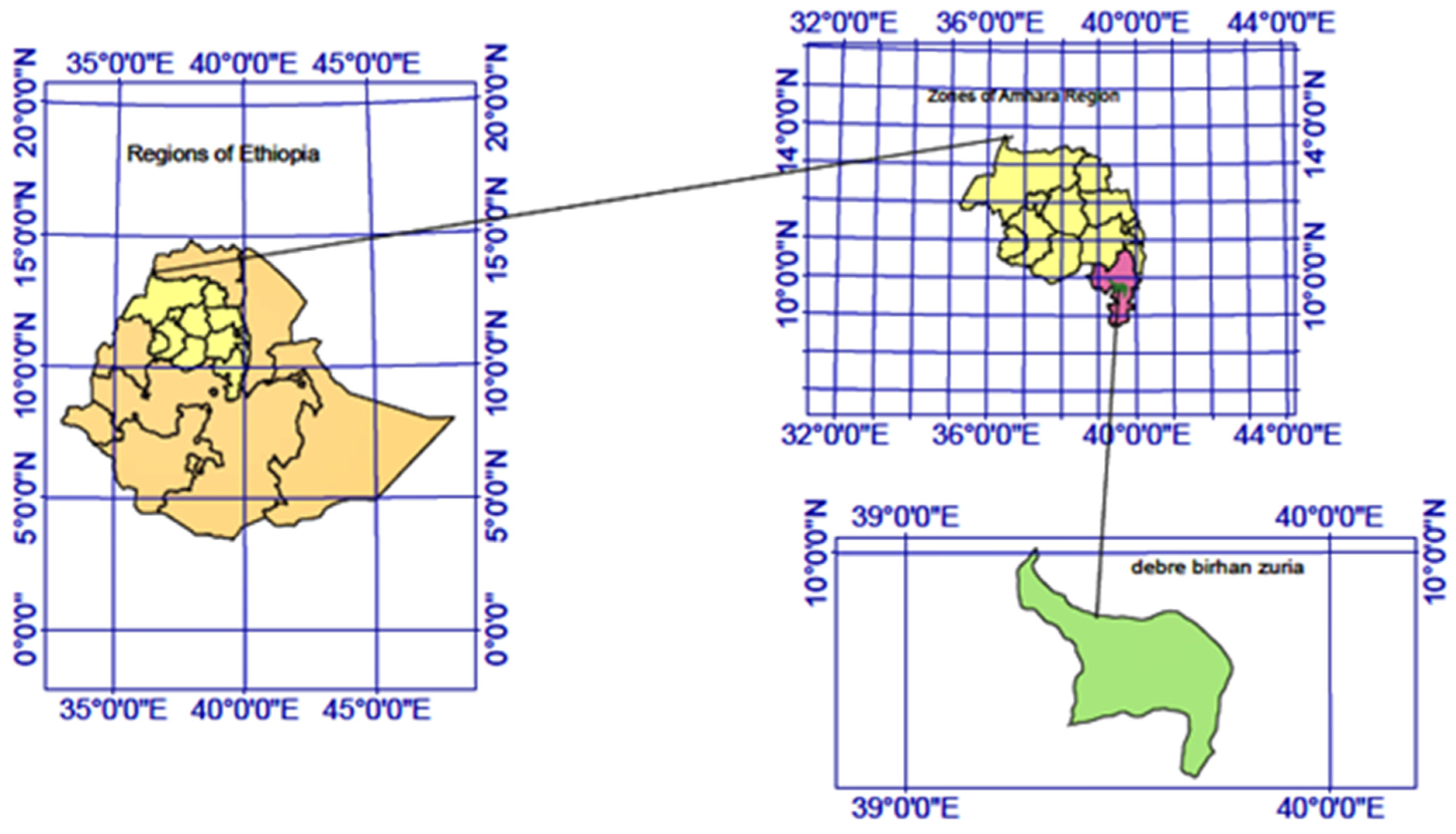
2.1. Study Area and Period

The study was conducted in Basona Werana woreda (District), Ethiopia (Figure 1). The Basona District is found in the north Shewa zone of the Amhara regional state, 130 Km from the capital, Addis Ababa. The district is divided into 22 small administrative subdistricts (Kebeles). The district has a population of 120,930 individuals living in 27,686 households. The households are distributed in urban and rural areas with an urban to a rural proportion of 0.01 [22]. It has an annual temperature range of 6 to 20 °C and the rainfall varies between 950 and 1200 mm. The farming practice is a mixed crop–livestock (crops, cattle, sheep, and goat) type where cattle predominate.

2.2. Study Design and Sampling Technique

A cross-sectional study design was used. The sample collection was carried out from October 2020 to March 2021, following the contact of veterinarians in each subdistrict. Farm owners were informed about the sampling through their milk union and requested to report the occurrence of diarrhea in calves. All types of farms with diarrheic calves were included. Each farm was visited once during the study period. Samples were taken from diarrheic calves between 0 and 10 weeks old. The age groups were 0–2, 2–4, 4–7, and 7–10 weeks. One fecal sample was collected from one calf per farm, except on one occasion when three diarrheic calves were sampled on a single farm. The sample size was set based on the financial constraints and a reasonable representation of the farms.
A total of 100 fecal samples from diarrheic calves were collected from 98 farms. Five ml feces were collected from the rectum of the calves. All samples were temporarily stored in phosphate-buffered saline (HiMEDIA, Mumbai, India) and transported to Addis Ababa University, the Institute of Biotechnology, health biotechnology laboratory, and processed within 24 h of the collection time.

2.3. Isolation and Characterization of E. coli

The isolation and characterization of E. coli were performed using standard bacteriological procedures [23]. Briefly, all of the collected samples were enriched in tryptose soya broth (TSB) (HiMedia, Mumbai, India) at 37 °C for 18–24 h. Subsequently, a loopful of this bacterial culture was streaked onto McConkey agar (HiMedia, Mumbai, India) and was grown at 37 °C for 18–24 h. The pink color colonies (lactose fermenter) from the McConkey agar culture were picked and transferred onto an Eosin Methylene Blue (EMB) (HiMedia, Mumbai, India) agar medium and incubated to grow at 37 °C for 18–24 h. The E. coli colonies were presumptively identified based on their appearance of a greenish metallic sheen on Eosin Methylene Blue (EMB) (HiMedia, Mumbai, India) medium. Three well-isolated presumptive E. coli colonies were randomly selected from each EMB plate and pure cultures from the selected isolates were used for further testing. The E. coli isolates were identified using the biochemical tests Indole, Methyl Red, Vogues–Proskauer test, and Citrate utilization (IMViC) [23].

2.4. DNA Extraction

The DNA was extracted from the E. coli isolates as described by Moore et al. [24] with a minor modification. Briefly, a single colony was subcultured in Luria Bertani (LB) broth media (HiMEDIA, Mumbai, India). A 1.5 mL bacterial culture was pelleted via centrifugation and subsequently resuspended and washed in 100 µL sterile deionized water. The 150 µL bacterial suspensions were lysed at 100 °C and then frozen at −20 °C. Finally, the suspensions were centrifuged at 10,000 rpm for 10 min, and 100 µL of supernatant containing DNA from each preparation was transferred into fresh Eppendorf tubes. The DNA preparations were quantified using a Nano-drop spectrophotometer and a ratio of 260 nm/280 nm was used to estimate the quality of the DNA. Moreover, gel electrophoresis was used to check the intactness of the isolated DNA.

2.5. E. coli Pathotyping Using PCR

The DNA from the E. coli isolates was subjected to a PCR amplification of ten virulence genes; bundle-forming Pili (bfp), attaching and effacing (eae), adhesion (daaA-E); shiga-toxin-1/2 (stx-1/2), heat-stable (st) and heat-labile (lt) toxins, hemolysin toxin (hly), virulence protein transporter (aatA), and invasion-associated protein (ial) using the primers listed in Table 1 and the E. coli isolates were classified into one of the six pathotypes based on their virulence genes.
All primers and PCR conditions used for the virulence gene amplification were performed as described previously [25,26,27,28,29,30,31,32,33]. All of the PCR reactions were performed in a singleplex platform in a Prima 96 plus Thermal Cycler (Himedia Laboratories, Mumbai, India).
Each PCR reaction was carried out in a 25 μL final volume reaction mixture containing nuclease-free water (Himedia, Mumbai, India), 10X PCR buffer with 17.5 mM of MgCl2 (Himedia, Mumbai, India), 0.35 mM of each dNTPs (Himedia, Mumbai, India), 100 pM for each of the virulence gene-specific forward and reverse primers (Bioneer; Daejeon, Republic of Korea), 500 U Taq polymerase enzyme (DELTA Biotechnology, Ethiopia), and a DNA template. DNA samples that carried the relevant virulence gene(s) and nuclease-free water (without template) were used as the positive and negative control, respectively. PCR amplification products were processed using 1.5% agarose gel electrophoresis at 100 Volt for one hour; the gels were stained with ethidium bromide in 1× TAE buffer (40 mM Tris-HCl, 20 mM Acetate, 0.5 mM EDTA, pH 8.3). A DNA ladder of 100 bp (Himedia, Mumbai, India) was run in parallel with the PCR products to determine the size of the amplicons. Separated PCR products were visualized, photographed, and documented in a gel documentation system (Bio-Rad, Feldkirchen, Germany).

2.6. Farm Data

A structured questionnaire was prepared and used to collect the relevant farm data (Supplementary Table S1). In the questionnaire, farm owners were given closed-ended questions about the overall management of the calves. The questions covered the sex, age, and breed of the calf, colostrum and supplementary feeding practice, housing type, and flooring. In addition to the questionnaire, data about the hygiene was collected through a direct observational judgment by the first author. To grade the hygiene of the housing, a four-point scale ranging from 1 to 4, 1—very poor, 2—poor, 3—good, and 4—very good, was used. For the housing hygiene evaluation, a checklist was used to minimize the judgment biases, containing the following aspects: proper manure disposal, dryness, aeration, and the absence of nearby waste, each with 1 point (Supplementary Table S2). The questionnaire and observational data as well as the fecal samples from diarrheic calves were collected during the same visit to the farm. Each farm was visited once between October 2020 and March 2021.

2.7. Data Processing and Analysis

Data related to the sex, age, breed, colostrum feeding status, time of the colostrum feeding, type of supplementary feed, housing type, and hygiene were coded and entered into a Microsoft® Excel spreadsheet. The responses to the multiple-choice questions were coded into categorical variables. Similarly, the “Yes” or “No” responses were categorized as “1” or “0”, respectively. The housing hygiene “Very poor” to “Very good” responses were coded into numbers ranging from 1 to 4.
Descriptive statistics were used to analyze the occurrence of virulence genes and E. coli pathotypes. The questionnaire responses and their association with the proportion of detected E. coli pathotypes were analyzed using the chi-square test, with p < 0.05 considered as a significant association. A pairwise analysis of the co-occurrences of virulence genes was carried out using a two-tailed chi-square test, with p < 0.05 seen as a strong correlation. The software SPSS (IBM SPSS Statistics 28.0.0.0) was used for the analyses.

3. Results

3.1. Farm Description and Calf Management

A total of 100 diarrheic calves from 98 farms of three different types, family (95), enterprise (2), and research (1) were sampled. The size of the farms ranged from 2 to 32 animals with an average of 6.5 and a median value of 6 animals. The farms were grouped into small farms (SF, n = 51) with 2–6 animals and medium farms (MF, n = 47) with 7–32 animals (Supplementary Table S3).
Of the sampled calves, 53% were male and of all of the calves, 3% were less than 1 week old, 30% were between 2 and 4 weeks old, 27% were between 4 and 7 weeks old, and 39% were between 7 and 10 weeks old. Crossbreds (local X Holstein Friesians) were the most common, accounting for 85% of the calves. About 89% of the calves were fed colostrum, and 43% of these were fed colostrum within 6 h after their birth. Allowing calves to suckle colostrum was the most common practice, with this occurring in 72% of farms. Twenty-seven (23%) of the calves were also provided additional feeds by grazing and hay, respectively. In 31% of the farms, calves were housed in the same barn with other animal species. The majority of the houses (74%) had soil flooring and 63% of the calving houses were judged to have poor hygienic conditions (Supplementary Table S1).

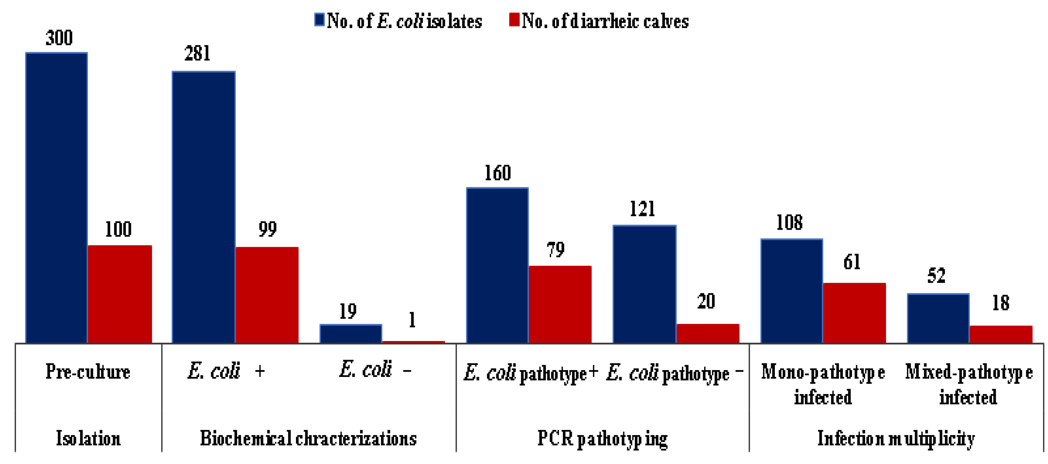
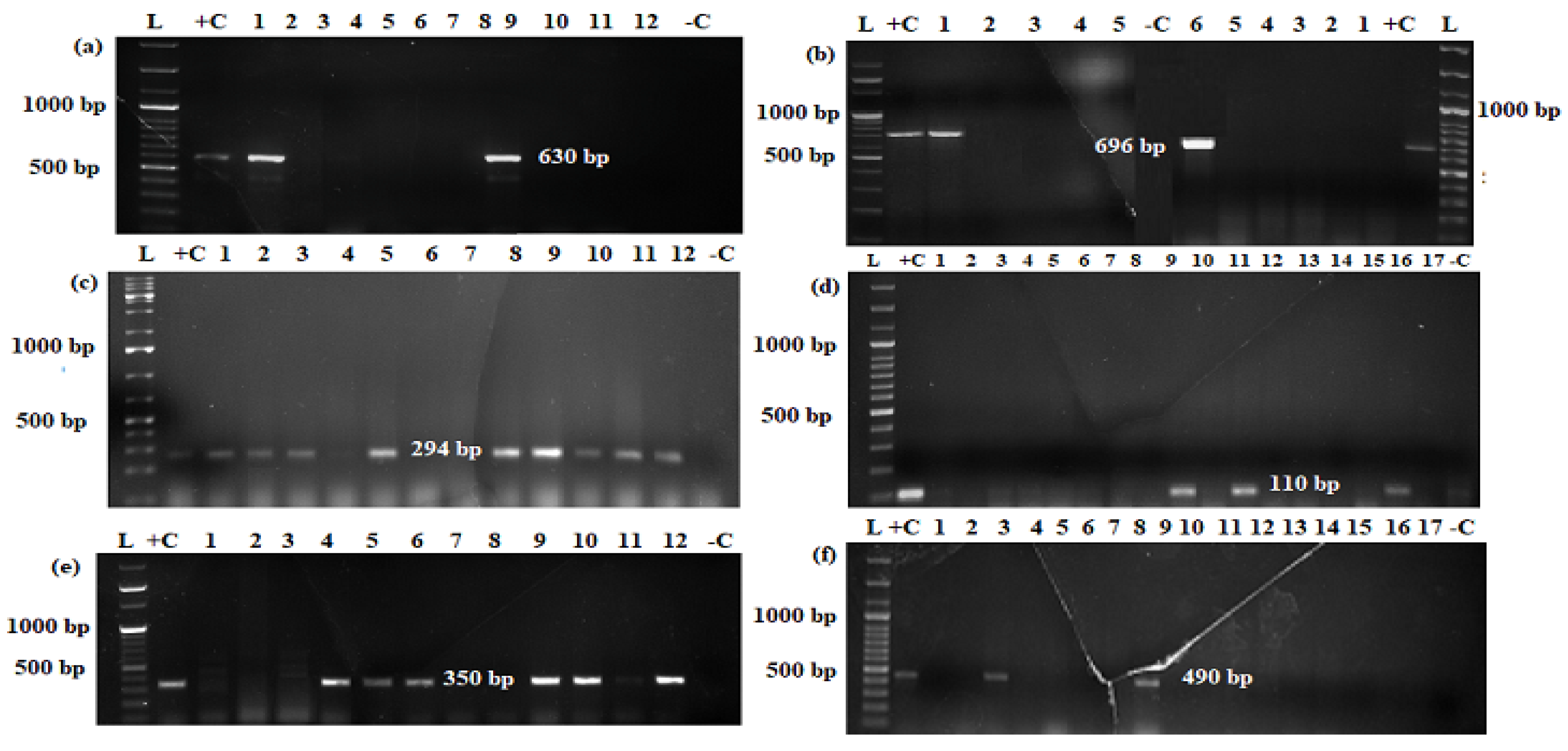
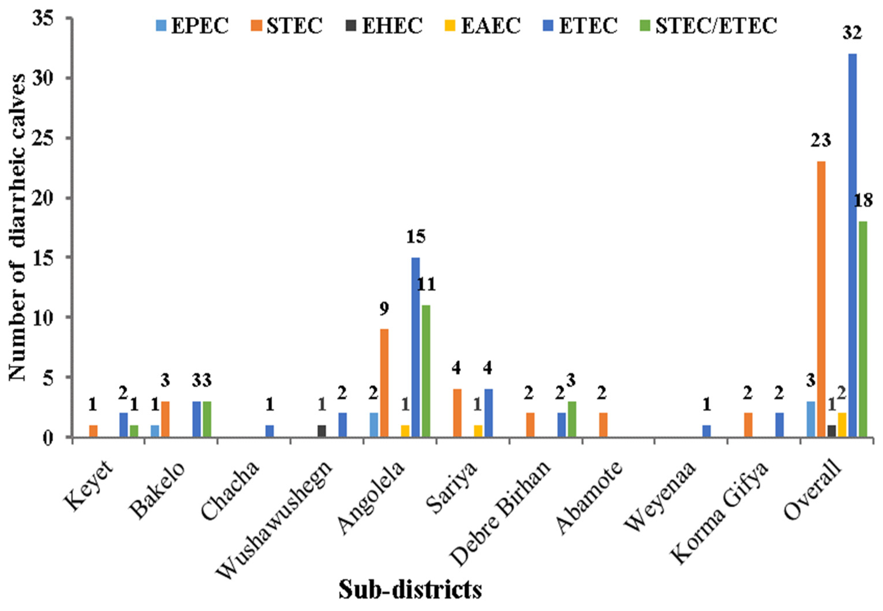
3.2. E. coli Isolation, Virulence Genes (VGs) Detection, and E. coli Pathotyping

The results of the E. coli isolation, PCR pathotyping, and infection multiplicity are shown in Figure 2. Of all the 300 presumptive E. coli isolates from 100 diarrheic calves, 281 were confirmed as E. col. Among the isolated E. coli, 160 were identified as an E. coli pathotype. E. coli was isolated from 99 sampled calves and, of these, 79 were found to be infected with at least one of the E. coli pathotypes. Of the infected calves, 61 were infected with single-pathotype and 18 had mixed E. coli pathotypes.
The gel electrophoresis demonstrating the PCR products of virulence genes are shown in Figure 3. The samples from 99 diarrheic calves were assessed for the presence of virulence gene/s (Table 2). The proportions of detected virulence genes were; eae 3 (3%), stx1 8 (8.1%), stx2 9 (9%), hly 1 (1%), aatA 2 (2%), and st 30 (30.3%). Several virulence genes were found in combinations; the most common combinations were stx2-st, 10.1% (n = 10), sx1-st, 6.1% (n = 6), stx1-stx2, 6.1% (n = 6), and lt-st, 2% (n = 2). Similarly, stx1-stx2-st and stx2-hly were each found in 1% (n = 1) of the diarrheic calves. The bfp, ial, and daaE virulence genes were not found in any of the tested samples. No virulence genes were found in the samples from 20.2% (n = 20) of the diarrheic calves.
Of the 79 diarrheic calves positive for E. coli pathotypes, ETEC 32.3% (n = 32), STEC 23.3% (n = 23), and STEC/ETEC 18.2% (n = 18) were the most prevalent pathotypes. On the other hand, EHEC, EAEC, and EPEC pathotypes were less common in diarrheic calves with 1, 2, and 3% detection rates, respectively. EPEC pathotypes were atypical, lacking the bfp virulence gene. In this study, no diarrheic calves were positive for the EIEC and DAEC pathotypes.
Table 3 shows the distribution of the E. coli pathotypes based on the different farm sizes, sex, age, and breed of the diarrheic calves. The number of detected E. coli pathotypes was higher on medium-size farms (MF), in female calves, calves under 2 weeks of age, and crossbred calves. In diarrheic calves from MF, 4.1% (2) EPEC and 20.8% (10) STEC/ETEC were found, the corresponding figures from small farms (SF) were EPEC 2.0% (1) and 15.7% (6) STEC/ETEC (15.7%). All of the detected EPEC pathotypes were from female calves (p = 0.059). Calves under 2 weeks of age had the highest incidence of STEC, 100%, (3/3), while the 2–4 weeks-old calves had the lowest incidence 13.3%, (4) (p = 0.006). Calves of an indigenous breed were more commonly infected with mixed STEC/ETEC, 33.3% (n = 5) than crossbreed calves 15.5% (n = 13) pathotypes.
The detected E. coli pathotypes among isolates from calves fed colostrum and a different supplementary feed are presented in Table 4. Among calves that had not been fed colostrum, the STEC, (27.3%, n = 3) and STEC/ETEC (36.4%, n = 4) pathotypes were more common; the corresponding results from the calves which were fed colostrum were 22.7% (n = 20) and 15.9% (n = 14), respectively. The detection rates of most E. coli pathotypes were higher in calves fed colostrum > 6 h from birth as compared to calves fed colostrum within 6 h after birth. STEC were detected in 8 of 17 (47.1%) of calves who were fed colostrum by hand, which was higher than in the 12 of 71 (16.9%) of calves who were allowed to suckle colostrum themselves (p = 0.008). The EAEC pathotypes were found in 2 of 17 calves who were fed colostrum by the hand method (p = 0.003). E. coli pathotypes were more frequently detected from calves that had not been started on a supplementary feed and calves who were allowed to graze than from calves who were fed concentrate and hay, but the observed differences were not significant.
The detection rate variations between E. coli pathotypes in calves from different housing types, flooring, and hygienic status were minor and no significant associations were seen (Table 5). E. coli pathotypes were present in 25 of 30 (83.3%) of the diarrheic calves housed in barns with other animals. Calves in houses with a soil flooring had higher E. coli pathotype proportions (80.6%) than calves on concrete (77.8%) and stone-lined (77.8%) floors. The E. coli pathotypes were found in 100%, 80%, 76.5%, and 73.3% of calves housed in very good, poor, very poor, and good hygienic conditions, respectively.
The distribution of pathotypes between the study subdistricts is shown in Figure 4. ETEC pathotypes were detected in 9 of the 10 subdistricts studied, with about 46.9% (n = 15) from Angolela and 53.1% (n = 17) of the pathotypes from eight other subdistricts. Compared to other subdistricts, STE/ETEC, 37.5% (n = 3), appeared more frequently among the isolates from the Debre Birhan subdistrict. EHEC (n = 1) was only found from the Wushawushign subdistrict (p = 0.00).
The results from the pairwise correlation analysis between the detected virulence genes are shown in Figure 5. The correlation analysis revealed that the co-occurrence of virulence genes stx2-lt, stx1-stx2, stx2-hly, and lt-st virulence genes had a weak positive correlation. The majority of virulence genes showed weak negative correlations. The observed correlations were not statistically significant.

4. Discussion

The present study intended to describe the occurrence of E. coli pathotypes and overall calving practices in a low-income setting. Livestock production is important in many Sub-Saharan countries like Ethiopia, for the national economy as well as for the livelihood of individual families, but the sector is facing various challenges. This study supports previous reports that diarrhea in calves is one of these challenges [15].
Virulence genes are extensively used in determining E. coli pathotypes and have been found in multiple combinations [34,35]. In our study, stx1, stx2, and st VGs were more frequent than eae, hly, aatA, and lt. In another report, however, stx1 and eae were found at a higher rate than st and stx2 [35]. In the isolates where virulence genes were found in combination, stx2-st, sx1-st, stx1-stx2, and lt-st represented the dominant combinations. In addition, stx1-stx2-st and stx2-hly were identified in a low proportion of samples from diarrheic calves. None of the samples were positive for the bfp, ial, and daaE virulence genes, in contrast to a previous report that indicated the presence of these genes in E. coli from diarrheic calves [36].
When translating the VRG combinations into pathotypes, ETEC was detected from 32.3% of the diarrheic calves as a sole pathogenic isolate. This result was in accordance with Dall et al. [37] who described ETEC as the leading cause of severe neonatal calf diarrhea in Brazil. All of the ETEC pathotypes carried the st virulence gene. It has been reported that st encodes structurally stable and highly virulent toxins [38]. Other studies also suggested that ETEC carries multiple chromosomal and plasmid virulence gene combinations that enhance the fitness and transmission between hosts [39,40]. Moreover, a study in Egypt described an st-associated ETEC pathotype in diarrheic calves [41,42]. The present finding and the previous studies indicate that ETEC plays a major role in causing calf diarrhea. However, the results presented in this study contradicted that of Umpiérrez et al. [34] and Khawaskar et al. [43] who reported only 10% and 1.9% ETEC from diarrheic calves in Uruguay and India.
In the present study, STEC was the second most frequent pathotype found in 23.2% of diarrheic calves. This is in agreement with previous reports that STEC is a key pathotype in calf diarrhea [2,43]. In addition, STEC has been detected at a higher rate in diarrheic calves [40] and other diarrheic animals than in healthy animals [44]. Several studies [44,45,46] have reported that STEC isolates with stx1 were more common than those that carried stx2, which contradicts the findings in the current study. Similar with the current study, Sobhy et al. [47] showed that the majority of STEC isolates had stx2 instead of stx1 and stx1-stx2 combinations. These inconsistent results indicate that stx1, stx2, and stx1-stx2 play a role in causing diarrhea among calves. In addition, STEC was reported in 34% of non-diarrheic water buffalo calves in Brazil [48] and 5.5% of healthy cattle calves in Korea [35]. This is in line with previous suggestions that animals are important reservoirs of STEC [35,49]. Moreover, the severity of the disease could be associated with the number and type of VGs present, suggesting that an infection by E. coli isolates with more VGs in various possible combinations are more likely to cause a disease.
This study revealed that 18.2% of the sampled calves had mixed STEC and ETEC (STEC/ETEC) infections. This result was in line with Awad et al. [41] who reported ETEC/STEC (14.7%) and ETEC/EPEC (2.7%) in diarrheic calves in Egypt. Other researchers showed that diarrheic calves could be infected with a hybrid STEC-ETEC pathotype [50,51]. These pathogroups are characterized as hetero-pathotypes comprised of virulence genes representing two or more E. coli pathotypes [52]. Other studies reported that various hetero-pathotypes, EAEC-STEC and EPEC-STEC, successfully colonized newborn calves and caused persistent diarrheal diseases [53,54].
In the present study, EPEC, EAEC, and EHEC pathotypes were found only in 3, 2, and 1% of diarrheic calves, respectively. These low percentages are in line with the findings from diarrheic neonatal calves sampled in India: EPEC (2.9%), EAEC (0.9%), and EHEC (0.3%) [43]. A study of diarrheic water buffalo diarrheic calves in Brazil detected EPEC (3.4%) and EHEC (3.4%) [48]. However, the 2% EAEC detection rate in our study is lower than the 39% EAEC detection rate in the same study [48]. This difference could be due to differences in the study’s design or target population (the species, health status, and geographical location).
In the current study, we attempted to relate the occurrence of pathotypes in the fecal samples to some basic farm and calf management practices. However, these observations must be interpreted with caution as the sampling focused on clinical cases with no healthy controls and was not randomized, which would have been needed for a solid epidemiological analysis. The incidence of E. coli pathotypes was higher in diarrheic calves sampled from medium sized farms. Regardless of the farm’s size, calves ≤ 2 weeks old were more frequently infected than other age groups. This is similar to previous studies that noted that younger calves were more frequently infected with different E. coli pathogroups than older calves [55,56]. This might be due to differences between younger and older calves regarding their immunity and rumen function. In addition, this could be due to age-related changes in the receptor expression for adhesins of the different pathotypes in the intestinal epithelial cells. In the present study, more female and crossbreed diarrheic calves were found to be infected with E. coli pathotypes. There is no plausible explanation for these differences, and the sex skewed finding is inconsistent with a report from India where the detection of STEC (8.0%) and ETEC (2.5%) were higher in samples from male neonatal calves. This was suggested due to the fact that male calves did not receive appropriate care as they were considered economically less important [43].
Our results show that E. coli pathotypes were more common in calves not fed colostrum than in calves fed colostrum. We also noted that calves fed colostrum 6–24 h after birth were more frequently infected by E. coli pathotypes than those fed within 0–6 h. Multiple studies show that the sufficient and timely colostrum feeding of calves improves the growth, immunity, and intestinal microbiota of the calves [57,58], as maternal antibodies in the colostrum prevent a bacterial attachment to the intestinal epithelium [59,60,61]. According to the findings in this study and multiple other studies, the colostrum feeding of calves should takes place within 6 h after their birth. Of the 17 diarrheic calves fed colostrum via hand, 47.1% (p = 0.008) and 11.8% (p = 0.003) were positive for STEC and EAEC, respectively. Similar research in Ethiopia has indicated that hand/bucket feeding was significantly correlated with a high incidence of E. coli [15]. The higher occurrence of pathotypes in samples from hand-fed calves compared with those that suckled colostrum may indicate a lack of appropriate hygiene routines during the hand-feeding of the colostrum to calves. This merits further study.
The detection of E. coli pathotypes was slightly higher in calves housed in barns (with other animals) and on soil floors. This result was in agreement with Fesseha et al. [15]. We also found that calves housed with very good hygiene had a higher frequency of E. coli pathotypes; however, those farms were comparatively larger farms, where a higher infectious pressure with E. coli pathotypes could be assumed. The interaction between the farm’s size and hygiene may have prevented the full exploration of these aspects in our limited number of samples.

5. Conclusions

The study showed that E. coli pathotypes could be frequently found in diarrheic calves in a low-income setting. The ETEC and STEC pathotypes were the majority of pathotypes found among the diarrheic calves. A significant number of calves were found to be infected with mixed STEC/ETEC pathotypes. Given the rearing conditions in these settings, where calves are kept close to human dwellings, this poses a significant zoonotic risk.
The presence of E. coli pathotypes was higher in calves under 2 weeks of age and crossbred calves. Calves that had not been fed colostrum and were late-fed colostrum appeared to be more vulnerable to E. coli pathotypes. This, together with the frequent occurrence of E. coli pathotypes, suggests there is a need for providing the better management for calves and younger calves in particular to improve their health and reduce the economic loss.

Supplementary Materials

The following supporting information can be downloaded at: https://www.mdpi.com/article/10.3390/pathogens12010042/s1, Table S1, Table S2, and Table S3.

Author Contributions

Conceptualization, W.S.C., H.A., S.S.-L., U.M. and T.S.T.; methodology, W.S.C., S.S.-L., U.M. and T.S.T.; software, W.S.C.; validation, H.A., S.S.-L., U.M. and T.S.T.; formal analysis, W.S.C.; investigation, W.S.C. and T.S.T.; resources, H.A., S.S.-L., U.M. and T.S.T.; data curation, W.S.C., S.S.-L., U.M. and T.S.T.; writing—original draft preparation, W.S.C.; writing—review and editing, W.S.C., H.A., S.S.-L., U.M. and T.S.T.; supervision, H.A., S.S.-L., U.M. and T.S.T.; project administration, H.A., S.S.-L., U.M. and T.S.T.; Funding acquisition, H.A., S.S.-L., U.M. and T.S.T. All authors have read and agreed to the published version of the manuscript.

Funding

The study was financially supported by the Swedish International Development Cooperation Agency (SIDA)’s Research and Training Grant AAU—SLU program, https://sida.aau.edu.et/index.php/international-comparative-education-phd-program/ accessed 30 April 2022.

Institutional Review Board Statement

The study was ethically cleared by the natural and computational sciences institutional review board committee, Addis Ababa University, Ethiopia (Ref. No. CNCSDO/423/14/2022).

Informed Consent Statement

Not applicable.

Data Availability Statement

The data generated will be provided upon the request of the corresponding author.

Acknowledgments

The authors acknowledge, SIDA for financing the project. The authors acknowledge AAU, IoB, and SLU, the Department of Clinical Sciences for all of their support provided during the study period. The authors are grateful to the veterinarian who supported us during fecal sample collection from diarrheic calves and Basona Werana Agricultural officers for direct and indirect support.

Conflicts of Interest

The authors declare no conflict of interest.

References

  1. Lugsomya, K.; Yindee, J.; Niyomtham, W.; Tribuddharat, C.; Tummaruk, P.; Hampson, D.J.; Prapasarakul, N. Antimicrobial Resistance in Commensal Escherichia Coli Isolated from Pigs and Pork Derived from Farms Either Routinely Using or Not Using In-Feed Antimicrobials. Microb. Drug Resist. 2018, 24, 1054–1066. [Google Scholar] [CrossRef] [PubMed] [Green Version]
  2. Angappan, M.; Ghatak, S.; Milton, A.A.P.; Verma, A.K.; Inbaraj, S.; Abhishek; Chaudhuri, P.; Agarwal, R.K.; Thomas, P. Detection of Novel Sequence Types and Zoonotic Transmission Potentiality among Strains of Shiga Toxigenic Escherichia Coli (STEC) from Dairy Calves, Animal Handlers and Associated Environments. Brazilian J. Microbiol. 2021, 52, 2541–2546. [Google Scholar] [CrossRef] [PubMed]
  3. Engelen, F.; Thiry, D.; Devleesschauwer, B.; Heyndrickx, M.; Mainil, J.; De Zutter, L.; Cox, E. Pathogenic Potential of Escherichia Coli O157 and O26 Isolated from Young Belgian Dairy Calves by Recto-Anal Mucosal Swab Culturing. J. Appl. Microbiol. 2021, 131, 964–972. [Google Scholar] [CrossRef]
  4. Pakbin, B.; Brück, W.M.; Rossen, J.W.A. Virulence Factors of Enteric Pathogenic Escherichia Coli: A Review. Int. J. Mol. Sci. 2021, 22, 9922. [Google Scholar] [CrossRef]
  5. World Health Organization. Who Estimates of the Global Burden of Foodborne Diseases 2007–2015. In FOODBORNE Disease Burden Epidemiology Reference Group 2007–2015; WHO: Geneva, Switzerland, 2015; p. 72. [Google Scholar]
  6. Abuelo, A. Investigation of an Outbreak of Neonatal Calf Diarrhoea in a Dairy Herd. Vet. Rec. Case Rep. 2016, 4, e000372. [Google Scholar] [CrossRef]
  7. Kayasaki, F.; Okagawa, T.; Konnai, S.; Kohara, J.; Sajiki, Y.; Watari, K.; Ganbaatar, O.; Goto, S.; Nakamura, H.; Shimakura, H.; et al. Direct Evidence of the Preventive Effect of Milk Replacer–Based Probiotic Feeding in Calves against Severe Diarrhea. Vet. Microbiol. 2021, 254, 108976. [Google Scholar] [CrossRef]
  8. Tora, E.; Abayneh, E.; Seyoum, W.; Shurbe, M. Longitudinal Study of Calf Morbidity and Mortality on Smallholder Farms in Southern Ethiopia. PLoS ONE 2021, 16, e0257139. [Google Scholar] [CrossRef]
  9. Caffarena, R.D.; Casaux, M.L.; Schild, C.O.; Fraga, M.; Castells, M.; Colina, R.; Maya, L.; Corbellini, L.G.; Riet-Correa, F.; Giannitti, F. Causes of Neonatal Calf Diarrhea and Mortality in Pasture-Based Dairy Herds in Uruguay: A Farm-Matched Case-Control Study. Brazilian J. Microbiol. 2021, 52, 977–988. [Google Scholar] [CrossRef]
  10. Ferede, Y.; Mazengia, H.; Bimrew, T.; Bitew, A.; Nega, M.; Kebede, A. Pre-Weaning Morbidity and Mortality of Crossbred Calves in Bahir Dar Zuria and Gozamen Districts of Amhara Region, Northwest Ethiopia. OALib 2014, 1, e600. [Google Scholar] [CrossRef]
  11. Fentie, T.; Guta, S.; Mekonen, G.; Temesgen, W.; Melaku, A.; Asefa, G.; Tesfaye, S.; Niguse, A.; Abera, B.; Kflewahd, F.Z.; et al. Assessment of Major Causes of Calf Mortality in Urban and Periurban Dairy Production System of Ethiopia. Vet. Med. Int. 2020, 2020, 3075429. [Google Scholar] [CrossRef]
  12. United States Department of Agriculture. Dairy 2007 Part II: Changes in the U.S. Dairy Cattle Industry, 1991–2007. In Fort Collins: USDA-APHIS-VS, CEAH; USDA: Washington, DC, USA, 2008; pp. 57–61. [Google Scholar]
  13. Schinwald, M.; Creutzinger, K.; Keunen, A.; Winder, C.B.; Haley, D.; Renaud, D.L. Predictors of Diarrhea, Mortality, and Weight Gain in Male Dairy Calves. J. Dairy Sci. 2022, 105, 5296–5309. [Google Scholar] [CrossRef] [PubMed]
  14. Østerås, O.; Gjestvang, M.S.; Vatn, S.; Sølverød, L. Perinatal Death in Production Animals in the Nordic Countries—Incidence and Costs. Acta Vet. Scand. 2007, 49, 4–7. [Google Scholar] [CrossRef] [Green Version]
  15. Fesseha, H.; Mathewos, M.; Aliye, S.; Mekonnen, E. Isolation and Antibiogram of Escherichia Coli O157: H7 from Diarrhoeic Calves in Urban and Peri-Urban Dairy Farms of Hawassa Town. Vet. Med. Sci. 2022, 8, 864–876. [Google Scholar] [CrossRef] [PubMed]
  16. Zhao, W.; Choi, C.Y.; Li, G.; Li, H.; Shi, Z. Pre-Weaned Dairy Calf Management Practices, Morbidity and Mortality of Bovine Respiratory Disease and Diarrhea in China. Livest. Sci. 2021, 251, 104608. [Google Scholar] [CrossRef]
  17. Endale, A.; Muktar, Y.; Tessema, T.S. Molecular Characterization of Diarrheagenic Escherichia Coli Strains Isolated from Dairy Calves in North Shoa Zone, Oromiya Region, Ethiopia. Res. Sq. 2020, 17, e0275229. [Google Scholar]
  18. Negeri, A.A.; Mamo, H.; Gurung, J.M.; Firoj Mahmud, A.K.M.; Fällman, M.; Seyoum, E.T.; Feleke Desta, A.; Francis, M.S. Antimicrobial Resistance Profiling and Molecular Epidemiological Analysis of Extended Spectrum β-Lactamases Produced by Extraintestinal Invasive Escherichia Coli Isolates from Ethiopia: The Presence of International High-Risk Clones ST131 and ST410 Reveal. Front. Microbiol. 2021, 12, 706846. [Google Scholar] [CrossRef]
  19. Ayenew, H.Y.; Mitiku, B.A.; Tesema, T.S. Occurrence of Virulence Genes and Antimicrobial Resistance of E. Coli O157:H7 Isolated from the Beef Carcass of Bahir Dar City, Ethiopia. Vet. Med. Int. 2021, 2021, 8046680. [Google Scholar] [CrossRef]
  20. Sebre, S.; Erku Abegaz, W.; Seman, A.; Awoke, T.; Mihret, W.; Desalegn, Z.; Abebe, T.; Mihret, A. Molecular Characterization of Extended-Spectrum Beta-Lactamase-Producing Enterobacteriaceae Isolates Collected from Inanimate Hospital Environments in Addis Ababa, Ethiopia. In Advances in Microbiology, Infectious Disease and Public Health; Springer: Berlin/Heidelberg, Germany, 2021. [Google Scholar] [CrossRef]
  21. Central Statistical Agency Central Statistical Agency (2015) Agricultural Sample Survey 2014/15. Report on Livestock and Livestock Characteristics. Stat. Bull. 2015, 2, 578. [Google Scholar]
  22. Central Statistical Agency. Statistical Report for Amhara Region; CSA: Addis Ababa, Ethiopia, 2007. [Google Scholar]
  23. Cheesbrough, M. Medical Laboratory Manual for Tropical Countries; Tropical Health Technology: London, UK, 1985; Volume II, ISBN 0950743429. [Google Scholar]
  24. Moore, E.; Arnscheidt, A.; KrÜger, A.; StrÖmpl, C.; Mau, M. Section 1 Update: Simplified Protocols for the Preparation of Genomic DNA from Bacterial Cultures. In Molecular Microbial Ecology Manual; Kluwer Academic: Amsterdam, The Netherlands, 2004. [Google Scholar]
  25. Khan, A.; Yamasaki, S.; Sato, T.; Ramamurthy, T.; Pal, A.; Datta, S.; Chowdhury, N.R.; Das, S.C.; Sikdar, A.; Tsukamoto, T.; et al. Prevalence and Genetic Profiling of Virulence Determinants of Non-O157 Shiga Toxin-Producing Escherichia Coli Isolated from Cattle, Beef, and Humans, Calcutta, India. Emerg. Infect. Dis. 2002, 8, 54–62. [Google Scholar] [CrossRef]
  26. Pal, A.; Ghosh, S.; Ramamurthy, T.; Yamasaki, S.; Tsukamoto, T.; Bhattacharya, S.K.; Takeda, Y. Shiga-Toxin Producing Escherichia Coli from Healthy Cattle in a Semi-Urban Community in Calcutta, India. Indian J. Med. Res. 1999, 110, 83. [Google Scholar]
  27. Selim, S.A.; Ahmed, S.F.; Abdel Aziz, M.H.; Zakaria, A.M.; Klena, J.D.; Pangallo, D. Prevalence and Characterization of Shiga-Toxin O157:H7 and Non-O157:H7 Enterohemorrhagic Escherichia Coli Isolated from Different Sources. Biotechnol. Biotechnol. Equip. 2013, 27, 3834–3842. [Google Scholar] [CrossRef] [Green Version]
  28. Havt, A.; Lima, I.F.; Medeiros, P.H.; Clementino, M.A.; Santos, A.K.; Amaral, M.S.; Veras, H.N.; Prata, M.M.; Lima, N.L.; Di Moura, A.; et al. Prevalence and Virulence Gene Profiling of Enteroaggregative Escherichia Coli in Malnourished and Nourished Brazilian Children. Diagn. Microbiol. Infect. Dis. 2017, 89, 98–105. [Google Scholar] [CrossRef] [PubMed]
  29. Gunzburg, S.T.; Tornieporth, N.G.; Riley, L.W. Identification of Enteropathogenic Escherichia Coli by PCR-Based Detection of the Bundle-Forming Pilus Gene. J. Clin. Microbiol. 1995, 33, 1375–1377. [Google Scholar] [CrossRef] [PubMed] [Green Version]
  30. Nada, R.A.; Shaheen, H.I.; Touni, I.; Fahmy, D.; Armstrong, A.W.; Weiner, M.; Klena, J.D. Design and Validation of a Multiplex Polymerase Chain Reaction for the Identification of Enterotoxigenic Escherichia Coli and Associated Colonization Factor Antigens. Diagn. Microbiol. Infect. Dis. 2010, 67, 134–142. [Google Scholar] [CrossRef]
  31. Schultsz, C.; Pool, G.J.; Van Ketel, R.; De Wever, B.; Speelman, P.; Dankert, J. Detection of Enterotoxigenic Escherichia Coli in Stool Samples by Using Nonradioactively Labeled Oligonucleotide DNA Probes and PCR. J. Clin. Microbiol. 1994, 32, 2393–2397. [Google Scholar] [CrossRef] [Green Version]
  32. Batista, C.; Carolina, M.; Souza, S.D.; Serra, P.T.; Santos, I.; Balieiro, A.; Pieri, F.A.; Nogueira, P.A.; Orlandi, P.P. Virulence Factors Associated with Pediatric Shigellosis in Brazilian Amazon. Biomed. Res. Int. 2014, 2014, 539697. [Google Scholar] [CrossRef] [Green Version]
  33. Vidal, M.; Kruger, E.; Durán, C.; Lagos, R.; Levine, M.; Prado, V.; Toro, C.; Vidal, R. Single Multiplex PCR Assay to Identify Simultaneously the Six Categories of Diarrheagenic Escherichia Coli Associated with Enteric Infections. J. Clin. Microbiol. 2005, 43, 5362–5365. [Google Scholar] [CrossRef] [Green Version]
  34. Umpiérrez, A.; Ernst, D.; Fernández, M.; Oliver, M.; Casaux, M.L.; Caffarena, R.D.; Schild, C.; Giannitti, F.; Fraga, M.; Zunino, P. Virulence Genes of Escherichia Coli in Diarrheic and Healthy Calves. Rev. Argent. Microbiol. 2021, 53, 34–38. [Google Scholar] [CrossRef]
  35. Ryu, J.H.; Kim, S.H.; Park, J.; Choi, K.S. Characterization of Virulence Genes in Escherichia Coli Strains Isolated from Pre-Weaned Calves in the Republic of Korea. Acta Vet. Scand. 2020, 62, 45. [Google Scholar] [CrossRef]
  36. Zhu, Z.; Wang, W.; Cao, M.; Zhu, Q.; Ma, T.; Zhang, Y.; Liu, G.; Zhou, X.; Li, B.; Shi, Y.; et al. Virulence Factors and Molecular Characteristics of Shigella Flexneri Isolated from Calves with Diarrhea. BMC Microbiol. 2021, 21, 214. [Google Scholar] [CrossRef]
  37. Dall Agnol, A.M.; Lorenzetti, E.; Leme, R.A.; Ladeia, W.A.; Mainardi, R.M.; Bernardi, A.; Headley, S.A.; Freire, R.L.; Pereira, U.P.; Alfieri, A.F.; et al. Severe Outbreak of Bovine Neonatal Diarrhea in a Dairy Calf Rearing Unit with Multifactorial Etiology. Brazilian J. Microbiol. 2021, 52, 2547–2553. [Google Scholar] [CrossRef] [PubMed]
  38. Zhu, Y.; Luo, Q.; Davis, S.M.; Westra, C.; Vickers, T.J. Molecular Determinants of Enterotoxigenic Escherichia Coli Heat-Stable Toxin Secretion and Delivery. ASM J. 2018, 86, e00526-18. [Google Scholar] [CrossRef] [PubMed]
  39. Von Mentzer, A.; Connor, T.R.; Wieler, L.H.; Semmler, T.; Iguchi, A.; Thomson, N.R.; Rasko, D.A.; Joffre, E.; Corander, J.; Pickard, D.; et al. Identification of Enterotoxigenic Escherichia Coli (ETEC) Clades with Long-Term Global Distribution. Nat. Genet. 2014, 46, 1321–1326. [Google Scholar] [CrossRef] [PubMed] [Green Version]
  40. Shepard, S.M.; Danzeisen, J.L.; Isaacson, R.E.; Seemann, T.; Achtman, M.; Johnson, T.J. Genome Sequences and Phylogenetic Analysis of K88- and F18- Positive Porcine Enterotoxigenic Escherichia Coli. J. Bacteriol. 2012, 194, 395–405. [Google Scholar] [CrossRef] [Green Version]
  41. Awad, W.S.; El-Sayed, A.A.; Mohammed, F.F.; Bakry, N.M.; Abdou, N.E.M.I.; Kamel, M.S. Molecular Characterization of Pathogenic Escherichia Coli Isolated from Diarrheic and In-Contact Cattle and Buffalo Calves. Trop. Anim. Health Prod. 2020, 52, 3173–3185. [Google Scholar] [CrossRef]
  42. Shabana, I.I.; Zaraket, H.; Suzuki, H. Molecular Studies on Diarrhea-Associated Escherichia Coli Isolated from Humans and Animals in Egypt. Vet. Microbiol. 2013, 167, 532–539. [Google Scholar] [CrossRef]
  43. Khawaskar, D.P.; Sinha, D.K.; Lalrinzuala, M.V.; Athira, V.; Kumar, M.; Chhakchhuak, L.; Mohanapriya, K.; Sophia, I.; Abhishek; Kumar, O.R.V.; et al. Pathotyping and Antimicrobial Susceptibility Testing of Escherichia Coli Isolates from Neonatal Calves. Vet. Res. Commun. 2021, 46, 353–362. [Google Scholar] [CrossRef]
  44. Leomil, L.; Aidar-Ugrinovich, L.; Guth, B.E.C.; Irino, K.; Vettorato, M.P.; Onuma, D.L.; De Castro, A.F.P. Frequency of Shiga Toxin-Producing Escherichia Coli (STEC) Isolates among Diarrheic and Non-Diarrheic Calves in Brazil. Vet. Microbiol. 2003, 97, 103–109. [Google Scholar] [CrossRef]
  45. Srivani, M.; Narasimha Reddy, Y.; Subramanyam, K.V.; Ramakoti Reddy, M.; Srinivasa Rao, T. Prevalence and Antimicrobial Resistance Pattern of Shiga Toxigenic Escherichia Coli in Diarrheic Buffalo Calves. Vet. World 2017, 10, 774–778. [Google Scholar] [CrossRef] [Green Version]
  46. Marashifard, M.; Aliabad, Z.K.; Ali, S.; Malek, A.; Darban-sarokhalil, D. Determination of Antibiotic Resistance Pattern and Virulence Genes in Escherichia Coli Isolated from Bovine with Subclinical Mastitis in Southwest of Iran. Trop. Anim. Health Prod. 2018, 51, 575–580. [Google Scholar] [CrossRef]
  47. Sobhy, N.M.; Yousef, S.G.A.; Aboubakr, H.A.; Nisar, M.; Nagaraja, K.V.; Mor, S.K.; Valeris-Chacin, R.J.; Goyal, S.M. Virulence Factors and Antibiograms of Escherichia Coli Isolated from Diarrheic Calves of Egyptian Cattle and Water Buffaloes. PLoS One 2020, 15, e0232890. [Google Scholar] [CrossRef] [PubMed]
  48. Coura, F.M.; Diniz, S.D.A.; Silva, M.X.; de Oliveira, C.H.S.; Mussi, J.M.S.; de Oliveira, C.S.F.; Lage, A.P.; Heinemann, M.B. Virulence Factors and Phylotyping of Escherichia Coli Isolated from Non-Diarrheic and Diarrheic Water Buffalo Calves. Cienc. Rural 2019, 49, 1–9. [Google Scholar] [CrossRef]
  49. Wani, S.A.; Bhat, M.A.; Samanta, I.; Nishikawa, Y.; Buchh, A.S. Isolation and Characterization of Shiga Toxin-Producing Escherichia Coli (STEC) and Enteropathogenic Escherichia Coli (EPEC) from Calves and Lambs with Diarrhoea in India. Lett. Appl. Microbiol. 2003, 37, 121–126. [Google Scholar] [CrossRef]
  50. Aref, N.E.M.; Abdel-Raheem, A.R.A.; Kamaly, H.F.; Hussien, S.Z. Clinical and Sero-Molecular Characterization of Escherichia Coli with an Emphasis on Hybrid Strain in Healthy and Diarrheic Neonatal Calves in Egypt. Open Vet. J. 2018, 8, 351–359. [Google Scholar] [CrossRef] [PubMed] [Green Version]
  51. Shen, J.; Zhi, S.; Guo, D.; Jiang, Y.; Xu, X.; Zhao, L.; Lv, J. Prevalence, Antimicrobial Resistance, and Whole Genome Sequencing Analysis of Shiga Toxin-Producing Escherichia Coli (STEC) and Enteropathogenic Escherichia Coli (EPEC) from Imported Foods in China during 2015–2021. Toxins 2022, 14, 68. [Google Scholar] [CrossRef]
  52. Santos, A.C.D.M.; Santos, F.F.; Silva, R.M.; Gomes, T.A.T. Diversity of Hybrid- and Hetero-Pathogenic Escherichia Coli and Their Potential Implication in More Severe Diseases. Front. Cell. Infect. Microbiol. 2020, 10, 339. [Google Scholar] [CrossRef]
  53. Badouei, M.A.; Morabito, S.; Najafifar, A.; Mazandarani, E. Molecular Characterization of Enterohemorrhagic Escherichia Coli Hemolysin Gene (EHEC-HlyA)-Harboring Isolates from Cattle Reveals a Diverse Origin and Hybrid Diarrheagenic Strains. Infect. Genet. Evol. 2016, 39, 342–348. [Google Scholar] [CrossRef]
  54. Frank, C.; Werber, D.; Cramer, J.P.; Askar, M.; Faber, M.; an der Heiden, M.; Bernard, H.; Fruth, A.; Prager, R.; Spode, A.; et al. Epidemic Profile of Shiga-Toxin–Producing Escherichia Coli O104:H4 Outbreak in Germany. N. Engl. J. Med. 2011, 365, 1771–1780. [Google Scholar] [CrossRef] [Green Version]
  55. Osman, K.M.; Mustafa, A.M.; Elhariri, M.; Abdelhamed, G.S. The Distribution of Escherichia Coli Serovars, Virulence Genes, Gene Association and Combinations and Virulence Genes Encoding Serotypes in Pathogenic E. Coli Recovered from Diarrhoeic Calves, Sheep and Goat. Transbound. Emerg. Dis. 2013, 60, 69–78. [Google Scholar] [CrossRef]
  56. Algammal, A.M.; El-Kholy, A.W.; Riad, E.M.; Mohamed, H.E.; Elhaig, M.M.; Al Yousef, S.A.; Hozzein, W.N.; Ghobashy, M.O.I.I. Genes Encoding the Virulence and the Antimicrobial Resistance in Enterotoxigenic and Shiga-Toxigenic E. Coli Isolated from Diarrheic Calves. Toxins 2020, 12, 383. [Google Scholar] [CrossRef]
  57. Liang, G.; Malmuthuge, N.; Bao, H.; Stothard, P.; Griebel, P.J.; Guan, L.L. Transcriptome Analysis Reveals Regional and Temporal Differences in Mucosal Immune System Development in the Small Intestine of Neonatal Calves. BMC Genomics 2016, 17, 602. [Google Scholar] [CrossRef] [Green Version]
  58. Nissen, A.; Andersen, P.H.; Bendixen, E.; Ingvartsen, K.L.; Røntved, C.M. Colostrum and Milk Protein Rankings and Ratios of Importance to Neonatal Calf Health Using a Proteomics Approach. J. Dairy Sci. 2017, 100, 2711–2728. [Google Scholar] [CrossRef] [PubMed]
  59. DuBourdieu, D. Colostrum Antibodies, Egg Antibodies and Monoclonal Antibodies Providing Passive Immunity for Animals. In Nutraceuticals in Veterinary Medicine; Springer: Cham, Switzerland, 2019; pp. 245–257. ISBN 9783030046248. [Google Scholar]
  60. Ma, T.; O’Hara, E.; Song, Y.; Fischer, A.J.; He, Z.; Steele, M.A.; Guan, L.L. Altered Mucosa-Associated Microbiota in the Ileum and Colon of Neonatal Calves in Response to Delayed First Colostrum Feeding. J. Dairy Sci. 2019, 102, 7073–7086. [Google Scholar] [CrossRef] [PubMed]
  61. Ghaffari, M.H.; Sadri, H.; Steinhoff-Wagner, J.; Hammon, H.M.; Sauerwein, H. Effects of Colostrum Feeding on the MRNA Abundance of Genes Related to Toll-like Receptors, Key Antimicrobial Defense Molecules, and Tight Junctions in the Small Intestine of Neonatal Dairy Calves. J. Dairy Sci. 2021, 104, 10363–10373. [Google Scholar] [CrossRef] [PubMed]
Figure 1. Study area map. The map on the left-hand side (yellow) shows Ethiopia; the upper right shows Amhara regional state and the lower r right in green shows the study area known as Basona Werena district (woreda).
Pathogens 12 00042 g001
Figure 2. Number of E. coli isolates and number of sampled calves after initial isolation, confirmation, and pathotyping and detected infection multiplicity.
Pathogens 12 00042 g002
Figure 3. Representative gel electrophoresis results of PCR products for virulence genes: (a) aatA (630 bp), (b) lt (696 bp), (c) st (294 bp), (d) stx1 (110 bp), (e) stx2 (350 bp), and (f) eae (490 bp). L-ladder DNA (marker size, 100 bp plus), Lane +C-positive control, lanes with numbers are PCR products of virulence genes from E. coli isolates (test) and lane −C-negative control.
Pathogens 12 00042 g003
Figure 4. Distributions of E. coli pathotypes in 10 study subdistricts from fecal samples of 99 diarrheic calves in Basona Werana district, Ethiopia. Pathotypes; EPEC—Enteropathogenic E. coli; STEC—Shiga-toxin E. coli; EAEC—Enteroaggregative E. coli; ETEC—Enterotoxigenic E. coli; and STEC/ETEC–STEC/ETEC (mixed Shiga and stable toxins) E. coli.
Pathogens 12 00042 g004
Figure 5. The pairwise correlation coefficient of detected E. coli virulence genes in the 99 diarrheic calve’ fecal isolates in Basona Werana district Ethiopia. eae-intimin; stx ½-Shiga toxin 1&2; hly-hemolysin; aatA-aggregative, st-stable toxin; lt-liable toxin.
Pathogens 12 00042 g005
Table 1. Escherichia coli pathotyping oligonucleotide primers: primer sequences for the ten (10) virulence genes and amplicon size for each of the six E. coli pathotypes are shown below.
Primer Oligonucleotide Sequence 5′ to 3′GenePathotypeProduct Size (bp)Reference
EAE1
EAE2
F:AAACAGGTGAAACTGTTGCC
R:CTCTGCAGATTAACCTCTGC
eaeEPEC/EHEC490[25]
EVS1
EVC2
F:ATCAGTCGTCACTCACTGGT
R:CTGCTGTCACAGTGACAAA
stx1STEC/EHEC110[26]
EVT1
EVT2
F:CAACACTGGATGATCTCAGC
R:CCCCCTCAACTGCTAATA
stx2STEC/EHEC350
EHEC
EHEC
F:ACGATGTGGTTTATTCTGGA
R:CTTCACGTCACCATACATAT
hlyEHEC167[27]
EAEC
EAEC
F: CTGGCGAAAGACTGTATCAT
R:CAATGTATAGAAATCCGCTGTT
aatAEAEC630[28]
BFP
BFP
F:AATGGTGCTTGCGCTTGCTGC
R:GCCGCTTTATCCAACCTGGTA
bfpATypical EPEC324[29]
ST1
ST2
F: TTT ATT TCT GTA TTG TCT T
R:GCAGGATTACAACACAATTC
StETEC294[30]
LT1
LT2
F: GGCGACAGATTATACCGTGC
R: CCGAATTCTGTTATATATGTC
lt
ETEC
696
[31]
IAL F
IAL R
F: CTGGATGGTATGGTGAGG
R:GGAGGCCAACAACATTATTTCC
ialEIEC320[32]
daaE 1
daaE 2
F:GAACGTTGGTTAATGTGGGGT
R:TATTCACCGGTCGGTTATCAG
daaEDAEC542[33]
F—forward primer, R—reverse primer, and bp—basepair.
Table 2. Virulence genes profiles and distribution of E. coli pathotypes in fecal samples from 99 diarrheic calves on 98 farms in Basona Werana district, Ethiopia.
PathotypeVirulence GenesDiarrheic Calves Total
eaestx1stx2hlyaatAltstn (%)n (%)
EPEC+ -----3 (3.0)3 (3.0)
STEC-+-----8 (8.1)23 (23.2)
-++----6 (6.1)
--+----9 (9.1)
EHEC--++---1 (1.0)1 (1.0)
EAEC----+--2 (2.0)2 (2.0)
ETEC-----++2 (2.0)32 (32.3)
------+30 (30.3)
STEC/ETEC-+----+6 (6.1)18 (18.2)
--+---+10 (10.1)
-++---+1 (1.0)
--+- ++1 (1.0)
TPD--------79 (79.8)
NPD--------20 (20.2)
Overall --------99 (100.0)
n &%—number and percentage of calves positive for tested virulence genes and E. coli pathotypes; TPD—total pathotypes detected; NPD—no pathotypes detected; virulence genes; eae- intimin; stx ½—Shiga toxin 1&2; hly—hemolysin; aatA—aggregative; st—stable toxin; lt—liable toxin); pathotypes; EPEC—Enteropathogenic E. coli; STEC—Shiga-toxin E. coli; EAEC—Enteroaggregative E. coli; ETEC—Enterotoxigenic E. coli and STEC/ETEC—mixed STEC and ETEC E. coli pathotype.
Table 3. Detection of E. coli pathotypes from different farm sizes, sex, age, and breed of 99 diarrheic calve fecal isolates in Basona Werana district, Ethiopia.
Pathotype Diarrheic Calves—n (%)
Farm SizeSexAge (Weeks)Breed
SFMFFM≤2(2–4](4–7](7–10]IndigenousCrossbreed
EPEC 1 (2.0)2 (4.2)0 (0.0 b)3 (6.5 b)0 (0.0)0 (0.0)1 (3.7)2 (5.1)0 (0.0)3 (3.6)
STEC 13 (25.5)10 (20.8)13 (24.5)10 (21.7)3 (100.0 a)4 (13.3 a)5 (18.5 a)11 (28.2 a)3 (20.0)20 (23.8)
EHEC 1 (2.0)0 (0.0)0 (0.0)1 (2.2)0 (0.0)1 (3.3)0 (0.0)0 (0.0)0 (0.0)1 (1.2)
EAEC 1 (2.0)1 (2.1)1 (1.9)1 (2.2)0 (0.0)0 (0.0)1 (3.7)1 (2.6)0 (0.0)2 (2.4)
ETEC 16 (31.4)16 (33.3)16 (30.2)16 (34.8)0 (0.0)12 (40.0)8 (29.6)12 (30.8)4 (26.7)28 (33.3)
STEC/ETEC 8 (15.7)10 (20.8)11 (20.8)7 (15.2)0 (0.0)7 (23.3)5 (18.5)6 (15.4)5 (33.3)13 (15.5)
NPD=11 (21.6)9 (18.8)12 (22.6)8 (17.4)0 (0.0)6 (20.0)7 (25.9)7 (17.9)3 (20.0)17 (20.2)
Total (n)5148534633027391584
row values with the same letter superscript a and b were significantly different; at p = 0.006 and 0.059, respectively. SF—Small farm (≤6 animals/farm); MF—medium farm (≥7 animals/farm); F—female; M—male; T (n)—total number of calves examined in the categories; n (%)—number and percentage of calves positive for respective E. coli pathotypes. NPD—no pathotypes detected; EPEC—Enteropathogenic E. coli; STEC—Shiga-toxin E. coli; EAEC—Enteroaggregative E. coli; ETEC—Enterotoxigenic E. coli and STEC/ETEC—mixed STEC and ETEC pathotypes.
Table 4. Distribution of E. coli pathotypes from diarrheic calves in relation with colostrum feeding status (n = 99), colostrum feeding time and method (n = 88), and supplementary feed (n = 99) in Basona Werana district of Ethiopia.
PathotypeDiarrheic Calves—n (%)
Colostrum Feeding Supplementary Feed
FeedingFeeding Time (Hours)Feeding Method
NoYes(0–6] (6–24] SuckleHandNSGzConHayCom
EPEC0 (0.0)3 (3.4)1 (2.3)2 (4.4)3 (4.2)0 (0.0)0 (0.0)2 (7.7)0 (0.0)0 (0.0)1 (4.0)
STEC3 (27.3)20 (22.7)8 (18.6)12 (26.7)12 (16.9 b)8 (47.1 b)3 (18.8)9 (34.6)0 (0.0)5 (21.7)6 (24.0)
EHEC0 (0.0)1 (1.1)0 (0.0)1 (2.2)1 (1.4)0 (0.0)0 (0.0)0 (0.0)0 (0.0)0 (0.0)1 (4.0)
EAEC0 (0.0)2 (2.3)0 (0.0)2 (4.4)0 (0.0)2 (11.8 a)0 (0.0)1 (4.0)0 (0.0)0 (0.0)1 (4.0)
ETEC2 (18.2)30 (34.1)16 (37.2)14 (31.1)27 (38.0)3 (17.6)7 (43.8)5 (19.2)4 (44.4)9 (39.1)7 (28.0)
STEC/ETEC4 (36.4)14 (15.9)5 (11.6)9 (20.0)11 (15.5)3 (17.6)4 (25.0)4 (15.4)2 (22.2)4 (17.4)4 (16.0)
NPD2 (18.2)18 (20.5)13 (30.2)5 (11.1)17 (23.9)1 (5.9)2 (12.5)5 (19.2)3 (33.3)5 (21.7)5 (20.0)
Total (n)118843457117162692325
row values with the same letter superscript a and b were significantly different at p = 0.003 and 0.008, respectively. NS—not started; GZ—grazing: Con—concentrates; Com—combined; T (n)—total number of calves examined in the categories; n (%)—number and percentage of calves positive for respective E. coli pathotypes; NPD—no pathotypes detected; EPEC—Enteropathogenic E. coli; STEC—Shiga-toxin E. coli; EAEC—Enteroaggregative E. coli; ETEC—Enterotoxigenic E. coli and STEC-ETEC—mixed STEC and ETEC pathotypes.
Table 5. The distribution of E. coli pathotypes from diarrheic calves kept in different house types and hygienic practices in Basona Werana district, Ethiopia. The variations in the distribution of E. coli pathotypes across housing situations were minor and no significant difference values were seen.
PathotypeDiarrheic Calves—n (%)
Housing TypeHousing FloorHousing Hygiene
PenBarnSoilConcreteOtherVery PoorPoorGoodVery Good
n%n%n%n%n%n%n%n%n%
EPEC 11.426.722.8111.100.0 0.023.300.0114.3
STEC 1521.7826.71520.8333.3527.8529.41321.7213.3342.9
EHEC 11.400.011.400.000.000.011.700.000.0
EAEC 22.900.011.400.015.600.023.300.000.0
ETEC 2434.8826.72534.7111.1633.3211.82236.7746.7114.3
STEC/ETEC 1115.9723.31419.4222.2211.1635.3813.3213.3228.6
NPD1521.7516.71419.4222.2444.4423.51220.0426.700.0
Total (n)69100.030100.072100.09100.018100.017100.060100.015100.07100.0
T (n)—total number of calves examined in the categories; n—number and %—the percentage of calves positive for respective E. coli pathotypes; NPD—no pathotypes detected; EPEC- Enteropathogenic E. coli; STEC- Shiga-toxin E. coli; EAEC—Enteroaggregative E. coli; ETEC—Enterotoxigenic E. coli and STEC/ETEC—mixed STEC and ETEC pathotypes.
Disclaimer/Publisher’s Note: The statements, opinions and data contained in all publications are solely those of the individual author(s) and contributor(s) and not of MDPI and/or the editor(s). MDPI and/or the editor(s) disclaim responsibility for any injury to people or property resulting from any ideas, methods, instructions or products referred to in the content.

